Optimizing Orbitrap Exploris 480 Parameter Settings for Robust Metabolomics: A Foundational Guide from Setup to Advanced Applications
This article provides a comprehensive guide for researchers and drug development professionals on configuring and optimizing the Thermo Scientific Orbitrap Exploris 480 mass spectrometer for metabolomics studies.
Optimizing Orbitrap Exploris 480 Parameter Settings for Robust Metabolomics: A Foundational Guide from Setup to Advanced Applications
Abstract
This article provides a comprehensive guide for researchers and drug development professionals on configuring and optimizing the Thermo Scientific Orbitrap Exploris 480 mass spectrometer for metabolomics studies. It covers foundational principles, from understanding core specifications like resolving power and mass accuracy to implementing advanced data acquisition modes such as DIA, DDA, and AcquireX. The content delivers practical methodologies for diverse applications, including single-cell and nano-flow LC-MS workflows, alongside systematic troubleshooting for common sensitivity and reproducibility challenges. Finally, it presents a comparative analysis of acquisition modes based on recent performance data, empowering scientists to establish robust, high-sensitivity metabolomics workflows for biomedical and clinical research.
Mastering the Orbitrap Exploris 480: Core Specifications and Principles for Metabolomics
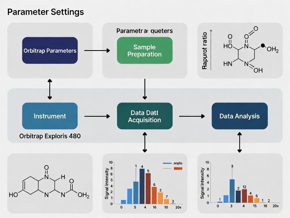
Mass spectrometry (MS) has evolved as the preferred analytical method for proteomics, lipidomics and metabolomics, allowing thousands of biologically active metabolites to be identified and quantified at trace levels in a wide range of matrices [1]. The success of untargeted metabolomics depends not only on instrument performance but also on the optimization of mass spectrometric parameters, which directly influence the quality and quantity of MS/MS spectra collected [1]. The Orbitrap Exploris 480 represents the pinnacle of high-resolution, accurate-mass (HR/AM) mass spectrometry, with exceptional resolution, mass accuracy, and sensitivity making it a go-to choice for labs pushing the boundaries of discovery metabolomics [2]. This application note details the key specifications and optimized parameters for the Orbitrap Exploris 480 to maximize metabolite coverage and data quality in metabolomics research.
Core Instrument Specifications for Metabolomics
The Thermo Scientific Orbitrap Exploris 480 is a hybrid quadrupole-Orbitrap MS instrument capable of providing high quality high energy collisional dissociation (HCD) mass spectra with resolving powers from 7500 to 480,000 at m/z 200 [1] [3]. The increased scan speed, high resolution, improved sensitivity and robustness of the instrument has made it a popular choice in untargeted metabolomics research [1].
Table 1: Key Technical Specifications of the Orbitrap Exploris 480 Mass Spectrometer
| Parameter | Specification | Significance for Metabolomics |
|---|---|---|
| Resolution | Up to 480,000 at m/z 200 [4] [3] | Enables separation of isobaric compounds with minimal mass differences |
| Scan Speed | Up to 40 Hz [4] [3] | Compatible with high-throughput separations and fast chromatography |
| Mass Accuracy | < 3 ppm RMS (external calibration)< 1 ppm RMS (internal calibration with EASY-IC) [3] [5] | Confident compound identification through accurate mass measurement |
| Mass Range | 40-6000 m/z (extendable to 8000 m/z with BioPharma option) [4] [5] | Captures low molecular weight fragments to high-mass metabolites |
| Sensitivity | MS/MS: 50 fg reserpine on-column S/N 100:1 [3] [5] | Detection of trace-level metabolites in complex biological matrices |
| Dynamic Range | >5000:1 within a single Orbitrap mass analyzer spectrum [3] | Quantification of abundant and rare metabolites within the same analysis |
The instrument incorporates a maintenance-free secondary ion source (EASY-IC) to deliver a regulated number of calibrant ions into the MS, enabling real-time fine adjustment of the instrument's m/z calibration. This corrects otherwise uncompensated errors due to temperature fluctuations and scan-to-scan variations, maintaining mass accuracy under 1 ppm for at least five days [4].
Optimized MS Parameters for Untargeted Metabolomics
Experimental Design for Parameter Optimization
A systematic optimization of mass spectrometric parameters for data dependent acquisition (DDA) experiments is essential to increase MS/MS coverage and metabolite identifications in untargeted metabolomics [1]. The optimization study utilized a one factor at a time (OFAT) approach on a Vanquish UHPLC coupled to an Orbitrap Exploris 480 mass spectrometer equipped with high flow and low flow HESI probes [1]. The experimental setup employed NIST SRM 1950 reference human plasma extracted using an in-house methanol extraction method, with chromatographic separations performed using an Acquity Premier CSH C18 column with a 15-minute gradient elution [1].
Table 2: Optimized MS Parameters for Untargeted Metabolomics on Orbitrap Exploris 480
| Parameter | Optimal Setting for Full MS | Optimal Setting for MS/MS |
|---|---|---|
| Resolution | 180,000 [1] | 30,000 [1] |
| RF Lens | 70% [1] | Not Applicable |
| Intensity Threshold | Not Applicable | 1 × 10⁴ [1] |
| Mass Isolation Width | Not Applicable | 2.0 m/z [1] |
| TopN (MS/MS Scans) | Not Applicable | 10 [1] |
| AGC Target | 5 × 10⁶ [1] | 1 × 10⁵ [1] |
| Maximum Ion Injection Time | 100 ms [1] | 50 ms [1] |
| Dynamic Exclusion | Not Applicable | 10 s [1] |
| Collision Energy | Not Applicable | Stepped HCD (20, 40, 60) [1] |
Detailed Methodology for Metabolomic Analysis
Sample Preparation Protocol
- Metabolite Extraction: Add 800 μL of cold methanol to 200 μL of frozen plasma in a 1.7 mL centrifuge tube [1].
- Incubation: Incubate the mixture for 15 minutes at 4°C on a ThermoMixer [1].
- Centrifugation: Centrifuge at 18,000g at 4°C for 10 minutes [1].
- Aliquoting and Drying: Divide the supernatant into 100 μL aliquots and dry using a vacuum concentrator [1].
- Reconstitution: Reconstitute extracts in 200 μL of water/methanol (95:5) modified with 0.1% formic acid [1].
- Storage: Store dried plasma extracts at -80°C until analysis [1].
Liquid Chromatography Conditions
- Column: Acquity Premier CSH C18 1.7 μm × 2.1 × 100 mm [1]
- Flow Rate: 0.3 mL min⁻¹ [1]
- Mobile Phase: A: Water with 0.1% formic acid; B: Acetonitrile with 0.1% formic acid [1]
- Gradient Elution: 0 min, 0% B; 2 min, 40% B; 8 min, 98% B; 10 min, 98% B; 10.5 min, 0% B; 15 min, 0% B [1]
- Column Temperature: 40°C [1]
- Injection Volume: 5.0 μL [1]
Mass Spectrometry Instrument Settings
- Ionization Mode: Positive ion mode with spray voltage of 3.6 kV [1]
- Source Gas Settings: Sheath gas: 35 Arb, Auxiliary gas: 10 Arb, Sweep gas: 1 Arb [1]
- Temperature Settings: Ion transfer tube: 350°C, Vaporizer: 350°C [1]
- Mass Scan Range: 50-750 m/z [1]
- Calibration: Perform mass spectrometer calibration in low and high mass range with Pierce FlexMix calibration solution [1]
Figure 1: Experimental workflow for untargeted metabolomics on the Orbitrap Exploris 480 mass spectrometer.
Advanced Applications and Technologies
FAIMS Technology for Enhanced Metabolite Detection
The front-end High Field Asymmetric Waveform Ion Mobility Spectrometry (FAIMS) Pro interface functions as an ion selection device and an electrospray filter that prevents neutrals from entering the orifice of the mass spectrometer while reducing chemical background noise [6]. This "purification" of the electrosprayed ions typically results in improved robustness and sensitivity for metabolomics experiments. The FAIMS Pro interface continuously selects and focuses ions at atmospheric pressure based on their differential mobilities in a high field versus a low electric field [6].
Combining Data Independent Acquisition (DIA) with FAIMS using single compensation voltages enables analysis of up to 2000 peptides per LC gradient minute, demonstrating the technology's capability for high-throughput analysis [6]. For sensitivity applications, the raw sensitivity of the instrument has been evaluated by analyzing 5 ng of a HeLa digest from which >1000 proteins were reproducibly identified with 5 min LC gradients using DIA-FAIMS [6].
Intelligent Acquisition Modes
The Orbitrap Exploris 480 incorporates intelligent data acquisition modes that leverage new levels of instrument performance to deliver high confidence and high throughput results [4]. These include:
- SureQuant IS Targeted Protein Quantitation: A data-aware quantitation scan mode that leverages internal standards to dynamically adjust scan parameters and automatically maximize data quality for targeted metabolome analysis in real-time [4].
- TurboTMT: Intelligent acquisition based on novel ΦSDM spectral processing increases resolution to baseline resolve TMT reporter ion isotopologues and speed up spectral acquisition for TMT experiments [4].
- Precursor Fit Filter: An algorithm that reduces co-isolated ion interferences that can mask true differences in metabolite abundance [4].
Figure 2: Data-dependent acquisition (DDA) workflow with optimized parameters for untargeted metabolomics.
The Scientist's Toolkit: Essential Research Reagents and Materials
Table 3: Key Research Reagent Solutions for Orbitrap Exploris 480 Metabolomics
| Reagent/Material | Function | Example Source/Product |
|---|---|---|
| NIST SRM 1950 Reference Plasma | Standardized reference material for method development and quality control | National Institute of Standards and Technology [1] |
| Pierce FlexMix Calibration Solution | Mass calibration in low and high mass range for instrument qualification | Thermo Fisher Scientific [1] |
| LC-MS Optima Grade Solvents | High-purity solvents for mobile phase preparation to minimize background noise | Thermo Fisher Scientific [1] |
| CSH C18 Chromatography Column | Reversed-phase separation of metabolites with high efficiency and resolution | Waters Acquity Premier CSH C18 [1] |
| Formic Acid (LC-MS Grade) | Mobile phase modifier for improved ionization efficiency in positive mode | Various suppliers [1] |
| Methanol (LC-MS Grade) | Protein precipitation and metabolite extraction solvent | Various suppliers [1] |
The Orbitrap Exploris 480 mass spectrometer, when configured with the optimized parameters detailed in this application note, provides exceptional performance for untargeted metabolomics studies. The combination of high resolving power (up to 480,000), fast scan rates (up to 40 Hz), and exceptional mass accuracy (<1 ppm with EASY-IC) enables comprehensive metabolite coverage and confident compound identification [4] [1] [3]. The parameter optimization study demonstrated that specific settings for resolution, RF level, intensity threshold, AGC target, and maximum injection time significantly influence metabolite annotations and should be carefully controlled for reproducible results [1]. These advanced capabilities, coupled with intelligent acquisition modes and FAIMS technology, position the Orbitrap Exploris 480 as a powerful platform for addressing the most challenging questions in metabolomics research and drug development.
For researchers utilizing the Orbitrap Exploris 480 mass spectrometer in metabolomics and proteomics, a deep understanding of the intrinsic relationship between transient length and mass resolution is fundamental to designing effective experiments. The Orbitrap mass analyzer generates high-resolution accurate-mass (HRAM) spectra by recording the image current of trapped ions—a signal known as a transient—and converting it into a mass spectrum using Fourier transformation (FT) [7]. The quality of this spectral data is not arbitrary but is governed by specific instrument parameters that involve significant trade-offs between resolution, acquisition speed, and sensitivity. This application note delineates these critical relationships within the context of Orbitrap Exploris 480 operation, providing structured data and protocols to guide researchers in making informed decisions that align with their experimental objectives. The fundamental principle underlying these trade-offs is that mass resolution in Orbitrap MS scales directly with the duration of the transient acquisition [7]. Consequently, higher resolution settings necessitate longer transient times, which in turn reduces the instrument's scan speed and impacts the overall cycle time of an experiment. Navigating this balance is particularly crucial in applications like untargeted metabolomics, where comprehensive metabolite coverage is desired, and in high-throughput proteomics, where rapid analysis is paramount.
The Fundamental Relationship: Transient Length and Resolution
The Orbitrap Exploris 480 achieves its exceptional mass resolution by measuring ion oscillation frequencies over a specific period known as the transient length. The direct correlation is simple yet profound: longer transient times enable higher mass resolution by allowing more precise frequency measurements. This enhanced resolution improves the ability to distinguish between ions with very similar mass-to-charge (m/z) ratios, a critical capability in complex sample analysis. However, this advantage comes at the direct cost of acquisition speed, as fewer scans can be completed per unit of time.
Table 1: Resolution Settings and Corresponding Transient Times on the Orbitrap Exploris 480
| Resolution at m/z 200 | Transient Length (ms) | Approximate Scan Speed (Hz) | "Free" Fill Time (ms) |
|---|---|---|---|
| 7,500 | 16 | 40 | N/A |
| 15,000 | 32 | 22 | 22 |
| 30,000 | 64 | 12 | 54 |
| 60,000 | 128 | 7 | 118 |
| 120,000 | 256 | 3 | 246 |
| 240,000 | 512 | 1.5 | 502 |
| 480,000 | 1024 | 0.7 | 1014 |
The data in Table 1, derived from instrument specifications [3], quantitatively defines this trade-off. For instance, increasing the resolution from 15,000 to 240,000 (a 16-fold increase) extends the transient length from 32 ms to 512 ms (also a 16-fold increase), while the scan rate plummets from 22 Hz to just 1.5 Hz. This has a direct impact on experimental design, particularly in chromatography-coupled workflows where the mass spectrometer must acquire enough data points across rapidly eluting peaks for accurate quantification. The "Free Fill Time" column represents the time available to fill the C-trap with ions for the next analysis while the current transient is being processed, a feature that enhances instrument efficiency [3].
Diagram 1: The relationship between transient length and its impact on key instrument capabilities and experimental applications. Increasing transient length directly enables higher mass resolution but reduces acquisition speed, leading to different experimental design considerations.
Impact on Experimental Design and Data Quality
The choice of resolution and corresponding transient length profoundly influences data quality and experimental outcomes. In untargeted metabolomics, higher resolution (e.g., 120,000-180,000 for MS1) provides superior mass accuracy and better differentiation of co-eluting isobaric compounds, leading to more confident metabolite annotations [1]. However, if the selected resolution is too high for the chromatographic peak width, insufficient data points may be collected across each peak, compromising quantitative accuracy. This is especially critical in high-throughput applications using short LC gradients.
Advanced Signal Processing: ΦSDM Technology
A significant advancement in mitigating the traditional trade-offs is the implementation of the phase-constrained spectrum deconvolution method (ΦSDM). This novel computational strategy for processing Orbitrap transients has the potential to double the mass resolving power at a given transient duration compared to standard enhanced Fourier transformation (eFT) [7]. For instance, ΦSDM can achieve a resolution comparable to a 256 ms transient in just 128 ms. This allows researchers to either obtain higher resolution data without sacrificing scan speed or maintain their required resolution at twice the acquisition rate. The benefits of ΦSDM are particularly pronounced in data-independent acquisition (DIA) proteomics and in applications using fast chromatographic gradients (e.g., 5-21 minutes), where it has been shown to increase the number of identified protein groups and peptides by over 15% [7]. This technology effectively provides a "best of both worlds" scenario, enhancing spectral quality in regions of high peptide density and improving the ability to resolve low-abundance signals without extending cycle times.
Optimized Protocols for Parameter Selection
Protocol 1: Optimizing for Untargeted Metabolomics via DDA
This protocol is adapted from a systematic optimization study for untargeted metabolomics using data-dependent acquisition (DDA) on the Orbitrap Exploris 480 [1].
Step 1: Sample Preparation
- Extract metabolites from your biological matrix (e.g., serum, plasma, tissues). For instance, precipitate 200 μL of plasma with 800 μL of cold methanol, incubate at 4°C for 15 min, and centrifuge at 18,000g for 10 min.
- Collect and dry the supernatant using a vacuum concentrator. Reconstitute in a compatible solvent like water/methanol (95:5) with 0.1% formic acid prior to LC-MS analysis [1].
Step 2: Liquid Chromatography
- Column: Employ a reversed-phase column (e.g., Acquity Premier CSH C18, 1.7 μm, 2.1 × 100 mm).
- Gradient: Use a binary solvent system (A: water + 0.1% formic acid; B: acetonitrile + 0.1% formic acid). A recommended gradient is: 0% B to 40% B over 2 min, 40% B to 98% B over 6 min, hold at 98% B for 2 min, then re-equilibrate [1].
- Flow Rate: 0.3 mL/min.
- Column Temperature: 40°C.
- Injection Volume: 5.0 μL.
Step 3: Mass Spectrometry - Full Scan (MS1)
- Ionization: Positive ion mode with HESI source. Set spray voltage to 3.6 kV, ion transfer tube temperature to 350°C, and vaporizer temperature to 350°C.
- Resolution: Set to 120,000 (at m/z 200) for optimal balance of mass accuracy and scan speed [1]. This corresponds to a transient length of 256 ms.
- Scan Range: 50–750 m/z.
- AGC Target:
5e6(Improved from "standard" setting) [1]. - Maximum Injection Time (MIT): 100 ms.
- RF Lens: 70%.
Step 4: Mass Spectrometry - Data-Dependent MS/MS (ddMS2)
- Resolution: Set to 30,000 (at m/z 200) [1].
- AGC Target:
1e5[1]. - Maximum Injection Time (MIT): 50 ms.
- Intensity Threshold:
1e4[1]. - Top N: 10 (i.e., perform ten MS/MS scans per cycle) [1].
- Isolation Window: 2.0 m/z [1].
- Collision Energy: Use stepped HCD energies (e.g., 20, 40, 60 eV) [1].
- Dynamic Exclusion: 10 seconds.
Protocol 2: Enhancing Sensitivity for Targeted Analytes via SIM
For targeted analysis of low-abundance metabolites or precise isotope ratio measurements in tracing studies, Selected Ion Monitoring (SIM) is highly beneficial. This protocol can be integrated with a full-scan method.
Step 1: LC Setup
- Use an amide column (e.g., Waters XBridge BEH Amide, 2.1 × 150 mm, 2.5 μm) with a 25-minute gradient for hydrophilic interaction liquid chromatography (HILIC) separation [8].
Step 2: Full Scan Acquisition
- Resolution: 120,000 at m/z 200.
- Scan Range: m/z 120–1000 (positive mode) or m/z 70–1000 (negative mode).
- AGC Target:
1e7. - Maximum Injection Time: 200 ms [8].
- This provides a broad, untargeted overview of the sample.
Step 3: SIM Acquisition for Targeted Ions
- Define narrow mass windows (e.g., ±1.5 Da) around the precursor m/z of your low-intensity target metabolites.
- AGC Target:
1e6(Lower than full scan to mitigate space-charge effects) [8]. - Maximum Injection Time: 200 ms [8].
- Note: SIM significantly enhances the signal-to-noise (S/N) ratio and measurement precision for low-intensity ions but must be used judiciously. Excessive ion accumulation in a narrow m/z window can cause space-charge effects, leading to signal loss and ion coalescence. Optimize AGC target and injection time to control ion accumulation [8].
Table 2: Decision Matrix for Resolution and Scan Mode Selection
| Experimental Goal | Recommended MS1 Resolution | Recommended Scan Mode | Rationale |
|---|---|---|---|
| Untargeted Metabolomics | 120,000 - 180,000 [1] | Full Scan DDA | Optimal balance of mass accuracy, coverage, and scan speed for metabolite ID. |
| High-Throughput Proteomics | 60,000 [7] | Full Scan DIA | Faster cycle times to adequately sample narrow chromatographic peaks. |
| Targeted Metabolite Quant | 120,000 [8] | Combined Full Scan + SIM | Broad coverage plus enhanced sensitivity/precision for specific low-level ions. |
| TMT Reporter Ion Quant | 120,000 (MS1) [3] | DDA with MS2 Res = 45,000 [3] | High MS1 resolution for precursor quant; High MS2 res to resolve reporter ions. |
The Scientist's Toolkit: Research Reagent Solutions
Table 3: Essential Materials for Orbitrap-Based Metabolomics
| Item | Function / Application |
|---|---|
| Standard Reference Material (SRM) 1950 | Commercially available reference human plasma used for method validation and standardization [1]. |
| Pierce FlexMix Calibration Solution | Contains a mixture of compounds for mass accuracy calibration in both low and high mass ranges [1]. |
| LC-MS Optima Grade Solvents | High-purity water, methanol, and acetonitrile modified with 0.1% formic acid for UHPLC mobile phases to minimize background noise and ion suppression [1]. |
| C18 Reversed-Phase UHPLC Columns | High-pressure stable stationary phase (e.g., 1.7 μm particle size) for efficient separation of complex metabolite mixtures [1] [9]. |
| Authenticated Chemical Standards | Pure metabolite compounds essential for validating metabolite identifications and retention times [1] [8]. |
The relationship between transient length and resolution is a cornerstone principle governing experimental design on the Orbitrap Exploris 480. The quantitative data presented herein provides a clear framework for selecting appropriate parameters based on specific analytical goals. For untargeted metabolomics seeking broad coverage, higher resolution settings (120,000-180,000) are advantageous, whereas high-throughput proteomics demands lower resolutions (15,000-60,000) to maintain fast cycle times. The emergence of technologies like ΦSDM and strategic application of scan modes like SIM offer powerful means to circumvent traditional limitations, enabling higher resolution at faster speeds or greater sensitivity for targeted analyses. By applying the structured protocols and decision matrices provided, researchers can systematically optimize their methods to maximize data quality and extract more biologically meaningful results from their experiments.
Within the framework of a broader thesis on parameter settings for Orbitrap Exploris 480 metabolomics research, this document details the essential hardware components and their operational protocols. The precision and depth of untargeted metabolomics are fundamentally governed by the mass spectrometric parameters, whose optimization is only possible on a robust and advanced hardware foundation [1]. The Thermo Scientific Orbitrap Exploris 480 mass spectrometer provides this foundation, integrating components like the OptaMax NG ion source and high-capacity ion transfer optics to deliver the sensitivity, resolution, and robustness required for modern translational science [4]. This application note provides a detailed examination of these critical hardware elements, placing them in the context of optimized experimental workflows for researchers, scientists, and drug development professionals. We summarize optimized parameters into structured tables and provide explicit protocols to empower scientists to achieve superior metabolite coverage and confidence in their results.
Core Hardware Architecture and Configuration
The Orbitrap Exploris 480 MS is engineered with a complete ground-up redesign focusing on system usability, technological advancements in pumping technology, control electronics, and ion optics [4]. The physical path of an ion from the sample to detection involves a series of critical components, each contributing to the system's overall performance, reliability, and data certainty.
The relationship between these components and their collective function in a data acquisition workflow is illustrated below.
Detailed Component Specifications
OptaMax NG Ion Source: This source supports multiple ionization modes, including Heated Electrospray Ionization (H-ESI), Atmospheric Pressure Chemical Ionization (APCI), and Atmospheric Pressure Photoionization (APPI), providing flexibility for a wide range of metabolite polarities and masses [10]. Its key function is to efficiently generate gas-phase ions from the liquid chromatograph effluent. In a typical metabolomics setup for positive mode, the spray voltage is set at 3.6 kV. The source also regulates gas temperatures (ion transfer tube and vaporizer at 350 °C) and gas flows (sheath gas: 35 Arb, auxiliary gas: 10 Arb, sweep gas: 1 Arb) to ensure optimal desolvation and ion yield [1].
Ion Transfer Tube (ITT) and High-Capacity Transfer Tube (HCTT): The ITT is a critical interface that conducts ions from the atmospheric pressure source region into the high-vacuum mass analyzer. The maintained temperature of 350 °C prevents condensation and ensures ions remain in the gas phase [1]. The system's improved ion routing, which includes a redesigned bent flatapole, significantly increases instrument robustness by reducing contamination [4].
Ion-Routing Multipole and HCD Cell: This multipole device performs Higher Collisional Dissociation (HCD) fragmentation and routes ions similarly to the Orbitrap Tribrid platform. Its design increases instrument robustness by significantly reducing contamination, which is vital for maintaining consistent performance in high-throughput metabolomics [4].
High-Field Orbitrap Mass Analyzer: This is the core detection component, capable of a resolution of up to 480,000 at m/z 200 and scan speeds of up to 40 Hz. This high resolution is crucial for confident metabolite annotation by providing accurate mass measurements [4] [1].
Experimental Protocol: Metabolite Extraction and LC-MS Analysis
Metabolite Extraction from Human Plasma
This protocol is adapted from the methodology used to optimize parameters on the Orbitrap Exploris 480 [1].
Materials:
- NIST SRM 1950 Reference Human Plasma.
- LC-MS optima grade water, methanol, and formic acid.
- ThermoMixer (e.g., Eppendorf).
- Refrigerated centrifuge (capable of 18,000×g).
- Vacuum concentrator (e.g., Thermo Scientific SpeedVac).
Procedure:
- Aliquot 200 µL of frozen plasma into a 1.7 mL microcentrifuge tube.
- Add 800 µL of cold methanol to the plasma.
- Incubate the mixture for 15 minutes at 4 °C on a ThermoMixer.
- Centrifuge the mixture at 18,000×g for 10 minutes at 4 °C.
- Carefully transfer the supernatant and divide it into 100 µL aliquots.
- Dry each aliquot using a vacuum concentrator.
- Store the dried extracts at -80 °C until analysis.
- For LC-MS analysis, reconstitute the dried extract in 200 µL of a solution of water/methanol (95:5) modified with 0.1% formic acid.
Liquid Chromatography and Mass Spectrometry
Chromatography:
- Column: Acquity Premier CSH C18 (1.7 µm, 2.1 mm × 100 mm).
- Flow Rate: 0.3 mL/min.
- Mobile Phase: A) Water with 0.1% formic acid; B) Acetonitrile with 0.1% formic acid.
- Gradient:
- 0 min: 0% B
- 2 min: 40% B
- 8 min: 98% B
- 10 min: 98% B
- 10.5 min: 0% B
- 15 min: 0% B (re-equilibration)
- Column Temperature: 40 °C.
- Injection Volume: 5.0 µL [1].
Mass Spectrometry - Global Settings:
- Instrument: Orbitrap Exploris 480 MS equipped with HESI probe.
- Ionization Mode: Positive.
- Spray Voltage: 3.6 kV.
- Sheath, Aux, Sweep Gas: 35, 10, 1 (Arb units).
- Ion Transfer Tube Temp: 350 °C.
- Vaporizer Temp: 350 °C.
- Mass Range: m/z 50–750 [1].
Parameter Optimization for Untargeted Metabolomics
Optimization of mass spectrometric parameters in Data Dependent Acquisition (DDA) is essential to increase MS/MS coverage and metabolite identifications [1]. The following parameters were systematically evaluated using a one-factor-at-a-time (OFAT) approach on the Orbitrap Exploris 480.
Optimized Parameter Settings
Table 1: Optimized MS and MS/MS parameters for untargeted metabolomics on the Orbitrap Exploris 480.
| Parameter | Optimized Value (Full MS) | Optimized Value (dd-MS/MS) |
|---|---|---|
| Mass Resolution | 180,000 [1] | 30,000 [1] |
| RF Lens (%) | 70% [1] | Not Applicable |
| Intensity Threshold | Not Applicable | 1 × 10⁴ [1] |
| Mass Isolation Width (m/z) | Not Applicable | 2.0 [1] |
| TopN (MS/MS Events) | Not Applicable | 10 [1] |
| AGC Target | 5 × 10⁶ [1] | 1 × 10⁵ [1] |
| Max. Injection Time (ms) | 100 [1] | 50 [1] |
| Collision Energy | Not Applicable | Stepped HCD (20, 40, 60) [1] |
| Dynamic Exclusion | Not Applicable | 10 s [1] |
Optimization Workflow Logic
The process for determining these optimal values follows a logical, sequential workflow to ensure each parameter is validated against its impact on metabolite coverage.
Advanced Intelligent Acquisition Strategies
Beyond standard DDA, the Orbitrap Exploris 480 platform enables more sophisticated, intelligent acquisition methods that integrate targeted and discovery approaches.
The Hybrid-DIA Workflow
The hybrid-DIA strategy uses an Application Programming Interface (API) within the Tune software to dynamically combine Data-Independent Acquisition (DIA) with triggered, multiplexed MS/MS (MSx) scans of predefined targets [11]. This is particularly valuable for quantifying low-abundance phosphopeptides or key metabolites while simultaneously acquiring a global profile.
- Principle: A standard DIA method runs continuously. A predefined list of target ions (e.g., from spiked-in heavy isotope-labeled standards) is monitored in real-time during the full MS scan. Upon detection of a target, the API triggers a high-sensitivity MSx scan for the standard and its endogenous counterpart, interleaving it with the DIA scans within the same cycle [11].
- Benefit: It maximizes information from a single injection, providing the breadth of discovery proteomics/metabolomics with the sensitivity and quantitative accuracy of targeted methods for critical pathways [11].
The Scientist's Toolkit: Research Reagent Solutions
Table 2: Essential materials and reagents for Orbitrap Exploris 480 metabolomics protocols.
| Item | Function / Application |
|---|---|
| NIST SRM 1950 Serum | Standardized reference material for method development, optimization, and inter-laboratory comparison [1]. |
| Pierce FlexMix Calibration Solution | Used for mass accuracy calibration in both low and high mass ranges to ensure sub-ppm mass accuracy [1]. |
| LC-MS Optima Grade Solvents (Water, Methanol, Acetonitrile) | High-purity solvents for mobile phase preparation and sample reconstitution to minimize background noise and ion suppression [1]. |
| Acquity Premier CSH C18 Column | Reversed-phase UHPLC column for high-resolution separation of complex metabolite mixtures prior to MS analysis [1]. |
| Heavy Stable Isotope-Labeled Standards | Used in intelligent acquisition methods (SureQuant, hybrid-DIA) for sensitive and accurate targeted quantification of predefined metabolites or pathway markers [11]. |
| Acid Modifier (e.g., Formic Acid) | Added to the mobile phase to improve protonation and ionization efficiency of metabolites, particularly in positive ion mode [1]. |
Mass accuracy is a cornerstone of reliable metabolomics data, directly influencing metabolite identification confidence. For high-resolution mass spectrometers like the Orbitrap Exploris 480, maintaining long-term mass accuracy presents a significant challenge due to potential instrumental drift caused by environmental fluctuations, such as variations in temperature and humidity. Effective calibration strategies are therefore essential for ensuring data integrity throughout long analytical sequences. This application note, framed within a broader thesis on parameter optimization for the Orbitrap Exploris 480, details robust calibration protocols using the integrated EASY-IC and FlexIC systems to achieve sustained sub-ppm mass accuracy, critical for confident metabolite annotation in drug development and biomedical research.
The Orbitrap Exploris 480 Mass Spectrometer
The Thermo Scientific Orbitrap Exploris 480 mass spectrometer is an advanced, intelligence-driven instrument designed for ultimate performance and ease of use. Its hardware architecture ensures maximum uptime and easy serviceability, which are fundamental requirements for long-term metabolomic studies [3]. A key feature of this system is the EASY-IC (Internal Calibration) source, which provides real-time internal mass calibration by delivering a constant flow of calibrant ions alongside the analyte stream. This enables automated, real-time fine adjustment of the mass calibration, achieving constant 1-ppm mass accuracy during data acquisition without manual intervention [12]. The instrument is capable of a wide resolving power, from 7,500 to 480,000 at m/z 200, and under external calibration, it can achieve a mass accuracy of < 3 ppm RMS drift over 24 hours. This is significantly improved to < 1 ppm RMS drift over the same period when internal calibration is employed [3].
Calibration Fundamentals and Performance
Understanding Mass Accuracy Specifications
Mass accuracy is typically reported as the root mean square (RMS) of the mass error drift over a specified time. The specifications for the Orbitrap Exploris 480 highlight the critical difference between external and internal calibration strategies, as shown in the table below.
Table 1: Mass Accuracy Specifications for the Orbitrap Exploris 480
| Calibration Type | Mass Accuracy (RMS) | Duration | Key Characteristic |
|---|---|---|---|
| External Calibration | < 3 ppm | Over 24 hours | Relies on initial calibration |
| Internal Calibration (EASY-IC) | < 1 ppm | Over 24 hours | Real-time, continuous calibration |
Resolving Power and Calibration
It is crucial to understand that higher mass resolution does not automatically translate to better mass accuracy. While higher resolution increases the ability to distinguish between ions of close m/z values, the Orbitrap Exploris 480 offers a range of resolution settings, each with an associated transient length and scan speed. The relationship between these parameters involves a trade-off; higher resolution requires longer transient times, reducing the number of spectra that can be acquired per second [3]. The EASY-IC system functions optimally across this entire range, ensuring high mass accuracy regardless of the chosen resolution-speed balance for the experiment.
Experimental Protocols for Long-Term Accuracy
Protocol 1: Leveraging the EASY-IC Source for Untargeted Metabolomics
This protocol is designed for broad, untargeted metabolomics profiling where sustained high mass accuracy is paramount for unknown metabolite identification.
1. Instrument Setup:
- Mass Spectrometer: Orbitrap Exploris 480 equipped with the EASY-IC source [12].
- LC System: Vanquish UHPLC system.
- Column: Waters XBridge BEH Amide column (2.1 × 150 mm, 2.5 µm) for HILIC separation [13] or equivalent C18 column for reversed-phase.
- Ionization Source: HESI-II probe.
2. EASY-IC Calibration:
- Ensure the EASY-IC source is activated within the instrument method.
- The system will automatically introduce the internal calibrant (e.g., Pierce FlexMix) during data acquisition [1].
- The instrument control software uses the known m/z of the calibrant ions to perform real-time, fine-scale calibration adjustments.
3. Recommended MS Parameters:
- Ion Mode: Positive and/or negative mode with fast polarity switching [14].
- Scan Range: m/z 70–1000 [13].
- MS1 Resolution: 120,000 [13] or 180,000 [1] at m/z 200.
- Spray Voltage: 3.5 kV (positive), 2.5 kV (negative) [14].
- Sheath Gas: 35 Arb [13] [14].
- Auxiliary Gas: 10 Arb [13].
- Ion Transfer Tube Temp: 300 °C [13] [14].
- Vaporizer Temp: 350 °C [14].
- RF Lens: 30% [14] or 60% [13] (optimization recommended).
- AGC Target: Standard or 1e7 [13].
- Maximum Injection Time: Auto or 200 ms [13].
4. Data Acquisition and Quality Control:
- Acquire data in profile mode.
- Monitor the mass error for the lock mass calibrant ions in real-time to verify continuous sub-ppm performance.
- Process data using software that can leverage the high-accuracy MS1 data, such as Compound Discoverer or Xcalibur.
Protocol 2: Targeted Quantitation with Selected Ion Monitoring (SIM)
For targeted analysis of low-abundance metabolites, such as in isotope-tracing studies, SIM can be combined with EASY-IC to enhance sensitivity and quantitative accuracy [13].
1. Sample Preparation:
- Matrix: Mouse liver, kidney, or other tissues.
- Extraction: Use 800 μL of cold methanol:acetonitrile:water (40:40:20) with 0.5% formic acid per ~25 mg of tissue powder.
- Neutralization: Add NH4HCO3 solution post-extraction to neutralize acid [13].
2. LC-MS Configuration with SIM:
- LC and MS Setup: As described in Protocol 1.
- Data Acquisition: Combine a full scan with SIM events for the targeted, low-intensity ions.
- Full Scan Parameters:
- Resolution: 120,000 at m/z 200.
- Scan Range: m/z 120–1000 (positive) or m/z 70–1000 (negative).
- SIM Parameters for a metabolite (e.g., 3-Phosphoglycerate):
3. Calibration and Quantitation:
- The EASY-IC source ensures mass accuracy for both the full scan and the SIM scans.
- The improved signal-to-noise (S/N) and precision (RSD) in SIM mode, underpinned by accurate mass measurement, allow for more reliable quantification and isotope ratio determination for low-abundance metabolites [13].
Figure 1: A simplified workflow for an untargeted metabolomics method with an embedded SIM scan, enabled by continuous EASY-IC calibration.
The Scientist's Toolkit: Research Reagent Solutions
Table 2: Essential Reagents and Materials for Metabolomics Calibration and Sample Preparation
| Item | Function / Application | Example / Specification |
|---|---|---|
| Pierce FlexMix Calibration Solution | Mass calibration in both low and high mass ranges; used for initial instrument calibration [1]. | ThermoFisher Scientific Pierce FlexMix |
| EASY-IC Calibrant | Provides the internal reference ions for real-time mass calibration during data acquisition [12]. | Proprietary calibrant for Orbitrap Exploris series |
| Standard Reference Material (SRM) 1950 | A standardized human plasma reference material for method validation and inter-laboratory comparison [1]. | National Institute of Standards and Technology (NIST) |
| LC-MS Optima Grade Solvents | High-purity solvents to minimize chemical noise and ion suppression, ensuring optimal performance [1]. | Water, Methanol, Acetonitrile, Formic Acid (Thermo Fisher) |
| Waters XBridge BEH Amide Column | Hydrophilic Interaction Liquid Chromatography (HILIC) for separation of polar metabolites [13]. | 2.1 × 150 mm, 2.5 µm particle size |
The combination of the Orbitrap Exploris 480's hardware stability and the intelligence of the EASY-IC internal calibration system provides a robust solution for achieving and maintaining long-term mass accuracy in metabolomics. The protocols outlined herein, from broad untargeted profiling to sensitive targeted SIM, offer researchers and drug development professionals clear pathways to generating high-fidelity, reproducible data. By ensuring mass accuracy remains below 1 ppm over extended periods, these strategies form a critical foundation for confident metabolite identification and quantification, thereby enhancing the overall validity and impact of metabolomics research.
Figure 2: A decision pathway for selecting the appropriate calibration strategy based on the required level of mass accuracy and experiment duration.
In mass spectrometry-based untargeted metabolomics, the reliability and depth of biological insight are fundamentally governed by three key performance metrics: sensitivity, dynamic range, and selectivity. For researchers using the Thermo Scientific Orbitrap Exploris 480 mass spectrometer, a precise understanding and optimization of these metrics is crucial for detecting low-abundance metabolites, quantifying compounds across a wide concentration spectrum, and confidently identifying analytes within complex biological matrices. This application note details the experimental protocols and performance data for characterizing these metrics, providing a framework for robust metabolomic method development within a broader thesis on parameter optimization for the Orbitrap Exploris 480 platform. The guidance is designed to empower researchers and drug development professionals to maximize the output and data quality of their metabolomics investigations.
Key Performance Metrics and Their Experimental Assessment
Sensitivity
Sensitivity refers to the instrument's ability to detect and measure low-abundance metabolites. It is often experimentally defined by the lowest concentration of an analyte that can be reliably distinguished from background noise, typically expressed as a signal-to-noise ratio [3].
Experimental Protocol for Determining Sensitivity:
- Stock Solution Preparation: Prepare a serial dilution of a certified standard metabolite (e.g., reserpine) across a range of concentrations, for instance, from 50 fg/µL to 1 pg/µL.
- LC-MS Analysis: Inject these solutions onto the Orbitrap Exploris 480 system using a suitable LC method. The system itself has demonstrated sensitivity of 50 fg of reserpine on-column with a signal-to-noise ratio of 100:1 for MS/MS spectra [3].
- Data Analysis: Measure the signal-to-noise (S/N) ratio for the analyte peak at each concentration level. The sensitivity limit is frequently defined as the concentration yielding a S/N ratio of 3:1 (for detection) or 10:1 (for quantification).
Parameters Influencing Sensitivity on the Orbitrap Exploris 480:
- Ion Source Parameters: Spray voltage, vaporizer temperature, and sheath/auxiliary gas flows must be optimized. One systematic evaluation found that a spray voltage of 3.5 kV, vaporizer and ion transfer tube temperatures of 350 °C, sheath gas of 35 arb, and auxiliary gas of 10 arb provided optimal results [1] [15].
- Ion Injection Times: Longer maximum injection times (MIT) allow more ions to be collected, boosting signal. For MS/MS scans, an MIT of 50 ms is recommended [1].
- Automatic Gain Control (AGC): A lower AGC target (e.g., 1e5 for MS/MS) can improve the detection of low-abundance ions by preventing the trap from being filled predominantly by high-abundance ions [1].
Dynamic Range
Dynamic range defines the span of concentrations over which an analyte can be quantified with acceptable accuracy and precision. It is the ratio between the highest concentration (where the response remains linear) and the lowest (the limit of quantification). The Orbitrap Exploris 480 has been documented to have a dynamic range of >5,000 within a single spectrum [3].
Experimental Protocol for Determining Dynamic Range:
- Calibration Curve: Prepare a calibration curve using a stable isotope-labeled internal standard, spiked into a representative biological matrix (e.g., plasma or urine extract). The concentration should cover several orders of magnitude (e.g., 0.01 ng/mL to 100 ng/mL).
- LC-MS Analysis: Analyze these samples in triplicate using the Orbitrap Exploris 480 with the intended acquisition method (e.g., DDA or DIA).
- Data Analysis: Plot the peak area ratio (analyte to internal standard) against the nominal concentration. Fit a linear regression curve and determine the range over which the coefficient of determination (R²) remains >0.99 and the accuracy is within 80-120%.
Selectivity
Selectivity is the ability of the method to accurately measure the analyte in the presence of interferences, such as isobars, isomers, and matrix components. High-resolution accurate mass (HRAM) instruments like the Orbitrap Exploris 480 achieve selectivity through high mass accuracy (routinely < 1 ppm with internal calibration) and high resolving power [4] [3].
Experimental Protocol for Assessing Selectivity:
- Matrix Spiking: Spike a known metabolite into a complex matrix (e.g., bovine liver total lipid extract or human plasma) at a low, physiologically relevant concentration (e.g., 1 ng/mL) [16].
- LC-MS/MS Analysis: Analyze the spiked matrix and a blank matrix using a high-resolution MS/MS method. The Orbitrap Exploris 480 can achieve resolving powers up to 480,000 at m/z 200 for MS and 120,000 for MS/MS, which is critical for separating nearly isobaric ions [4] [1].
- Data Analysis: Confirm the identity of the analyte by examining the following:
- Mass Accuracy: The measured mass of the precursor ion should be within 5 ppm of the theoretical mass.
- Isotopic Pattern: The observed isotopic distribution should match the theoretical pattern.
- Fragmentation Spectrum: The MS/MS spectrum should match a reference spectrum with high confidence.
Table 1: Optimized Mass Spectrometric Parameters for Untargeted Metabolomics on the Orbitrap Exploris 480 [1]
| Parameter | Full MS Scan | Data-Dependent MS/MS (ddMS2) |
|---|---|---|
| Resolution | 180,000 | 30,000 |
| RF Lens (%) | 70 | N/A |
| AGC Target | 5e6 | 1e5 |
| Maximum Injection Time | 100 ms | 50 ms |
| Intensity Threshold | N/A | 1e4 |
| Top N | N/A | 10 |
| Mass Isolation Window | N/A | 2.0 m/z |
| Dynamic Exclusion | N/A | 10 s |
Comparative Performance of Acquisition Modes
The choice of acquisition mode—Data-Dependent Acquisition (DDA), Data-Independent Acquisition (DIA), or others like AcquireX—significantly impacts the effective sensitivity, dynamic range, and selectivity in an untargeted metabolomics experiment.
A systematic comparison of these modes on the Orbitrap Exploris 480 revealed distinct performance characteristics [16]:
- DIA demonstrated superior performance, detecting the highest number of metabolic features (averaging 1,036 over three measurements) and exhibiting the best reproducibility (CV of 10%).
- DDA detected 18% fewer features than DIA and showed lower reproducibility (CV of 17%).
- AcquireX detected 37% fewer features than DIA but offered moderate reproducibility (CV of 15%).
Table 2: Performance Comparison of Acquisition Modes for Metabolite Detection in a Complex Matrix [16]
| Acquisition Mode | Average Number of Metabolic Features Detected | Reproducibility (Coefficient of Variance) | Identification Consistency (Overlap Between Days) |
|---|---|---|---|
| Data-Independent Acquisition (DIA) | 1036 | 10% | 61% |
| Data-Dependent Acquisition (DDA) | 18% fewer than DIA | 17% | 43% |
| AcquireX | 37% fewer than DIA | 15% | 50% |
The following workflow diagram illustrates the logical decision process for selecting an acquisition mode based on the primary research objectives:
The Scientist's Toolkit: Essential Research Reagent Solutions
The following table lists key materials and reagents referenced in the optimized protocols for the Orbitrap Exploris 480.
Table 3: Essential Research Reagent Solutions for Metabolomics
| Item | Function / Application | Example / Source |
|---|---|---|
| NIST SRM 1950 | Standard Reference Material of human plasma used for method development, validation, and ensuring inter-laboratory reproducibility. | National Institute of Standards and Technology (NIST) [1] [15] |
| Pierce FlexMix | Calibration solution used for mass accuracy calibration in both low and high mass ranges on the Orbitrap Exploris 480. | Thermo Fisher Scientific [1] |
| C18 Reverse-Phase Columns | Workhorse columns for chromatographic separation of a wide range of metabolites in untargeted metabolomics. | e.g., Acquity Premier CSH C18 [1] |
| HILIC Columns | (Hydrophilic Interaction Liquid Chromatography) Used to retain and separate highly polar metabolites not retained by reverse-phase C18. | e.g., Zwitterionic HILIC columns [15] |
| Stable Isotope-Labeled Standards (AQUA) | Used as internal standards for precise targeted quantification, correcting for matrix effects and recovery losses. | Thermo Fisher Scientific [17] |
| Eicosanoid Standard Mix | A set of specific metabolite standards used in system suitability tests (SST) to evaluate instrument detection power and performance over time. | Commercially available from various vendors [16] |
Detailed Experimental Protocol: A Reproducible Workflow
This integrated protocol summarizes the optimal parameters and steps for a robust untargeted metabolomics run on the Orbitrap Exploris 480.
Step 1: Sample Preparation
- Extract metabolites from your sample matrix (e.g., using a methanol-based protein precipitation for plasma [1]).
- Reconstitute the dried extract in a suitable solvent (e.g., 95:5 water/methanol with 0.1% formic acid).
- Use a system suitability test (SST), such as a spiked eicosanoid standard mix, to verify instrument performance prior to the analysis [16].
Step 2: Liquid Chromatography
- Column: Use a C18 column (e.g., 2.1 x 100 mm, 1.7 µm) for reverse-phase separation [1].
- Mobile Phase: (A) Water with 0.1% formic acid; (B) Acetonitrile with 0.1% formic acid.
- Gradient: Employ a linear gradient from 0% B to 40% B over 2 min, then to 98% B by 8 min, hold for 2 min, and re-equilibrate [1].
- Flow Rate & Temperature: 0.3 mL/min and 40 °C.
Step 3: Ion Source Optimization (Orbitrap Exploris 480 with HESI)
- Spray Voltage: 3.5 kV (Positive Ion Mode) [1] [15].
- Vaporizer & Ion Transfer Tube Temp: 350 °C [1].
- Sheath Gas: 35 arb [1].
- Auxiliary Gas: 10 arb [1].
- Sweep Gas: 1 arb [1].
Step 4: Mass Spectrometry Data Acquisition
- Acquisition Mode: For comprehensive coverage and reproducibility, DIA is recommended. For more targeted hypothesis testing, DDA can be used [16].
- Apply the optimized parameters from Table 1 for full scan and MS/MS settings.
- Enable EASY-IC for internal calibration to maintain mass accuracy below 1 ppm [4] [3].
The complete experimental journey from sample to insight is captured in the following workflow:
From Theory to Practice: Implementing Robust DIA, DDA, and Targeted Metabolomics Workflows
Configuring Data-Independent Acquisition (DIA) for Maximum Feature Detection and Reproducibility
Within the broader scope of optimizing parameter settings for Orbitrap Exploros 480 metabolomics research, the selection and configuration of the data acquisition mode is a foundational decision. This Application Note provides a detailed protocol for implementing Data-Independent Acquisition (DIA) on the Orbitrap Exploris 480 platform, an approach demonstrated to maximize feature detection and quantitative reproducibility in untargeted metabolomics. Compared to the more traditional Data-Dependent Acquisition (DDA), DIA systematically fragments all ions within pre-defined isolation windows, thereby reducing the stochasticity and intensity bias inherent in DDA [16] [18]. Recent evidence obtained on the Orbitrap Exploris 480 shows that DIA not only detects a higher number of metabolic features but also delivers superior consistency in compound identification across repeated measurements, making it particularly suitable for large-scale cohort studies and longitudinal research where reproducibility is paramount [16].
Key Advantages of DIA on the Orbitrap Exploris 480
The Orbitrap Exploris 480 mass spectrometer is engineered with several features that make it exceptionally suitable for DIA-based metabolomics. Its high-field Orbitrap mass analyzer provides a resolution of up to 480,000 at m/z 200 and an extended mass range, which is critical for resolving complex metabolic features [4]. The instrument's ion-routing multipole (IRM) and improved C-Trap design enhance ion transmission and reduce contamination, contributing to robust long-term performance and minimal downtime [4]. Furthermore, the optional FAIMS Pro interface (high-field asymmetric waveform ion mobility spectrometry) can be integrated to add an ion mobility separation dimension, effectively reducing spectral complexity and chemical noise in DIA analyses, which leads to cleaner MS2 spectra and improved identification rates [4] [18].
A direct comparative study evaluating DIA, DDA, and AcquireX on the Orbitrap Exploris 480 for untargeted metabolomics revealed clear performance benefits for DIA, as summarized in Table 1 [16].
Table 1: Performance Comparison of Acquisition Modes in Untargeted Metabolomics on the Orbitrap Exploris 480
| Performance Metric | DIA | DDA | AcquireX |
|---|---|---|---|
| Average Number of Metabolic Features Detected | 1,036 | 18% fewer than DIA | 37% fewer than DIA |
| Reproducibility (Coefficient of Variance) | 10% | 17% | 15% |
| Compound Identification Consistency (Overlap between Days) | 61% | 43% | 50% |
| Detection Power for Spiked Eicosanoids (10 & 1 ng/mL) | Best | Good | Good |
| Fragmentation Spectrum Consistency | High | Moderate | High |
Experimental Protocol: DIA Method Configuration for Metabolomics
The following section provides a step-by-step protocol for configuring a DIA method for untargeted metabolomics on an Orbitrap Exploris 480 system coupled to a Vanquish UHPLC.
Sample Preparation and Chromatography
- Sample Extraction: Extract metabolites from your biological matrix (e.g., plasma, tissue, cells) using a suitable method. As an example, for human plasma, a methanol precipitation protocol can be used. Briefly, add 800 µL of cold methanol to 200 µL of plasma, incubate at 4°C for 15 min, and centrifuge at 18,000g for 10 min at 4°C. Collect the supernatant, dry it using a vacuum concentrator, and reconstitute the pellet in 200 µL of water/methanol (95:5) with 0.1% formic acid prior to analysis [1].
- Chromatography:
- Column: Acquity Premier CSH C18 (1.7 µm, 2.1 mm × 100 mm) or equivalent [1].
- Mobile Phase: A) Water with 0.1% formic acid; B) Acetonitrile with 0.1% formic acid [1] [16].
- Gradient: 0-2 min: 0% B; 2-8 min: 0-40% B; 8-10 min: 40-98% B; 10-10.5 min: 98-0% B; 10.5-15 min: 0% B (re-equilibration) [1].
- Flow Rate: 0.3 mL/min [1].
- Column Temperature: 40°C [1].
- Injection Volume: 5 µL [1].
Mass Spectrometry DIA Method Setup
Configure the Orbitrap Exploris 480 mass spectrometer with the following source and acquisition parameters. The method can be built using the Thermo Scientific Method Editor, leveraging pre-defined templates as a starting point [4].
Ion Source Conditions:
Full MS1 Scan (Survey Scan) Parameters:
DIA Segment MS2 Parameters:
- Resolution: 30,000 (at m/z 200) [1].
- AGC Target:
1e5[1]. - Maximum Injection Time: 50 ms [1].
- Collision Energy: Stepped HCD; optimal values can be 20, 40, 60 eV [1] or a single energy such as 35 eV.
- Isolation Window Scheme: This is the most critical DIA parameter. The entire m/z range of interest (e.g., 150-750) should be covered with consecutive, slightly overlapping windows. A scheme with ~60 variable windows of 10-20 m/z each has been successfully used in proteomics on this platform and is a robust starting point for metabolomics [20]. For ultimate specificity, emerging "narrow-window DIA" (nDIA) using 2 Th windows is powerful but requires ultra-fast scanners like the Astral analyzer [19].
The following diagram illustrates the logical workflow for setting up and executing a DIA metabolomics experiment on the Orbitrap Exploris 480:
DIA Metabolomics Experimental Workflow
The Scientist's Toolkit: Essential Reagents and Materials
To replicate the protocols cited in this note and ensure high-quality results, researchers should consider the following key research reagent solutions.
Table 2: Essential Research Reagents and Materials
| Item | Function / Application | Example / Source |
|---|---|---|
| Standard Reference Material (SRM) 1950 | Quality control; method benchmarking and monitoring long-term system performance. | National Institute of Standards and Technology (NIST) [1]. |
| Eicosanoid Standard Mixture | System suitability test (SST) to evaluate detection power and sensitivity for low-abundance metabolites. | Commercially available purified standards [16]. |
| LC-MS Optima Grade Solvents | Mobile phase preparation; ensures minimal background noise and ion suppression. | Thermo Fisher Scientific or equivalent [1]. |
| Pierce FlexMix Calibration Solution | Mass accuracy calibration in low and high mass ranges. | Thermo Fisher Scientific [1]. |
| C18 Core-Shell UHPLC Column | High-efficiency chromatographic separation of complex metabolite mixtures. | Acquity Premier CSH C18, 1.7 µm, 2.1x100 mm [1]. |
Data Analysis and Interpretation
The primary challenge of DIA data is its complexity, as each MS2 spectrum contains fragment ions from multiple co-eluting precursors. Successful analysis, therefore, relies on sophisticated computational deconvolution.
- Spectral Libraries: The most common approach involves using spectral libraries generated from authentic standards or data-dependent acquisition (DDA) runs on fractionated samples [21] [18]. These libraries allow software to extract fragment ion chromatograms for specific metabolites and match them against reference spectra.
- Library-Free Analysis: It is also possible to process DIA data using a library-free approach, which relies on in silico-predicted spectra or direct extraction from the DIA data itself, making the workflow more exploratory and less reliant on prior knowledge [21].
- Software Tools: Powerful software packages like Spectronaut (Biognosys), DIA-NN [19], and Skyline are widely used for this purpose. These tools can perform both library-based and library-free analysis, and they are essential for achieving the high reproducibility and deep coverage that DIA promises [18].
The fundamental difference in acquisition strategy between DDA and DIA, which underpins the performance gains shown in Table 1, is visualized below.
DIA vs DDA Acquisition Logic
Configuring Data-Independent Acquisition on the Orbitrap Exploris 480 mass spectrometer as detailed in this protocol provides a robust framework for untargeted metabolomics studies that demand high feature detection and superior reproducibility. The empirical evidence clearly indicates that DIA outperforms DDA in both the number of metabolic features detected and the consistency of those measurements across time [16]. By leveraging the high resolution and speed of the Orbitrap Exploris 480, along with a carefully optimized DIA method and advanced computational tools, researchers can achieve a more comprehensive and reliable view of the metabolome, thereby strengthening findings in biomarker discovery, drug development, and systems biology.
Optimizing Data-Dependent Acquisition (DDA) for In-Depth Metabolite Identification
This application note provides a detailed protocol for optimizing Data-Dependent Acquisition (DDA) parameters on the Orbitrap Exploris 480 mass spectrometer for comprehensive metabolite identification. We present systematically evaluated instrumental parameters including collision energy, fragment spectrum resolution, and maximum ion injection time to maximize metabolite detection and identification confidence in complex biological matrices. Our optimized methods demonstrate robust performance across various sample types ranging from cell lines to plasma, enabling researchers to achieve superior metabolome coverage with high analytical reproducibility.
Data-Dependent Acquisition (DDA) represents a cornerstone methodology in untargeted metabolomics, enabling the simultaneous detection and identification of hundreds to thousands of metabolites in a single analytical run. The Orbitrap Exploris 480 mass spectrometer, with its high-field Orbitrap mass analyzer, delivers resolving power up to 480,000 and scan speeds up to 40Hz, providing the technical foundation for advanced metabolomic investigations [4]. However, achieving optimal performance requires careful parameter optimization tailored to specific biological matrices and analytical objectives. This protocol details the systematic optimization of DDA parameters for metabolomics applications, framed within our broader thesis that intelligent parameter configuration is fundamental to unlocking the full potential of high-resolution mass spectrometry in metabolite identification.
Experimental Design and Optimization Strategy
The Orbitrap Exploris 480 platform incorporates several technological advancements critical for metabolomics research. The high-field Orbitrap mass analyzer doubles both resolving power and acquisition speed compared to previous generations, while maintaining exceptional mass accuracy below 1 ppm with the EASY-IC internal calibration source [4]. The ion-routing multipole and bent flatapole designs significantly reduce contamination, enhancing instrument robustness for complex matrix analyses. For metabolomics applications where sample amounts may be limited, the system provides single-cell sensitivity, making it suitable for precious clinical samples and minute biological specimens [4].
Parameter Optimization Approach
Our optimization strategy employed a systematic approach to evaluate three critical DDA parameters: collision energy, fragment spectrum resolution, and maximum ion injection time. We assessed parameter performance using bovine liver total lipid extract spiked with eicosanoid standards at decreasing concentrations (10-0.01 ng/mL) to evaluate detection power across abundance ranges [16]. Analytical reproducibility was determined across three independent measurements spaced one week apart to ensure method robustness.
Table 1: Key Optimized DDA Parameters for Metabolite Identification
| Parameter | Suboptimal Setting | Optimized Setting | Impact on Performance |
|---|---|---|---|
| Collision Energy | 25-35 (broad range) | 27 (normalized) | Improved fragmentation efficiency without excessive precursor annihilation |
| MS/MS Resolution | 7,500-30,000 | 15,000 | Optimal balance between spectral quality and acquisition speed |
| Maximum Ion Injection Time | 10-54 ms | 22 ms | Sufficient ion accumulation without compromising duty cycle |
| Mass Accuracy | >3 ppm | <1 ppm | Enabled by EASY-IC internal calibration source [4] |
| Detection Sensitivity | Variable | Single-cell level | Suitable for trace samples [4] |
Optimized DDA Protocol for Metabolite Identification
Sample Preparation Considerations
For ultra-low samples ranging from 200 pg to 5 ng, individual mass spectrometer parameters require careful consideration to maintain detection sensitivity [20]. Our experiments identified 1,259 and 1,725 proteins in 200 pg and 500 pg of HeLa cell lysate respectively, demonstrating the system's capability for single-cell proteomics, which translates well to metabolomic applications requiring high sensitivity [20].
- Sample Extraction: Use methanol:acetonitrile:water (4:4:2) extraction for comprehensive metabolite recovery from biological matrices
- Standard Addition: Incorporate internal standards including deuterated metabolites for quality control
- Matrix Considerations: For complex matrices like plasma, consider depletion strategies to enhance low-abundance metabolite detection
Liquid Chromatography Conditions
- Column: C18-Kinetex Core-Shell column (2.1 × 100 mm, 1.7 μm)
- Mobile Phase A: Water with 0.1% formic acid
- Mobile Phase B: Acetonitrile with 0.1% formic acid
- Gradient: 5-95% B over 60 minutes (or extend to 150 minutes for increased coverage)
- Flow Rate: 0.3 mL/min
- Temperature: 45°C
Orbitrap Exploris 480 Mass Spectrometer Settings
- Ion Source: H-ESI (Heated Electrospray Ionization)
- Spray Voltage: 3.5 kV (positive), 3.0 kV (negative)
- Capillary Temperature: 320°C
- Sheath Gas: 45 arb
- Aux Gas: 15 arb
- Sweep Gas: 2 arb
- Vaporizer Temperature: 350°C
Data-Dependent Acquisition Parameters
- Full Scan Resolution: 120,000
- Scan Range: m/z 70-1050
- AGC Target: Standard
- Maximum Injection Time: 100 ms
- MS/MS Resolution: 15,000 [20]
- Collision Energy: 27 (normalized) [20]
- Isolation Window: m/z 1.2
- Maximum Injection Time for MS/MS: 22 ms [20]
- Loop Count: 10
- Minimum AGC Trigger: 1e3
- Dynamic Exclusion: 10 s
- Isotope Exclusion: Enabled
DDA Workflow: Method optimization process
Performance Evaluation and Comparative Analysis
System Suitability Testing
Implement a system suitability test (SST) using eicosanoid standards to evaluate instrumental performance prior to untargeted metabolomics analyses [16]. Our SST protocol utilizes 14 eicosanoid standards at known concentrations to monitor long-term system performance and ensure analytical reproducibility.
Comparison with Alternative Acquisition Modes
In comparative evaluations across acquisition modes, DDA demonstrated robust performance for metabolite identification:
Table 2: Performance Comparison of Acquisition Modes in Metabolomics
| Performance Metric | DDA | DIA | AcquireX |
|---|---|---|---|
| Feature Detection | 18% fewer than DIA | 1036 features (reference) | 37% fewer than DIA |
| Reproducibility (CV) | 17% | 10% | 15% |
| Identification Consistency | 43% overlap between days | 61% overlap between days | 50% overlap between days |
| Fragmentation Quality | Moderate | High consistency | Variable |
| Low Abundance Detection | Limited at <0.1 ng/mL | Best at 1-10 ng/mL | Limited at <0.1 ng/mL |
DIA detected and identified the highest number of metabolic features, averaging 1,036 metabolic features over three measurements, followed by DDA (18% fewer) and AcquireX (37% fewer) [16]. Moreover, DIA demonstrated superior reproducibility with a coefficient of variance of 10% across detected compounds over three measurements, compared to 17% for DDA and 15% for AcquireX [16]. DIA further exhibited better compound identification consistency, with 61% overlap between two days, compared to 43% for DDA and 50% for AcquireX [16].
Enhanced DDA with FAIMS Technology
Incorporating the FAIMS Pro interface with DDA acquisition significantly improves metabolite identification. For 60-90 minute gradients, use a single compensation voltage of -45V; for extended gradients (120-150 minutes), implement CV combinations (-45V to -65V) to maximize identifications [20]. This approach boosted protein identifications to 6,300, 6,994, and 7,500 in 60, 120, and 150 minutes from 293T proteome respectively, demonstrating the value of ion mobility separation for complex samples [20].
Data Processing and Analysis Workflow
Software Tools for Metabolite Identification
- Compound Discoverer: Process raw files using untargeted metabolomics workflows
- ProteoWizard: Convert vendor files to open formats using msConvert [22]
- Skyline: Targeted method development for validation of key metabolites [22]
- XCMS/CAMERA: Open-source alternative for peak picking and annotation
Quality Control Measures
- Mass Accuracy: Monitor deviation (<1 ppm with EASY-IC) [4]
- Retention Time Stability: <0.1 min shift across runs
- Peak Area CV: <20% for quality control standards
- Background Contamination: Monitor and subtract blank signals
SST Validation: System suitability workflow
The Scientist's Toolkit: Essential Research Reagents and Materials
Table 3: Essential Research Reagents and Materials for DDA Metabolomics
| Reagent/Material | Function | Example Application |
|---|---|---|
| C18-Kinetex Core-Shell Column | Chromatographic separation of metabolites | Reversed-phase separation of complex lipid extracts [16] |
| Eicosanoid Standard Mixture | System suitability testing and quantification | Monitoring instrumental performance [16] |
| Tandem Mass Tags | Multiplexed quantitative analysis | Precise measurement of metabolite abundance [4] |
| Deuterated Internal Standards | Quality control and normalization | Correction for matrix effects and ion suppression |
| Bovine Liver Total Lipid Extract | Complex matrix for method validation | Evaluating detection power in biological matrix [16] |
| FAIMS Pro Interface | Ion mobility separation | Enhancing metabolite coverage in complex samples [4] |
| Methanol, Acetonitrile (HPLC grade) | Metabolite extraction and mobile phase | Sample preparation and chromatographic separation [16] |
| Formic Acid (MS grade) | Mobile phase additive | Promoting protonation in positive ion mode [16] |
Discussion and Concluding Remarks
The optimized DDA protocol presented here enables comprehensive metabolite identification using the Orbitrap Exploris 480 mass spectrometer. Our systematic parameter optimization demonstrates that collision energy of 27, fragment spectrum resolution of 15K, and maximum ion injection time of 22 ms represent the optimal configuration for DDA experiments [20]. While DIA shows superior feature detection and reproducibility for certain applications, DDA remains a powerful approach for metabolite identification, particularly when combined with FAIMS technology for complex samples.
The Orbitrap Exploris 480 platform provides the technical capabilities necessary for advanced metabolomics research, including high resolution (up to 480,000), accurate mass measurement (<1 ppm) with EASY-IC, and extended mass range up to m/z 6000 [4]. These features, combined with the optimized parameters detailed in this protocol, empower researchers to push the boundaries of metabolite identification in complex biological systems.
For applications requiring the highest sensitivity at physiologically relevant concentrations (below 0.1 ng/mL), researchers should consider that none of the currently assessed acquisition modes – DDA, DIA, or AcquireX – consistently detected eicosanoids at these levels [16]. This highlights an important limitation in current metabolomics methodologies and indicates an area for future technological development.
The comprehensive analysis of complex biological matrices presents a significant challenge in untargeted metabolomics, where the sheer diversity and dynamic range of metabolites necessitate advanced analytical strategies. Data-dependent acquisition (DDA) has traditionally been the cornerstone of untargeted analysis on high-resolution mass spectrometers like the Orbitrap Exploris 480, but it often struggles with comprehensive coverage in complex samples due to stochastic precursor selection and the predominance of high-abundance ions [1]. The optimization of mass spectrometric parameters—including resolution, automatic gain control (AGC), maximum injection time (MIT), and intensity thresholds—is crucial for increasing MS/MS coverage and subsequent metabolite identifications [1]. However, even optimized traditional DDA can miss low-abundance compounds in the presence of complex background matrices. The AcquireX Intelligent Data Acquisition Workflow addresses these fundamental limitations by introducing an intelligence-driven, connected experimental approach that extends beyond single-sample analysis. This application note details how AcquireX workflows, when implemented on the Orbitrap Exploris 480 platform and framed within a broader thesis on parameter optimization, can systematically enhance metabolite coverage and identification confidence in complex matrices for drug development and biomedical research.
Experimental Protocols
Materials and Reagents
The following research reagent solutions are essential for implementing the described AcquireX metabolomics workflows:
- LC-MS Optima Grade Solvents: Water, methanol, and acetonitrile with 0.1% formic acid are required for chromatographic separation to minimize background interference and ion suppression [1].
- Standard Reference Material (SRM) 1950: Commercially available from the National Institute of Standards and Technology (NIST), this human plasma SRM serves as a standardized complex matrix for method development and validation [1].
- Pierce FlexMix Calibration Solution: Used for instrument calibration in both low and high mass ranges to ensure mass accuracy below 1 ppm, a critical requirement for confident metabolite annotation [1].
- Thermo Scientific mzCloud Library: A high-quality, curated mass spectral fragmentation library essential for confident metabolite identification and structural annotation via spectral matching [23].
- Thermo Scientific Compound Discoverer Software: A comprehensive data analysis platform that integrates with AcquireX data, enabling automated processing, metabolite identification, and statistical analysis [23].
Sample Preparation Protocol
- Protein Precipitation: Add 800 μL of cold methanol to 200 μL of thawed plasma in a 1.7 mL microcentrifuge tube.
- Incubation: Vortex mix and incubate for 15 minutes at 4°C on a ThermoMixer.
- Centrifugation: Centrifuge the mixture at 18,000× g for 10 minutes at 4°C to pellet precipitated proteins.
- Aliquoting and Drying: Transfer the supernatant to a new tube and divide into 100 μL aliquots. Dry completely using a vacuum concentrator (SpeedVac).
- Reconstitution: Reconstitute the dried metabolite extract in 200 μL of water/methanol (95:5) modified with 0.1% formic acid. Vortex thoroughly before LC-MS analysis [1].
Instrumentation and Base Method Parameters
All analyses were performed using a Vanquish UHPLC system coupled to an Orbitrap Exploris 480 mass spectrometer equipped with a HESI-II probe [1] [4].
Chromatography:
- Column: Acquity Premier CSH C18 (1.7 μm, 2.1 × 100 mm)
- Flow Rate: 0.3 mL/min
- Mobile Phase: A) Water + 0.1% formic acid; B) Acetonitrile + 0.1% formic acid
- Gradient: 0% B (0 min) → 40% B (2 min) → 98% B (8 min) → 98% B (10 min) → 0% B (10.5 min) → 0% B (15 min)
- Column Temperature: 40°C
- Injection Volume: 5.0 μL [1]
Orbitrap Exploris 480 Base MS Parameters (Positive Mode):
- Spray Voltage: 3.6 kV
- Sheath, Auxiliary, Sweep Gas: 35, 10, 1 (arbitrary units)
- Ion Transfer Tube Temp.: 350 °C
- Vaporizer Temp.: 350 °C
- Scan Range: 50–750 m/z [1]
AcquireX Workflow Configuration
The core of the methodology involves selecting and configuring the appropriate AcquireX routine. The workflow is set up and automated within the Thermo Scientific Method Editor, which provides pre-defined templates for AcquireX [23]. The following parameters are critical for all AcquireX modes:
- Full MS Scan Settings: A resolution of 120,000 (at m/z 200) provides a balance between speed and accurate mass measurement for precursor ion selection.
- MS/MS Settings: A resolution of 30,000, an isolation window of 1.5 m/z, and stepped HCD collision energies (e.g., 20, 40, 60 eV) are recommended for generating high-quality, searchable fragmentation spectra [1].
- Intelligent Precursor Selection: The system is configured to prioritize [M+H]+ adducts and group related ions (e.g., [M+Na]+) to the same compound, increasing the efficiency of coverage for sample-specific metabolites [23].
Results and Discussion: A Comparative Analysis of AcquireX Workflows
The AcquireX platform offers several distinct data acquisition routines, each designed to address specific challenges in untargeted analysis. The choice of workflow depends on the study objectives, sample complexity, and available time. The table below provides a structured comparison of these modes, highlighting their operational logic and optimal use cases.
Table 1: Comparative Analysis of AcquireX Intelligent Data Acquisition Workflows
| Workflow Mode | Core Mechanism | Key Applications | Data Outcome |
|---|---|---|---|
| Background Exclusion [23] | Automatically creates and applies a study-specific exclusion list from a representative blank injection. | Profiling samples with high and consistent background matrix (e.g., plasma, urine). | Preferential fragmentation of sample-specific ions, increasing coverage of low-abundance metabolites. |
| Background Exclusion & Component Inclusion [23] | Creates an exclusion list from a blank and an inclusion list from a pooled sample. | Studies with known compound groups or specific metabolite classes of interest. | Targets MS/MS acquisition on predefined ions of interest while still filtering out background. |
| Iterative Precursor Exclusion [23] | Dynamically updates an exclusion list after each DDA scan in a single injection, preventing re-selection. | Deep, comprehensive profiling of individual complex samples with limited instrument time. | Maximizes the number of unique precursors fragmented in a single analysis. |
| Deep Scan [23] | Manages replicate injections with dynamic list management, comparing sample and blank ion intensities. | Ultimate coverage for ultra-complex samples, requiring the highest level of annotation confidence. | Achieves near-comprehensive MS/MS coverage by combining data from multiple iterative runs. |
Enhanced Metabolite Annotation with AcquireX
Traditional DDA on the Orbitrap Exploris 480, even with optimized parameters (e.g., AGC target of 1×10⁵, MIT of 50 ms for MS/MS), typically results in a significant portion of detected features lacking MS/MS spectra [1] [23]. The implementation of any AcquireX workflow directly addresses this bottleneck. For instance, the Deep Scan workflow has been demonstrated to dramatically reduce the number of compounds without MS/MS spectra and significantly increase the number of compounds with confident identifications and ranked putative annotations compared to traditional DDA [23]. This is achieved by systematically and iteratively targeting precursors that would otherwise be missed due to signal suppression or stochastic selection.
The Role of Parameter Optimization in AcquireX Success
The intelligence of AcquireX is built upon the foundational performance of the Orbitrap Exploris 480. The optimized parameters established in basic DDA experiments are directly relevant and crucial for maximizing the output of AcquireX workflows. The high mass accuracy (< 3 ppm with EASY-IC source) and resolving power (up to 480,000) of the Exploris 480 are critical for distinguishing isobaric compounds and generating clean MS/MS spectra [4]. Furthermore, parameters like the RF lens setting at 70% and an intensity threshold of 1×10⁴ have been shown to improve annotation rates in metabolomics, ensuring that the instrument is sensitized to biologically relevant metabolites before the AcquireX intelligence layer is even applied [1].
Workflow Visualization
The following diagram illustrates the logical structure and decision-making pathway for selecting the most appropriate AcquireX workflow based on experimental goals.
AcquireX Workflow Selection Guide
The operational sequence of the Deep Scan workflow, which offers the most comprehensive coverage, is detailed below.
Deep Scan Workflow Process
The AcquireX Intelligent Data Acquisition Workflow represents a paradigm shift in untargeted metabolomics for complex matrices. By moving beyond single-injection DDA, it leverages experimental connectivity and real-time, selective data acquisition to overcome the fundamental limitations of coverage and stochasticity. When deployed on the optimized platform of the Orbitrap Exploris 480 mass spectrometer—where parameters such as resolution, AGC, and RF level have been fine-tuned for metabolomics—AcquireX enables researchers to achieve a depth of metabolite annotation previously unattainable. For scientists and drug development professionals, this translates to more comprehensive biological insights, a higher confidence in metabolite identifications, and a powerful, streamlined workflow for tackling the most challenging analytical problems in translational research.
Integrating metabolomics and proteomics from a single sample presents a powerful approach for obtaining comprehensive molecular profiles while conserving valuable biological material and reducing technical variability. This protocol details a robust method for simultaneous extraction and analysis of metabolites and proteins from a single sample source using nano-liquid chromatography mass spectrometry (nLC-MS) on an Orbitrap Exploris 480 platform. The coordinated analysis of these complementary molecular layers provides unique insights into cellular processes, pathway activities, and functional states in biological systems, with particular relevance to drug development and biomarker discovery.
The success of dual-omics integration hinges on optimized sample preparation that preserves both metabolite and protein integrity, coupled with mass spectrometric parameters carefully balanced to capture the diverse physicochemical properties of these molecular classes. Parameter optimization is particularly critical in data dependent acquisition (DDA) experiments to maximize coverage and identification confidence in untargeted approaches [1]. This protocol establishes standardized procedures within the context of a broader thesis on parameter settings for Orbitrap Exploris 480 research, enabling researchers to implement a harmonized workflow that ensures data quality and reproducibility across experiments.
Experimental Design and Workflow
The integrated metabolomics and proteomics workflow encompasses coordinated sample preparation, chromatographic separation, mass spectrometric analysis, and data processing steps. Special consideration is given to parameter optimization based on established methodologies for Orbitrap Exploris 480 instrumentation [1]. The complete workflow is visualized in Figure 1, illustrating the parallel processing streams for both molecular classes from a single sample source.
Dual Omics Workflow Visualization
Figure 1. Integrated workflow for metabolomics and proteomics analysis from a single sample. The diagram illustrates the parallel processing of metabolite and protein fractions from a single biological source through optimized nLC-MS parameters for each molecular class, culminating in integrated data analysis.
Materials and Methods
Research Reagent Solutions
The following table details essential materials and reagents required for implementing the dual omics protocol, along with their specific functions in the workflow.
Table 1: Essential Research Reagents and Materials for Dual Omics Analysis
| Item | Function/Purpose | Examples/Specifications |
|---|---|---|
| Extraction Solvents | Simultaneous metabolite/protein extraction | LC-MS grade methanol, acetonitrile, water [1] |
| Protein Digestion Reagents | Protein processing for proteomics | Trypsin/Lys-C mixture, urea, DTT, iodoacetamide |
| Chromatography Columns | Nano-scale separation | C18 reversed-phase column (e.g., 75µm × 250mm, 1.7µm) |
| Mobile Phase Additives | Chromatographic separation | Mass spec-grade formic acid (0.1%) [1] |
| Internal Standards | Quality control and quantification | Isotopically labeled metabolites/proteins |
| Quality Control Materials | Monitoring analytical performance | Pooled QC samples, procedural blanks [24] |
| Calibration Solutions | Mass accuracy calibration | Pierce FlexMix (low/high mass range) [1] |
Sample Preparation Protocol
Dual Extraction Procedure
The sequential extraction protocol maximizes recovery of both metabolites and proteins from a single sample:
Sample Homogenization: Begin with 50-100µL of biological sample (cell lysate, plasma, or tissue homogenate). For tissues, use bead-beating or sonication in ice-cold PBS.
Metabolite-Protein Co-precipitation: Add 400µL of cold methanol (-20°C) to 100µL of sample. Vortex vigorously for 30 seconds and incubate at 4°C for 15 minutes with shaking [1].
Phase Separation: Centrifuge at 18,000×g for 10 minutes at 4°C to separate supernatant (metabolite fraction) from pellet (protein fraction).
Metabolite Processing: Transfer supernatant to a clean tube and evaporate to dryness using a vacuum concentrator. Store dried metabolite extracts at -80°C until analysis. For LC-MS analysis, reconstitute in 200µL of water/methanol (95:5) with 0.1% formic acid [1].
Protein Processing: Wash protein pellet with cold methanol and solubilize in 50µL of 8M urea/100mM Tris buffer (pH 8.0). Reduce with 5mM DTT (30 minutes, 37°C), alkylate with 15mM iodoacetamide (30 minutes, room temperature in dark), and digest with trypsin/Lys-C mixture (1:50 enzyme:protein, 37°C, overnight). Desalt peptides using C18 solid-phase extraction.
Quality Control Samples
Implement a comprehensive QC strategy as recommended by QComics guidelines [24]:
- Procedural Blanks: Prepare extraction blanks by replacing biological sample with water.
- Pooled QC Samples: Create a representative QC pool by combining equal aliquots from all samples.
- Sequence Design: Inject 5 initial blank samples for system equilibration, followed by 5-10 QC samples for system conditioning. Analyze study samples in randomized order, intercalating a QC sample after every 10 study samples. Conclude with 5 blank samples to assess carryover.
nLC-MS Analysis Parameters
Chromatographic Separation
Utilize a nano-flow liquid chromatography system coupled to the Orbitrap Exploris 480 mass spectrometer:
- Column: C18 reversed-phase column (75µm × 250mm, 1.7µm particle size)
- Flow Rate: 300 nL/min
- Mobile Phase: A) Water with 0.1% formic acid; B) Acetonitrile with 0.1% formic acid
- Gradient: 2-40% B over 60 minutes, 40-98% B over 10 minutes, hold at 98% B for 5 minutes
- Column Temperature: 40°C
- Injection Volume: 5µL [1]
Mass Spectrometric Parameters
The following tables summarize optimized parameters for the Orbitrap Exploris 480 based on systematic optimization studies [1]. These parameters balance the analytical requirements for both metabolomics and proteomics applications.
Table 2: Full Scan MS1 Parameters for Dual Omics Analysis
| Parameter | Metabolomics Settings | Proteomics Settings | Notes |
|---|---|---|---|
| Mass Resolution | 180,000 [1] | 120,000 | Higher resolution beneficial for metabolite separation |
| Scan Range | 50-750 m/z [1] | 300-1650 m/z | |
| RF Level | 70% [1] | 60% | |
| AGC Target | 5×10^6 [1] | 1×10^6 | |
| Maximum IT | 100 ms [1] | 50 ms | |
| Data Type | Profile [1] | Profile |
Table 3: Data-Dependent MS/MS Parameters for Dual Omics Analysis
| Parameter | Metabolomics Settings | Proteomics Settings | Notes |
|---|---|---|---|
| Resolution | 30,000 [1] | 30,000 | |
| Top N | 10 [1] | 15 | |
| Intensity Threshold | 1×10^4 [1] | 5×10^3 | |
| Mass Isolation Window | 2.0 m/z [1] | 1.4 m/z | |
| Collision Energy | Stepped: 20, 40, 60 eV [1] | Stepped: 28, 32, 36 eV | |
| Dynamic Exclusion | 10 s [1] | 30 s | |
| AGC Target | 1×10^5 [1] | 5×10^4 | |
| Maximum IT | 50 ms [1] | 35 ms |
Quality Control and Data Assessment
Quality Control Visualization
A systematic quality control workflow is essential for ensuring data reproducibility in dual-omics studies. The QComics framework provides a robust approach for monitoring and controlling data quality throughout the analytical process [24]. Figure 2 illustrates the sequential steps for comprehensive quality assessment.
Figure 2. Sequential quality control workflow for dual omics data. Based on QComics guidelines, this multi-step process ensures data reproducibility and identifies potential analytical issues [24].
Quality Assessment Metrics
Implement the following quality assessment procedures based on QComics recommendations [24]:
Chemical Descriptors: Select a set of representative metabolites and peptides spanning different chemical classes, molecular weights, and chromatographic regions to monitor system performance.
Retention Time Stability: Assess retention time drift for chemical descriptors across QC injections; acceptable variation should be <0.1 minutes.
Peak Area Precision: Calculate relative standard deviation (RSD) for peak areas of chemical descriptors across QC samples; target RSD <15-20% for metabolites and <10% for peptides.
Mass Accuracy: Monitor mass measurement errors throughout acquisition; maintain accuracy <3 ppm for reliable identification.
Feature Detection Consistency: Track the number of detected features across QC samples; variation should be <20% between injections.
Data Processing and Integration
Data Processing Workflow
Process raw data using specialized tools for each molecular class:
Metabolomics Data: Use software such as MS-DIAL, XCMS, or Compound Discoverer for peak detection, alignment, and annotation. Employ spectral matching against databases like HMDB, MassBank, or GNPS for metabolite identification.
Proteomics Data: Process using tools such as MaxQuant, Proteome Discoverer, or FragPipe for database searching against appropriate protein sequences. Use FDR thresholds <1% at both protein and peptide levels.
Data Repository: Deposit processed data and metadata in public repositories such as Metabolomics Workbench (metabolomics data) and PRIDE (proteomics data) following journal guidelines [25] [26].
Multi-Omics Integration Approaches
Integrate processed metabolomics and proteomics data using:
Pathway Analysis: Joint pathway enrichment using tools such as MetaboAnalyst and Ingenuity Pathway Analysis to identify significantly altered pathways.
Correlation Networks: Construct molecular correlation networks to identify key regulator molecules connecting metabolic and proteomic changes.
Multivariate Statistics: Apply multivariate methods such as DIABLO or MOFA to identify coordinated molecular patterns across omics layers.
Troubleshooting and Optimization
Common Issues and Solutions
Low Metabolite Coverage: Optimize MS parameters, particularly mass resolution (180,000 for MS1), RF level (70%), and intensity threshold (1×10^4) as identified in optimization studies [1].
Poor Protein Identification Rates: Ensure complete protein digestion and optimize collision energy settings for peptide fragmentation.
Systematic Variation: Implement rigorous QC procedures including randomized injection orders and regular system conditioning with QC samples [24].
Data Quality Issues: Monitor key performance metrics including retention time stability, mass accuracy, and peak intensity precision across QC injections.
Method Adaptation
This protocol provides a foundation for dual omics analysis that can be adapted to specific research needs:
Sample Types: Adjust extraction protocols for different sample matrices (cells, tissues, biofluids).
Instrument Platforms: While optimized for Orbitrap Exploris 480, core principles apply to other high-resolution MS platforms.
Study Designs: Scale quality control measures based on study size and complexity.
This integrated protocol enables comprehensive molecular profiling from limited samples, providing a robust framework for dual omics investigations in basic research and drug development applications.
Method Templates and Quick-Start Guides for High-Throughput Targeted and Untargeted Screening
Mass spectrometry-based metabolomics has become a cornerstone in modern biological research, enabling the comprehensive profiling of metabolites in diverse fields such as drug discovery, biomarker identification, and precision medicine [1]. The Orbitrap Exploris 480 mass spectrometer, with its high-resolution accurate-mass (HRAM) capabilities, has emerged as a powerful platform for both untargeted and targeted screening applications [1]. However, the successful implementation of high-throughput screening methodologies requires careful optimization of mass spectrometric parameters and selection of appropriate acquisition modes to maximize metabolite coverage and reproducibility. This application note provides detailed methodological frameworks and optimized parameters for high-throughput screening on the Orbitrap Exploris 480 platform, addressing a critical need in the metabolomics community for standardized, reproducible protocols.
The performance of untargeted metabolomics studies depends not only on instrumental capabilities but also on the optimization of numerous acquisition parameters that collectively influence data quality [1]. Published literature reveals significant discrepancies in parameter usage for untargeted metabolomic analysis, with essential MS parameters sometimes omitted entirely from methodological descriptions [1]. This document aims to address this challenge by providing comprehensively optimized method templates that researchers can immediately implement for both targeted and untargeted screening applications.
Experimental Protocols
Sample Preparation and Extraction
Protocol: Methanol-Based Extraction from Biological Matrices
This protocol is optimized for serum/plasma samples but can be adapted for other biological matrices with minimal modifications [1].
Materials Required:
- Frozen biological sample (e.g., 200 μL of human plasma)
- Cold methanol (LC-MS optima grade, 800 μL)
- Centrifuge tubes (1.7 mL capacity)
- ThermoMixer or similar temperature-controlled mixing device
- Refrigerated centrifuge capable of 18,000×g
- Vacuum concentrator (SpeedVac or equivalent)
Procedure:
- Transfer 200 μL of thawed plasma to a 1.7 mL centrifuge tube.
- Add 800 μL of cold methanol (pre-chilled to 4°C).
- Incubate the mixture for 15 minutes at 4°C on a ThermoMixer with gentle agitation.
- Centrifuge at 18,000×g for 10 minutes at 4°C to pellet precipitated proteins.
- Carefully transfer the supernatant to a new tube.
- Divide the supernatant into 100 μL aliquots.
- Dry the aliquots using a vacuum concentrator without heating.
- Store dried extracts at -80°C until analysis.
- Prior to LC-MS analysis, reconstitute dried extracts in 200 μL of water/methanol (95:5) modified with 0.1% formic acid.
Liquid Chromatography Separation
Protocol: Reversed-Phase UHPLC Separation for Metabolites
- Chromatography System: Vanquish UHPLC system or equivalent
- Column: Acquity Premier CSH C18 (1.7 μm, 2.1 × 100 mm)
- Mobile Phase:
- Solvent A: Water with 0.1% formic acid
- Solvent B: Acetonitrile with 0.1% formic acid
- Gradient Elution Program:
- 0-2 min: 0% to 40% B
- 2-8 min: 40% to 98% B
- 8-10 min: 98% B (column cleaning)
- 10-10.5 min: 98% to 0% B (re-equilibration)
- 10.5-15 min: 0% B (column stabilization)
- Flow Rate: 0.3 mL/min
- Column Temperature: 40°C
- Injection Volume: 5.0 μL
Mass Spectrometry Data Acquisition
Protocol: System Suitability Testing (SST) for Quality Control
Implement a system suitability test based on eicosanoid standards to evaluate instrumental performance before conducting untargeted metabolomics analyses [16].
- SST Standards: Prepare a mixture of 14 eicosanoid standards at concentrations ranging from 0.01 to 10 ng/mL.
- Matrix: Spike standards into bovine liver total lipid extract (TLE) to simulate complex biological matrix.
- Frequency: Perform SST at the beginning of each analytical batch and periodically throughout analysis to monitor system performance.
- Evaluation Criteria: Monitor retention time stability, peak area reproducibility, and mass accuracy.
Optimized Mass Spectrometric Parameters
Data-Dependent Acquisition (DDA) Parameters
Extensive optimization studies using the one-factor-at-a-time (OFAT) approach have identified optimal parameters for DDA experiments on the Orbitrap Exploris 480 [1]. The table below summarizes the recommended settings for untargeted metabolomics.
Table 1: Optimized DDA Parameters for Untargeted Metabolomics on Orbitrap Exploris 480
| Parameter | Full MS Scan | MS/MS Scan |
|---|---|---|
| Mass Resolution | 180,000 | 30,000 |
| RF Level | 70% | Not Applicable |
| Signal Intensity Threshold | Not Applicable | 1.0 × 10⁴ |
| Mass Isolation Window | Not Applicable | 2.0 m/z |
| Number of MS/MS Events (TopN) | Not Applicable | 10 |
| AGC Target | 5.0 × 10⁶ | 1.0 × 10⁵ |
| Maximum Ion Injection Time | 100 ms | 50 ms |
| Dynamic Exclusion | Not Applicable | 10 seconds |
| Collision Energy | Not Applicable | Stepped: 20, 40, 60 eV |
These parameters were optimized using a standard reference material (SRM 1950) human plasma extract and have demonstrated improved metabolite coverage and annotation rates compared to default settings [1]. The mass resolution of 180,000 for full MS scans provides optimal balance between spectral accuracy and scan speed, while the RF level of 70% improves ion transmission and signal intensity.
Comparison of Acquisition Modes
For high-throughput screening applications, the choice of acquisition mode significantly impacts detection power and reproducibility. A comprehensive comparison of three acquisition modes reveals distinct advantages for each approach.
Table 2: Performance Comparison of MS Acquisition Modes in Untargeted Metabolomics
| Performance Metric | DDA | DIA | AcquireX |
|---|---|---|---|
| Average Feature Detection | 18% fewer than DIA | 1036 features | 37% fewer than DIA |
| Reproducibility (CV) | 17% | 10% | 15% |
| Identification Consistency | 43% overlap | 61% overlap | 50% overlap |
| Low-Abundance Detection | Moderate | Best at 1-10 ng/mL | Moderate |
| Physiological Relevance | Limited at <0.1 ng/mL | Limited at <0.1 ng/mL | Limited at <0.1 ng/mL |
Data-Independent Acquisition (DIA) demonstrates superior performance in multiple metrics, including feature detection, reproducibility, and identification consistency across independent measurements [16]. However, all acquisition modes face limitations in detecting metabolites at physiologically relevant concentrations (<0.1 ng/mL), highlighting a persistent challenge in untargeted metabolomics [16].
Workflow Visualization
The following diagram illustrates the complete experimental workflow for high-throughput screening, from sample preparation to data acquisition and analysis:
High-Throughput Screening Workflow
The Scientist's Toolkit
Research Reagent Solutions
Table 3: Essential Materials and Reagents for Metabolomics Screening
| Item | Function | Specifications |
|---|---|---|
| NIST SRM 1950 | Reference material for method validation | Certified metabolomic reference human plasma |
| LC-MS Optima Grade Solvents | Mobile phase preparation | Water, methanol, acetonitrile with 0.1% formic acid |
| Eicosanoid Standard Mix | System suitability testing | 14 eicosanoid standards at 0.01-10 ng/mL |
| Pierce FlexMix Calibration Solution | Mass spectrometer calibration | Low and high mass range calibration standards |
| Bovine Liver Total Lipid Extract | Matrix for spike-in experiments | Complex biological matrix for detection power assessment |
| Acquity Premier CSH C18 Column | Chromatographic separation | 1.7 μm, 2.1 × 100 mm column dimensions |
Software Tools for Data Analysis
The Orbitrap Exploris 480 platform is supported by comprehensive software solutions for data processing and analysis [27]:
- Compound Discoverer: Primary software for small molecule identification in untargeted metabolomics
- XCalibur: Instrument control and basic data processing
- TraceFinder: Targeted and untargeted analysis with simplified workflows
- LipidSearch: Specialized software for accurate lipid identification
- Thermo Fisher Cloud: Secure platform for data storage, analysis, and sharing
This application note provides comprehensively optimized method templates for high-throughput targeted and untargeted screening on the Orbitrap Exploris 480 platform. The parameters and protocols presented here address the critical need for standardized methodologies in metabolomics research, enabling researchers to achieve improved metabolite coverage and annotation rates. The optimized DDA parameters, particularly mass resolution of 180,000 for MS1 and 30,000 for MS2, RF level of 70%, and intensity threshold of 1.0×10⁴, have demonstrated significant improvements in metabolite identifications [1].
When selecting acquisition modes, researchers should consider the demonstrated advantages of DIA in feature detection and reproducibility, while recognizing the limitations of all current approaches in detecting low-abundance metabolites at physiologically relevant concentrations [16]. Implementation of the system suitability testing protocol using eicosanoid standards provides a robust framework for monitoring instrumental performance and ensuring data quality throughout large-scale screening campaigns.
These method templates provide researchers with immediately implementable protocols that address the well-documented challenges in metabolomics parameter optimization and methodological reporting [1]. By adopting these standardized approaches, the metabolomics community can advance toward more reproducible and comparable datasets across laboratories and instrumental platforms.
Solving Real-World Challenges: A Troubleshooting Guide for Sensitivity, Reproducibility, and Contamination
In the context of optimizing parameter settings for Orbitrap Exploris 480 metabolomics research, maintaining peak instrument sensitivity is paramount for data quality. Sensitivity loss is frequently a direct consequence of ion source contamination, a issue that can be systematically diagnosed and resolved through a structured protocol focusing on maintenance and sample preparation.
In untargeted metabolomics, the goal is to reliably measure the comprehensive metabolome profile, where sensitivity and reproducibility are foundational [28]. The Thermo Scientific Orbitrap Exploris 480 mass spectrometer is engineered for high sensitivity, which is a prerequisite for detecting low-abundance metabolites [4]. However, this performance can be compromised by the accumulation of non-volatile residues in the ion source and related components, leading to a gradual but significant decline in signal intensity. Adherence to quality assurance (QA) and quality control (QC) practices, as championed by the Metabolomics Quality Assurance and Quality Control Consortium (mQACC), is essential for demonstrating the quality and reproducibility of measurements [28]. This document provides a detailed protocol for diagnosing and remediating sensitivity loss rooted in ion source contamination, ensuring data quality within a robust metabolomics framework.
Diagnosis: Identifying Contamination-Related Sensitivity Loss
A systematic approach to diagnosis is crucial before initiating maintenance procedures. The symptoms and their likely causes are summarized in the table below.
Table 1: Symptoms and Diagnostic Checks for Ion Source Contamination
| Observed Symptom | Potential Underlying Cause | Diagnostic Action |
|---|---|---|
| Gradual or sudden drop in signal intensity across samples | Contamination of the H-ESI spray needle, ion transfer tube, or associated lenses by non-volatile deposits [29]. | Inspect Tune software for pressure and stability metrics; check for elevated background noise in spectra [30]. |
| Presence of large, persistent background ions in mass spectra | General contamination of the ion source assembly and surrounding optics [30]. | Compare current system suitability test (SST) results with historical baselines for sensitivity and peak width. |
| Unstable spray current or pressure fluctuations | Partial clogging of the spray needle or fluidic path [29]. | Review sample preparation logs for use of non-volatile buffers or incomplete clean-up. |
The following workflow provides a logical diagram for the diagnostic process:
Protocol: Ion Source Cleaning and Maintenance
This protocol outlines the steps for cleaning key components of the ion source to restore sensitivity. Always consult your instrument's manual and adhere to local laboratory safety guidelines.
Materials and Safety
Research Reagent Solutions and Essential Materials
Table 2: Key Materials for Ion Source Maintenance and Contamination Prevention
| Item | Function / Purpose |
|---|---|
| LC-MS Grade Methanol, Acetonitrile, and Water [31] | High-purity solvents for cleaning and flushing components without introducing new contaminants. |
| Formic Acid (LC-MS Grade) [31] | Volatile additive to solvents to aid in the removal of organic residues. |
| Non-volatile Buffers (e.g., phosphate buffers) | TO BE AVOIDED in mobile phases and sample preparation to prevent clogging and contamination [29]. |
| Solid-Phase Extraction (SPE) Cartridges [31] [32] | For sample clean-up to remove salts and other non-volatile components from samples prior to injection. |
| Ultrasonic Bath | For thorough cleaning of disassembled metal components. |
Safety: Always wear appropriate personal protective equipment (PPE), including a lab coat, safety goggles, and chemical-resistant gloves. Handle all organic solvents and acids in a properly ventilated fume hood [31].
Step-by-Step Cleaning Procedure
- Power Down and Vent the Source: Safely shut down the mass spectrometer according to the manufacturer's guidelines. Vent the ion source region.
- Disassemble the Ion Source: Carefully remove the H-ESI probe and the ion transfer tube. Refer to the manufacturer's documentation for specific disassembly instructions for your source model.
- Clean the Spray Needle:
- If clogged, the spray needle can be sonicated in a series of solvents: first in a 50:50 mixture of water and methanol, then in pure methanol, and finally in LC-MS grade water, for 10-15 minutes each [29].
- Alternatively, flush the needle with a syringe filled with a compatible solvent. If cleaning is ineffective, replace the needle with a new one.
- Clean the Ion Transfer Tube and Lenses:
- Sonicate the ion transfer tube and the RF lens located behind it in the same solvent series as the spray needle [30].
- For other removable ion optics (e.g., S-lens, sweep cone), follow the same sonication procedure. Inspect all components for any residual particulate matter and dry completely with a stream of nitrogen gas before reassembly.
- Reassemble and Power On: Reinstall all cleaned components in the reverse order of disassembly. Close the source and initiate pump-down. Allow the system to reach operating vacuum and temperature before proceeding.
Preventive Measures and Quality Control
Preventing contamination is more efficient than restoring a contaminated system. Integrate the following practices into your routine.
Robust Sample Preparation
The use of solid-phase micro-extraction (SPME) or other solid-phase extraction (SPE) techniques is highly recommended for metabolite cleaning and enrichment. These methods effectively remove non-volatile salts and matrix components that contribute to source contamination, thereby protecting the capillary column and the MS optics [31] [32]. Always use LC-MS grade solvents and volatile buffers (e.g., ammonium formate or acetate) in mobile phases.
Systematic Quality Control Monitoring
Incorporate quality control (QC) samples, such as pooled QC samples, into your analytical sequence. These are essential for monitoring system stability and performance over time [28]. Regular system suitability testing (SST) with a reference standard should be performed to establish a sensitivity baseline and detect early signs of performance degradation. The consistent use of the EASY-IC ion source can further ensure long-term mass accuracy by providing internal calibration, correcting for instrumental drift [4].
Table 3: Preventive Maintenance Schedule for Sustained Sensitivity
| Activity | Frequency | Purpose |
|---|---|---|
| Visual inspection of ion source | Weekly | Check for visible salt deposits or contamination. |
| System Suitability Testing (SST) | With every batch | Quantitatively track sensitivity, resolution, and mass accuracy against baseline. |
| Analysis of Pooled QC Samples | Throughout analytical batch | Monitor instrumental stability and perform batch correction if needed [28]. |
| Full ion source cleaning | As needed (based on SST/symptoms) or prophylactically after heavy use. | Restore performance and prevent severe contamination. |
Troubleshooting Guide
If sensitivity issues persist after cleaning, consult the following table for further guidance.
Table 4: Advanced Troubleshooting for Persistent Sensitivity Issues
| Problem | Possible Cause | Solution |
|---|---|---|
| Sensitivity loss after source cleaning | Vacuum leak or improper reassembly. | Use the Tune software to check vacuum levels and for error messages. Verify all electrical and fluidic connections are secure [29]. |
| Poor sensitivity accompanied by mass calibration drift | Contamination further down the ion path (e.g., C-Trap, Orbitrap analyzer) or calibrant issue. | Check calibrant spray stability. If stable, run instrument diagnostics for Orbitrap transmission and mass calibration. If problems persist, contact a service engineer [33]. |
| Frequent spray needle clogging despite sample clean-up | Use of a divert valve causing solvent evaporation in a static needle. | Configure a second HPLC pump to supply a make-up flow of clean solvent to the needle when the eluent is diverted to waste [29]. |
Liquid chromatography-mass spectrometry (LC-MS) has become the cornerstone of modern untargeted metabolomics, enabling the high-throughput analysis of thousands of metabolites in complex biological samples [1]. The performance of an LC-MS platform, particularly when using high-resolution instruments like the Orbitrap Exploris 480, is profoundly dependent on the careful optimization of the liquid chromatography separation parameters. The coupling between the LC component and the MS detector is not merely a technical connection but a critical functional interface where separation efficiency directly dictates the quality and quantity of metabolite identifications [34]. This application note provides a detailed protocol for optimizing column selection, gradient profiles, and flow rates specifically for metabolite separation, framed within broader parameter optimization for Orbitrap Exploris 480 metabolomics research.
Critical LC Parameters for Metabolite Separation
Column Selection Strategy
The choice of chromatographic column is a primary determinant of metabolite separation. Different column chemistries exploit distinct chemical properties of metabolites to achieve resolution.
- Reversed-Phase C18 Columns: Ideal for separating non-polar to medium-polarity metabolites. In metabolomics, they are extensively used for profiling a wide range of metabolites, including lipids, bile acids, and many secondary metabolites. As demonstrated in one optimization study, an Acquity Premier CSH C18 1.7 µm × 2.1 × 100 mm Column provided excellent results for a plasma metabolome analysis [1].
- HILIC (Hydrophilic Interaction Liquid Chromatography) Columns: Essential for capturing the highly polar metabolite fraction that is poorly retained on reversed-phase columns. This includes key central carbon pathway intermediates like organic acids, sugar phosphates, and amino acids. For instance, a Waters XBridge BEH Amide column (2.1 × 150 mm, 2.5 µm particle size) has been successfully employed for the LC separation of hundreds of metabolites, with a dedicated 25-minute gradient [13].
Table 1: Guide to Column Selection for Metabolomics
| Column Type | Retention Mechanism | Ideal Metabolite Classes | Example Column Specifications |
|---|---|---|---|
| Reversed-Phase C18 | Hydrophobic interaction | Lipids, non-polar secondary metabolites, steroids [35] | 1.7 µm, 2.1 × 100 mm [1] |
| HILIC/Amide | Polar surface interaction | Sugars, amino acids, organic acids, nucleotide bases [13] [35] | 2.5 µm, 2.1 × 150 mm [13] |
Gradient Profile Optimization
The gradient profile—the change in organic solvent composition over time—is crucial for eluting metabolites with a wide range of polarities with optimal peak shape and resolution.
- Standard Binary Gradients: A common and effective gradient for reversed-phase metabolomics uses acidified water and acetonitrile. An optimized protocol for serum metabolomics employs the following profile at a flow rate of 0.3 mL/min: 0-2 min (0-40% B), 2-8 min (40-98% B), 8-10 min (98% B), 10.5-15 min (0% B) for column re-equilibration [1].
- Complex and Long Gradients: For highly complex samples, longer gradients and multi-segmented profiles can significantly enhance peak capacity—the number of peaks separated in a given time. Computer-driven optimization algorithms have shown that multi-segmented and "shifting gradients" can be rapidly and effectively developed to improve the separation of complex mixtures, such as a tryptic digest of a monoclonal antibody, moving beyond traditional "trial-and-error" approaches [36]. In proteomics, which presents analogous complexity challenges, a 720-minute gradient (10–45% acetonitrile) on a long column (100 µm × 150 cm) achieved a peak capacity of ~700, enabling the identification of over 10,000 proteins [37]. This principle of increasing separation space is directly transferable to challenging metabolomics applications.
Table 2: Exemplary Gradient Protocols for Metabolite Separation
| Application Context | Gradient Profile | Flow Rate | Analysis Time | Key Outcome |
|---|---|---|---|---|
| Serum Metabolomics (RPLC) | 0-40-98-0% B over 10.5 min [1] | 0.3 mL/min | 15 min | Optimized for high MS/MS coverage on an Orbitrap Exploris 480 |
| Polar Metabolites (HILIC) | Custom 25 min gradient [13] | Not Specified | 25 min | Enables detection of ~600 metabolites |
| High-Peak Capacity (Proteomics) | 10-45% B over 720 min [37] | Not Specified | 720 min | Peak capacity of ~700 |
Flow Rate Considerations
The flow rate must be optimized in conjunction with the column internal diameter (ID) and the ionization source parameters.
- Nano vs. Micro-Flow: While conventional LC-MS methods use flow rates of 0.2-0.5 mL/min, nano-flow LC (using columns with <100 µm ID and nL/min flow rates) can offer enhanced sensitivity due to more efficient ionization and reduced ion suppression [37]. However, micro-flow systems (e.g., 0.3 mL/min on a 2.1 mm ID column) are more robust and commonly used in high-throughput metabolomics [1].
- Source Parameter Interdependence: The flow rate directly impacts the efficiency of the ionization process in the electrospray ionization (ESI) source. Parameters such as the nebulizing gas flow rate, drying gas requirements, and sprayer position relative to the MS inlet must be re-optimized whenever the eluent system or flow rate is significantly altered [34].
Coupling LC Separation with MS Detection on the Orbitrap Exploris 480
Ion Source and In-Source Parameter Tuning
The interface between the LC and the MS is critical. For the Orbitrap Exploris 480 equipped with a HESI (Heated Electrospray Ionization) source, the following parameters were optimized for positive mode metabolomics [1]:
- Spray Voltage: 3.6 kV
- Sheath, Auxiliary, and Sweep Gases: 35, 10, and 1 arbitrary units, respectively.
- Ion Transfer Tube and Vaporizer Temperature: 350 °C
Efficient ion sampling requires attention to the sprayer's position (both axially and laterally) relative to the sampling orifice. Furthermore, the capillary voltage should be assessed for different analyte types and eluent systems, as it is a frequently overlooked variable that can significantly impact sensitivity and reproducibility [34].
MS Parameter Synergy with LC Performance
The data acquisition settings on the mass spectrometer must be synchronized with the chromatographic peak width to ensure sufficient data points per peak for accurate quantification and to maximize MS/MS acquisitions.
- Automatic Gain Control (AGC) and Maximum Injection Time (MIT): These parameters control the number of ions accumulated in the mass analyzer. For the Orbitrap Exploris 480, optimal annotation results were obtained with an AGC target of 5 × 10⁶ and an MIT of 100 ms for MS¹ scans, and an AGC target of 1 × 10⁵ and an MIT of 50 ms for MS/MS scans [1].
- Mass Resolution and Scan Speed: A balance must be struck between resolution and acquisition speed. A resolution of 120,000 for MS¹ is often used for its good trade-off [13], while the cited optimization study found optimal results at 180,000 for MS¹ and 30,000 for MS/MS [1]. Higher resolution provides better mass accuracy and isomer separation but requires longer scan times, which can reduce the number of data points across a chromatographic peak.
Integrated Experimental Protocol for LC-MS Metabolomics
Workflow for Untargeted Metabolomics on an Orbitrap Exploris 480
The following diagram illustrates the end-to-end workflow for an optimized LC-MS metabolomics experiment, from sample preparation to data acquisition.
Detailed Step-by-Step Protocol
Step 1: Sample Preparation
- Protocol: Extract metabolites from biological matrix (e.g., 200 µL plasma) using 800 µL of cold methanol. Incubate at 4°C for 15 min, then centrifuge at 18,000×g for 10 min at 4°C. Collect supernatant, dry, and reconstitute in 200 µL of water/methanol (95:5) with 0.1% formic acid [1].
Step 2: Liquid Chromatography
- Column: Install either a reversed-phase C18 (e.g., Acquity Premier CSH C18, 1.7 µm, 2.1 × 100 mm) or a HILIC column (e.g., XBridge BEH Amide, 2.5 µm, 2.1 × 150 mm).
- Mobile Phase: Solvent A: Water with 0.1% formic acid; Solvent B: Acetonitrile with 0.1% formic acid.
- Gradient (for RPLC): Inject 5 µL. Use a gradient: 0% B at 0 min → 40% B at 2 min → 98% B at 8 min → hold until 10 min → 0% B at 10.5 min → re-equilibrate until 15 min.
- Flow Rate: 0.3 mL/min.
- Column Temperature: 40°C [1].
Step 3: Mass Spectrometry on Orbitrap Exploris 480
- Ion Source (HESI): Set spray voltage to 3.6 kV (positive mode). Sheath gas: 35 Arb, Aux gas: 10 Arb, Sweep gas: 1 Arb. Ion transfer tube and vaporizer temperature: 350°C [1].
- Full MS Scan: Set resolution to 120,000-180,000 at m/z 200. Scan range: 50-750 m/z. AGC target: 5e6, Maximum injection time: 100 ms [1] [13].
- Data-Dependent MS/MS: Use top N (e.g., 10) scans per cycle. Set resolution to 30,000. Use a stepped normalized collision energy (e.g., 20, 40, 60). AGC target: 1e5, Maximum injection time: 50 ms. Set intensity threshold to 1e4 and use a mass isolation window of 2.0 m/z. Enable dynamic exclusion for 10 seconds [1].
The Scientist's Toolkit: Essential Research Reagents and Materials
Table 3: Key Reagents and Materials for LC-MS Metabolomics
| Item | Function / Application | Example / Specification |
|---|---|---|
| Standard Reference Material (SRM) 1950 | Quality control and method validation; a pooled human plasma sample with characterized values. | Available from the National Institute of Standards and Technology (NIST) [1]. |
| LC-MS Optima Grade Solvents | Ensure low background noise and prevent instrument contamination in mobile phase preparation. | Water, methanol, acetonitrile, formic acid [1]. |
| Pierce FlexMix Calibration Solution | Mass calibration of the instrument in both low and high mass ranges. | For Thermo Scientific Orbitrap instruments [1]. |
| MxP Quant Kits | Targeted quantitative metabolomics kits for standardized profiling of hundreds of pre-defined metabolites. | e.g., MxP Quant 1000 kit [38]. |
| Authenticated Chemical Standards | Required for definitive metabolite identification (Level 1 confidence) and retention time confirmation. | Individual purified metabolite standards [1] [13]. |
Effective coupling of liquid chromatography with the Orbitrap Exploris 480 mass spectrometer requires a systematic and integrated approach to parameter optimization. The selection of an appropriate chromatographic column and the careful design of gradient profiles and flow rates form the foundation for separating complex metabolite mixtures. These LC parameters must then be seamlessly integrated with optimized ion source and mass spectrometer settings to maximize sensitivity, coverage, and confidence in metabolite identification. The protocols and data summarized herein provide a actionable framework for researchers to enhance their metabolomics workflows, thereby deepening the biological insights attainable from their studies.
Advanced Quadrupole Technology (AQT) Settings for Superior Precursor Isolation in Complex Matrices
The analysis of complex biological matrices represents a significant challenge in mass spectrometry-based metabolomics. Superior precursor isolation is critical to reduce spectral complexity, minimize ion interference, and enable accurate compound identification and quantification. The Orbitrap Exploris 480 mass spectrometer, when equipped with Advanced Quadrupole Technology (AQT), provides researchers with an advanced platform for addressing these challenges in metabolomics research. This technical note details optimized AQT parameters and methodologies specifically validated for metabolomic applications, enabling researchers to achieve exceptional analytical performance when profiling complex samples such as biofluids, tissues, and cell cultures. The protocols presented herein are framed within the broader context of optimizing Orbitrap Exploris 480 parameter settings for metabolomics, with particular emphasis on maintaining system robustness during high-throughput analyses of complex biological specimens [6] [3].
The Orbitrap Exploris 480 platform incorporates a compact quadrupole-Orbitrap mass analyzer designed for high performance in proteomics and metabolomics applications. The system features a quadrupole mass filter with advanced precursor selection capabilities, Higher Energy Collisional Dissociation (HCD) for fragmentation, and an EASY-IC source for internal calibration, providing exceptional mass accuracy of <1 ppm RMS with internal calibration over 24 hours [3].
Table 1: Key Instrument Specifications for Orbitrap Exploris 480
| Parameter | Specification | Implication for Metabolomics |
|---|---|---|
| Resolution | Up to 480,000 at m/z 200 | Enables separation of isobaric metabolites with minimal mass differences |
| Scan Rate | Up to 40 Hz at resolution 7,500 at m/z 200 | Facilitates rapid profiling of co-eluting metabolites in complex samples |
| Mass Accuracy | <1 ppm RMS with internal calibration over 24 hours | Provides confident metabolite identification without need for frequent calibration |
| Dynamic Range | >5,000 within a single Orbitrap mass analyzer spectrum | Allows simultaneous quantification of abundant and low-abundance metabolites |
| Fragmentation | HCD with multiplexing up to 20 precursors/scan | Increases MS/MS coverage for structural elucidation of unknown metabolites |
The front-end High Field Asymmetric Waveform Ion Mobility Spectrometry (FAIMS) interface, when coupled with the Orbitrap Exploris 480, functions as an ion selection device that prevents neutrals from entering the orifice while reducing chemical background noise. This "purification" of electrosprayed ions significantly improves robustness and sensitivity for metabolomics experiments, particularly for complex biological matrices [6].
Experimental Protocols
Sample Preparation and Extraction for Complex Matrices
Proper sample preparation is fundamental for achieving superior precursor isolation in complex matrices. The following protocol is adapted from established untargeted metabolomics methods for biofluids, with modifications to enhance compatibility with AQT settings [39].
Materials:
- LC/MS-grade water, methanol, and acetonitrile
- Formic acid (99.0+%, LC/MS-grade)
- Ammonium formate
- Stable isotope-labeled internal standards (e.g., l-Phenylalanine-d8, l-Valine-d8)
Extraction Protocol for Biofluids (Plasma/Urine/CSF):
- Internal Standard Extraction Solution Preparation:
- Prepare extraction solvent: acetonitrile:methanol:formic acid (74.9:24.9:0.2, v/v/v)
- Add stable isotope-labeled internal standards to achieve final concentrations of 0.1 μg/mL l-Phenylalanine-d8 and 0.2 μg/mL l-Valine-d8
- Vortex thoroughly and store at -20°C for up to one month
- Sample Extraction:
- Aliquot 50 μL of biofluid into a microcentrifuge tube
- Add 200 μL of ice-cold Internal Standard Extraction Solution
- Vortex vigorously for 30 seconds
- Incubate at -20°C for 60 minutes to precipitate proteins
- Centrifuge at 14,000 × g for 15 minutes at 4°C
- Transfer supernatant to a fresh LC vial for analysis
This extraction method effectively precipitates proteins while maintaining a broad range of hydrophilic and semi-hydrophilic metabolites in solution, crucial for comprehensive metabolomic profiling [39].
Liquid Chromatography Separation Conditions
Effective chromatographic separation significantly reduces matrix effects and simplifies precursor isolation. The hydrophilic interaction liquid chromatography (HILIC) method described below is optimized for polar metabolite separation prior to mass analysis [39].
Mobile Phase Preparation:
- Mobile Phase A: 0.1% formic acid, 10 mM ammonium formate in LC/MS-grade water
- Mobile Phase B: 0.1% formic acid in LC/MS-grade acetonitrile
HILIC Chromatography Conditions:
- Column: Waters Atlantis HILIC Silica (3 μm, 2.1 × 150 mm)
- Flow Rate: 0.3 mL/min
- Injection Volume: 5-10 μL
- Column Temperature: 30°C
- Gradient Program:
- 0-2 min: 95% B
- 2-15 min: 95% to 65% B (linear gradient)
- 15-18 min: 65% to 40% B (linear gradient)
- 18-21 min: Hold at 40% B
- 21-22 min: 40% to 95% B (linear gradient)
- 22-30 min: Re-equilibrate at 95% B
This HILIC method effectively separates polar metabolites by increasing hydrophilicity throughout the analysis, ensuring optimal ionization conditions and reducing ion suppression effects in the electrospray source [39].
Optimized AQT Settings for Complex Matrices
The following AQT parameters have been optimized specifically for metabolomic analysis of complex matrices using the Orbitrap Exploris 480 platform.
Table 2: Optimized AQT-DDA Parameters for Metabolomics
| Parameter | Recommended Setting | Rationale |
|---|---|---|
| Full MS Resolution | 120,000 @ m/z 200 | Optimal balance between mass accuracy and scan speed for metabolite detection |
| Full MS Scan Range | m/z 70-1,000 | Covers most endogenous metabolites while excluding background ions |
| AGC Target (Full MS) | Standard (4e5 ions) | Prevents overfilling while maintaining sensitivity |
| Maximum IT (Full MS) | 50 ms | Ensures adequate cycle times for co-eluting metabolites |
| MS/MS Resolution | 30,000 @ m/z 200 | Provides sufficient fragment ion information for structural elucidation |
| MS/MS AGC Target | 1e5 ions | Optimizes fragment ion spectra quality |
| Maximum IT (MS/MS) | 54 ms | Corresponds to "free" fill time at 30,000 resolution |
| Isolation Window | 1.0 m/z | Balances selectivity and sensitivity for precursor isolation |
| NCE | 20, 40, 60 eV stepped | Provides comprehensive fragmentation across metabolite classes |
| Dynamic Exclusion | 15 seconds | Prevents repeated fragmentation of abundant ions |
For data-independent acquisition (DIA) methods, which are particularly valuable for complex matrices, the following BoxCar DIA settings are recommended:
- MS1 Scans: 4-8 BoxCar windows across the m/z range
- MS2 Isolation Windows: 10-20 m/z windows with 1 m/z overlap
- Collision Energies: Stepped NCE (20, 40, 60 eV)
The combination of DIA with FAIMS using single compensation voltages has been demonstrated to enable identification of over 2,500 peptides per minute in proteomic applications, suggesting similar benefits for metabolomic analyses of complex samples [6].
Research Reagent Solutions
Table 3: Essential Research Reagents for AQT-Optimized Metabolomics
| Reagent | Function | Application Notes |
|---|---|---|
| Stable Isotope-Labeled Internal Standards (l-Phenylalanine-d8, l-Valine-d8) | Quality control for extraction efficiency and ionization stability | Monitor sample preparation consistency; correct for ion suppression [39] |
| Ammonium Formate | Mobile phase additive for HILIC chromatography | Promotes protonation/deprotonation; improves chromatographic separation of polar metabolites [39] |
| Formic Acid | Mobile phase modifier | Enhances positive ion formation in ESI; improves chromatographic peak shape [39] |
| LC/MS-grade Acetonitrile and Methanol | Extraction and mobile phase solvents | Minimal impurity levels reduce chemical noise and background interference [39] |
| SDS Buffer (for tissue samples) | Protein denaturation and extraction | Effective for comprehensive metabolite extraction from complex tissue matrices [6] |
Workflow Visualization
Metabolomics Analysis Workflow
Performance Optimization and Troubleshooting
Resolution and Scan Rate Considerations
The relationship between resolution and transient length directly impacts method design for metabolomics applications. Higher resolution settings provide better separation of isobaric compounds but require longer acquisition times, reducing the number of data points across chromatographic peaks.
Table 4: Resolution and Transient Length Relationships
| Resolution at m/z 200 | Transient Length (ms) | Approx. Scan Speed (Hz) | Application in Metabolomics |
|---|---|---|---|
| 7,500 | 16 | 40 | High-speed profiling for very high sample throughput |
| 15,000 | 32 | 22 | General untargeted metabolomics with good sensitivity |
| 30,000 | 64 | 12 | Targeted analysis of isobaric compounds |
| 60,000 | 128 | 7 | Complex mixture analysis with challenging interferences |
| 120,000 | 256 | 3 | Detailed structural characterization of key metabolites |
For most untargeted metabolomics applications, a resolution setting of 15,000-30,000 provides the optimal balance between mass accuracy, sensitivity, and scan speed, particularly when analyzing complex matrices with rapidly eluting chromatographic peaks [3].
FAIMS Integration for Enhanced Precursor Isolation
The FAIMS Pro interface significantly improves precursor isolation in complex matrices by reducing chemical noise and filtering interfering ions before they enter the mass analyzer. For metabolomics applications, the following FAIMS parameters are recommended:
- Compensation Voltage (CV): -45 V to -65 V for positive ion mode (optimal transmission of diverse metabolite classes)
- Carrier Gas: Nitrogen or clean, dry air
- Interface Temperature: 100°C (standard) to 300°C (for challenging matrices)
The integration of FAIMS with DIA methods has demonstrated particular utility for complex sample analysis, resulting in identification of more molecular features while maintaining identification numbers, crucial for comprehensive metabolomic coverage [6].
The implementation of optimized Advanced Quadrupole Technology settings on the Orbitrap Exploris 480 platform enables superior precursor isolation in complex matrices, addressing a critical challenge in mass spectrometry-based metabolomics. The protocols and parameters detailed in this application note provide researchers with a validated framework for achieving high-quality metabolomic data from diverse biological samples. By leveraging the combination of AQT precision, FAIMS filtration, and HILIC separation, scientists can overcome significant analytical hurdles in characterizing complex metabolomes, ultimately advancing research in biomarker discovery, drug development, and systems biology.
Mitigating Carryover and Matrix Effects in High-Throughput Clinical and Biofluid Samples
Carryover and matrix effects represent two significant challenges in liquid chromatography-mass spectrometry (LC-MS) based analysis of clinical and biofluid samples, potentially compromising data integrity in high-throughput metabolomics. Carryover, defined as the contribution of analyte response from a previous injection that elutes in subsequent runs, can limit the dynamic range of quantification and lead to false positives [40]. Matrix effects, caused by co-eluting components from complex biological samples, can suppress or enhance ionization, adversely affecting the accurate quantification of analytes [41]. Within the context of parameter optimization for Orbitrap Exploris 480 metabolomics research, this application note provides detailed protocols for identifying, quantifying, and mitigating these critical issues to ensure data quality and reproducibility.
Theoretical Background
Classification and Impact of Carryover
Carryover in LC-MS systems can be systematically categorized based on its behavior and underlying mechanism:
- Classic vs. Constant Carryover: Classic carryover diminishes over successive blank injections, while constant carryover presents with consistent peak intensities across multiple blanks, typically indicating system contamination rather than true carryover [40].
- Dilution vs. Dilution-Adsorption Carryover: Dilution carryover results from analyte trapped in system cavities, while dilution-adsorption carryover involves analyte adsorption onto component surfaces, making it more challenging to eliminate [40].
Carryover becomes particularly problematic in regulated bioanalysis, where regulatory bodies like the FDA require it to be less than 20% of the lower limit of quantification (LLOQ) and less than 5% of the internal standard [40]. For untargeted metabolomics on sensitive instruments like the Orbitrap Exploris 480, even minimal carryover can significantly impact the detection of low-abundance metabolites.
Matrix Effects in Clinical Samples
Matrix effects arise from various components in biological samples that interfere with analyte detection. Clinical samples such as serum, plasma, urine, and saliva contain diverse components that can inhibit analytical reactions. Research has demonstrated that serum and plasma can inhibit reporter production by >98%, urine by >90%, while saliva shows relatively less interference (~70% inhibition for luciferase, 40% for sfGFP) [41]. These effects are primarily mediated through:
- RNase Activity: Degradation of RNA in cell-free systems and potentially interfering with MS analysis [41].
- Protein Adsorption: Binding of analytes to proteins or system components [40].
- Ionic Interference: Salts and phospholipids affecting ionization efficiency [40].
Systematic Approaches for Carryover Investigation
Carryover Source Identification
Implementing a systematic workflow for identifying carryover sources is essential for effective troubleshooting. The following diagram illustrates this investigative process:
Figure 1: Systematic workflow for identifying carryover sources in LC-MS systems
Experimental Protocol for Carryover Assessment
Protocol 1: Carryover Quantification and Source Identification
Preparation of Solutions:
- Prepare a high concentration standard (upper limit of quantification, ULOQ) and a blank matrix (or mobile phase for neat standards)
- Ensure wash solvents are prepared fresh and degassed
Injection Sequence:
- Inject blank to establish baseline
- Inject ULOQ standard in triplicate
- Inject at least three consecutive blanks
- Monitor analyte peaks in blank injections
Carryover Calculation:
- Calculate carryover as percentage: (Peak area in first blank / Average peak area in ULOQ) × 100
- Regulatory acceptance: <20% of LLOQ response [40]
Source Identification:
Mitigation Strategies for Carryover and Matrix Effects
Autosampler-Related Carryover Mitigation
The autosampler represents the most common source of carryover in LC-MS systems. Effective mitigation involves both hardware maintenance and wash solvent optimization:
Protocol 2: Autosampler Wash Solvent Optimization
Evaluate Analyte Properties:
- Determine analyte hydrophobicity, ionization characteristics, and solubility
- For hydrophobic compounds (e.g., steroids), increase organic solvent percentage
- For basic compounds, add acid to prevent ionic coordination with metal surfaces [42]
Wash Solvent Preparation:
- Option A (Broad Spectrum): 25:25:25:25 [v/v] methanol/acetonitrile/isopropanol/water with 1% formic acid [42]
- Option B (Hydrophobic Compounds): 45:45:10 [v/v] acetonitrile/isopropanol/acetone with 1% formic acid [42]
- Volume: Prepare 1L of wash solution, ensuring compatibility with instrument components
Needle Wash Configuration:
- Program both internal and external needle washing
- For Shimadzu systems, utilize capability to wash needle exterior with multiple solvents [42]
- Set appropriate wash volume and duration (typically 10-30 seconds)
Validation:
- Inject ULOQ standard followed by three blanks
- Verify carryover is <20% of LLOQ
- If unacceptable, adjust wash solvent composition based on analyte properties
LC System Maintenance to Minimize Carryover
Protocol 3: Preventive Maintenance Schedule
Monthly Maintenance:
- Inspect and replace needle wash solvents
- Check for needle damage or misalignment
- Flush system with strong solvent (e.g., 90% organic) for 30-60 minutes
Quarterly Maintenance:
- Replace injection valve rotor seal
- Inspect and replace tubing ferrules
- Check for leaks and pressure fluctuations
As-Needed Maintenance:
- Replace worn needles or needle seats
- Address buffer salt crystallization in seals and fittings [43]
Matrix Effects Mitigation for Biofluid Samples
Matrix effects vary significantly across different biofluid types. Systematic evaluation and mitigation are essential for accurate quantification:
Table 1: Matrix Effects Across Different Clinical Samples
| Sample Type | Inhibition (%) sfGFP | Inhibition (%) Luciferase | Major Interfering Components | Effective Mitigation Strategies |
|---|---|---|---|---|
| Serum | >98% | >98% | Proteins, phospholipids, salts | RNase inhibitor, extensive sample preparation, stable-labeled internal standards |
| Plasma | >98% | >98% | Proteins, anticoagulants, lipids | RNase inhibitor, phospholipid removal plates, protein precipitation |
| Urine | >90% | >90% | Salts, metabolites, urea | Dilution, solid-phase extraction, RNase inhibitor |
| Saliva | ~40% | ~70% | Bacteria, enzymes, food residues | Centrifugation, filtration, RNase inhibitor |
Protocol 4: Mitigation of Matrix Effects in Biofluid Samples
Sample Preparation Optimization:
RNase Inhibition:
- Add commercial RNase inhibitor to samples (0.1-0.2 μL/μL)
- Note: Commercial inhibitors contain glycerol (up to 50%) which can inhibit reactions; account for this in experimental design [41]
- Alternative: Use E. coli strains engineered to produce endogenous RNase inhibitor during extract preparation [41]
Chromatographic Separation:
- Optimize gradient to separate analytes from matrix components
- Use longer analytical columns (e.g., 100mm vs. 50mm) for complex matrices
- Incorporate delay time for early-eluting matrix components
Instrument Parameter Optimization for Orbitrap Exploris 480
Optimal parameter configuration on the Orbitrap Exploris 480 is essential for minimizing carryover and matrix effects while maximizing sensitivity in untargeted metabolomics:
Table 2: Optimized MS Parameters for Orbitrap Exploris 480 in Untargeted Metabolomics
| Parameter | Full MS Scan | Data-Dependent MS/MS | Rationale |
|---|---|---|---|
| Mass Resolution | 120,000-180,000 | 30,000 | Balances sensitivity and specificity; higher resolution improves peak separation |
| RF Level (%) | 70% | N/A | Optimal ion transmission and focusing |
| Intensity Threshold | N/A | 1×10⁴ | Ensures fragmentation of low-abundance metabolites without excessive triggering |
| Top N | N/A | 10 | Optimal balance between coverage and cycle time |
| Mass Isolation Width | N/A | 2.0 m/z | Captures entire isotopic pattern without reducing specificity |
| AGC Target | 5×10⁶ | 1×10⁵ | Optimal ion accumulation without space charge effects |
| Maximum Injection Time | 100 ms | 50 ms | Balances sensitivity and scan speed |
| Collision Energy | N/A | Stepped: 20, 40, 60 eV | Generates comprehensive fragmentation patterns |
| Dynamic Exclusion | N/A | 10 seconds | Prevents repeated fragmentation of abundant ions |
Protocol 5: Orbitrap Exploris 480 Method Configuration for Biofluid Analysis
Ion Source Parameters:
- Spray Voltage: 3.6 kV (positive mode)
- Sheath Gas: 35 Arb
- Auxiliary Gas: 10 Arb
- Sweep Gas: 1 Arb
- Ion Transfer Tube Temperature: 350°C
- Vaporizer Temperature: 350°C [1]
Data Acquisition Settings:
- Acquisition Range: 50-750 m/z
- Scan Type: Data-Dependent Acquisition (DDA) or Data-Independent Acquisition (DIA)
- DIA demonstrates superior reproducibility (10% CV vs. 17% for DDA) and feature detection [16]
- Microscans: 1 for both full MS and MS/MS
System Suitability Testing (SST):
- Implement SST using eicosanoid standards (10-0.01 ng/mL) [16]
- Monitor retention time stability, peak area reproducibility, and mass accuracy
- Perform SST before each batch to ensure system performance
The Scientist's Toolkit: Essential Research Reagents and Materials
Table 3: Essential Research Reagents and Materials for Carryover and Matrix Effects Mitigation
| Category | Product/Component | Function/Application |
|---|---|---|
| Wash Solvents | Methanol, Acetonitrile, Isopropanol | Removes hydrophobic and hydrophilic contaminants from autosampler components |
| Acetone | Effective for highly non-polar compounds | |
| Formic Acid (1%) | Prevents adsorption of basic compounds to metal surfaces | |
| Matrix Mitigation | RNase Inhibitor | Prevents RNA degradation in cell-free systems and biofluid samples |
| Cold Methanol | Protein precipitation for plasma/serum samples | |
| Phospholipid Removal Plates | Selective removal of phospholipids that cause matrix effects | |
| Stable Isotope-Labeled Internal Standards | Compensates for matrix effects and recovery variations | |
| LC System Components | Bioinert Autosampler Parts | Reduces analyte adsorption for metal-sensitive compounds |
| Ghost Trap DS/DS-HP | Traps mobile phase impurities in reverse-phase LC | |
| C18 Chromatography Columns | Core-shell technology (e.g., CSH C18) provides improved separation efficiency |
Effective management of carryover and matrix effects is fundamental to success in high-throughput clinical metabolomics using the Orbitrap Exploris 480 platform. Through systematic investigation of carryover sources, implementation of optimized wash protocols, strategic sample preparation to mitigate matrix effects, and careful instrument parameter configuration, researchers can significantly improve data quality and reproducibility. The protocols and strategies outlined in this application note provide a comprehensive framework for addressing these challenges, ultimately supporting more reliable biomarker discovery and clinical research outcomes.
System Suitability Testing (SST) serves as a critical quality assurance practice in mass spectrometry-based metabolomics, ensuring that analytical instruments perform within specified parameters to generate reliable and reproducible data. For high-resolution platforms like the Orbitrap Exploris 480, implementing robust SST protocols is essential for maintaining data integrity across long-term studies, particularly in drug development and clinical research where analytical consistency directly impacts result validity. SST involves the systematic analysis of reference standards and quality control samples to monitor key performance metrics including sensitivity, resolution, mass accuracy, and retention time stability. This proactive approach to performance verification enables researchers to detect analytical drift before it compromises data quality, thereby supporting the longitudinal comparability essential for multi-year metabolomic investigations [16].
Within the context of Orbitrap Exploris 480 metabolomics research, SST establishes the foundational framework for method validation and instrument qualification. The platform's advanced capabilities, including high-resolution accurate mass (HRAM) measurements up to 480,000 at m/z 200 and scan rates of up to 40 Hz, provide exceptional analytical performance for comprehensive metabolite profiling [4] [3]. However, without regular performance verification through SST, this technical sophistication cannot guarantee consistent data quality over extended timelines. The implementation of standardized SST protocols directly addresses the metabolomics field's pressing need for improved data comparability across studies and laboratories, a challenge recently highlighted by initiatives such as the Metabolomics Quality Assurance and Quality Control Consortium (mQACC) [44].
Key Performance Metrics and Benchmark Establishment
Quantitative SST Benchmarks for Orbitrap Exploris 480
Establishing performance benchmarks for the Orbitrap Exploris 480 requires defining acceptable ranges for critical parameters that directly impact metabolomic data quality. These benchmarks should be determined during instrument qualification and regularly monitored through SST protocols. The following table summarizes core performance metrics and their established benchmarks based on manufacturer specifications and experimental data:
Table 1: System Suitability Testing Performance Benchmarks for Orbitrap Exploris 480 in Metabolomics
| Performance Parameter | Target Benchmark | Monitoring Frequency | Acceptance Criterion |
|---|---|---|---|
| Mass Accuracy | < 1 ppm (with EASY-IC) | Each SST run | RMS drift < 1 ppm over 24 hours with internal calibration [3] |
| Resolution | 240,000 at m/z 200 | Weekly | ≤ 10% deviation from initial qualification value [3] |
| Retention Time Stability | < 0.1 min deviation | Each SST run | Coefficient of variance (CV) < 1% for internal standards [16] |
| Signal Intensity | S/N > 150:1 for 50 fg reserpine | Monthly | ≤ 20% deviation from established baseline [3] |
| Peak Area Reproducibility | CV < 10% for targeted metabolites | Each SST run | CV < 15% for known standards [16] [45] |
| Dynamic Range | > 5,000 within single spectrum | Quarterly | Consistent across calibration curve levels [3] |
The mass accuracy benchmark of < 1 ppm is achievable through the EASY-IC internal calibration source, which introduces calibrant ions during analysis to correct for uncompensated errors from temperature fluctuations and scan-to-scan variations [4]. This exceptional mass accuracy is crucial for confident metabolite identification in complex biological samples. Resolution settings should be appropriate for the specific experiment, with higher resolutions (240,000-480,000) beneficial for distinguishing isobaric compounds in untargeted metabolomics, while lower resolutions (15,000-60,000) may suffice for targeted analyses where scan speed is prioritized [3].
SST Experimental Protocol for Metabolomics
SST Sample Preparation: The SST protocol employs a standardized mixture of 14 eicosanoid compounds spanning a concentration range of 0.01-10 ng/mL to evaluate system performance across various abundance levels [16]. This approach assesses the instrument's detection power for both high-abundance and low-abundance metabolites. Prepare the SST sample as follows:
- Obtain commercially available eicosanoid standard mix containing compounds such as hydroxyeicosatetraenoic acids (HETEs), hydroxyoctadecadienoic acids (HODEs), and prostaglandins
- Reconstitute standards in appropriate solvent (typically methanol:water mixture)
- Prepare serial dilutions in bovine liver total lipid extract (TLE) matrix to mimic complex biological samples
- Aliquot and store at -80°C to maintain stability
- Include isotopically labeled internal standards for quantification normalization
Chromatographic Conditions:
- Column: C18-Kinetex Core-Shell column (2.6 μm, 100 × 2.1 mm) or equivalent
- Mobile Phase A: Water with 0.1% formic acid
- Mobile Phase B: Acetonitrile with 0.1% formic acid
- Gradient: Optimized linear gradient from 5% B to 95% B over 15 minutes
- Flow Rate: 0.4 mL/min
- Column Temperature: 40°C
- Injection Volume: 5 μL
Mass Spectrometry Parameters:
- Ionization Mode: Heated electrospray ionization (HESI) in negative mode for eicosanoids
- Spray Voltage: 3.0 kV
- Capillary Temperature: 320°C
- Sheath Gas Flow: 45 arbitrary units
- Aux Gas Flow: 15 arbitrary units
- Sweep Gas Flow: 1 arbitrary unit
- Resolution Settings: 120,000 for full MS scans, 30,000 for MS/MS scans
- Scan Range: m/z 200-500 for eicosanoid analysis
- Data Acquisition: Data-Independent Acquisition (DIA) mode preferred for superior reproducibility [16]
Performance Evaluation: After each SST analysis, compare the results against established benchmarks:
- Verify mass accuracy < 1 ppm for all detected eicosanoid standards
- Confirm retention time stability with CV < 1% for each compound
- Assess peak area reproducibility with CV < 10% across replicated injections
- Evaluate detection limits by verifying identification of lowest concentration standards (0.01 ng/mL)
- Monitor chromatographic peak shape (asymmetry factor 0.8-1.2)
The entire SST procedure should be performed weekly or whenever significant maintenance is performed on the instrument. Documentation of all SST results creates a longitudinal performance record essential for identifying gradual system drift [46].
SST Implementation Workflow
The following workflow diagram illustrates the comprehensive SST implementation process for long-term performance monitoring of the Orbitrap Exploris 480 in metabolomics studies:
Data Acquisition Mode Performance Comparison
Selecting appropriate data acquisition modes is essential for obtaining high-quality metabolomics data. Different acquisition strategies offer distinct advantages for various experimental designs. The following table compares the performance characteristics of three primary acquisition modes available on the Orbitrap Exploris 480:
Table 2: Performance Comparison of Data Acquisition Modes for Metabolomics on Orbitrap Exploris 480
| Performance Characteristic | Data-Dependent Acquisition (DDA) | Data-Independent Acquisition (DIA) | AcquireX |
|---|---|---|---|
| Number of Metabolic Features | 18% fewer than DIA [16] | Highest (avg. 1036 features) [16] | 37% fewer than DIA [16] |
| Reproducibility (CV) | 17% across measurements [16] | 10% across measurements [16] | 15% across measurements [16] |
| Identification Consistency | 43% overlap between days [16] | 61% overlap between days [16] | 50% overlap between days [16] |
| Fragmentation Quality | High quality for selected precursors | Reproducible fragmentation patterns [16] | Variable depending on preset |
| Best Application | Hypothesis-driven targeted analysis | Untargeted discovery studies [16] | Specialized targeted workflows |
| Detection Power | Moderate at low concentrations (0.1-0.01 ng/mL) [16] | Superior at medium concentrations (1-10 ng/mL) [16] | Limited at low concentrations [16] |
DIA demonstrates superior performance for comprehensive metabolomic profiling, particularly in discovery-phase studies where reproducible detection of a broad range of metabolites is essential. The consistency of DIA across measurements (10% CV) makes it particularly valuable for long-term studies where analytical stability is crucial [16]. For targeted analyses focusing on specific metabolite panels, DDA or specialized methods like AcquireX may be appropriate, though with potentially reduced feature detection.
Research Reagent Solutions for SST
Implementing robust SST requires carefully selected reagents and reference materials. The following table details essential research reagents for establishing SST protocols on the Orbitrap Exploris 480:
Table 3: Essential Research Reagents for System Suitability Testing in Metabolomics
| Reagent/Standard | Function in SST | Application Context | Quality Specifications |
|---|---|---|---|
| Eicosanoid Standard Mix | Performance verification for low-abundance metabolites [16] | Detection power assessment across 0.01-10 ng/mL range | Minimum 14 compounds including HETEs, HODEs, prostaglandins |
| Bovine Liver Total Lipid Extract (TLE) | Complex matrix for spiking standards [16] | Mimics biological sample complexity for realistic assessment | Certified reference material from reputable supplier |
| Isotopically Labeled Internal Standards | Normalization for quantification accuracy [44] | Corrects for extraction efficiency and ion suppression | (^{13})C or (^{2})H labeled analogs of target metabolites |
| Reserpine Solution | Sensitivity verification [3] | System sensitivity testing at 50 fg on column | HPLC grade, purity >98% |
| QC4L Digest | Proteomics-based system qualification [46] | Longitudinal performance tracking across multiple labs | Standardized tryptic digest reference material |
| Mobile Phase Additives | Chromatographic performance | Optimal separation and ionization | LC-MS grade acids (formic, acetic) and buffers |
| Calibration Solution | Mass accuracy verification | EASY-IC internal calibration system [4] | Manufacturer-recommended calibration mixture |
The eicosanoid standard mix serves as a particularly valuable SST component due to the physiological relevance of eicosanoids in inflammatory processes and their challenging analytical properties, including low abundance and structural diversity [16]. Incorporating these compounds in SST protocols ensures the system can detect biologically important metabolites at physiologically relevant concentrations.
Data Analysis and Longitudinal Performance Tracking
SST Data Processing Workflow
The following diagram illustrates the data processing and decision pathway for SST results interpretation and corrective action implementation:
Advanced Performance Tracking Strategies
Advanced SST implementations incorporate multivariate statistical process control to monitor complex interactions between multiple performance parameters simultaneously. Techniques such as Analysis of Variance Simultaneous Component Analysis (ASCA-ANOVA) enable decomposition of variability into different sources, distinguishing technical variation from biological effects in longitudinal studies [47]. This approach is particularly valuable for identifying subtle performance drifts that might not exceed individual parameter thresholds but collectively indicate emerging issues.
For multicenter studies or core facilities supporting multiple researchers, establishing community reference values for key SST parameters enhances cross-laboratory comparability. The Core for Life alliance demonstrated the effectiveness of this approach in proteomics, establishing harmonized quality control frameworks that accommodate instrumental diversity while maintaining performance standards [46]. Similar community efforts are emerging in metabolomics through initiatives like the Metabolomics Quality Assurance and Quality Control Consortium (mQACC), which works to define best practices for quality assurance [44].
When performance deviations are detected through SST, systematic troubleshooting should follow a hierarchical approach:
- Verify sample preparation and handling procedures
- Assess chromatographic performance (retention time stability, peak shape)
- Evaluate source and ionization components (signal intensity, noise levels)
- Check mass analyzer performance (resolution, mass accuracy)
- Review data system and processing parameters
Documenting all corrective actions and their outcomes builds an institutional knowledge base that accelerates future troubleshooting and enhances overall laboratory efficiency.
Implementing comprehensive System Suitability Testing protocols for the Orbitrap Exploris 480 mass spectrometer establishes essential safeguards for data quality in metabolomics research. By defining appropriate performance benchmarks, regularly monitoring key parameters through standardized SST analyses, and maintaining detailed longitudinal records, researchers can ensure the analytical consistency required for reliable biological interpretation. The SST framework presented here—incorporating eicosanoid standards in a complex matrix, leveraging DIA for superior reproducibility, and implementing multivariate performance tracking—provides a robust foundation for quality assurance in drug development and clinical metabolomics. As the field advances toward increased standardization, such rigorous SST practices will be indispensable for generating metabolomic data that withstands scrutiny across laboratories and over time, ultimately strengthening the translation of metabolomic discoveries into clinical applications.
Benchmarking Performance: A Data-Driven Comparison of Acquisition Modes and Quantitative Rigor
In untargeted metabolomics, the choice of data acquisition mode is a critical determinant for the depth of coverage, reproducibility, and consistency of compound identification. This is particularly true for research conducted on high-resolution accurate-mass (HRAM) platforms like the Orbitrap Exploris 480, where optimal parameter settings are essential for unlocking the instrument's full potential [48] [1]. While Data-Dependent Acquisition (DDA) has been a traditional mainstay, Data-Independent Acquisition (DIA) and newer intelligent acquisition technologies like AcquireX present powerful alternatives, each with distinct operational philosophies and performance outcomes [48] [49] [50].
This application note provides a structured, evidence-based comparison of these three acquisition modes. We focus specifically on their performance in feature detection power and compound identification consistency within metabolomics workflows, synthesizing quantitative experimental data into actionable insights and protocols for researchers and drug development professionals.
Comparative Performance Analysis
A recent systematic study directly compared DDA, DIA, and AcquireX using a robust experimental design: a bovine liver total lipid extract (TLE) matrix spiked with a mix of 14 eicosanoid standards at decreasing concentrations (10 to 0.01 ng/mL). The analysis was performed on an Orbitrap Exploris 480 mass spectrometer, with reproducibility assessed over three independent measurements spaced one week apart [48].
Table 1: Quantitative Performance Comparison of DIA, DDA, and AcquireX [48]
| Performance Metric | DIA | DDA | AcquireX |
|---|---|---|---|
| Average Number of Metabolic Features Detected | 1036 | 18% fewer than DIA | 37% fewer than DIA |
| Reproducibility (Coefficient of Variance) | 10% | 17% | 15% |
| Identification Consistency (Inter-day Overlap) | 61% | 43% | 50% |
| Detection Power (10 and 1 ng/mL spiking levels) | Best | Good | Good |
| Detection Power (0.1 and 0.01 ng/mL spiking levels) | General cut-off for all modes; none detected physiologically relevant eicosanoids | General cut-off for all modes; none detected physiologically relevant eicosanoids | General cut-off for all modes; none detected physiologically relevant eicosanoids |
Key Performance Insights
- DIA demonstrated superior performance across all key metrics. Its comprehensive acquisition strategy resulted in the highest number of detected features, the best reproducibility (lowest CV), and the most consistent compound identifications across different days [48]. The parallel fragmentation of all ions within sequential mass windows minimizes the undersampling bias inherent to DDA, contributing to this robust performance [49] [50].
- DDA showed limitations in reproducibility and consistency. Its method, which selects the most intense precursor ions for fragmentation in real-time, is inherently biased and can lead to stochastic data gaps where low-abundance ions are missed in some runs but identified in others [48] [51] [50]. This is reflected in the lower inter-day identification overlap (43%) and higher technical variation (17% CV).
- AcquireX, an intelligent acquisition technology designed to enhance coverage, performed intermediately. It detected more features than DDA but 37% fewer than DIA. Its reproducibility and identification consistency were better than DDA but did not match the performance of DIA [48].
Experimental Protocols
Protocol: System Suitability Test and Comparative Analysis
This protocol outlines the key experimental steps for conducting a performance comparison of acquisition modes, as described in the primary literature [48].
Sample Preparation
- Matrix: Use Bovine Liver Total Lipid Extract (TLE) as a complex background matrix.
- Spiking Standard: Prepare a mix of 14 eicosanoid standards.
- Spiking Procedure: Spike the eicosanoid standard mix into the TLE matrix at decreasing concentrations: 10, 1, 0.1, and 0.01 ng/mL. This allows for the evaluation of detection power and dynamic range.
Instrumental Analysis
- Chromatography:
- Column: C18-Kinetex Core-Shell column.
- Mobile Phase: Standard reversed-phase solvents.
- Mass Spectrometry:
- Instrument: Orbitrap Exploris 480 Mass Spectrometer.
- Ionization: Electrospray Ionization (ESI).
- Acquisition Modes:
- DDA: Set to select the top N most abundant precursors from the MS1 survey scan for fragmentation.
- DIA: Set to cycle through consecutive, wide mass isolation windows (e.g., 20-25 Da) covering the entire m/z range of interest.
- AcquireX: Utilize the instrument's built-in AcquireX intelligent acquisition workflow.
- System Suitability Test: Implement a system suitability test based on the 14 eicosanoid standards to monitor long-term instrument performance and validate the setup prior to untargeted analysis.
Data Analysis and Reproducibility Assessment
- Feature Detection: Process the raw data using software to extract and align metabolic features.
- Compound Identification: Perform database matching using acquired MS/MS spectra.
- Reproducibility Calculation: Analyze three independent replicates, measured one week apart. Calculate the Coefficient of Variance (CV) for detected compounds across these replicates.
- Consistency Calculation: Determine the percentage overlap of identified compounds between measurements taken on different days.
Protocol: DDA Parameter Optimization
Optimal parameter setting is crucial for DDA performance. The following recommendations are synthesized from optimization studies on the Orbitrap Exploris 480 [1] and established guidelines [51].
Table 2: Key Mass Spectrometric Parameters for DDA on Orbitrap Exploris 480
| Parameter | Recommended Setting | Impact on Performance |
|---|---|---|
| MS1 Resolution | 120,000 - 180,000 [1] | Higher resolution improves mass accuracy and feature detection but increases cycle time. |
| MS2 Resolution | 30,000 [1] | Provides a good balance between spectral quality and acquisition speed. |
| Top N | 10 [1] | A higher number increases MS/MS coverage but can lead to longer cycle times and undersampling of narrow chromatographic peaks. |
| Intensity Threshold | 1 × 10⁴ [1] | Prevents fragmentation of low-quality, noisy signals, improving spectral quality. |
| Mass Isolation Window | 2.0 m/z [1] | A wider window can increase sensitivity but may lead to co-fragmentation. |
| Dynamic Exclusion | 10 s [1] | Prevents repeated fragmentation of the same abundant ions, allowing less intense ions to be selected. |
| Maximum Ion Injection Time (MIT) | MS: 100 ms; MS/MS: 50 ms [1] | Balances sensitivity and cycle time. |
| AGC Target | MS: 5 × 10⁶; MS/MS: 1 × 10⁵ [1] | Controls the number of ions accumulated, affecting dynamic range and spectral quality. |
| Collision Energy | Stepped (e.g., 20, 40, 60 eV) [1] | Provides more comprehensive fragmentation patterns for better compound identification. |
Workflow and Logical Relationship Diagrams
The following diagram illustrates the core operational logic and decision pathways of DDA, DIA, and AcquireX acquisition modes, highlighting their fundamental differences.
The Scientist's Toolkit: Essential Research Reagents and Materials
Table 3: Key Reagents and Materials for Untargeted Metabolomics Acquisition Studies
| Item | Function / Application |
|---|---|
| Bovine Liver Total Lipid Extract (TLE) | A complex biological matrix used to mimic the challenging chemical background of real-world samples, essential for testing acquisition mode performance under realistic conditions [48]. |
| Eicosanoid Standard Mix | A set of known metabolite standards spiked at trace levels (e.g., 0.01-10 ng/mL) into the TLE matrix to quantitatively evaluate the detection power and sensitivity of different acquisition modes [48]. |
| System Suitability Test (SST) Standards | A defined mix of compounds (e.g., the 14 eicosanoids) used to verify instrument performance, sensitivity, and stability before and during untargeted metabolomics campaigns [48]. |
| Pierce FlexMix Calibration Solution | Used for mass calibration of the Orbitrap Exploris 480 in both low and high mass ranges, ensuring high mass accuracy which is foundational for confident compound identification [1]. |
| NIST SRM 1950 Reference Plasma | A standardized reference material from the National Institute of Standards and Technology. It is often used as a benchmark sample for method development, optimization, and inter-laboratory comparison in metabolomics [1]. |
| C18 Chromatography Column | (e.g., C18-Kinetex Core-Shell [48] or Acquity Premier CSH C18 [1]). The core component for liquid chromatographic separation of complex metabolite mixtures prior to mass spectrometric detection. |
The comparative data clearly positions DIA as the superior acquisition mode for untargeted metabolomics studies where maximizing feature detection, quantitative reproducibility, and identification consistency are the primary goals. Its unbiased nature makes it particularly suited for large-scale discovery projects and biomarker validation [48] [52] [50].
DDA remains a valuable tool, especially when optimized parameters are applied, offering cleaner MS/MS spectra that can be easier to interpret and are sufficient for many applications [1] [51]. AcquireX technology provides a strategic middle ground, leveraging intelligent learning to deepen coverage beyond standard DDA.
The choice of acquisition mode should be a deliberate decision based on the specific research objectives. However, for the most comprehensive and reliable results in untargeted metabolomics on the Orbitrap Exploris 480 platform, DIA currently sets the benchmark for performance.
In mass spectrometry-based untargeted metabolomics, the choice of data acquisition mode is a critical parameter that directly impacts the reproducibility and detection power of an analysis. This is especially true when working with complex biological matrices, which introduce significant analytical challenges. For researchers utilizing the Orbitrap Exploris 480 mass spectrometer, optimizing acquisition parameters is essential for generating reliable, high-quality data. This application note, framed within broader thesis research on parameter optimization for this instrument, provides a quantitative evaluation of the reproducibility of three primary acquisition modes: Data-Dependent Acquisition (DDA), Data-Independent Acquisition (DIA), and AcquireX. We present a detailed protocol and quantitative data, demonstrating that DIA exhibits superior reproducibility in complex matrices, as evidenced by a lower coefficient of variation (CV%) across replicate measurements.
Experimental Design and Workflow
The following diagram outlines the core experimental workflow used to generate the reproducibility data discussed in this note.
Key Experimental Protocols
Sample Preparation and System Suitability Testing
A system suitability test (SST) based on 14 eicosanoid standards was implemented prior to untargeted analysis to monitor long-term instrument performance [16]. The core sample preparation protocol is as follows:
- Matrix Preparation: A bovine liver total lipid extract (TLE) was used as a complex biological matrix to mimic real-world sample conditions [16].
- Standard Spiking: The TLE matrix was spiked with a standard mixture (StdMix) of eicosanoids at decreasing concentrations ranging from 10 ng/mL down to 0.01 ng/mL to evaluate detection power and reproducibility at physiologically relevant levels [16].
- Quality Control: The SST was run consistently to ensure the instrumental setup was suitable for the untargeted metabolomics analysis and to track system performance over time [16].
Liquid Chromatography and Mass Spectrometry Parameters
Chromatographic separation and mass spectrometry analysis were performed under controlled conditions to ensure data comparability.
- Liquid Chromatography:
- Mass Spectrometry:
- Instrument: All data were acquired using an Orbitrap Exploris 480 mass spectrometer [16] [6].
- Acquisition Modes: The performance of three acquisition modes was directly compared: DDA, DIA, and AcquireX [16].
- Longitudinal Assessment: Reproducibility was evaluated over three independent measurements, spaced one week apart, to assess both intra- and inter-laboratory variability and identify potential performance drift [16] [46].
Quantitative Data and Reproducibility Analysis
Performance Comparison of Acquisition Modes
The table below summarizes the key quantitative metrics for evaluating the reproducibility and performance of DDA, DIA, and AcquireX acquisition modes in a complex matrix.
Table 1: Quantitative Comparison of Acquisition Mode Reproducibility
| Performance Metric | Data-Dependent Acquisition (DDA) | Data-Independent Acquisition (DIA) | AcquireX |
|---|---|---|---|
| Average Number of Metabolic Features [16] | 18% fewer than DIA | 1036 (average over 3 measurements) | 37% fewer than DIA |
| Reproducibility (CV% across compounds) [16] | 17% | 10% | 15% |
| Identification Consistency (Overlap between days) [16] | 43% | 61% | 50% |
| Detection Power (10 & 1 ng/mL spiking) [16] | Good | Best | Good |
| Detection Power (0.1 & 0.01 ng/mL spiking) [16] | Cut-off observed for all modes | Cut-off observed for all modes | Cut-off observed for all modes |
Analysis of Reproducibility Results
The quantitative data clearly demonstrates that DIA mode provides superior reproducibility compared to DDA and AcquireX. This is evidenced by its lowest CV% (10%) across detected compounds over three replicate measurements [16]. The higher identification consistency (61% overlap between days) further confirms that DIA generates more stable and reliable data, which is crucial for longitudinal studies and for ensuring the integrity of data in large-scale analyses [16] [46]. The superior performance of DIA is attributed to its systematic approach of fragmenting all ions within predetermined isolation windows, which avoids the stochastic sampling inherent to DDA and leads to more consistent fragmentation spectra [16] [6]. It is critical to note that while DIA showed the best detection power at higher spiking levels (10 and 1 ng/mL), none of the acquisition modes could reliably detect or identify the spiked eicosanoids at the lowest concentrations (0.1 and 0.01 ng/mL), highlighting a general sensitivity cut-off for these untargeted methods at physiologically relevant concentrations [16].
The Scientist's Toolkit: Essential Research Reagents and Materials
Successful execution of reproducible metabolomics studies requires carefully selected materials and reagents. The following table details key solutions used in the featured research.
Table 2: Essential Research Reagent Solutions for Metabolomics on Orbitrap Exploris 480
| Research Reagent Solution | Function and Application in Metabolomics |
|---|---|
| Bovine Liver Total Lipid Extract (TLE) [16] | A complex biological matrix used to mimic the chemical background of real tissue samples, allowing for realistic assessment of method performance in a challenging environment. |
| Eicosanoid Standard Mixture (StdMix) [16] | A set of 14 known eicosanoid standards used for system suitability testing (SST) and for evaluating the detection power and reproducibility of the method by spiking into the TLE matrix at defined concentrations. |
| C18-Kinetex Core-Shell Chromatography Column [16] | Used for high-performance liquid chromatographic (HPLC) separation of metabolites prior to mass spectrometry analysis, providing robust and efficient separation of complex samples. |
| QC4L Digest Standard [46] | A standardized peptide digest used in quality control procedures for longitudinal assessment of LC-MS system performance, enabling monitoring of intra- and inter-laboratory variability and instrument drift over time. |
| Orbitrap Exploris 480 Mass Spectrometer [16] [6] [3] | The core analytical instrument providing high-resolution accurate mass (HRAM) MS and MS/MS data. Its robust design and advanced ion optics are key for sensitive and reproducible metabolomic profiling. |
This application note provides conclusive evidence that for untargeted metabolomics using the Orbitrap Exploris 480 platform, the Data-Independent Acquisition (DIA) mode offers the highest reproducibility and most consistent compound identification in complex matrices. The quantitative data, showing a 10% CV for DIA versus 17% for DDA and 15% for AcquireX, makes a strong case for selecting DIA in experimental designs where data reliability and longitudinal consistency are paramount. Researchers should implement a robust system suitability test, such as the eicosanoid standard mix described, to continuously monitor instrument performance. While DIA excels in reproducibility, it is important to recognize the inherent sensitivity limitations of untargeted methods for detecting very low-abundance metabolites, which may require targeted assays for comprehensive coverage.
The comprehensive detection of low-abundance metabolites remains a significant challenge in untargeted metabolomics. This application note investigates the performance boundaries of the Orbitrap Exploris 480 mass spectrometer for identifying metabolites at physiologically relevant concentrations. Through a systematic comparison of three acquisition modes—Data-Dependent Acquisition (DDA), Data-Independent Acquisition (DIA), and AcquireX—we demonstrate that DIA provides superior reproducibility and detection power for mid-range spiking levels (1-10 ng/mL). However, a critical sensitivity threshold was observed, as none of the acquisition modes could reliably detect eicosanoids spiked at concentrations of 0.1 and 0.01 ng/mL in a complex bovine liver lipid extract matrix. These findings establish clear performance boundaries for untargeted metabolomics and underscore the need for further technological advancements to probe the lowest physiological concentration ranges.
Untargeted metabolomics requires analytical methods with exceptional sensitivity and reproducibility to comprehensively profile the vast dynamic range of metabolites present in biological systems. The detection of low-abundance but biologically critical metabolites, such as eicosanoids, presents particular challenges in complex matrices. The Orbitrap Exploris 480 mass spectrometer, with its advanced ion optics and scanning capabilities, represents a technological platform capable of addressing some of these challenges, yet its performance limits for trace-level metabolites require systematic characterization.
This study frames its investigation within the broader context of optimizing parameter settings for Orbitrap Exploris 480 metabolomics research. We employ a rigorous system suitability testing (SST) approach using eicosanoid standards to evaluate the detection power of three primary acquisition modes across decreasing concentration levels. The results provide critical benchmarking data to guide researchers in selecting appropriate acquisition parameters for metabolomics studies targeting low-abundance compounds, while clearly delineating the current technological limitations of untargeted approaches for detecting metabolites at the lowest physiological concentrations.
Experimental Design and Methodology
Instrumentation and Platform Configuration
All experiments were conducted using an Orbitrap Exploris 480 mass spectrometer (Thermo Fisher Scientific) equipped with a heated electrospray ionization (HESI) source. The instrument was coupled to a high-performance liquid chromatography (HPLC) system with chromatographic separation achieved using a C18-Kinetex Core-Shell column. This specific hardware configuration provides the foundation for high-resolution accurate mass tandem mass spectrometry (HRAM-MS/MS) analyses essential for untargeted metabolomics [16].
The Orbitrap Exploris 480 platform features several technologies critical for sensitive detection: an Ion Routing Multipole (IRM) for effective trapping and focusing of ions, a quadrupole mass filter for precursor selection, and the Orbitrap mass analyzer capable of achieving resolutions up to 480,000 at m/z 200. The instrument also incorporates EASY-IC for internal calibration, maintaining mass accuracy below 1 ppm RMS drift over 24 hours with internal calibration [3].
Sample Preparation and Experimental Matrix
A bovine liver Total Lipid Extract (TLE) was employed as a complex biological matrix to mimic real-world sample conditions. A standardized mixture of 14 eicosanoids (StdMix) was spiked into the TLE at decreasing concentrations ranging from 10 ng/mL down to 0.01 ng/mL. This design enabled systematic evaluation of detection power across physiologically relevant concentration ranges. Eicosanoids were selected as model analytes due to their biological significance as oxidative metabolites of polyunsaturated fatty acids and their characteristically low abundance in biological systems [16].
Sample preparation incorporated 2,6-di-tert butyl-4-methylphenol (BHT) as an antioxidant to prevent oxidative degradation of target analytes. Mobile phases consisted of water (H2O), acetonitrile (ACN), isopropanol (IPA), methanol (MeOH), and formic acid (FA) for optimal chromatographic separation and ionization efficiency [16].
Data Acquisition Modes and Parameters
Three acquisition modes were evaluated for their performance characteristics:
Data-Dependent Acquisition (DDA): In this traditional approach, the instrument first performs an MS1 scan to detect precursor ions, then automatically selects the most abundant ions for subsequent MS2 fragmentation. While powerful for compound identification, DDA can suffer from stochastic sampling limitations and undersampling of lower abundance ions [16].
Data-Independent Acquisition (DIA): This mode fragments all ions within predetermined isolation windows across the entire mass range, regardless of intensity. DIA provides more comprehensive fragmentation data but generates complex spectra that require advanced deconvolution algorithms [16] [20].
AcquireX: This intelligent acquisition mode leverages background subtraction and library information to target compounds of interest, potentially enhancing sensitivity for specific analyte classes [16].
For all acquisition modes, MS2 resolution was typically set to 15,000-30,000 as these settings provide an optimal balance between scan speed and spectral quality for metabolomics applications [3] [20]. Maximum ion injection time was set to 22 ms for fragment spectra to maximize signal while maintaining reasonable cycle times [20].
Data Processing and Analysis
Raw data were processed using Compound Discoverer software (Thermo Fisher Scientific), which enabled alignment, peak detection, compound identification, and statistical analysis. Reproducibility was assessed across three independent measurements conducted one week apart to evaluate long-term performance stability. The coefficient of variance (CV) was calculated across technical and biological replicates to quantify reproducibility [16] [27].
Table 1: Key Instrument Parameters for Acquisition Mode Comparison
| Parameter | DDA | DIA | AcquireX |
|---|---|---|---|
| MS1 Resolution | 120,000 | 120,000 | 120,000 |
| MS2 Resolution | 15,000-30,000 | 15,000-30,000 | 15,000-30,000 |
| Maximum Injection Time | 22 ms (MS2) | 22 ms (MS2) | 22 ms (MS2) |
| Isolation Window | 1.2 m/z | Variable (e.g., 13.7 m/z) | Variable |
| Collision Energy | 27-30% | 27-30% | 27-30% |
Results and Performance Evaluation
Feature Detection and Identification Consistency
The comprehensive comparison of acquisition modes revealed significant differences in metabolic feature detection and identification consistency. DIA demonstrated superior performance, detecting an average of 1036 metabolic features across three measurements—18% more than DDA and 37% more than AcquireX. This enhanced detection capability in DIA mode is attributed to its unbiased fragmentation approach, which captures data for all ions within selected mass windows regardless of intensity [16].
Consistency in compound identification across multiple measurements was notably higher for DIA, with 61% overlap between two different days compared to 43% for DDA and 50% for AcquireX. The superior consistency of DIA translates to more reliable biomarker discovery in longitudinal studies where technical variance can compromise biological interpretation [16].
Table 2: Performance Comparison of Acquisition Modes for Untargeted Metabolomics
| Performance Metric | DDA | DIA | AcquireX |
|---|---|---|---|
| Average Feature Detection | 18% fewer than DIA | 1036 features | 37% fewer than DIA |
| Reproducibility (CV) | 17% | 10% | 15% |
| Identification Consistency | 43% overlap | 61% overlap | 50% overlap |
| Detection at 1 ng/mL | Good | Best | Moderate |
| Detection at 0.1 ng/mL | Limited | Limited | Limited |
| Fragmentation Quality | High | Highest | High |
Reproducibility Assessment
Method reproducibility was rigorously evaluated through repeated measurements over one-week intervals. DIA exhibited exceptional reproducibility with a coefficient of variance of 10% across all detected compounds, significantly lower than DDA (17%) and AcquireX (15%). The superior reproducibility of DIA stems from its consistent fragmentation patterns across runs, reducing stochastic sampling variability [16].
This level of reproducibility is critical for large-scale metabolomics studies where analytical drift can compromise data quality and biological interpretation. The implementation of system suitability testing (SST) using eicosanoid standards provided robust monitoring of long-term system performance, establishing a framework for quality control in untargeted metabolomics [16].
Detection Power at Low Abundance Levels
The detection power of each acquisition mode was evaluated across decreasing spiking levels of eicosanoid standards in the complex TLE matrix. DIA showed superior detection capability for all spiked eicosanoids at concentrations of 10 ng/mL and 1 ng/mL. However, a critical cutoff was observed at lower concentrations, with none of the acquisition modes able to reliably detect or identify eicosanoids at 0.1 ng/mL and 0.01 ng/mL concentrations [16].
This finding establishes a clear sensitivity boundary for current untargeted metabolomics approaches using the Orbitrap Exploris 480 platform. The inability to detect eicosanoids at physiologically relevant concentrations explains their frequent omission in routine untargeted analyses and highlights the need for either targeted approaches or technological advancements for comprehensive coverage of low-abundance metabolomes [16].
Advanced Scanning Strategies for Enhanced Sensitivity
Preaccumulation and Scanning Speed Enhancements
Recent technological innovations have addressed scanning speed limitations in Orbitrap instruments through implementation of a preaccumulation feature. This novel scanning strategy enables the storage of ions in the bent flatapole in parallel with the operation of the C-trap/IRM, significantly improving ion beam utilization and enabling scanning speeds of approximately 70 Hz on hybrid Orbitrap instruments [32].
The preaccumulation approach is particularly beneficial for conditions with reduced signal input, as it maximizes the number of ions available for analysis without requiring hardware modifications. When combined with the phase-constrained spectrum deconvolution method (ΦSDM), preaccumulation enables shorter transient lengths while maintaining spectral quality, thereby enhancing sensitivity for high-throughput applications [32].
FAIMS Interface for Enhanced Selectivity
The integration of the FAIMS Pro (high-field asymmetric waveform ion mobility spectrometry) interface with the Orbitrap Exploris 480 provides an additional dimension of separation based on the ion mobility of gas-phase ions. This technology improves dynamic range and peak capacity by reducing chemical noise and separating isobaric compounds that would otherwise co-elute [53].
In proteomics applications, FAIMS has demonstrated remarkable sensitivity improvements, enabling identification of approximately 750 proteins from just a single nanoPOTS digested HeLa cell and approximately 2000 protein groups from 1 ng of HeLa digest in 2-hour gradients. For metabolomics applications, specific compensation voltages (CV) can be optimized—typically CV-45V for shorter gradients (60-90 min) and CV-45V-65V combinations for longer gradients (120-150 min)—to maximize feature detection [20].
The Scientist's Toolkit: Essential Research Reagents and Materials
Table 3: Key Research Reagent Solutions for Sensitive Metargeted Metabolomics
| Reagent/Material | Function and Application | Specification Notes |
|---|---|---|
| C18-Kinetex Core-Shell Column | Chromatographic separation of metabolites | Core-shell technology for enhanced efficiency |
| Eicosanoid Standard Mixture | System suitability testing and quantification | 14 eicosanoids for performance monitoring |
| Bovine Liver Total Lipid Extract | Complex matrix for method validation | Mimics challenging biological samples |
| BHT (2,6-di-tert butyl-4-methylphenol) | Antioxidant protection | Prevents oxidative degradation of analytes |
| FlexMix Calibration Solution | Mass accuracy calibration | Essential for <1 ppm mass accuracy |
| HeLa Cell Digest | System performance qualification | Standardized sample for cross-platform comparison |
| SepPak C18 Cartridges | Solid-phase extraction | Sample cleanup and concentration |
Protocol for Sensitive Metabolite Detection
Step-by-Step Workflow for Maximum Sensitivity
System Suitability Testing: Begin by injecting a eicosanoid standard mixture (1-10 ng/mL) to verify system performance. The system should detect at least 12 of 14 eicosanoids with peak area CV <15% across triplicate injections [16].
Sample Preparation:
- Add BHT antioxidant (0.1 mg/mL) to all samples to prevent oxidation.
- For complex matrices, perform protein precipitation with cold acetonitrile (2:1 ratio).
- Concentrate samples using SepPak C18 cartridges with elution in 80% methanol [16].
Chromatographic Separation:
- Column: C18-Kinetex (2.6 μm, 100 × 2.1 mm)
- Mobile Phase A: Water with 0.1% formic acid
- Mobile Phase B: Acetonitrile:isopropanol (1:1) with 0.1% formic acid
- Gradient: 5-100% B over 25 minutes, 5-minute re-equilibration
- Flow Rate: 0.3 mL/min [16]
Mass Spectrometry Acquisition:
Data Processing:
- Use Compound Discoverer with the following key parameters:
- Alignment maximum shift: 0.5 min
- Signal-to-noise threshold: 3
- Mass tolerance: 5 ppm
- Use mzVault and GNPS libraries for metabolite identification [27]
This systematic benchmarking of the Orbitrap Exploris 480 mass spectrometer establishes clear performance parameters for detecting low-abundance metabolites in complex matrices. While DIA acquisition demonstrates superior reproducibility and detection power compared to DDA and AcquireX, all acquisition modes face fundamental sensitivity limitations at concentrations below 0.1 ng/mL for eicosanoids in lipid-rich matrices.
These findings delineate the current boundaries of untargeted metabolomics and highlight the necessity for complementary targeted approaches when investigating low-abundance metabolites at physiological concentrations. The integration of advanced scanning strategies such as preaccumulation and FAIMS technology shows promise for pushing these sensitivity boundaries further, potentially enabling deeper coverage of the metabolome in future applications.
Experimental Workflows and Signaling Pathways
Mass spectrometry (MS)-based omics technologies are fundamental to advancing systems medicine, enabling the precise identification and quantification of biomolecules in complex clinical specimens [54]. The Orbitrap Exploris 480 mass spectrometer, with its high resolution, sensitivity, and robust quantitative performance, has become a cornerstone for these applications, particularly in challenging fields like single-cell proteomics and cancer cell metabolomics [4]. However, the sophistication of this instrument necessitates rigorous optimization of mass spectrometric parameters to ensure that the data generated is both comprehensive and reproducible [1] [54]. This application note details validated methodologies and parameter settings for the Orbitrap Exploris 480, providing ready-to-use protocols framed within the context of a broader thesis on parameter optimization for metabolomics and proteomics research. We present two detailed case studies demonstrating the application of these optimized methods in single-cell proteomics and cancer cell metabolomics, complete with workflows, reagent solutions, and structured data to empower researchers, scientists, and drug development professionals in their experimental design.
Optimized Mass Spectrometric Parameters for Orbitrap Exploris 480
Optimization of mass spectrometric parameters is critical for maximizing coverage, sensitivity, and quantitative accuracy in untargeted analyses. A systematic investigation of parameters for data-dependent acquisition (DDA) on the Orbitrap Exploris 480 has identified specific settings that significantly enhance metabolite identifications [1]. The table below summarizes the optimized parameters for both full MS (MS1) and data-dependent MS/MS (ddMS2) scans.
Table 1: Optimized DDA Parameters for Untargeted Metabolomics on Orbitrap Exploris 480
| Parameter | Optimized Setting for Full MS (MS1) | Optimized Setting for ddMS2 |
|---|---|---|
| Mass Resolution | 180,000 [1] | 30,000 [1] |
| RF Lens (%) | 70% [1] | Not Applicable |
| AGC Target | 5 × 10⁶ [1] | 1 × 10⁵ [1] |
| Maximum Injection Time (MIT) | 100 ms [1] | 50 ms [1] |
| Intensity Threshold | Not Applicable | 1 × 10⁴ [1] |
| Number of MS/MS Events (TopN) | Not Applicable | 10 [1] |
| Mass Isolation Window | Not Applicable | 2.0 m/z [1] |
| Dynamic Exclusion | Not Applicable | 10 s [1] |
| Collision Energy | Not Applicable | Stepped HCD (e.g., 20, 40, 60 eV) [1] |
These parameters were found to optimally balance spectral quality, scan speed, and the number of confident annotations. The high resolution of 180,000 for MS1 ensures accurate mass measurement, while the 30,000 setting for MS/MS allows for rapid acquisition without sacrificing critical fragment ion information [1]. The combination of AGC target and Maximum Injection Time settings ensures efficient filling of the ion traps, leading to improved signal-to-noise ratios. Furthermore, a 10-second dynamic exclusion prevents repeated fragmentation of the most abundant ions, thereby increasing the coverage of lower-abundance species [1].
For proteomics applications, the Orbitrap Exploris 480 offers advanced features like the Precursor Fit Filter, which improves isolation specificity and quantitative accuracy by reducing co-isolated ion interferences, and TurboTMT, which accelerates acquisition for TMT multiplexing experiments [4]. The instrument's real-time internal calibration (EASY-IC) ensures mass accuracy below 1 ppm for over five days, which is crucial for confident compound identification in large-scale studies [4].
Case Study 1: Single-Cell Proteomics Analysis
Experimental Protocol
Objective: To identify and quantify proteins from limited cell inputs, simulating a single-cell proteomics workflow, using optimized DDA parameters on the Orbitrap Exploris 480.
Materials & Reagents:
- Sample: HeLa cell digest (Pierce HeLa Protein Standard, Thermo Scientific) [54].
- LC System: Vanquish UHPLC system (ThermoFisher Scientific).
- MS Instrument: Orbitrap Exploris 480 mass spectrometer (ThermoFisher Scientific) [54].
- Column: Reversed-phase C18 column (e.g., 75 µm i.d. × 50 cm length, 1.7 µm particle size).
- Mobile Phase A: 0.1% Formic acid in water.
- Mobile Phase B: 0.1% Formic acid in acetonitrile.
- iRT Kit: PROCAL iRT peptides (JPT Peptide Technologies GmbH) for retention time alignment [54].
Sample Preparation:
- Protein Digestion: Follow a standardized protocol (e.g., filter-aided sample preparation or in-solution digestion) using trypsin as the proteolytic enzyme.
- Peptide Cleanup: Desalt peptides using C18 solid-phase extraction tips or columns.
- Standard Addition: Spike in iRT peptides (e.g., 25-100 fmol per injection) to facilitate retention time normalization [54].
LC-MS/MS Data Acquisition:
- Chromatography: Use a linear gradient from 2% to 35% Mobile Phase B over 120 minutes at a flow rate of 300 nL/min.
- MS1 Settings: Apply parameters from Table 1: Resolution: 120,000; RF Lens: 70%; AGC: 5 × 10⁶; MIT: 100 ms. Scan range: 375-1500 m/z.
- MS2 Settings: Use data-dependent acquisition with the optimized settings from Table 1: Resolution: 30,000; AGC: 1 × 10⁵; MIT: 50 ms; Intensity Threshold: 1 × 10⁴; TopN: 10; Dynamic Exclusion: 10 s. Use stepped HCD collision energy at 27, 30, 33 eV.
Data Analysis:
- Process raw data using software such as MaxQuant (v2.0.3.0 or later) [54].
- Search spectra against the appropriate UniProt database (e.g., UP000005640 for human) [54].
- Enable match between runs to maximize protein identifications across replicates [54].
- Use the R package
mpwRfor standardized performance comparison, assessing metrics like the number of protein identifications, data completeness, and quantitative precision [54].
Workflow Diagram
Key Research Reagent Solutions
Table 2: Essential Reagents for Single-Cell Proteomics
| Reagent / Solution | Function |
|---|---|
| Pierce HeLa Protein Standard | A well-characterized standard used as a quality control to assess LC-MS system performance and quantitative accuracy [54]. |
| Trypsin (Proteomic Grade) | Protease that cleaves proteins at lysine and arginine residues, generating peptides suitable for LC-MS/MS analysis. |
| PROCAL iRT Kit | A set of synthetic peptides with known retention times used to normalize retention times across different LC-MS runs, improving quantification consistency [54]. |
| C18 Solid-Phase Extraction Tips | For desalting and concentrating peptide samples prior to LC-MS analysis, which improves signal and prevents ion source contamination. |
| Formic Acid (LC-MS Grade) | Mobile phase additive that aids in peptide protonation during electrospray ionization, improving sensitivity in positive ion mode. |
Case Study 2: Cancer Cell Metabolomics
Experimental Protocol
Objective: To achieve extensive coverage of the metabolome in cancer cell lines using optimized parameters for untargeted metabolomics on the Orbitrap Exploris 480.
Materials & Reagents:
- Sample: Metabolites extracted from cultured cancer cells (e.g., MCF-7 breast cancer cells).
- Extraction Solvent: Cold methanol (LC-MS grade) [1].
- LC System: Vanquish UHPLC system (ThermoFisher Scientific) [1].
- MS Instrument: Orbitrap Exploris 480 mass spectrometer equipped with a HESI-II probe [1].
- Column: Acquity Premier CSH C18 Column (1.7 µm, 2.1 × 100 mm, Waters) [1].
- Mobile Phase A: 0.1% Formic acid in water.
- Mobile Phase B: 0.1% Formic acid in acetonitrile.
Metabolite Extraction:
- Quenching and Extraction: Rapidly rinse cells with cold saline and add 800 µL of cold methanol per million cells.
- Incubation: Incubate the cell suspension for 15 minutes at 4°C on a thermomixer [1].
- Centrifugation: Centrifuge at 18,000×g for 10 minutes at 4°C to pellet proteins and cell debris [1].
- Collection and Storage: Collect the supernatant (metabolite extract), divide into aliquots, and dry using a vacuum concentrator. Store dried extracts at -80°C until analysis [1].
- Reconstitution: Reconstitute dried extracts in 100 µL of water/methanol (95:5) with 0.1% formic acid immediately before LC-MS analysis [1].
LC-MS/MS Data Acquisition:
- Chromatography: Use the following gradient at a flow rate of 0.3 mL/min and 40°C: 0% B (0 min), 40% B (2 min), 98% B (8-10 min), 0% B (10.5-15 min) [1].
- Ionization: HESI-II source in positive ion mode with spray voltage of 3.6 kV, sheath gas: 35 Arb, auxiliary gas: 10 Arb, ion transfer tube temperature: 350°C [1].
- MS1 Settings: Precisely follow the optimized parameters from Table 1: Resolution: 180,000; RF Lens: 70%; AGC: 5 × 10⁶; MIT: 100 ms. Scan range: 50-750 m/z [1].
- MS2 Settings: Precisely follow the optimized parameters from Table 1: Resolution: 30,000; AGC: 1 × 10⁵; MIT: 50 ms; Intensity Threshold: 1 × 10⁴; TopN: 10; Mass Isolation Window: 2.0 m/z; Dynamic Exclusion: 10 s. Use stepped HCD collision energy (e.g., 20, 40, 60 eV) [1].
Data Analysis:
- Use software such as Compound Discoverer (Thermo Fisher Scientific) or XCMS for peak picking, alignment, and compound annotation.
- Annotate metabolites by matching acquired MS/MS spectra against databases like mzCloud or HMDB.
Workflow Diagram
Key Research Reagent Solutions
Table 3: Essential Reagents for Cancer Cell Metabolomics
| Reagent / Solution | Function |
|---|---|
| Methanol (LC-MS Grade) | A robust solvent for metabolite extraction, effectively precipitating proteins while solubilizing a broad range of intracellular metabolites [1]. |
| Formic Acid (LC-MS Grade) | An additive for mobile phases that improves chromatographic peak shape and enhances ionization efficiency in positive electrospray mode [1]. |
| Acetonitrile (LC-MS Grade) | An organic mobile phase for reversed-phase UHPLC that provides excellent metabolite separation and low background noise. |
| CSH C18 UHPLC Column | A charged surface hybrid column that provides superior separation for a wide range of metabolites, including acidic and basic compounds [1]. |
| Standard Reference Material (SRM) 1950 | A standardized human plasma reference material from NIST, useful for assessing method performance and reproducibility in metabolomic assays [1]. |
Discussion and Concluding Remarks
The case studies presented herein demonstrate that the application of optimized mass spectrometric parameters on the Orbitrap Exploris 480 directly translates to enhanced data quality in demanding applications like single-cell proteomics and cancer metabolomics. The parameters detailed in Table 1, particularly the high MS1 resolution (180,000), tailored AGC/MIT targets, and appropriate dynamic exclusion, are foundational for increasing coverage of low-abundance analytes—a common challenge in both fields [1]. Furthermore, the emphasis on standardized protocols and quality controls, such as the use of HeLa standards and iRT peptides, is critical for ensuring interlaboratory reproducibility, a key requirement for translating research findings into clinically actionable insights [54].
A central theme of a broader thesis on this subject is that instrument parameter optimization is not a one-time task but an iterative process. The round-robin study conducted by the CLINSPECT-M consortium powerfully illustrates this point; after an initial evaluation and transparent exchange of protocols, laboratories that adjusted their methods saw marked improvements in performance during a second measurement round [54]. This underscores the immense value of collaborative knowledge sharing in advancing the entire field. For researchers, this means that while the parameters provided here are an excellent starting point, continuous refinement and validation against one's specific sample matrix and research question are paramount. The Orbitrap Exploris 480, with its combination of high performance and intelligent data acquisition features like SureQuant and FAIMS Pro, provides a powerful platform to support these endeavors, ultimately accelerating the path to impactful results in drug development and biomedical research [4].
Establishing Best Practices for Data Analysis and Software Tools (e.g., Compound Discoverer, MS-DIAL)
Untargeted mass spectrometry-based metabolomics, particularly using advanced instruments like the Orbitrap Exploris 480, generates exceptionally complex datasets that require sophisticated processing to extract biologically meaningful information [39] [55]. The transition from raw instrument data to biological insight hinges on the selection of appropriate software tools and their parameter settings, making data processing a pivotal determinant of research outcomes. This protocol establishes best practices for data analysis within the specific context of Orbitrap Exploris 480 metabolomics research, addressing the critical need for standardized methodologies that ensure reproducibility, accuracy, and depth of metabolite detection [56] [16].
The fundamental challenge in untargeted metabolomics lies in the physiochemical diversity of the metabolome, which no single analytical method can comprehensively capture [39]. Data processing strategies must therefore be optimized to address the specific characteristics of the data generated by the Orbitrap Exploris 480 platform, which offers high-resolution accurate mass measurements, exceptional sensitivity, and multiple acquisition modes including Data-Dependent Acquisition (DDA) and Data-Independent Acquisition (DIA) [4] [16]. By establishing rigorous protocols for software tool selection and parameter configuration, this guide aims to empower researchers to maximize the analytical potential of their metabolomics studies while maintaining rigorous quality standards essential for drug development and other translational research applications.
Experimental Design and Workflow Considerations
Sample Preparation and LC-MS Configuration
A robust untargeted metabolomics workflow begins with meticulous sample preparation and chromatographic separation optimized for the Orbitrap Exploris 480 platform. For comprehensive coverage of hydrophilic metabolites relevant to mitochondrial biology and energy pathways, hydrophilic interaction liquid chromatography (HILIC) is recommended [39]. The following protocol details a standardized approach for biofluids (plasma, urine, cerebral spinal fluid):
Mobile Phase Preparation:
- Mobile Phase A: 0.1% formic acid, 10 mM ammonium formate in LC/MS-grade water (prepare fresh, expires approximately 1 month after preparation)
- Mobile Phase B: 0.1% formic acid in LC/MS-grade acetonitrile (prepare fresh, expires approximately 1 month after preparation)
Sample Extraction Protocol:
- Prepare extraction solvent: acetonitrile:methanol:formic acid (74.9:24.9:0.2, v/v/v)
- Add internal standard extraction solution containing stable isotope-labeled standards (e.g., l-Phenylalanine-d8 at 0.1 μg/mL and l-Valine-d8 at 0.2 μg/mL) for quality control
- Precipitate proteins using 3:1 ratio of extraction solvent to sample volume
- Vortex vigorously for 30 seconds and incubate on ice for 10 minutes
- Centrifuge at 14,000 × g for 10 minutes at 4°C
- Transfer supernatant to LC/MS vials for analysis [39]
Chromatographic Conditions:
- Column: Waters Atlantis HILIC Silica column (or equivalent)
- Gradient: Optimized for separation of polar metabolites with a 15-minute analytical run
- Flow Rate: 0.4 mL/min
- Column Temperature: 40°C
- Injection Volume: 5-10 μL (sample-dependent) [39]
Mass Spectrometry Configuration on Orbitrap Exploris 480
The Orbitrap Exploris 480 provides multiple acquisition modes, each with distinct advantages for untargeted metabolomics. Configuration should align with specific research objectives:
DDA Mode Parameters:
- Full Scan Resolution: 120,000 at m/z 200
- Scan Range: m/z 70-1000
- Automatic Gain Control (AGC) Target: 1e6
- Maximum Injection Time: 100 ms
- Fragmentation: Top 10 most intense ions
- HCD Collision Energies: Stepped (20, 40, 60 eV) [16]
DIA Mode Parameters:
- Full Scan Resolution: 120,000 at m/z 200
- DIA Windows: 20-30 m/z windows covering entire mass range
- AGC Target: 1e6
- Maximum Injection Time: 100 ms
- HCD Collision Energy: 30 eV [16]
Instrument Performance Optimization:
- Utilize the EASY-IC ion source for real-time internal calibration maintaining mass accuracy <1 ppm
- Enable advanced features like Precursor Fit Filter for improved isolation specificity
- For multiplexed analyses, implement TurboTMT for increased throughput and quantification confidence [4]
Table 1: Performance Comparison of Acquisition Modes on Orbitrap Exploris 480
| Parameter | DDA | DIA | AcquireX |
|---|---|---|---|
| Features Detected | ~850 | ~1036 | ~650 |
| Reproducibility (CV%) | 17% | 10% | 15% |
| Identification Consistency | 43% | 61% | 50% |
| Fragmentation Consistency | Moderate | High | Moderate |
| Best Application | Novel metabolite identification | Comprehensive profiling | Specialized applications |
Data adapted from comparative studies of acquisition modes [16].
Data Processing Software Landscape
Comparative Performance of Major Platforms
Selection of appropriate data processing software significantly impacts feature detection, metabolite identification, and ultimately, biological conclusions. Recent comparative studies evaluating four major platforms using spiked standards reveal substantial differences in performance characteristics:
Table 2: Software Tool Performance Metrics for Untargeted Metabolomics
| Software | Features Detected | Precision (vs. Manual) | Blank Filtering Efficiency | Best Use Cases |
|---|---|---|---|---|
| XCMS | High | Good | Effective with optimization | General purpose, large datasets |
| Compound Discoverer | Moderate | Challenged with high baseline peaks | Moderate | Thermo ecosystem integration |
| MS-DIAL | High | Excellent (closest to manual) | Highly effective | Lipidomics, complex mixtures |
| MZmine | High | Good | Effective with optimization | Flexible, customizable workflows |
Data summarized from modular comparison study [56].
The analysis revealed limited overlap between platforms, with only approximately 8% of detected features common to all four software tools, highlighting both the complementarity of different approaches and the challenge of comprehensive metabolome coverage [56]. This underscores the importance of selecting processing tools aligned with specific analytical goals and sample types.
Processing Workflows and Parameter Optimization
Feature Detection and Alignment:
- Set mass tolerance according to instrument performance (typically 2-5 ppm for Orbitrap Exploris 480)
- Adjust retention time tolerance based on chromatographic stability (typically 0.1-0.3 minutes)
- Implement gap filling with careful consideration of potential false positives
- Apply minimum peak intensity thresholds to reduce noise [56]
Blank Filtering Strategy:
- Utilize study-specific blank samples for contamination identification
- Implement low threshold filtering (e.g., 5-10× intensity in samples vs. blanks)
- Balance stringency to remove technical artifacts while retaining low-abundance biological signals [56]
Missing Value Imputation:
- Replace missing values with small values (e.g., 1/5 of minimum positive value) for low-abundance metabolites
- Avoid aggressive imputation methods that may introduce bias
- Document imputation strategy for reproducibility [56]
Data Scaling and Transformation:
- Apply auto scaling (mean-centered and divided by standard deviation) for general applications
- Evaluate data distribution before determining necessity of transformation
- Maintain raw data for alternative processing pathways [56]
Figure 1: Untargeted Metabolomics Data Processing Workflow
Advanced Data Analysis and Visualization Strategies
Network-Based Approaches for Data Interpretation
Network and graph-based methods provide powerful frameworks for interpreting complex metabolomics data by representing relationships between metabolites. Two primary network types facilitate biological interpretation:
Experimental Networks: Built directly from metabolomics data, these include:
- Correlation networks: Identify statistically co-varying metabolites
- Spectral similarity networks: Connect metabolites with similar fragmentation patterns
- Mass difference networks: Reveal potential biochemical transformations [57]
Knowledge Networks: Derived from prior biological information, these include:
- Metabolic reaction networks: Represent known biochemical pathways
- Genome-scale metabolic networks: Incorporate genomic information
- Ontology-based networks: Formalize hierarchical relationships [57]
The integration of experimental and knowledge networks enables hypothesis generation about unknown metabolic reactions and pathway activities, significantly enhancing data interpretation beyond conventional statistical approaches [57].
Visualization Best Practices
Effective data visualization is crucial for exploration, analysis, and communication of metabolomics results. The following strategies optimize interpretability and accessibility:
Color Selection Guidelines:
- For qualitative data (distinct categories): Use colorblind-friendly palettes with sufficient contrast
- For sequential data (low to high values): Use single-hue gradients with varying intensity
- For diverging data (deviation from reference): Use contrasting hues with neutral midpoint
- Test visualizations with color blindness simulators (e.g., Color Oracle) [58]
Multidimensional Data Representation:
- Combine scatter plots with additional encoding (size, shape, color) for multiple dimensions
- Utilize interactive visualizations for exploration of complex datasets
- Implement heatmaps with hierarchical clustering for pattern recognition [55]
Visualization Validation:
- Ensure legends accurately represent data relationships and groupings
- Maintain consistent color schemes across related figures
- Provide grayscale-compatible visualizations for publication requirements [58]
Innovative Applications: Isotopologue Similarity Networking
Stable-isotope tracing experiments combined with advanced networking strategies enable discovery of previously uncharacterized metabolic reactions. The Isotopologue Similarity Networking (IsoNet) approach leverages the principle that reaction-paired metabolites share similar isotopologue patterns when labeled with stable isotopes (e.g., ¹³C) [59].
IsoNet Workflow Implementation:
- Experimental Design: Administer ¹³C-labeled substrates (e.g., [U-¹³C]-glutamine, [U-¹³C]-glucose) to biological systems
- Data Acquisition: Perform LC-MS analysis on Orbitrap Exploris 480 with high mass accuracy
- Isotopologue Extraction: Identify and quantify labeled isotopologues for detected features
- Similarity Networking: Construct networks where edges represent isotopologue pattern similarity
- Reaction Deduction: Infer novel metabolic reactions from highly connected metabolite pairs [59]
Application Insights: This approach has successfully identified approximately 300 previously unknown metabolic reactions in living cells and mice, including novel transsulfuration reactions within glutathione metabolism that underscore glutathione's role as a sulfur donor [59]. The method demonstrates that reaction-paired metabolites show significantly higher isotopologue similarity scores (>60% with scores >0.7) compared to non-reaction-paired metabolites (<20% with scores >0.7), validating the underlying principle [59].
Figure 2: Isotopologue Similarity Networking (IsoNet) Workflow
Research Reagent Solutions
Table 3: Essential Research Reagents for Orbitrap Exploris 480 Metabolomics
| Reagent Category | Specific Examples | Function/Purpose | Quality Requirements |
|---|---|---|---|
| Chromatography Solvents | LC/MS-grade water, acetonitrile, methanol | Mobile phase preparation, sample extraction | LC/MS-grade, low background signals |
| Mobile Phase Additives | Formic acid, ammonium formate, ammonium acetate | Ion pair formation, pH adjustment | High purity (>99%), LC/MS-grade |
| Internal Standards | l-Phenylalanine-d8, l-Valine-d8 | Quality control, normalization | Stable isotope-labeled (>98% purity) |
| Metabolite Standards | Biocrates AbsoluteIDQ p400 HR kit | Method validation, quantification | Certified reference materials |
| System Suitability Test Mix | Eicosanoid standard mix (14 compounds) | Instrument performance verification | Analytically validated |
| Stable Isotope Tracers | [U-¹³C]-glucose, [U-¹³C]-glutamine, [U-¹³C]-acetate | Metabolic flux studies, novel reaction discovery | >99% isotope enrichment |
Reagent information compiled from multiple methodological sources [39] [16] [59].
Establishing best practices for data analysis in Orbitrap Exploris 480 metabolomics requires careful consideration of the entire workflow from experimental design through biological interpretation. The protocols outlined herein provide a framework for maximizing the quality, reproducibility, and biological insight of untargeted metabolomics studies. As the field continues to evolve with new computational approaches and experimental methodologies, maintaining rigorous standards for data processing and validation will remain essential for generating biologically meaningful and translatable results in basic research and drug development applications.
The integration of advanced networking strategies, such as isotopologue similarity networking, with high-resolution accurate mass spectrometry presents exciting opportunities for expanding our knowledge of cellular biochemistry and discovering novel metabolic reactions [59]. By adopting standardized protocols while remaining open to methodological innovations, metabolomics researchers can continue to push the boundaries of what is possible in systems-level biochemical analysis.
Conclusion
The Orbitrap Exploris 480 stands as a versatile and powerful platform for modern metabolomics, capable of supporting a wide range of applications from high-throughput screening to sensitive single-cell analyses. By mastering its core specifications, meticulously applying optimized methodological workflows, and proactively addressing potential pitfalls, researchers can unlock its full potential. The comparative data strongly supports DIA as a highly reproducible and feature-rich acquisition mode for untargeted studies, though the choice of method must align with specific project goals. As the field advances, the integration of intelligent data acquisition, robust system suitability testing, and sophisticated bioinformatics will be crucial for translating deep metabolomic profiling into meaningful biomedical discoveries and clinically actionable insights.
References
- 1. Optimization of Mass Spectrometric Parameters in Data ... [https://pmc.ncbi.nlm.nih.gov/articles/PMC10402710/]
- 2. Best Mass Spectrometers of 2025: Top Picks for Research ... [https://www.labx.com/resources/best-mass-spectrometers-of-2025-top-picks-for-research-clinical-industry-labs/4808]
- 3. Orbitrap Exploris 480 [https://proteomicsresource.washington.edu/instruments/orbitrapexploris480.php]
- 4. Orbitrap Exploris 480 Mass Spectrometers [https://www.thermofisher.com/us/en/home/industrial/mass-spectrometry/liquid-chromatography-mass-spectrometry-lc-ms/lc-ms-systems/orbitrap-lc-ms/orbitrap-exploris-mass-spectrometers/orbitrap-exploris-480-mass-spectrometers.html]
- 5. Exploris 480 Orbitrap [https://www.well-healthcare.com/en/products_cont_66.html]
- 6. A Compact Quadrupole-Orbitrap Mass Spectrometer with ... [https://pmc.ncbi.nlm.nih.gov/articles/PMC7124470/]
- 7. Full Mass Range ΦSDM Orbitrap Mass Spectrometry for ... [https://pmc.ncbi.nlm.nih.gov/articles/PMC10851225/]
- 8. Selected Ion Monitoring for Orbitrap-Based Metabolomics [https://www.mdpi.com/2218-1989/14/4/184]
- 9. UHPLC-Q-Orbitrap metabolomics of Syrah red wines ... [https://www.sciencedirect.com/science/article/pii/S0308814624031674]
- 10. Optamax NG Ion Source User | PDF [https://www.scribd.com/document/835726462/Optamax-ng-ion-source-user]
- 11. Hybrid-DIA: intelligent data acquisition integrates targeted ... [https://pmc.ncbi.nlm.nih.gov/articles/PMC10276052/]
- 12. Extraordinary Simplified - Orbitrap Exploris 480 Mass Spectrometer [https://usermanual.wiki/m/d61f950cd215ddc0d4e999069d31c1e2ededec9fc7279fe89ae102afdd6dc0d0]
- 13. Selected Ion Monitoring for Orbitrap-Based Metabolomics - PMC [https://pmc.ncbi.nlm.nih.gov/articles/PMC11051813/]
- 14. Summary of Study ST001670 [https://www.metabolomicsworkbench.org/data/DRCCMetadata.php?Mode=Study&DataMode=MSData&StudyID=ST001670&StudyType=MS&ResultType=1]
- 15. How to optimize ion source parameters and LC methods ... [https://www.linkedin.com/posts/dr-biswapriya-misra-724ba51aa_ionsource-parameters-lc-activity-7319931197877301249-0YuO]
- 16. mass spectrometry based- untargeted metabolomics ... [https://www.sciencedirect.com/science/article/pii/S2001037025002090]
- 17. Targeted Proteomic Quantification on Quadrupole-Orbitrap ... [https://pmc.ncbi.nlm.nih.gov/articles/PMC3518128/]
- 18. Acquisition and Analysis of DIA-Based Proteomic Data [https://pmc.ncbi.nlm.nih.gov/articles/PMC10847697/]
- 19. Ultra-fast label-free quantification and comprehensive ... [https://www.nature.com/articles/s41587-023-02099-7]
- 20. Optimization and Evaluation of Orbitrap Exploris 480 Mass ... [https://www.pibb.ac.cn/pibben/article/abstract/20200245]
- 21. Unleash the power DIA combined with short gradients [https://www.evosep.com/applications/dia/]
- 22. Resources [https://maccosslab.org/resources/]
- 23. AcquireX Intelligent Data Acquisition Workflow [https://www.thermofisher.com/us/en/home/industrial/mass-spectrometry/liquid-chromatography-mass-spectrometry-lc-ms/lc-ms-software/lc-ms-data-acquisition-software/acquirex-intelligent-data-acquisition-workflow.html]
- 24. QComics: Recommendations and Guidelines for Robust ... [https://pmc.ncbi.nlm.nih.gov/articles/PMC10809278/]
- 25. Submission guidelines | Metabolomics [https://link.springer.com/journal/11306/submission-guidelines]
- 26. NIH Data Repository : Upload / Manage Studies [https://www.metabolomicsworkbench.org/data/DRCCDataDeposit.php]
- 27. Orbitrap Exploris 480 Mass Spectrometer Support [https://www.thermofisher.com/us/en/home/technical-resources/technical-reference-library/mass-spectrometry-support-center/liquid-chromatography-mass-spectrometry-instruments-support/orbitrap-exploris-480-mass-spectrometer-support.html]
- 28. Establishing a framework for best practices for quality ... [https://pmc.ncbi.nlm.nih.gov/articles/PMC10861687/]
- 29. Orbitrap Exploris 480 Mass Spectrometer Support [https://www.thermofisher.com/us/en/home/technical-resources/technical-reference-library/mass-spectrometry-support-center/liquid-chromatography-mass-spectrometry-instruments-support/orbitrap-exploris-480-mass-spectrometer-support/orbitrap-exploris-480-mass-spectrometer-support-troubleshooting.html]
- 30. Orbitrap Elite Hybrid Ion Trap Mass Spectrometer Support [https://www.thermofisher.com/us/en/home/technical-resources/technical-reference-library/mass-spectrometry-support-center/liquid-chromatography-mass-spectrometry-instruments-support/orbitrap-elite-hybrid-ion-trap-orbitrap-mass-spectrometer-support/orbitrap-elite-hybrid-ion-trap-orbitrap-mass-spectrometer-support-troubleshooting.html]
- 31. Protocol for dual metabolomics and proteomics using ... [https://pmc.ncbi.nlm.nih.gov/articles/PMC12001105/]
- 32. Enhancing Tandem MS Sensitivity and Peptide ... [https://pmc.ncbi.nlm.nih.gov/articles/PMC12322952/]
- 33. Orbitrap Tribrid Mass Spectrometer Support Troubleshooting [https://www.thermofisher.com/us/en/home/technical-resources/technical-reference-library/mass-spectrometry-support-center/liquid-chromatography-mass-spectrometry-instruments-support/orbitrap-fusion-lumos-tribrid-mass-spectrometer-support/orbitrap-fusion-lumos-tribrid-mass-spectrometer-support-troubleshooting.html]
- 34. Optimizing LC–MS and LC–MS-MS Methods [https://www.chromatographyonline.com/view/optimizing-lc-ms-and-lc-ms-ms-methods-1]
- 35. Metabolomics in Endocrinology: The Way Forward - PMC [https://pmc.ncbi.nlm.nih.gov/articles/PMC12410955/]
- 36. Computer-driven optimization of complex gradients in ... [https://www.sciencedirect.com/science/article/pii/S0021967323005319]
- 37. Systematic Optimization of Long Gradient Chromatography ... [https://pmc.ncbi.nlm.nih.gov/articles/PMC4324436/]
- 38. Metabolomics 2025 | A conference worth 1000 words [https://biocrates.com/metabolomics-society-2025/]
- 39. A Protocol for Untargeted Metabolomic Analysis: From Sample ... [https://pmc.ncbi.nlm.nih.gov/articles/PMC9284939/]
- 40. Critical strategies to pinpoint carryover problems in liquid ... [https://www.sciencedirect.com/science/article/abs/pii/S0026265X22002922]
- 41. Evaluating and mitigating clinical samples matrix effects on ... [https://pmc.ncbi.nlm.nih.gov/articles/PMC9374283/]
- 42. How to Monitor and Prevent Sample Carryover during ... [https://www.biotage.com/blog/how-to-monitor-and-prevent-sample-carryover-during-method-development]
- 43. Minimizing HPLC Carryover [https://www.labmanager.com/minimizing-hplc-carryover-1850]
- 44. Mass-spectrometry based metabolomics: an overview of ... [https://proteomesci.biomedcentral.com/articles/10.1186/s12953-025-00241-8]
- 45. Semi-Targeted Metabolomics To Bridge Targeted and ... [https://www.arome-science.com/semi-targeted-metabolomics-to-bridge-targeted-and-untargeted-approaches/]
- 46. A Multiyear Longitudinal Harmonization Study of Quality ... [https://pmc.ncbi.nlm.nih.gov/articles/PMC11811999/]
- 47. Serum metabolomics approach to monitor the changes in ... [https://www.nature.com/articles/s41598-020-74245-z]
- 48. untargeted metabolomics: Reproducibility and detection ... [https://pubmed.ncbi.nlm.nih.gov/40529182/]
- 49. DDA vs. DIA vs. MRM vs. PRM [https://www.metwarebio.com/mass-spectrometry-acquisition-modes-dda-vs-dia-vs-mrm-vs-prm/]
- 50. Data-dependent vs. Data-independent Proteomic Analysis [https://www.technologynetworks.com/proteomics/lists/data-dependent-vs-data-independent-proteomic-analysis-331712]
- 51. Eight key rules for successful data‐dependent acquisition in ... [https://pmc.ncbi.nlm.nih.gov/articles/PMC10078780/]
- 52. a comparative study of DIA and DDA mass spectrometry [https://www.sciencedirect.com/science/article/pii/S2667145X25000240]
- 53. Label-free proteomics performance with a mass ... [https://www.sepscience.com/label-free-proteomics-performance-with-a-mass-spectrometer-with-single-cell-sensitivity-6689]
- 54. Multicenter Collaborative Study to Optimize Mass ... [https://pmc.ncbi.nlm.nih.gov/articles/PMC10775142/]
- 55. Effective data visualization strategies in untargeted ... [https://pmc.ncbi.nlm.nih.gov/articles/PMC11610048/]
- 56. Modular comparison of untargeted metabolomics ... [https://www.sciencedirect.com/science/article/pii/S0003267024012923]
- 57. Networks and Graphs Discovery in Metabolomics Data ... [https://www.frontiersin.org/journals/molecular-biosciences/articles/10.3389/fmolb.2022.841373/full]
- 58. Guidelines color blind friendly figures [https://www.nki.nl/about-us/responsible-research/guidelines-color-blind-friendly-figures]
- 59. Charting unknown metabolic reactions by mass ... [https://www.nature.com/articles/s41467-025-60258-7]