Advanced Strategies for Optimizing UV-Vis Sensitivity in Low Concentration Sample Analysis
This article provides a comprehensive guide for researchers and drug development professionals on enhancing the sensitivity of UV-Vis spectroscopy for analyzing low-concentration samples.
Advanced Strategies for Optimizing UV-Vis Sensitivity in Low Concentration Sample Analysis
Abstract
This article provides a comprehensive guide for researchers and drug development professionals on enhancing the sensitivity of UV-Vis spectroscopy for analyzing low-concentration samples. It covers foundational principles of band shape analysis and instrumental selection, advanced methodological approaches including derivative spectroscopy and curve fitting, practical troubleshooting for sample preparation and interference minimization, and rigorous validation protocols against established standards. By integrating theoretical insights with practical applications from recent scientific literature, this resource aims to equip scientists with the knowledge to achieve reliable, precise, and sensitive quantification in pharmaceutical and biological matrices, ultimately supporting robust analytical outcomes in research and quality control.
Mastering Core Principles: The Science Behind UV-Vis Sensitivity and Low Concentration Detection
Understanding the Beer-Lambert Law and its Limitations at Low Concentrations
The Beer-Lambert Law is a fundamental principle in absorption spectroscopy, forming the basis for quantitative analysis of substances in solution. It states that the absorbance of light by a sample is directly proportional to the concentration of the absorbing species and the path length the light travels through [1]. While this relationship provides a powerful tool for concentration determination, researchers working with low-concentration samples often encounter significant deviations from ideal behavior. This technical guide explores the limitations of the Beer-Lambert Law at low concentrations and provides practical troubleshooting methodologies to enhance measurement accuracy and reliability in sensitive analytical applications.
Core Principles and Fundamental Limitations
The Beer-Lambert Law: Foundation and Formulation
The Beer-Lambert Law establishes a linear relationship between absorbance and concentration, expressed mathematically as A = εcl, where A is absorbance, ε is the molar absorptivity (in L·mol⁻¹·cm⁻¹), c is the concentration (in mol·L⁻¹), and l is the path length (in cm) [1]. Absorbance itself is defined as A = log₁₀(I₀/I), where I₀ is the incident light intensity and I is the transmitted light intensity [2].
At its core, the law assumes that the absorbing species act independently, the light is monochromatic, the sample is homogeneous, and no chemical interactions occur that would alter the absorption characteristics [3]. While these conditions are often approximately met in ideal systems, they frequently break down in real-world analytical scenarios, particularly when working with low-concentration samples or complex matrices.
Key Limitations at Low Concentrations
| Limitation | Underlying Cause | Impact on Low Concentration Measurements |
|---|---|---|
| Deviations from Linearity | Instrumental noise, stray light, chemical interactions | Reduced accuracy in quantification; non-linear calibration curves [4] |
| Interference Effects | Multiple reflections in cuvette; light scattering | Fluctuations in measured absorbance due to wave interference [3] |
| Molecular Environment Effects | Solvent-solute interactions; proximity of other molecules | Changes in molar absorptivity (ε) at different concentrations [3] |
| Sample Heterogeneity | Micro-inhomogeneities in sample matrix | Non-representative absorption measurements [3] |
| Instrumental Limitations | Detector sensitivity; source stability; spectral bandwidth | Poor signal-to-noise ratio at low absorbance values [5] |
Troubleshooting Guides
Guide 1: Addressing Non-Linearity in Calibration Curves
Problem: Calibration curve shows significant deviation from linearity at low concentrations, making accurate quantification difficult.
Solution Steps:
- Implement a Blank Correction Strategy: Use an appropriate reference solution that matches the sample matrix to account for solvent and cuvette contributions [3].
- Limit Absorbance Range: Keep measured absorbance values preferably between 0.1 and 1.0 AU to operate within the instrument's optimal detection range [5].
- Apply Higher-Order fitting: For concentrations where linearity fails, consider using a quadratic fit (y = Ax - Bx²) to better model the behavior at low concentrations [4].
- Validate with Standard Additions: Use the method of standard additions to account for matrix effects that may cause non-linearity.
Verification: Prepare independent validation standards at concentrations spanning your range of interest. The predicted concentrations should fall within ±5% of the actual values for acceptable method performance.
Guide 2: Minimizing Interference and Scattering Effects
Problem: Interference patterns or scattering effects distort absorption spectra, particularly problematic for thin films or turbid samples.
Solution Steps:
- Use Matched Cuvettes: Ensure reference and sample cuvettes have identical optical properties to minimize interference from light reflections [3].
- Employ Integrating Spheres: For highly scattering samples, use instrumentation with integrating spheres to capture both transmitted and scattered light.
- Apply Dual-Wavelength Methods: For samples with background interference, use two wavelengths—one at the analyte peak and another as reference—to compensate for turbidity [6].
- Optimize Path Length: Use shorter path length cuvettes (e.g., 1 mm instead of 10 mm) for concentrated samples to avoid exceeding the linear range of detection [5].
Verification: Compare the absorption spectrum of your sample in different path length cuvettes. The spectra should maintain the same features when normalized for path length if interference effects are minimal.
Frequently Asked Questions (FAQs)
Q1: Why does the Beer-Lambert Law often fail at very low concentrations? The law assumes ideal conditions that break down at low concentrations due to several factors: instrumental noise becomes significant relative to the signal, molecular interactions with the solvent environment cause changes in molar absorptivity, and minor impurities or background absorption contribute proportionally more to the total signal [3] [4]. Additionally, the logarithmic relationship between transmittance and absorbance magnifies errors in intensity measurement at low concentrations.
Q2: What is the optimal absorbance range for accurate quantitative measurements? For most UV-Vis instruments, the optimal absorbance range for quantitative work is between 0.1 and 1.0 absorbance units [5]. Below 0.1 AU, the signal-to-noise ratio typically decreases, while above 1.0 AU, the transmitted light intensity is too low (less than 10% of incident light) for reliable detection, and deviations from linearity become more pronounced due to instrumental limitations.
Q3: How can I improve detection limits for low-concentration samples? Several strategies can enhance detection limits: (1) use cuvettes with longer path lengths to increase the effective absorption path; (2) employ derivative spectroscopy to resolve overlapping peaks and enhance sensitivity [7]; (3) utilize area under the curve (AUC) measurements across a wavelength range rather than single-point absorbance [7]; (4) ensure proper sample preparation to eliminate interfering substances; and (5) use high-sensitivity instrumentation with low-stray light and reduced noise characteristics.
Q4: What are the most common sources of error in UV-Vis measurements at low concentrations? The predominant error sources include: (1) instrumental noise from the detector and light source instability; (2) chemical effects such as association/dissociation equilibria or molecular interactions that alter absorptivity; (3) optical effects including light scattering, fluorescence, or refractive index changes; (4) positioning errors with cuvettes; and (5) dilution errors during sample preparation [3] [5]. Proper method validation and quality control measures help identify and minimize these error sources.
Experimental Protocols for Low Concentration Analysis
Protocol 1: Dual-Wavelength Method for Compensating Background Interference
Principle: This method uses two wavelengths to distinguish analyte signal from background interference, with one wavelength at the analyte's maximum absorption and another where the analyte shows minimal absorption but background interference is similar [6].
Procedure:
- Prepare a series of standard solutions spanning the expected concentration range.
- Scan the absorption spectrum of each standard to identify the optimal analytical wavelength (λanalyte) and reference wavelength (λreference).
- Measure absorbance of all standards and samples at both wavelengths.
- Calculate the corrected absorbance: Acorrected = Aanalyte - A_reference.
- Prepare a calibration curve using A_corrected versus concentration.
- For soil or complex matrices with organic matter interference, research indicates that the wavelength pair 235 nm/275 nm often provides better performance than the traditional 220 nm/275 nm combination for nitrate detection [6].
Validation: Analyze certified reference materials or spiked recovery samples. Percent recovery should fall between 95-105% for acceptable method accuracy.
Protocol 2: First-Order Derivative Spectroscopy for Enhanced Resolution
Principle: Derivative spectroscopy transforms normal absorption spectra into their first derivatives, which enhances the resolution of overlapping peaks and reduces the effect of baseline offsets [7].
Procedure:
- Record the zero-order absorption spectrum of both standards and samples with high wavelength resolution (e.g., 1 nm or less).
- Apply the first-derivative transformation using the spectrometer's software or mathematical processing.
- Identify the zero-crossing points or peak-to-peak measurements in the derivative spectrum that correspond to concentration.
- Construct a calibration curve using the derivative amplitude values versus concentration.
- For tafamidis meglumine analysis, this approach has demonstrated excellent precision with percent recovery ranging from 98.8% to 101.3% [7].
Applications: Particularly useful for analyzing drugs in biological fluids or complex formulations where excipients or metabolites cause spectral interference.
Critical Reagents and Materials
Research Reagent Solutions for Low Concentration UV-Vis Analysis
| Reagent/Material | Function | Critical Specifications |
|---|---|---|
| High-Purity Solvents | Dissolve analytes without introducing UV-absorbing impurities | UV-cutoff below measurement wavelength; e.g., HPLC-grade methanol for ~200 nm cutoff [7] |
| Reference Standards | Calibration curve establishment | Certified purity >98%; appropriate stability for reliable quantification |
| Matched Quartz Cuvettes | Contain samples for measurement | Precise path length (typically 1 cm); transparent in UV-Vis range; strain-free [5] |
| Buffer Systems | Maintain constant pH environment | Non-absorbing in spectral region of interest; appropriate ionic strength |
| Chemical Derivatization Agents | Enhance absorption characteristics for poor chromophores | Selective for target analyte; yield stable, highly absorbing products |
Method Optimization Workflow
The following diagram illustrates the systematic approach to optimizing UV-Vis methods for low concentration analysis:
Advanced Correction Techniques
For complex matrices with significant background interference, the following correction methodology can be implemented:
Successfully applying the Beer-Lambert Law to low-concentration samples requires a comprehensive understanding of its limitations and the implementation of appropriate correction strategies. By utilizing the troubleshooting guides, experimental protocols, and method optimization workflows presented in this technical resource, researchers can significantly improve the accuracy and reliability of their UV-Vis spectroscopic analyses. The key to success lies in systematic method validation, proper instrumentation care, and selecting the most appropriate analytical approach for each specific application.
Technical Support Center: Pekarian Function Implementation
This technical support center provides troubleshooting guides and FAQs to help researchers successfully implement the Pekarian function for analyzing UV-Vis spectra of conjugated molecules and proteins in solution. These resources address common experimental challenges when working with low-concentration samples.
Troubleshooting Guide: Common Experimental Issues
Issue: Poor Fit Quality with Pekarian Function
- Problem Statement: The Pekarian function fit does not converge or shows high residuals when fitting UV-Vis spectra of conjugated molecules.
- Symptoms: High chi-squared values, systematic residual patterns, parameter uncertainty exceeding 15%.
- Environment: Solution spectra with overlapping bands, typically organic conjugated compounds in toluene or similar solvents.
Possible Causes:
- Incorrect initial parameter estimates
- Excessive noise in baseline region
- Overlooking minor overlapping transitions
- Temperature effects on band shape
Resolution Process:
- Set Initial Parameters: Start with literature values: S ≈ 0.8-1.2, Ω ≈ 1300-1400 cm⁻¹ for conjugated systems [8]
- Baseline Correction: Truncate spectrum to avoid interference from solvent absorption (<370 nm for toluene) [8]
- Check Components: Fit requiring 1-3 Pekarian functions for overlapping bands [9]
- Temperature Control: Maintain constant temperature; σ0 increases from 437 to 500 with temperature rise from 5°C to 90°C [8]
Validation: Weighted average 〈νge*〉 = ν0 + Ω×S should match TD-DFT calculations [8]
- Escalation: If uncertainty persists, use homemade PekarFit Python script for detailed output [9]
Issue: Low Sensitivity for Protein Concentration Analysis
- Problem Statement: Inaccurate protein quantification at low concentrations using direct UV absorbance at 280 nm.
- Symptoms: Poor correlation at concentrations <0.1 mg/mL, high variability in replicate measurements.
- Environment: Protein solutions, particularly HBOCs (hemoglobin-based oxygen carriers) with low encapsulation efficiency.
Possible Causes:
- Interference from nucleic acids or other contaminants
- Protein composition variations affecting absorbance
- Exceeding linear range of spectrophotometer
Resolution Process:
- Method Selection: Implement SLS-Hb method for specificity and sensitivity [10]
- Path Length Adjustment: Use 100 mm cell path instead of 10 mm for low concentration samples [11]
- Sample Purification: Remove contaminants through additional purification steps [12]
- Replicate Measurements: Perform minimum triplicate readings with statistical analysis [12]
Validation: Compare with orthogonal methods (BCA, Bradford) for validation [10]
- Escalation: For persistent issues, employ microvolume spectrophotometry (NanoDrop) for limited samples [12]
Issue: Band Shape Distortion at Low Temperatures
- Problem Statement: Non-centrosymmetric band shapes appearing at temperatures below 150°C.
- Symptoms: Asymmetric bands, poor Gaussian/Lorentzian fits, temperature-dependent parameters.
- Environment: Low-temperature measurements of organic conjugated derivatives in solution.
Possible Causes:
- Vibronic effects becoming prominent at lower temperatures
- Internal molecular rotation restrictions
- Solvatochromic shifts being overlooked
Resolution Process:
- Function Selection: Apply modified Pekarian function with five parameters [8]
- Parameter Monitoring: Track temperature dependence of σ0 and δ parameters [8]
- Vibrational Modes: Use k=0-8 progression terms in PF equations [8]
- Solvent Effects: Account for solvatochromic shifts in parameter interpretation
Validation: Verify S parameter remains temperature-independent (S=0.87 for rubrene) [8]
- Escalation: For gas-phase spectra, incorporate multiple vibrational modes [8]
Pekarian Function Parameters and Experimental Values
Table 1: Optimized Pekarian Function Parameters for Rubrene in Toluene [8]
| Parameter | Description | Value at 20°C | Temperature Dependence |
|---|---|---|---|
| S | Huang-Rhys factor (average phonon number) | 0.87 | Temperature-independent |
| ν₀ | Zero-phonon line position | 18,941 cm⁻¹ | Increases from 18,923 to 19,030 cm⁻¹ (5-90°C) |
| Ω | Effective vibrational wavenumber | 1,353.7 cm⁻¹ | Weak dependence: 1,352-1,365 cm⁻¹ (5-90°C) |
| σ₀ | Gaussian broadening | 448.3 | Increases from 437 to 500 (5-90°C) |
| δ | Global correction for other modes | 15.1 | Decreases from 20 to 0 (5-90°C) |
Table 2: Comparison of Hb Quantification Methods for Low Concentration Samples [10]
| Method | Specificity | Linear Range (mg/mL) | Advantages | Limitations |
|---|---|---|---|---|
| SLS-Hb | High | 0-2.0 | Specific, cost-effective, safe | Requires specific reagent |
| Cyanmet-Hb | High | 0-1.5 | Hb-specific, standardized | Uses toxic cyanide reagents |
| BCA Assay | Low | 0-1.5 | Sensitive, compatible with additives | Affected by other proteins |
| Bradford (CB) | Low | 0-1.0 | Rapid, simple procedure | Interference from detergents |
| A₂₈₀ | Low | 0-2.0 | Non-destructive, simple | Nucleic acid contamination |
Frequently Asked Questions (FAQs)
Q1: When should I use the Pekarian function instead of Gaussian or Lorentzian functions for UV-Vis spectra?
Use the Pekarian function when analyzing spectra of conjugated organic compounds that show vibronic progression, particularly at lower temperatures (<150°C) where bands become non-centrosymmetric [8]. PF is specifically superior for donor-acceptor substituted dyes exhibiting varying resolution in solution and solid state [8]. Gaussian/Lorentzian functions risk misinterpretation as they don't account for the intrinsic asymmetry in single absorption and emission bands at low temperatures [8].
Q2: What software tools can I use to implement Pekarian function fitting?
You can use commercial software like PeakFit or Origin with user-defined functions [9] [8]. Alternatively, a homemade PekarFit Python script is available that provides identical fitting results with more detailed outputs for deeper insight into the fitting process [8]. The Python implementation is particularly valuable for batch processing multiple spectra and customizing output parameters.
Q3: How does the Pekarian function improve sensitivity for low-concentration protein analysis?
While the Pekarian function primarily addresses band shape analysis, its accurate decomposition of overlapping signals enables better quantification of minor components in mixtures [9] [8]. For direct protein concentration measurement at low concentrations, combine PF analysis with sensitivity enhancement techniques including extended path length cells (100mm), method-specific assays (SLS-Hb), and orthogonal validation [12] [10] [11].
Q4: What are the critical parameters to monitor for reproducibility in Pekarian function fitting?
The five critical parameters requiring optimization are: S (Huang-Rhys factor), ν₀ (zero-phonon line), Ω (vibrational wavenumber), σ₀ (Gaussian broadening), and δ (global correction) [8]. For reproducibility, maintain constant temperature as σ₀ and δ show significant temperature dependence. The S parameter should remain temperature-independent, providing a reliability check [8].
Q5: How can I distinguish true protein signal from interference in low-concentration UV-Vis measurements?
First, obtain the full absorbance spectrum and check for characteristic protein peaks (280 nm for aromatic amino acids, Soret band ~410 nm for hemoglobin) [12] [10]. Use Hb-specific methods like SLS-Hb or CN-Hb that minimize interference from non-protein contaminants [10]. For conjugated molecules, the Pekarian function helps separate overlapping electronic transitions from noise or background interference [9] [8].
Experimental Workflow and Diagnostic Procedures
Parameter Relationships in Pekarian Function
The Scientist's Toolkit: Essential Research Materials
Table 3: Research Reagent Solutions for UV-Vis Spectral Analysis [12] [10] [11]
| Item | Function | Application Notes |
|---|---|---|
| Quartz Cuvettes | Sample holder for UV measurements | High transparency down to 200 nm; required for UV range measurements |
| SLS Reagent | Hemoglobin-specific quantification | Enables specific Hb measurement without toxic cyanide reagents |
| BCA Assay Kit | General protein quantification | Use for total protein measurement; sensitive to 0.1 mg/mL |
| Toluene Solvent | For conjugated organic compounds | High purity; truncate spectrum <370 nm to avoid solvent interference |
| TRIS Buffer | Protein stabilization and pH control | High-purity to minimize UV absorbance interference |
| PekarFit Python Script | PF spectral deconvolution | Provides detailed fitting parameters beyond commercial software |
Troubleshooting Guides and FAQs
FAQ: Pathlength and Sensitivity
How does pathlength affect my detection limit for low-concentration samples? According to the Beer-Lambert law (A = εcl), absorbance (A) is directly proportional to pathlength (l). Therefore, increasing the pathlength increases the measured absorbance for a given sample concentration, improving the signal and lowering your detection limit. For example, moving from a 1 mm to a 10 mm pathlength can provide a ten-fold increase in absorbance signal [5]. However, practical limitations like sample volume and instrument geometry exist. Alternative strategies to effectively increase pathlength in microfluidic systems include using U- or Z-shaped flow cells [13].
My sample absorbance is too high (>1.5 AU). What should I do? An absorbance value above 1.5 often falls outside the optimal linear range and can lead to detector saturation and unreliable data [14]. You have two primary options:
- Dilute the sample to bring it within the ideal 0.1-1.0 AU range [14].
- Use a shorter pathlength cuvette. For instance, switching from a standard 10 mm cuvette to a 1 mm cuvette will theoretically lower the absorbance by a factor of 10 [5]. Variable pathlength technology, which automates this process, can rapidly determine the optimal pathlength without manual dilution [15].
What is the relationship between pathlength and dynamic range? Pathlength directly determines the effective concentration range your instrument can measure. A longer pathlength enhances sensitivity for dilute samples but causes concentrated samples to exceed the upper absorbance limit quickly. A shorter pathlength sacrifices sensitivity for low concentrations but allows measurement of much higher concentrations without dilution. Variable pathlength cells exploit this principle, effectively expanding the dynamic range by allowing a single sample to be measured at multiple pathlengths within one setup [16].
How can I validate that my absorbance readings are reliable and within the linear range? A powerful method for internal data validation is to use a variable pathlength cell. By measuring the absorbance of your sample at multiple, continuous pathlengths in a single experiment, you can directly verify the linearity of the absorbance versus pathlength relationship as dictated by the Beer-Lambert law. If the data is linear, your measurement is reliable. Any non-linearity indicates a problem, such as detector saturation at high absorbance or stray light at low absorbance [16].
FAQ: Detector Performance
What are the key differences between a Photomultiplier Tube (PMT) and a Photodiode detector? The choice of detector impacts sensitivity and suitability for different applications. The key differences are summarized in the table below.
Table: Comparison of UV-Vis Detector Types
| Detector Type | Principle of Operation | Best For | Sensitivity |
|---|---|---|---|
| Photomultiplier Tube (PMT) [17] | Based on the photoelectric effect, ejecting electrons and multiplying the signal [5]. | Applications requiring detection of very low light levels. | Very High |
| Photodiode / CCD (as in PDA/DAD) [17] | Semiconductor-based; generates a current proportional to light intensity [5]. | Routine quantitative analysis, simultaneous multi-wavelength detection, and peak purity assessment [17]. | High (sufficient for most applications) |
My baseline is noisy. What could be the cause? Baseline noise can stem from several sources related to the detector and other components:
- Old Light Source: A degrading deuterium or tungsten/halogen lamp is a common cause of increased noise and should be replaced [14].
- Dirty Optical Components: Dust or contaminants on the flow cell windows, mirrors, or lenses can scatter light [14].
- Electrical Interference: Ensure the instrument is on a stable power supply and properly grounded.
- Stray Light: This is light reaching the detector at wavelengths outside the selected band, often caused by internal reflections or external light leaks, and it severely impacts linearity at high absorbance [17].
Experimental Protocols
Protocol 1: Implementing a Variable Pathlength Measurement for Data Validation
This protocol is adapted from research on variable pathlength cells for computer vision and can be conceptually applied to spectrophotometers with variable pathlength capabilities [16].
1. Objective: To verify the linearity of absorbance with pathlength for a given sample, ensuring data reliability and identifying the optimal quantification pathlength.
2. Materials:
- Variable pathlength cell (e.g., a triangular cell or a commercial system like the Solo VPE) [16] [15].
- Stable light source (e.g., LED pad) [16].
- Detector (e.g., CMOS camera, spectrophotometer) [16].
- Sample and reference blank solutions.
3. Methodology:
- Step 1: Setup. Place the variable pathlength cell on the light source. Ensure the detector is fixed and settings (e.g., ISO, aperture, white balance for a camera; or slit width for a spectrometer) are locked [16].
- Step 2: Blank Measurement. Fill the cell with the blank solution (e.g., solvent). Acquire an image or spectrum to record the reference light intensity (I₀) across the pathlength gradient [16].
- Step 3: Sample Measurement. Introduce the sample into the cell, ensuring it fills the entire pathlength gradient. Acquire a second image or spectrum under identical conditions to record the transmitted light intensity (I) [16].
- Step 4: Data Processing. For each position (and thus each pathlength) along the gradient, calculate the absorbance: A = -log(I / I₀). Plot absorbance (A) versus pathlength (l) [16].
4. Data Interpretation: A linear plot of A vs. l confirms that the absorbance readings are reliable and follow the Beer-Lambert law. The slope of the line is equal to εc. Non-linearity at longer pathlengths suggests the absorbance is too high for the detector, indicating a need for dilution or a shorter pathlength for quantification. Non-linearity at the shortest pathlengths may indicate the signal is too close to the system's noise floor [16].
Protocol 2: Enhancing Sensitivity in Microfluidic Systems via Extended Pathlength
This protocol is based on research for enhancing sensitivity in droplet microfluidics by converting droplets into a single-phase flow for measurement in a long-pathlength cell [13].
1. Objective: To overcome sensitivity limitations imposed by short optical pathlengths (typically <1 mm) in microfluidic channels.
2. Materials:
- T-junction droplet microfluidic chip [13].
- Reagents for assay (e.g., phosphate assay reagents) and sample [13].
- Carrier oil (e.g., Fluorinert FC-40) [13].
- PTFE membrane for oil removal [13].
- 3D-printed U-shape flow cell with integrated optical fibers (e.g., 5-20 mm pathlength) [13].
- LED light source and light-to-voltage converter (LVC) or spectrometer [13].
3. Methodology:
- Step 1: Droplet Generation and Reaction. Generate droplets containing the sample and reagents at a T-junction. Allow adequate residence time in a delay line for the reaction to complete [13].
- Step 2: Oil Removal. Direct the droplet stream through a PTFE membrane module. The hydrophobic membrane allows the continuous oil phase to be extracted, leaving a single-phase aqueous stream [13].
- Step 3: Long-Pathlength Absorbance Measurement. Guide the merged aqueous stream into a U-shape flow cell with a significantly longer pathlength (e.g., 5-20 mm). Measure the absorbance using the integrated optical fibers coupled to an LED and detector [13].
- Step 4: Calibration. Perform a calibration curve with standard concentrations under the same flow conditions.
4. Data Interpretation: This method combines the benefits of rapid, segregated mixing in droplets with the enhanced sensitivity of a long-pathlength measurement in a continuous flow, effectively lowering the limit of detection for colorimetric assays in microfluidic systems [13].
The Scientist's Toolkit: Research Reagent Solutions
Table: Essential Materials for Advanced UV-Vis Pathlength Experiments
| Item | Function / Application | Example / Specification |
|---|---|---|
| Variable Pathlength Cuvette | Allows continuous or discrete change of pathlength for data validation and dynamic range extension. | Triangular glass-plastic 3D-printed cell [16]; Commercial Solo VPE system [15]. |
| Long-Pathlength U/Z-Cell | Increases optical pathlength in flow systems to enhance sensitivity for low-concentration analytes. | 3D-printed U-shape flow cell with 5-20 mm pathlength [13]. |
| PTFE Membrane | Used to separate carrier oil from droplets in microfluidics, enabling single-phase analysis in long-pathlength cells. | Hydrophobic membrane for oil extraction [13]. |
| Quartz Cuvettes | Hold samples for analysis; quartz is transparent down to ~190 nm, essential for UV range measurements. | Standard 10 mm pathlength; various volumes available [5]. |
| Optical Filters & Monochromators | Select specific wavelengths of light for measurement, critical for isolating analyte signal. | Monochromators with 1200+ grooves/mm; bandpass filters [5]. |
| Certified Reference Materials | Used for regular instrument calibration to ensure accuracy and data integrity. | Potassium dichromate solutions [14]. |
Workflow and Relationship Diagrams
Diagram 1: Pathlength Validation Workflow
Diagram 2: Pathlength vs. Performance
Selecting Optimal Solvents and Wavelengths to Maximize Signal-to-Noise Ratio
Frequently Asked Questions (FAQs)
Q1: Why is the signal-to-noise ratio (SNR) critical for my UV-Vis analysis? A high SNR is fundamental for obtaining precise and accurate data, especially when analyzing low-concentration samples. The SNR quantifies how much your desired signal stands above the background statistical noise. [18] A low SNR can mask your analyte's signal, leading to poor detection limits and unreliable quantification. [19]
Q2: How does my choice of solvent affect the SNR? The solvent must be transparent in the wavelength region where your analyte absorbs. If the solvent itself absorbs significantly, it creates a high background signal, which increases noise and reduces the light available to reach the detector, thereby severely degrading the SNR. [5] [20] Always use high-purity, spectrophotometric-grade solvents.
Q3: What is the simplest way to find the best wavelength for maximum SNR? Perform an absorbance wavelength scan to identify your analyte's peak absorbance wavelength ((\lambda{\text{max}})). Operating at (\lambda{\text{max}}) provides the strongest signal. [20] [19] For complex mixtures, choose a wavelength where the analyte's absorption is distinct to minimize interference from other compounds. [20]
Q4: My absorbance reading is too high (>1 AU). What should I do? Absorbance values above 1 can lead to detector saturation and unreliable data because insufficient light reaches the detector. [5] To fix this, you can either dilute your sample or use a cuvette with a shorter path length. [21] [5] This simple step reduces the probability of light scattering and brings the measurement back into the instrument's optimal dynamic range.
Troubleshooting Guide: Improving Signal-to-Noise Ratio
| Problem Area | Symptom | Possible Cause | Solution |
|---|---|---|---|
| Solvent Selection | High baseline absorbance/noise. | Solvent absorbs in measurement range; contaminated or low-purity solvents. [20] | Use UV-transparent solvents (e.g., quartz cuvettes, HPLC-grade solvents). [20] [19] |
| Wavelength Selection | Low signal, poor sensitivity. | Not measuring at analyte's (\lambda_{\text{max}}). [20] | Perform wavelength scan to find and use (\lambda_{\text{max}}). [20] |
| Sample Preparation | Absorbance >1.0 AU, non-linear response. | Sample concentration too high. [21] [5] | Dilute sample or use cuvette with shorter path length. [21] [5] [20] |
| Instrumentation | Baseline drift, inconsistent readings. | Dirty cuvettes, scratched cuvettes, unstable light source, or lack of warm-up time. [21] [20] [22] | Clean/handle cuvettes properly; allow lamp warm-up (20 mins for halogen). [21] [20] |
| Detection Limit | Inability to quantify low-concentration analytes. | Inherent detector noise dominates signal. [19] | Increase signal via path length/wavelength; reduce noise via signal averaging/temperature control. [19] |
Experimental Protocols for SNR Optimization
Protocol 1: Initial Instrument Setup and Verification
- Power On and Warm-up: Turn on the spectrophotometer and allow the light source to stabilize. Tungsten halogen or arc lamps typically require around 20 minutes to achieve consistent output. [21]
- Blank Measurement: Fill a clean, appropriate cuvette with your pure solvent (blank) and perform the baseline correction. This step is critical for establishing the 0 Absorbance reference and accounts for any solvent or cuvette absorbance. [5] [20]
- Baseline Stability Check: Scan the blank solution over your desired wavelength range. The baseline should be flat and stable. Significant drift or noise indicates an instrument issue that must be resolved before sample measurement. [20] [22]
Protocol 2: Wavelength Scan to Determine (\lambda_{\text{max}})
- Objective: To identify the wavelength of maximum absorbance for your analyte.
- Procedure:
- Prepare a sample of your analyte at a concentration that gives an absorbance below 1.0.
- Place the sample in the spectrophotometer.
- Run an absorbance scan across a relevant wavelength range (e.g., 200-800 nm).
- Identify the peak(s) in the resulting spectrum. The highest point is the (\lambda_{\text{max}}), which should be used for quantitative measurements to maximize the signal. [20]
Protocol 3: Solvent Compatibility Testing
- Objective: To verify that your chosen solvent does not create excessive background noise.
- Procedure:
- Perform a wavelength scan with your proposed solvent in the sample cuvette and an air or water blank (depending on instrument type).
- Examine the resulting spectrum. The solvent should show minimal absorbance across your measurement range. High absorbance (e.g., >1.5 AU) indicates the solvent is unsuitable for those wavelengths. [5] [20] Refer to the solvent transparency table below for guidance.
Data Presentation: Solvent and Wavelength Guidelines
Table 1: UV Cutoff of Common Solvents
The "UV cutoff" is the wavelength below which the solvent absorbs too much light (Absorbance >1.0) for reliable measurements. Always choose a solvent with a cutoff well below your analyte's (\lambda_{\text{max}}).
| Solvent | UV Cutoff (nm) | Notes |
|---|---|---|
| Water | ~190 nm | Excellent for far-UV, use high-purity HPLC grade. [5] |
| Acetonitrile | ~190 nm | Preferred for HPLC UV-Vis detection. [19] |
| n-Hexane | ~200 nm | Common for non-polar analytes. |
| Methanol | ~205 nm | Common solvent, transparent in most visible ranges. |
| Ethanol | ~210 nm | Common solvent, transparent in most visible ranges. |
| Chloroform | ~245 nm | Use with care, absorbs significantly in mid-UV. |
| Acetone | ~330 nm | Avoid for UV measurements above its cutoff. |
Table 2: Troubleshooting SNR with Quantitative Data
This table connects practical issues with measurable SNR parameters.
| Issue | Impact on Signal | Impact on Noise | Corrective Action & Expected Outcome |
|---|---|---|---|
| Overly Concentrated Sample (A > 1) | Signal saturates, becomes non-linear. [5] | Increased light scattering can raise noise. | Dilute sample. Outcome: Absorbance reading returns to linear range (0.1-1.0 A), improving quantitation. [5] [20] |
| Solvent Absorbance | Attenuates light, reducing signal. [5] | High background absorption increases noise. [19] | Change solvent. Outcome: Lower baseline absorbance, leading to a direct increase in SNR. [20] |
| Sub-Optimal Wavelength | Lower signal strength. [20] [19] | Noise level remains constant. | Measure at (\lambda_{\text{max}}). Outcome: Maximum analyte signal is achieved, thus maximizing SNR. [20] |
| Dirty or Scratched Cuvette | Scatters light, reducing signal. | Scattered light contributes to noise. [20] | Clean or replace cuvette. Outcome: Reduced light scattering, restoring signal and lowering noise. [20] |
The Scientist's Toolkit: Essential Research Reagents & Materials
| Item | Function & Importance |
|---|---|
| Quartz Cuvettes | Essential for UV range measurements as quartz is transparent down to ~190 nm. Plastic and glass cuvettes absorb most UV light. [5] |
| HPLC-Grade Solvents | High-purity solvents minimize UV-absorbing contaminants that contribute to background noise and baseline drift. [19] |
| Potassium Dichromate | A common standard reference material (SRM) used for regular calibration of the spectrophotometer to ensure wavelength accuracy and photometric linearity. [20] |
| Certified Spectral Fluorescence Standards | Dye solutions with certified emission spectra (e.g., BAM F001b-F005b) used to determine the wavelength-dependent spectral responsivity of the instrument, which is critical for obtaining accurate, instrument-independent fluorescence data. [23] |
| Stable Light Source | A properly warmed-up and stable lamp (xenon, tungsten halogen, or deuterium) is crucial for consistent illumination and a stable baseline, which directly reduces noise. [21] [5] |
| Integrating Sphere (for ΦPL) | An accessory required for measuring quantitative photoluminescence quantum yield (ΦPL), a key parameter for evaluating the efficiency of luminescent compounds. [21] [24] |
Workflow for SNR Optimization
The diagram below outlines a systematic workflow for diagnosing and resolving common SNR issues in UV-Vis spectroscopy.
Advanced Techniques and Practical Protocols for Enhanced Sensitivity
Implementing Derivative and Area Under the Curve (AUC) Spectrophotometry
This technical support center provides targeted guidance for researchers implementing advanced UV-Vis spectrophotometric techniques to enhance sensitivity for low-concentration samples in pharmaceutical and bioanalytical research.
Core Principles and Methodologies
Fundamental Principles of Advanced Spectrophotometry
Derivative Spectrophotometry transforms traditional absorption spectra into their first, second, or higher-order derivatives. This transformation helps resolve overlapping spectral peaks that are indistinguishable in zero-order (conventional) absorption spectra, thereby improving selectivity in multi-component analysis [7] [25]. The first-order derivative spectrum represents the rate of change of absorbance with wavelength, effectively pinpointing wavelengths where the slope of the absorption spectrum is steepest [7].
Area Under the Curve (AUC) Spectrophotometry involves calculating the integrated area under the absorption spectrum across a selected wavelength range, rather than relying on absorbance at a single wavelength [7]. This approach offers enhanced analytical sensitivity and more robust measurements for drug quantification, as it is less affected by minor instrumental shifts or baseline noise [7].
Detailed Experimental Protocols
Protocol 1: Implementing First-Order Derivative Spectrophotometry
- Instrument Setup: Use a double-beam UV-Vis spectrophotometer with derivative capability. Ensure the instrument is calibrated and a baseline correction is performed with a blank solvent [7] [26].
- Preliminary Spectral Scan: Obtain the zero-order absorption spectrum (e.g., from 400-200 nm) of the standard analyte solution to identify the characteristic absorption peak [7].
- Derivative Parameter Selection: Access the derivative mode on your instrument software. Common initial parameters are a Δλ (delta lambda) of 8 nm and a scaling factor of 10 to generate the first-order derivative spectrum [25].
- Wavelength Selection for Quantification: In the first-derivative spectrum, identify the wavelength corresponding to a distinct peak, trough, or a zero-crossing point (where the derivative value is zero for an interfering substance) [7] [25].
- Calibration Curve: Measure the derivative absorbance values (amplitude from baseline to the peak or trough) at the selected wavelength for a series of standard solutions. Plot these values against concentration to construct the calibration curve [7] [25].
Protocol 2: Implementing Area Under the Curve (AUC) Spectrophotometry
- Spectral Collection: Record the zero-order absorption spectrum of the standard and sample solutions within a suitable range [7].
- Wavelength Range Selection: Choose a specific, broad wavelength range over which the integration will be performed. This range should be on the flank of a major absorption band where the analyte exhibits significant and consistent absorption [7].
- Area Calculation: Use the spectrophotometer's software to calculate the area under the curve (AUC) for the selected wavelength range. The software typically uses mathematical algorithms for numerical integration.
- Calibration Curve: Plot the calculated AUC values for standard solutions against their respective concentrations. This calibration curve is used to determine the concentration of unknown samples [7].
Troubleshooting Common Experimental Issues
Table 1: Common Issues and Solutions in Derivative and AUC Spectrophotometry
| Problem | Possible Cause | Solution |
|---|---|---|
| Noisy or unstable derivative spectrum [26] | Instrument drift; Dirty cuvettes; Incorrect Δλ setting. | Perform baseline correction; Use clean, scratch-free cuvettes; Increase the Δλ value to smooth the spectrum [25] [26]. |
| Poor linearity in calibration curve [26] | Sample concentration too high; Incorrect path length; Stray light. | Dilute samples to ensure absorbance is within the ideal 0.1-1.0 range; Verify cuvette path length is correct and accounted for in calculations [27] [26]. |
| Inaccurate AUC measurement | Incorrect baseline or poorly selected wavelength range. | Always zero the instrument with a blank; Re-evaluate and adjust the integration wavelength range to ensure it is on a relevant part of the spectrum [7] [27]. |
| Low sensitivity in derivative mode | Suboptimal wavelength or scaling factor. | Experiment with different wavelengths (e.g., peak-to-trough measurements) and adjust the scaling factor to enhance signal response [25]. |
| Irreproducible results between runs [26] | Temperature fluctuations; Inconsistent sample volumes; Solvent effects. | Use a thermostatic cell holder; Ensure consistent sample volumes in the cuvette; Use high-purity solvents that do not absorb in the measured range [26]. |
Frequently Asked Questions (FAQs)
Q1: When should I choose derivative spectrophotometry over the conventional (zero-order) method? Use derivative spectrophotometry when you need to resolve and quantify individual components in a mixture with significantly overlapping absorption spectra, or to eliminate interference from sample matrices [7] [25].
Q2: What is the main advantage of the AUC approach compared to single-wavelength measurement? The AUC method offers enhanced analytical sensitivity because it utilizes the absorbance information across a range of wavelengths. This makes the measurement more robust against minor baseline shifts, background noise, or instrumental drift that can affect a single data point [7].
Q3: My derivative spectrum shows a lot of noise. What is the first parameter I should adjust? The first parameter to adjust is Δλ (delta lambda). Increasing the Δλ value will smooth the derivative spectrum and reduce noise, though it may slightly decrease resolution. Finding a balance is key [25].
Q4: Can these advanced techniques be used for analysis in biological matrices like plasma or urine? Yes. These methods have been successfully validated for analyzing drugs in spiked human urine and other biological fluids, demonstrating excellent accuracy and precision with percent recoveries close to 100% [7].
Q5: How do I select the optimal wavelength range for AUC analysis? The range should be selected on a portion of the absorption spectrum where the analyte has significant and characteristic absorption, typically on the ascending or descending flank of a major peak. The range should be broad enough for a reliable measurement but specific to the analyte [7].
The Scientist's Toolkit: Essential Research Reagents and Materials
Table 2: Key Reagents and Materials for Advanced Spectrophotometry
| Item | Function | Specification Notes |
|---|---|---|
| High-Purity Solvent [27] [26] | Dissolves the analyte without interfering with absorption in the measured range. | Use spectroscopic grade (e.g., methanol, water). Check that the solvent's cutoff wavelength is below your measurement range [7] [27]. |
| Standard Reference Material [7] [26] | Used for instrument calibration and method validation. | Certified reference standard of the target analyte with known purity (e.g., pharmaceutical-grade Tafamidis Meglumine) [7]. |
| Quartz Cuvettes [27] [26] | Holds the sample for analysis. | Must be used for UV range measurements. Ensure they are clean, scratch-free, and have a known path length (typically 1 cm) [27]. |
| Buffer Salts [26] | Maintains a stable pH in the sample solution, which can critical for the stability of some analytes. | Use high-purity salts to prepare buffers that do not absorb in the spectral region of interest. |
| Volumetric Flasks & Pipettes [7] | For accurate preparation and dilution of standard and sample solutions. | Use Class A glassware and calibrated pipettes to ensure precision in concentration. |
Experimental Workflow and Decision Pathway
The following diagram illustrates the logical workflow for developing and troubleshooting an analytical method using derivative or AUC spectrophotometry.
Proper sample preparation is a critical foundation for obtaining reliable and reproducible data in UV-Vis spectroscopy, especially when working with low-concentration samples. This guide provides comprehensive troubleshooting and methodological support for researchers preparing both solution and thin-film samples. The protocols and FAQs presented here are framed within the broader context of optimizing UV-Vis sensitivity, where proper sample handling directly influences detection limits, signal-to-noise ratios, and overall analytical accuracy for drug development and material science applications.
Experimental Workflows
The sample preparation process differs significantly between solution and thin-film analyses. The following workflows outline the key steps for each method.
Detailed Methodologies
Solution-Based Sample Preparation
Objective: Prepare a homogeneous solution with optimal concentration for UV-Vis analysis, ensuring the absorbance falls within the instrument's dynamic range (typically 0.1-1.0 AU) [5] [28].
Materials:
- Analytical balance (precision ±0.1 mg)
- Volumetric flasks (appropriate size for dilution series)
- Precision pipettes and clean tips
- UV-transparent cuvettes (quartz for UV range, see Section 5.1)
- Appropriate solvent (HPLC grade or higher)
- Syringe filters (0.45 µm porosity, compatible with solvent) [29]
Procedure:
- Calculate required concentrations: Use Beer-Lambert law (A = ε × c × l) to estimate the concentration needed to achieve absorbance between 0.1-1.0 AU for your analyte. Always prepare slightly more volume than needed to account for handling losses [30].
Weigh solid samples: Using an analytical balance, weigh the precise amount of analyte needed. Record the exact mass to calculate final concentration.
Dissolve in appropriate solvent: Transfer the analyte to a volumetric flask and add solvent to approximately 80% of the final volume. Mix until completely dissolved, then dilute to the final volume.
Filter if necessary: For samples that may contain particulates, filter through a 0.45 µm syringe filter compatible with your solvent system. Pre-rinse the filter with 1 mL of solvent to remove potential leachates [29].
Transfer to cuvette: Pipette the solution into a clean quartz cuvette, filling it to the appropriate level (typically ¾ full). Handle cuvettes only by the opaque sides with gloved hands to prevent fingerprints [21].
Inspect for air bubbles: Gently tap the cuvette or invert it carefully if necessary to dislodge any air bubbles that could scatter light [21].
Thin-Film Sample Preparation
Objective: Prepare uniform, high-quality thin films on appropriate substrates for optical analysis, with controlled thickness and minimal defects [31] [32].
Materials:
- Plasma cleaner or UV-ozone treatment system
- Precursor materials (e.g., metal salts, polymers)
- High-purity solvents (e.g., ethylene glycol monomethyl ether - EGME)
- Acetic acid (to suppress hydrolysis)
- Spin coater with programmable parameters
- Hotplate or furnace for annealing
- Filtering system (13 mm/0.45 µm filter) [32]
Procedure (adapted from multi-component oxide film preparation) [32]:
- Substrate preparation:
- Cut glass or silicon substrates to appropriate size (e.g., 1 × 1.5 cm)
- Clean substrates sequentially with detergent, deionized water, and organic solvents
- Perform plasma treatment for 5 minutes to improve adhesion
Precursor solution preparation:
- Dissolve precursor materials in EGME at desired concentration (0.6-1.0 M)
- Add acetic acid to suppress hydrolysis of metal ions
- Stir the solution in atmosphere for 8 hours
- Filter through 0.45 µm filter
- Age the filtered precursor for 24 hours before use
Spin-coating parameters:
- Dispense 50 µL of precursor onto substrate center
- First stage: 1000 rpm for 6 seconds (spread phase)
- Second stage: 5000 rpm for 30 seconds (thinning phase)
- Optimize parameters based on desired thickness and material properties
Thermal treatment:
- Pre-anneal at 130°C to remove residual solvent
- Final anneal at 300°C for 1.5 hours in air to form final film structure
- Control heating and cooling rates to prevent stress and cracking
Troubleshooting Guides
Common Preparation Issues and Solutions
Table 1: Troubleshooting Solution Sample Preparation
| Problem | Possible Causes | Solutions | Prevention Tips |
|---|---|---|---|
| Unexpected peaks in spectrum [21] | Contaminated cuvette or sample | Thoroughly clean cuvettes with appropriate solvents; prepare fresh samples | Handle cuvettes with gloved hands; use high-purity reagents |
| Absorbance too high (>1.0 AU) [5] [28] | Sample concentration too high | Dilute sample; use cuvette with shorter path length | Calculate expected absorbance using Beer-Lambert law before preparation |
| Noisy or unstable baseline [21] [28] | Air bubbles in light path; insufficient lamp warm-up | Tap cuvette gently; allow lamp to warm up for 20+ minutes (halogen/tungsten) | Let light source warm up properly; degas solutions if necessary |
| Sample leaking from container [30] | Incorrect container size; overfilling | Use appropriately sized container; fill to ¾ capacity | Pre-label appropriately sized containers before starting preparation |
| Inconsistent results between replicates [33] | Improper mixing; evaporation | Mix solutions thoroughly; cap containers when not in use | Standardize mixing times and techniques; work in controlled environment |
Table 2: Troubleshooting Thin-Film Sample Preparation
| Problem | Possible Causes | Solutions | Prevention Tips |
|---|---|---|---|
| Non-uniform film thickness [31] | Improper spin speed; unstable substrate | Optimize spin coating parameters; ensure secure substrate mounting | Calibrate spin coater regularly; use clean, flat substrates |
| Pinholes or defects [21] | Dirty substrate; particulate contamination | Improve substrate cleaning; filter precursor solutions | Use plasma cleaning; work in clean environment |
| Poor adhesion to substrate [32] | Incorrect surface energy; contamination | Implement plasma treatment; optimize substrate preparation | Standardize substrate cleaning protocol; test adhesion early |
| Incorrect film thickness [32] | Wrong solution concentration/viscosity | Adjust concentration; characterize solution properties | Measure solution viscosity and surface tension before deposition |
| Cracking during annealing [32] | Too rapid heating/cooling; stress mismatch | Optimize thermal profile; consider graded annealing | Program controlled heating/cooling rates; match CTE of film and substrate |
Advanced Troubleshooting: Low Concentration Samples
Table 3: Optimizing Sensitivity for Low Concentration Samples
| Challenge | Optimization Strategy | Expected Improvement |
|---|---|---|
| Signal below detection limit | Pre-concentration techniques (TFME, SPME) [34] | 10-100x sensitivity improvement |
| High background noise | Use high-purity solvents; proper blank subtraction | Improved signal-to-noise ratio |
| Matrix interference | Selective extraction; standard addition method [34] | More accurate quantification |
| Path length limitations | Use longer path length cuvettes or liquid waveguides | Increased effective absorbance |
| Scattering effects | Filter samples; use integrating spheres | Reduced measurement artifacts |
Frequently Asked Questions (FAQs)
General Preparation Questions
Q1: What type of cuvette should I use for UV-Vis measurements? A: Quartz cuvettes are required for UV range measurements (below 350 nm) as they transmit UV light effectively. For visible-only measurements, glass or plastic cuvettes may be used. Disposable plastic cuvettes should only be used with compatible solvents, as some solvents can dissolve plastics [21] [5].
Q2: How can I improve reproducibility in my sample preparation? A: Implement strict protocols for each step: use calibrated pipettes, maintain consistent mixing times and techniques, pre-label all containers, and allow instruments to warm up properly (20+ minutes for tungsten/halogen lamps) [21] [33]. Digital tracking systems (LIMS) can also improve documentation and traceability [30].
Q3: Why is my blank measurement not zero? A: This can indicate contaminated solvent, dirty cuvettes, or incorrect blank selection. Ensure your blank contains all solution components except the analyte. Use high-purity solvents and meticulously clean cuvettes between measurements [21].
Solution-Specific Questions
Q4: My sample concentration is too high (A > 1.0). What are my options? A: You can either dilute the sample or use a cuvette with a shorter path length. If sample volume is limited, consider specialized micro-cuvettes designed for small volumes [21] [5].
Q5: How does solvent choice affect UV-Vis measurements? A: Solvents can have significant UV cutoffs (wavelengths below which they absorb strongly). Water and acetonitrile are transparent to about 190 nm, while chloroform absorbs below 245 nm. Always ensure your solvent doesn't absorb at your measurement wavelengths [5].
Thin-Film Specific Questions
Q6: How can I control thin-film thickness in solution-based preparation? A: Film thickness is primarily controlled by solution concentration, viscosity, and spin-coating parameters (speed, time, acceleration). Higher concentrations and viscosities generally produce thicker films, as do lower spin speeds [32].
Q7: What substrate treatments improve thin-film quality? A: Plasma treatment for 5 minutes significantly improves film adhesion and uniformity by increasing surface energy and removing organic contaminants. Chemical etching and UV-ozone treatment are also effective for specific substrate materials [32].
The Scientist's Toolkit: Essential Materials
Table 4: Essential Research Reagents and Materials
| Item | Function | Application Notes |
|---|---|---|
| Quartz cuvettes [21] [5] | Sample holder for UV-Vis measurements | Required for UV measurements; handle with gloves to prevent fingerprints |
| Syringe filters (0.45 µm) [29] [32] | Remove particulates from solutions | Choose membrane material compatible with solvent (PVDF for organics) |
| High-purity solvents | Dissolve analytes without interference | Check UV cutoff wavelength; use HPLC grade or better |
| Precision pipettes | Accurate liquid handling | Calibrate regularly; use appropriate size for volume being measured |
| Plasma cleaner [32] | Substrate surface treatment | Improves thin-film adhesion and uniformity |
| Programmable spin coater [32] | Thin-film deposition | Allows control of thickness through rotational speed |
| Analytical balance | Precise weighing of solids | Calibrate regularly; use in draft-free environment |
| Hotplate/annealing oven [32] | Thermal treatment of films | Programmable temperature profiles improve reproducibility |
Spectral Deconvolution with Modified Pekarian Functions for Overlapping Bands
The Challenge of Overlapping Bands in UV-Vis Spectroscopy
UV-Vis spectroscopy is a fundamental technique for sample characterization across chemical and biological disciplines, yet analysts frequently encounter complex spectra where multiple electronic transitions produce overlapping absorption bands. [21] [8] This overlap obscures crucial information about individual chromophores, reaction intermediates, or distinct electronic states, particularly when analyzing conjugated organic molecules, pharmaceuticals, or environmental samples with multiple absorbing species. [8] Traditional decomposition methods relying on symmetric Gaussian or Lorentzian functions often prove inadequate because real absorption and emission bands are inherently non-centrosymmetric, especially at lower temperatures typically encountered in laboratory environments. [8]
The Modified Pekarian Function Approach
The modified Pekarian Function (PF) provides a physically grounded alternative for fitting UV-Vis absorption and fluorescence spectra with high accuracy and reproducibility. [8] Originally developed to describe absorption bands associated with F-centers in crystals, the approach has been successfully adapted for analyzing conjugated organic compounds in solution. [8] Unlike purely mathematical fitting functions, PF incorporates vibronic coupling through the Huang–Rhys factor S, which represents the mean number of phonons accompanying an optical transition. [8] This physical basis makes it particularly suitable for analyzing conjugated molecules whose electronic properties are highly temperature-dependent and often exhibit significant solvatochromic shifts. [8]
Implementing Pekarian Function Fitting: A Step-by-Step Methodology
Mathematical Formulation
The modified Pekarian functions for absorption (PFa) and fluorescence (PFf) spectra are expressed as follows: [8]
For absorption spectra (PFa):
For fluorescence spectra (PFf):
Where the summation runs from k=0 to k=8, which proves sufficient for most practical applications. [8]
Parameter Definitions and Physical Significance
The PF fitting process optimizes five key parameters that define the band shape, each with distinct physical meaning:
Table 1: Key Parameters in Pekarian Function Fitting
| Parameter | Physical Significance | Role in Spectral Fitting |
|---|---|---|
| S | Huang–Rhys factor representing mean number of vibration quanta dissipated during relaxation | Determines relative intensities of vibronic progression peaks |
| ν₀ | Position of the 0-0 transition (cm⁻¹) | Sets the electronic transition energy without vibrational contributions |
| Ω | Wavenumber of the principal vibrational mode (cm⁻¹) | Defines spacing between successive vibronic peaks |
| σ₀ | Gaussian broadening parameter (cm⁻¹) | Accounts for homogeneous and inhomogeneous broadening effects |
| δ | Global correction factor (cm⁻¹) | Compensates for contributions from secondary vibrational modes |
Experimental Workflow for Spectral Deconvolution
The following workflow outlines the complete process for implementing PF fitting to resolve overlapping bands:
Software Implementation Options
Researchers can implement PF fitting through multiple software pathways:
- Commercial Software: Use PeakFit or Origin with user-defined functions [8]
- Custom Scripting: Employ the homemade PekarFit Python script [8]
- Quantum Mechanical Validation: Compare weighted averages 〈νge*〉 = ν₀ + Ω × S with TD-DFT calculations [8]
Troubleshooting Guide & FAQs
Common Fitting Issues and Solutions
Q: The fitting algorithm fails to converge or produces physically unrealistic parameters. What should I check?
A: This typically indicates issues with initial parameter estimation or experimental artifacts:
- Verify baseline stability and ensure no unintended absorption features from contaminants [21]
- Check solvent absorption cutoff – truncate spectra where solvent interference occurs [8]
- Validate initial S parameter estimates (typically 0.5-2.0 for conjugated organic molecules) [8]
- Ensure Ω values fall within expected vibrational modes (500-3000 cm⁻¹ for organic compounds) [8]
Q: My deconvoluted spectra show unexpected residual peaks not assignable to my sample. What could cause this?
A: Unexpected peaks often originate from experimental artifacts rather than sample properties:
- Confirm cuvette cleanliness using solvent blanks [21]
- Check for sample contamination during preparation or deposition [21]
- Verify solvent purity and absence of fluorescent impurities [21]
- Ensure light source has stabilized (20+ minutes for halogen/arc lamps) [21]
Q: How does temperature affect PF fitting parameters, and how should I control for this?
A: Temperature significantly impacts several fitting parameters:
- σ₀ (broadness) shows strong temperature dependence (e.g., increases from 437 to 500 cm⁻¹ between 5-90°C for rubrene) [8]
- δ parameter decreases with rising temperature and may reach zero [8]
- ν₀ exhibits linear temperature dependence [8]
- Maintain consistent temperature control (±0.5°C) during measurements for comparable results [21]
Q: The resolved bands show poor reproducibility between replicate measurements. Where should I look for the issue?
A: Poor reproducibility typically stems from instrumental or sample presentation factors:
- Confirm consistent sample positioning within the beam path [21]
- Check for solvent evaporation during extended measurements, which increases concentration [21]
- Verify spectrometer warm-up time (20+ minutes for lamp stability) [21]
- Ensure consistent sample concentration within optimal absorbance range (0.1-1.0 AU) [35]
Optimization for Low-Concentration Samples
Q: How can I enhance sensitivity when working with low-concentration samples typical in pharmaceutical development?
A: Several strategies can improve signal quality for dilute samples:
- Use cuvettes with longer path lengths to increase absorption signal [21]
- Employ advanced detection methods like peptide-modified nanoparticles for specific analyte enrichment [36]
- Implement hybrid machine learning approaches that combine classification and regression algorithms to extract more information from noisy spectra [37]
- Consider electrochemical enrichment techniques when appropriate for your analyte [38]
Q: What specific considerations apply when using PF fitting for weakly absorbing species?
A: Weak signals present distinct challenges for quantitative fitting:
- Increase integration times while monitoring for photodegradation [21]
- Apply Savitzky-Golay smoothing before fitting to improve signal-to-noise without distorting band shapes [37]
- Use constraint parameters during fitting to prevent unphysical results from noise amplification
- Validate results with complementary techniques when possible [36] [38]
Advanced Applications & Integration with Complementary Techniques
Machine Learning Enhancement of Spectral Deconvolution
Recent advances demonstrate how machine learning can augment traditional PF fitting, particularly for complex environmental samples: [37]
- Hybrid Classification-Regression Models: Samples are first classified by concentration ratio categories, then processed through specialized regression submodels [37]
- Characteristic Wavelength Selection: Stability and Variable Permutation (SVP) algorithms identify optimal wavelength subsets, eliminating redundant spectral regions [37]
- Foreign Ion Interference Compensation: Integrated classification algorithms (Support Vector Machines, Logistic Regression, Random Forest) enhance specificity in complex matrices [37]
Research Reagent Solutions for Enhanced Sensitivity
Table 2: Essential Materials for Advanced UV-Vis Analysis of Low-Concentration Samples
| Material/Reagent | Function | Application Notes |
|---|---|---|
| Quartz Cuvettes | Sample containment | Essential for UV transmission; reusable preferred for cost efficiency [21] |
| Peptide-modified Fe₃O₄@Au Nanoparticles | Analyte enrichment and specific detection | Enables detection of specific analytes down to 0.074 pg mL⁻¹ [36] |
| 2-Amino Nicotinamide Modified Electrodes | Electrochemical pre-concentration | Enhances sensitivity for phenolic contaminants via π-π interactions and hydrogen bonding [38] |
| Organic Solvents (ACN, TBATFB) | Electrochemical media | Enable electropolymerization for sensor fabrication [38] |
| Multivariate Calibration Standards | Instrument calibration | Essential for quantifying overlapping spectra of compounds like nitrate/nitrite [37] |
Experimental Protocols
Standardized Protocol for PF Spectral Deconvolution
Materials and Equipment:
- UV-Vis spectrophotometer with temperature control [21]
- Quartz cuvettes (path length appropriate for sample concentration) [21]
- High-purity solvents matched to sample compatibility [21]
- PeakFit, Origin, or custom PekarFit Python software [8]
Procedure:
- Instrument Preparation: Allow light source to warm up for ≥20 minutes until stable [21]
- Baseline Collection: Measure reference spectrum with pure solvent in matched cuvette [35]
- Sample Measurement: Acquire spectrum ensuring maximum absorbance between 0.1-1.0 AU [35]
- Data Preprocessing: Truncate spectrum to remove solvent absorption regions [8]
- Initial Parameter Estimation: Estimate S, ν₀, Ω, σ₀, and δ based on similar compounds or theoretical predictions [8]
- Fitting Iteration: Apply PF fitting algorithm with appropriate constraints [8]
- Validation: Compare derived transition energies with quantum mechanical calculations [8]
Quality Control Considerations:
- Maintain constant temperature throughout measurement series [21]
- Replicate measurements to assess fitting reproducibility [8]
- Verify residuals show random distribution without systematic deviations [8]
Workflow for Complex Mixture Analysis
For samples with multiple overlapping components, the analysis requires additional steps as shown in the following decision pathway:
The modified Pekarian function approach provides a robust physically grounded methodology for deconvoluting overlapping bands in UV-Vis spectroscopy, particularly valuable for analyzing conjugated molecules and complex mixtures in pharmaceutical and environmental applications. When integrated with proper sample preparation, instrumental optimization, and machine learning enhancements, PF fitting enables researchers to extract maximum information from challenging spectra, advancing sensitivity and specificity in low-concentration analysis. The troubleshooting guidelines presented here address common experimental pitfalls while maintaining the physical significance of fitting parameters essential for meaningful spectroscopic interpretation.
Leveraging Microvolume UV-Vis for Scarce and Precious Samples
Troubleshooting Guide: Common Microvolume UV-Vis Issues and Solutions
Sample Measurement Problems
Problem: Unusual or Unexpected Peaks in Spectrum
- Potential Cause: Sample or measurement surface contamination [21]
- Solution: Thoroughly clean measurement pedestals with a soft, lint-free lab wipe moistened with deionized water. Handle only with gloved hands to avoid fingerprint contamination [21] [39]
- Potential Cause: Use of inappropriate cuvette type [21]
- Solution: Use quartz cuvettes for UV range measurements (below 310 nm). Plastic or glass cuvettes are only suitable for visible wavelength measurements [21] [39]
Problem: Low Absorbance Signal
- Potential Cause: Incorrect pathlength setting in software [40]
- Solution: Understand that UV-Vis data is typically reported at a normalized 1.0 mm pathlength, while Nucleic Acid or Protein A280 applications normalize to 10 mm, resulting in 10-fold higher absorbance values in the latter [40]
- Potential Cause: Sample concentration too low for detection limit [21]
- Solution: Concentrate sample if possible, or use the instrument's autopathlength function if available to optimize signal detection [40]
Problem: Inconsistent Replicate Measurements
- Potential Cause: Evaporation of sample during measurement [21]
- Solution: Work quickly with volatile samples, ensure proper droplet formation, and maintain consistent environmental conditions [21] [41]
- Potential Cause: Pipetting inconsistencies [41]
- Solution: Implement proper pipetting technique including prewetting steps, maintain consistent pipetting angle (<20° deviation), use appropriate tips that fit properly, and ensure pipettes are regularly calibrated [41]
Instrument Performance Issues
Problem: Noisy or Unstable Baseline
- Potential Cause: Insufficient instrument warm-up time [21]
- Solution: Allow tungsten halogen or arc lamps to warm up for approximately 20 minutes after turning on before taking measurements [21]
- Potential Cause: Environmental fluctuations [21]
- Solution: Ensure consistent room temperature and minimize drafts that might affect sensitive microvolume measurements
Problem: Inability to Initialize or Calibrate Instrument
- Potential Cause: Software communication error [42]
- Solution: Restart instrument, ensure correct software version, and verify proper connectivity via USB or Bluetooth [42]
- Potential Cause: Dirty calibration surface [39]
- Solution: Meticulously clean both upper and lower measurement surfaces before calibration
Frequently Asked Questions (FAQs)
Q1: When should I choose microvolume measurement over traditional cuvette measurement? Microvolume mode (using only 0.5-2 μL) is ideal for precious samples with limited volume, high-concentration nucleic acids and proteins that would require dilution in cuvettes, and routine quality control workflows. Cuvette mode remains preferable for volatile organic solvents (to reduce evaporation), OD600 microbial measurements, kinetic assays requiring continuous monitoring, and samples where the larger pathlength provides better detection limits for very dilute solutions [43] [44].
Q2: Why are my sample concentrations different between microvolume and cuvette measurements? Concentration calculations rely on accurate pathlength values. Microvolume instruments use software-determined pathlengths based on sample absorbance characteristics and physical properties, while cuvettes have a fixed pathlength (typically 10 mm). Ensure you understand how your instrument reports pathlength and whether it normalizes absorbance values [40]. Consistent sample preparation and pipetting technique are also critical [41].
Q3: How does Beer-Lambert law apply to microvolume measurements? The Beer-Lambert law (A = ε · b · c) remains fundamental, where absorbance (A) equals the molar absorptivity (ε) times pathlength (b) times concentration (c). Microvolume spectrophotometers use precisely known extinction coefficients (e.g., 50 ng·cm/μL for dsDNA) and automatically determine the actual pathlength (which can be as low as 0.05 mm) to calculate concentration without dilution [40] [44].
Q4: What are the key sample requirements for reliable microvolume measurements?
- Volume: Typically 0.5-2 μL depending on the instrument
- Surface Tension: Aqueous solutions work best as they form stable droplets
- Concentration: Should fall within the instrument's dynamic range (e.g., 2-3700 ng/μL for dsDNA)
- Purity: Avoid particulates that can scatter light and contaminants that affect surface tension [44]
Q5: How can I improve reproducibility with viscous or volatile samples? For viscous samples: Use reverse pipetting mode, employ wide-bore tips, allow longer aspiration times, and keep tips in the liquid longer during dispensing. For volatile samples: Work quickly, use rapid dispense modes, program prewetting steps on electronic pipettes, and consider using sealed containers when possible [41].
Quantitative Data Reference Tables
Dynamic Range of Microvolume Measurements for Common Analytes
| Analyte Type | Wavelength (nm) | Typical Dynamic Range | Minimum Sample Volume | Optimal A260/A280 Ratio |
|---|---|---|---|---|
| dsDNA | 260 nm | 2-3700 ng/μL [44] | 0.5-1 μL [43] | 1.8-2.0 [40] |
| RNA | 260 nm | 4-3000 ng/μL | 0.5-1 μL [43] | 2.0-2.2 [40] |
| Proteins (A280) | 280 nm | 0.1-100 mg/mL | 1-2 μL [43] | Varies by protein |
| Bacterial OD600 | 600 nm | 0.1-1.2 AU | 1 mL (cuvette) [43] | N/A |
Troubleshooting Reference Table for Absorbance Anomalies
| Symptom | Potential Causes | Immediate Actions | Preventive Measures |
|---|---|---|---|
| Abnormal A260/A280 ratios | Protein/phenol contamination [40] | Re-purify sample | Ensure complete protein removal during extraction |
| High baseline noise | Dirty pedestals, unstable light source [21] [39] | Clean measurement surfaces, allow lamp warm-up [21] [39] | Regular maintenance, proper warm-up time [21] |
| Irreproducible replicates | Pipetting error, evaporation [41] | Check pipette calibration, work faster with volatiles [41] | Implement reverse pipetting, use quality tips [41] |
| Unexpected peaks | Contaminants, cuvette material interference [21] | Check solvent purity, verify cuvette type [21] | Use high-purity solvents, appropriate cuvettes [21] |
Experimental Workflow for Optimal Results
The Scientist's Toolkit: Essential Research Reagent Solutions
| Reagent/Consumable | Function/Purpose | Application Notes |
|---|---|---|
| Quartz Cuvettes | UV-range measurements (<310 nm) [21] | Essential for accurate protein and nucleic acid quantification in UV range; reusable with proper cleaning |
| High-Quality Pipette Tips | Precise microvolume delivery [41] | Use manufacturer-recommended tips to ensure proper fit and prevent leaking/dripping [41] |
| Nuclease-Free Water | Blank calibration and sample dilution | Essential for nucleic acid work to prevent degradation |
| BSA Standard Solutions | Protein assay calibration | Used for creating standard curves for protein quantification |
| DNA/RNA Standard Solutions | Nucleic acid quantification calibration | Verify instrument performance and create standard curves |
| Lint-Free Laboratory Wipes | Measurement surface cleaning | Critical for maintaining clean optical surfaces without introducing fibers |
| Deionized Water | Routine surface cleaning | Effectively removes salt crystals and water-soluble contaminants |
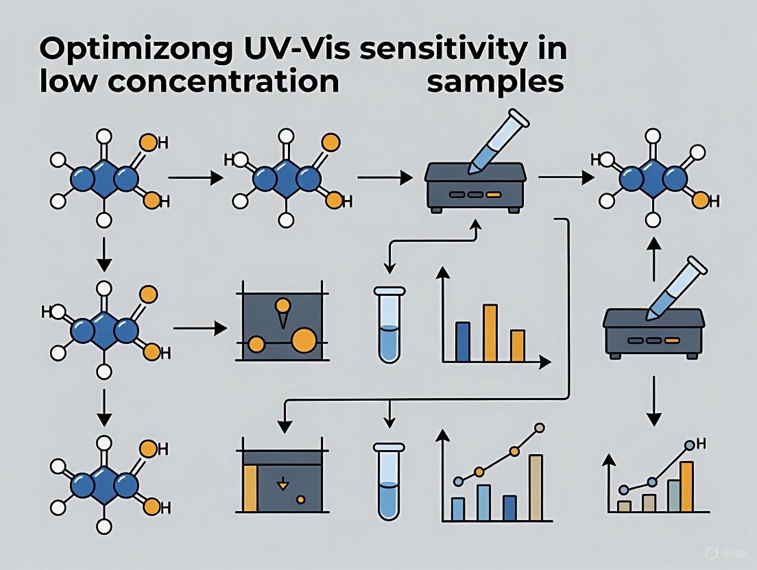
Chemical Derivatization Strategies for Analytes Lacking Strong Chromophores
FAQs and Troubleshooting Guides
Frequently Asked Questions (FAQs)
Q1: Why is chemical derivatization necessary for analyzing compounds with weak or no chromophores?
Chemical derivatization is required because many analytical techniques, particularly HPLC-UV/Vis, rely on a compound's ability to absorb ultraviolet or visible light for detection. Analytes lacking strong chromophores (UV-absorbing groups) produce poor or no signal with standard UV/Vis detectors. Derivatization chemically modifies the target analyte to introduce a strong chromophore or fluorophore, thereby significantly enhancing detection sensitivity and selectivity [45] [46] [47]. This process is crucial for accurately quantifying compounds like triterpenoids, sulforaphane, and various pharmaceuticals in complex matrices such as biological and environmental samples [45] [46] [48].
Q2: What are the main functional groups targeted for derivatization, and which reagents are commonly used?
The most frequently targeted functional groups are hydroxyl (-OH) and carboxyl (-COOH), which are common in many analytes lacking chromophores, such as triterpenoids and alcohols [45] [49]. Other groups include amines and thiols.
Table 1: Common Derivatization Reagents and Their Targets
| Functional Group | Derivatization Reagent | Key Characteristics of Derivative |
|---|---|---|
| Hydroxyl (-OH) [45] | Acyl chlorides (e.g., Benzoyl Chloride) [45] | Introduces a chromophore for UV detection. |
| Rhodamines [45] | Can introduce fluorophores for fluorescent detection. | |
| Isocyanates [45] | Used for strong acylation. | |
| Carboxyl (-COOH) [45] | Amines [45] | Used to target the carboxyl group. |
| Isothiocyanate (-N=C=S) [46] | Thiols (e.g., 2-Naphthalenethiol) [46] | Forms a stable dithiocarbamate ester with strong UV absorption. |
| Various active H (e.g., -OH, -COOH, -NH) [49] | Silylation reagents (e.g., for GC) [49] | Replaces active hydrogens to increase volatility and thermal stability for Gas Chromatography. |
Q3: What is the difference between pre-column and post-column derivatization?
The choice between pre-column and post-column derivatization is a key methodological decision [47].
- Pre-column derivatization is performed before the sample is injected into the chromatographic system. It can improve the chromatographic behavior and separation of analytes. However, it may require reaction time and the separation of unreacted reagents [47].
- Post-column derivatization occurs after the analytes have been separated by the column but before they reach the detector. This method is primarily used to enhance detectability and requires that the derivatization reaction be rapid and that the eluent is compatible with the reaction conditions without interfering with detection [47].
Q4: My derivatization reaction yield is low. What could be the cause?
Low derivatization yield can stem from several factors related to the reaction conditions [46]:
- pH and Buffer: The reaction must be conducted at an optimal pH. For example, the derivatization of sulforaphane with 2-Naphthalenethiol is best performed at pH 7.4 [46].
- Reagent Concentration: Using an insufficient amount of derivatization reagent. An excess of reagent is often required to drive the reaction to completion and achieve high yield [45].
- Temperature and Time: The reaction may require a specific temperature and a sufficient incubation period to proceed efficiently. For instance, some reactions require heating at 80°C for 2 hours [45], while others are optimal at 37°C for 60 minutes [46].
- Catalyst or Co-solvent: Some reactions require a specific environment, such as a weak alkaline medium provided by pyridine, to proceed effectively and prevent reagent hydrolysis [45].
Q5: How can I troubleshoot high background noise or interfering peaks in my chromatogram after derivatization?
Interfering peaks are often due to side reactions or excess reagent [21] [47].
- Cleanup: Implement a sample cleanup step (e.g., solid-phase extraction, liquid-liquid extraction) after derivatization to remove excess derivatization reagents or by-products that can cause high background noise [45] [48].
- Reaction Specificity: Ensure the derivatization reagent is specific to your analyte and does not react with other components in the sample matrix [47].
- Purity: Check the purity of your solvents and reagents, as impurities can introduce unexpected peaks [21].
Troubleshooting Guide
Table 2: Common Derivatization Issues and Solutions
| Problem | Potential Causes | Recommended Solutions |
|---|---|---|
| Low or No Signal | • Analyte lacks chromophore.• Derivatization reaction failed or is incomplete.• Wrong detection wavelength. | • Confirm the derivatized product's absorption maximum.• Optimize reaction conditions (pH, time, temperature, reagent excess) [46].• Use a reagent that introduces a strong chromophore [45]. |
| Poor Chromatographic Separation | • Derivatization may have altered analyte polarity insufficiently.• Matrix interference. | • Choose a derivatizing agent that significantly changes the analyte's hydrophobicity [49].• Improve sample cleanup prior to analysis [48]. |
| Low Reaction Yield | • Suboptimal pH, temperature, or time.• Insufficient reagent.• Unsuitable reaction medium. | • Systematically optimize all reaction parameters [46].• Use a large excess of derivatization reagent [45].• Use a catalyst or change the solvent (e.g., use pyridine for acylation) [45]. |
| Artifact Peaks or High Background | • Side reactions with matrix.• Decomposition of reagent or product.• Unreacted reagent. | • Purify the sample after derivatization [45].• Ensure stability of the derivative and store it appropriately.• Use high-purity reagents. |
Experimental Protocols and Data
Detailed Protocol: Derivatization of Sulforaphane with 2-Naphthalenethiol for HPLC-UV/Vis Analysis
This protocol is adapted from a study that developed a highly sensitive method for quantifying sulforaphane in rat plasma [46].
1. Principle: The isothiocyanate (-N=C=S) group of sulforaphane reacts with the thiol group of 2-Naphthalenethiol (2-NT) to form a stable dithiocarbamate ester derivative (2-NT-SFN). This derivative has strong UV absorption, allowing for sensitive detection at 234 nm [46].
2. Reagents and Materials:
- Sulforaphane (SFN) standard
- 2-Naphthalenethiol (2-NT)
- Acetonitrile (HPLC grade)
- Phosphate buffer (pH 7.4)
- Formic acid
- Kinetex C18 column (150 mm × 4.6 mm, 5 μm)
3. Step-by-Step Procedure: a. Derivatization Reaction:
- Prepare a solution of 0.3 M 2-NT in acetonitrile [46].
- Mix the sample (e.g., plasma extract containing SFN) with the 2-NT solution and phosphate buffer (pH 7.4) [46].
- Incubate the mixture at 37°C for 60 minutes to complete the derivatization [46].
b. HPLC-UV/Vis Analysis:
- Mobile Phase: Use 0.1% formic acid in water (Solvent A) and acetonitrile with 0.1% formic acid (Solvent B) [46].
- Elution: Perform gradient elution. The specific gradient should be optimized, but a typical run time for this method is around 10-15 minutes, with 2-NT-SFN eluting at approximately 7.5 minutes [46].
- Detection: Set the UV/Vis detector to 234 nm [46].
- Injection: Inject the derivatized sample onto the HPLC system.
4. Validation Data: The method was validated for the analysis of sulforaphane in biological samples [46]:
- Linearity: The calibration curve was linear over the range of 10–2000 ng/mL (R² > 0.999) [46].
- Accuracy and Precision: Intra- and inter-assay accuracy were within ±10% of the nominal value, and precision (%RSD) was <15% [46].
- Sensitivity: The lower limit of quantification (LLOQ) was 10 ng/mL [46].
Detailed Protocol: Benzoylation of Hydroxyl-Containing Triterpenoids
This protocol is typical for the analysis of compounds like astragaloside IV and ginsenosides, which have multiple hydroxyl groups but lack strong chromophores [45].
1. Principle: Acyl chloride reagents, such as benzoyl chloride (BC), react with nucleophilic hydroxyl groups (-OH) on the triterpenoid under basic conditions to form UV-absorbing esters [45].
2. Reagents and Materials:
- Triterpenoid standard (e.g., Astragaloside IV)
- Benzoyl Chloride (BC)
- Anhydrous Pyridine
3. Step-by-Step Procedure: a. Derivatization Reaction:
- Dissolve the triterpenoid sample in anhydrous pyridine. Pyridine acts as both a solvent and a base to scavenge the acid (HCl) produced during the reaction [45].
- Add a significant excess (e.g., more than 10-fold) of benzoyl chloride to the solution [45].
- Heat the reaction mixture in a water bath at 80°C for 2 hours to ensure complete derivatization of all hydroxyl groups [45].
b. Analysis:
- The derivatized product can be analyzed by HPLC-UV. The introduced benzoyl group allows for sensitive detection at UV wavelengths around 230 nm or 254 nm [45].
- A cleanup step (e.g., using solid-phase extraction) may be necessary before HPLC injection to remove excess reagents and reaction by-products [45].
Workflow and Signaling Pathways
The following diagram illustrates the logical decision-making workflow for selecting and optimizing a chemical derivatization strategy.
The Scientist's Toolkit: Research Reagent Solutions
Table 3: Essential Reagents and Materials for Derivatization
| Item | Function/Application | Key Considerations |
|---|---|---|
| 2-Naphthalenethiol (2-NT) [46] | Derivatization of isothiocyanate groups (e.g., in Sulforaphane). Introduces a strong chromophore for UV detection. | Offers high molar absorptivity and allows detection at longer wavelengths (≥230 nm) to reduce matrix interference [46]. |
| Benzoyl Chloride (BC) [45] | Acylation reagent for hydroxyl groups in compounds like triterpenoids. Introduces a benzoyl chromophore. | Requires a basic environment (e.g., pyridine) and excess reagent for high yield. Reaction often requires heating [45]. |
| Pyridine [45] | Solvent and base for acylation reactions with acid chlorides. Neutralizes HCl produced, driving the reaction forward. | Must be anhydrous. Acts as a catalyst and prevents hydrolysis of the derivatization reagent [45]. |
| Quartz Cuvettes [21] [50] | Sample holder for UV-Vis spectroscopy measurements. | Essential for UV region measurements due to high transparency. Must be kept clean and handled with gloves to avoid fingerprints [21]. |
| Solid-Phase Extraction (SPE) Cartridges [45] [48] | Sample cleanup to isolate the analyte or derivatized product from a complex matrix and remove interfering substances. | Crucial for analyzing samples in biological or environmental matrices to reduce background noise and protect the analytical column [48]. |
Solving Common Challenges: A Practical Guide to Optimization and Error Reduction
Optimizing Cuvette Selection and Cleaning to Minimize Contamination
A foundational guide for researchers aiming to maximize the sensitivity and accuracy of UV-Vis spectroscopy for low-concentration samples.
The integrity of your UV-Vis data for low-concentration samples—such as nucleic acids, dilute proteins, or environmental pollutants—is fundamentally tied to two critical, yet often overlooked, practices: the correct selection of cuvettes and their meticulous cleaning. Contamination or an inappropriate cuvette can introduce significant errors, obscuring the true signal of your valuable samples. This guide provides targeted protocols and troubleshooting advice to safeguard your data quality.
Frequently Asked Questions (FAQs)
Q1: How does cuvette material choice impact measurements of low-concentration samples?
The cuvette material dictates the range of light that can pass through to your sample. Using the wrong material can block the specific wavelength you need to measure, drastically reducing sensitivity and leading to inaccurate readings.
- UV-Transparent Quartz (Fused Silica): This is the gold standard for low-concentration assays in the UV range. It offers excellent transmission from deep UV (∼190 nm) through the visible and into the near-infrared spectrum [51]. This is absolutely essential for quantifying DNA (260 nm), RNA (280 nm), and proteins (280 nm) without signal loss [51] [52]. Quartz also exhibits very low autofluorescence, which is critical for sensitive fluorescence measurements where background glow can mask weak signals [51].
- Optical Glass: Suitable only for measurements in the visible range (∼340 nm and above) [51] [53]. It blocks UV light, making it useless for nucleic acid or protein quantification at their standard UV wavelengths. Its moderate autofluorescence also makes it less ideal for sensitive fluorescence work [51].
- Plastic (PS, PMMA): Generally transparent only in the visible range (380-850 nm) and blocks UV light [51] [53]. They often have high autofluorescence and poor chemical resistance, making them suitable only for disposable, educational, or simple colorimetric assays in the visible spectrum [51].
Table 1: Cuvette Material Properties and Their Impact on Sensitivity
| Feature | Quartz (Fused Silica) | Optical Glass | Plastic (e.g., PS) |
|---|---|---|---|
| UV Transmission | Excellent (down to ~190 nm) [51] | Poor (blocks below ~340 nm) [51] [53] | None (blocks below ~380 nm) [51] [53] |
| Autofluorescence | Very Low [51] | Moderate [51] | High [51] |
| Best Use Cases | UV-Vis absorbance, fluorescence, solvent use [51] | Visible-light-only absorbance [51] | Teaching labs, disposable visible assays [51] |
| Impact on Low-Concentration Samples | Prevents signal loss at UV wavelengths; minimizes background noise. | Can completely obliterate UV signals from dilute nucleic acids/proteins. | High background fluorescence can swamp weak signals. |
Q2: What is the relationship between path length and sensitivity for dilute samples?
According to the Beer-Lambert Law (A=εlc), the absorbance (A) is directly proportional to the path length (l) [44] [52]. For low-concentration samples, a longer path length increases the interaction distance between light and the analyte, thereby increasing the measured absorbance and improving the signal-to-noise ratio [53].
- Standard Path Length: 10 mm is the global calibration standard and a good starting point [51].
- Long Path Length Cuvettes: For very dilute samples, cuvettes with path lengths of 20 mm, 50 mm, or even 100 mm are available to enhance sensitivity [52] [53].
- Micro-Volume Considerations: While microvolume measurements (using 1 µL droplets) are excellent for sample conservation, they use a very short path length (typically 0.2-1 mm) [44]. This makes them best suited for samples with relatively high concentration, like DNA from mini-preps. For truly dilute samples, the signal in a microvolume mode may be too weak, making a longer path length cuvette the better choice [44].
Q3: What is the definitive cleaning protocol to prevent cross-contamination and residue buildup?
A rigorous and sequential cleaning protocol is non-negotiable for high-sensitivity work.
- Rinse Immediately: After use, immediately rinse the cuvette with a generous amount of the solvent used for your sample (e.g., water, buffer, or ethanol) to remove the bulk of the analyte [27].
- Wash with Rinsing Agent: Rinse thoroughly with a pure, volatile solvent such as acetone, isopropanol (IPA), or deionized water [27]. This helps remove residual solvent and any organic contaminants.
- Final Rinse: Perform a final rinse with high-purity solvent (e.g., HPLC-grade) that will be used in your next sample or measurement [27]. This ensures no cleaning solvent residues remain that could contaminate the next reading.
- Air Dry: Allow the cuvette to air-dry upside down on a clean lint-free wipe or in a rack. Avoid wiping the optical windows with anything that could scratch them. If wiping is necessary, use a certified lint-free tissue or an inert gas duster.
- Storage: Store clean, dry cuvettes in a closed container to prevent dust accumulation.
Table 2: Cleaning Solvent Compatibility Guide
| Chemical | Quartz Cuvette | Optical Glass Cuvette | Plastic Cuvette |
|---|---|---|---|
| Acetone | + [51] | + [51] | – [51] |
| Ethanol / IPA | + [51] | + [51] | + [51] |
| Hydrochloric Acid (36%) | + [51] | + [51] | + [51] |
| Sodium Hydroxide | + (Short-term, room temp) [51] | – (Causes corrosion) [51] | + [51] |
| DMSO | + [51] | + [51] | + [51] |
| Chloroform | + [51] | + [51] | – [51] |
| Hydrofluoric Acid (any %) | – (Severely Damages) [51] | – (Severely Damages) [51] | + [51] |
Key: + = Suitable, – = Not Recommended / Damaging
Troubleshooting Guides
Problem: Consistently High Background Absorbance or Unusual Spectral Peaks
This is a classic sign of cuvette contamination or improper cleaning.
Possible Causes and Solutions:
- Cause 1: Residual Contaminants from Incomplete Cleaning.
- Solution: Implement the multi-step cleaning protocol outlined in FAQ #3. Ensure the final rinse is with the same high-purity solvent that will be used for your sample.
- Cause 2: Scratched or Etched Optical Windows.
- Solution: Visually inspect cuvettes under bright light. Scratches scatter light, increasing apparent absorbance. Etching from strong bases (e.g., NaOH) creates a frosted surface with the same effect [51]. Prevention is key: avoid abrasive contact and do not use strong bases for prolonged periods.
- Cause 3: Cuvette Material Not Suited for the Wavelength.
- Solution: Confirm you are using a quartz cuvette for any measurements below 300 nm. A simple test is to run a baseline or blank scan with your clean, empty cuvette or one filled with pure solvent. A quartz cuvette should show a flat, low baseline in the UV, while glass or plastic will show a steep rise in absorbance as the wavelength decreases.
Problem: Inconsistent Replicate Measurements or Poor Data Reproducibility
This often points to issues with cuvette handling, meniscus effects, or volume inconsistencies.
Possible Causes and Solutions:
- Cause 1: Meniscus Effects Causing Path Length Variability.
- Solution: A concave meniscus acts as a lens, creating a non-uniform path length [54]. Always position the cuvette in the holder with the same orientation (e.g., marked window facing the light source). Fill the cuvette sufficiently so that the light beam passes well below the meniscus.
- Cause 2: Inconsistent Fill Volumes.
- Solution: Use a calibrated pipette to ensure the same sample volume is used for every replicate. The path length, and therefore the absorbance, is dependent on the fill height in a cuvette [54].
- Cause 3: Cuvette Not Placed Consistently in Holder.
- Solution: Ensure the cuvette is seated firmly in the same position in the holder every time. Frosted or marked sides can help maintain consistent orientation.
The Scientist's Toolkit: Essential Research Reagent Solutions
Table 3: Key Reagents and Materials for Cuvette-Based Experiments
| Item | Function / Application | Critical Notes for Low-Concentration Work |
|---|---|---|
| Quartz Cuvettes (10 mm path) | Standard for UV-Vis absorbance measurements [51]. | Ensure they are "UV-grade" fused silica for best UV transmission down to 190 nm [51]. |
| Fluorescence Quartz Cuvettes (4 windows) | Essential for fluorescence spectroscopy [51] [53]. | Four polished windows allow for 90° detection. Low autofluorescence is critical for signal-to-noise ratio [51]. |
| HPLC-Grade Solvents | For sample preparation, dilution, and final cuvette rinsing. | High purity minimizes UV-absorbing contaminants that can elevate background. |
| Spectrophotometer Cuvette Standards | For instrument validation and wavelength calibration. | Use NIST-traceable standards to ensure instrument accuracy before analyzing precious low-concentration samples. |
| Lint-Free Laboratory Wipes | For handling and drying cuvettes. | Prevents scratches on optical surfaces and avoids lint contamination. |
Controlling Aggregation and Solvent Effects in Dilute Solutions
Molecular aggregation and solvent effects are critical phenomena in dilute solutions that can significantly influence data obtained from characterization techniques like Ultraviolet-Visible (UV-Vis) spectroscopy. Aggregation occurs when molecules associate through interactions such as π-π stacking, hydrophobic effects, or hydrogen bonding, altering their electronic environment. Simultaneously, the solvent environment can modulate molecular conformation, stability, and spectral properties through polarity, hydrogen-bonding capacity, and specific solvent-solute interactions. For researchers aiming to optimize UV-Vis sensitivity for low-concentration samples, controlling these factors is paramount to obtaining accurate, reproducible, and interpretable data. This guide provides targeted troubleshooting and methodological support to address these specific experimental challenges.
Troubleshooting Guides
Common Problems and Solutions
| Problem Category | Specific Issue | Possible Causes | Recommended Solutions |
|---|---|---|---|
| Sample Preparation | Unexpected peaks in spectrum | Contaminated sample or unclean cuvettes [21] | Thoroughly wash cuvettes/substrates; handle only with gloved hands [21]. |
| Signal too high (Absorbance >1.0) | Sample concentration too high [21] | Dilute sample; use a cuvette with a shorter path length [21]. | |
| Light scattering | Particulates, bubbles, or molecular aggregates in solution [55] | Filter sample; ensure complete dissolution; degas solution [55]. | |
| Solvent Effects | Spectral shifts (vs. reference) | Solvent polarity differences (solvatochromism) [55] | Use the same solvent for sample and reference; document solvent identity [55]. |
| Obscured sample absorption | Solvent absorbs in same spectral region [55] | Select a UV-compatible solvent that does not overlap with sample's λmax (e.g., avoid acetone for carbonyl analysis) [55]. | |
| Altered aggregation state | Solvent hydrogen-bonding capacity [56] | Control solvent composition precisely; be aware of co-solvent complexes (e.g., DMSO/water) [57]. | |
| Instrumentation & Measurement | Low transmission signal | High concentration scattering light [21]; Damaged optical fibers [21] | Reduce concentration/path length; check and replace damaged fibers [21]. |
| Noisy or drifting baseline | Light source not stabilized [21] | Allow lamp warm-up time (20 mins for tungsten halogen/arc lamps) [21]. | |
| Non-linear Beer-Lambert response | Absorbance outside linear range (typically 0.1-1.0) [55]; Instrumental stray light [55] | Ensure absorbance is within 0.1-1.0; use appropriate dilutions [55]. |
Advanced Aggregation Analysis
For a deeper investigation into aggregation, the UV-Vis spectrum can be deconvoluted to quantify different aggregate species. The table below summarizes findings from a study on reactive dyes, illustrating how aggregation distribution changes with concentration [58].
| Dye | Concentration (mM) | Monomer (%) | Dimer (%) | Higher Aggregates (%) |
|---|---|---|---|---|
| O-13 | 1 | 33.2 | 31.8 | 27.0 |
| 10 | 32.0 | 30.0 | 30.4 | |
| 100 | 30.6 | 28.8 | 33.5 | |
| R-24:1 | 1 | 40.8 | 38.1 | 21.1 |
| 10 | 40.4 | 38.1 | 21.5 | |
| 100 | 39.4 | 38.5 | 22.1 | |
| R-218 | 1 | 41.4 | 37.8 | 20.8 |
| 10 | 39.2 | 39.9 | 20.9 | |
| 100 | 34.0 | 42.8 | 23.2 |
This data shows that different molecular structures exhibit distinct aggregation profiles. For instance, R-218 shows a strong concentration dependence, with dimer populations increasing significantly from 37.8% to 42.8% as concentration rises from 1 mM to 100 mM [58]. The molecular structure, particularly the size and position of non-conjugated side chains, plays a crucial role; a smaller side chain linked at the β-position of a naphthalene ring (as in O-13) can promote stronger aggregation compared to bulkier α-linked chains [58].
Experimental Protocols
Protocol 1: Assessing Aggregation in Concentrated Solutions via UV-Vis
Purpose: To characterize the aggregation behavior of a chromophore (e.g., a reactive dye or conjugated polymer) in aqueous solution across a wide concentration range [58].
Materials:
- Test compound (e.g., C.I. Reactive Orange 13, Red 24:1, or R-218).
- Deionized water or chosen buffer.
- UV-Vis spectrometer.
- Cuvettes with varying path lengths (e.g., 10 mm, 1 mm, 0.1 mm, and 0.01 mm).
- Volumetric flasks and pipettes.
Procedure:
- Sample Preparation: Prepare a stock solution of the dye at a high concentration (e.g., 100 mM). Serially dilute this stock to create a series of solutions spanning concentrations from 10⁻³ mM to 100 mM.
- Cuvette Selection: Based on the expected absorbance, select an appropriate cuvette path length to ensure the measured absorbance falls within the instrument's linear range (typically 0.1-1.0). For highly concentrated solutions (e.g., >1 mM), a short path length cuvette (e.g., 0.01 mm) is essential [58].
- Spectral Acquisition: Collect the UV-Vis absorption spectrum for each concentration-step solution using the corresponding optimal path length cuvette.
- Data Analysis: Normalize the spectra for comparative analysis. Observe shifts in the wavelength of maximum absorption (λmax), changes in the spectral shape, and the appearance of new peaks. The distribution of monomers, dimers, and higher aggregates can be quantified by deconvoluting the absorption curves using a Gaussian curve-fitting program [58].
Protocol 2: Investigating Solvent-Polymer Interactions
Purpose: To study the effect of solvent polarity and hydrogen-bonding on the solution properties and aggregation of a polymer like Poly(Vinyl Alcohol) or a conjugated polythiophene [57] [56].
Materials:
- Polymer (e.g., PVA, Mw = 124,000–186,000, or isothiouronium polythiophene).
- Solvents of different polarities and H-bonding capacities (e.g., Water, DMSO, 2-propanol, Tetrahydrofuran (THF), 1,4-Dioxane (DI)).
- UV-Vis spectrometer and Fluorometer.
- Standard glassware and heating/stirring equipment.
Procedure:
- Solution Preparation: Prepare a series of solvent mixtures with varying volume fractions (e.g., DMSO/water from ΦDMSO = 0 to 1). Dissolve the polymer in each solvent mixture at a fixed concentration, ensuring complete dissolution, potentially with heating (e.g., 95°C for PVA) [57].
- Spectroscopic Measurement: Allow solutions to equilibrate at the measurement temperature (e.g., 25°C). Acquire UV-Vis absorption and fluorescence emission spectra for each sample.
- Data Interpretation:
- Analyze the Stokes shift (difference between absorption and emission maxima) as a function of solvent polarity [56].
- Identify solvent compositions that lead to phenomena like cononsolvency (where a polymer becomes less soluble in a mixed solvent than in either pure solvent), often indicated by increased turbidity, phase separation, or changes in spectral intensity [57].
- For PVA in DMSO/water, note that the strongest aggregation and fastest gelation often occur near a specific DMSO mole fraction (X~0.28), attributed to the formation of DMSO/(water)₂ complexes [57].
Frequently Asked Questions (FAQs)
Q1: My sample is at a very low concentration, but the absorbance is still too high. What can I do? A: The most straightforward solution is to reduce the concentration further. If that is not possible due to detection limits, switch to a cuvette with a shorter path length. Reducing the path length from 10 mm to 1 mm decreases the absorbance by a factor of 10, bringing it into the optimal range without altering your sample composition [21].
Q2: How does solvent choice specifically affect my UV-Vis spectrum? A: The solvent can cause several effects:
- Spectral Shifts: Solvent polarity can induce bathochromic (red) or hypsochromic (blue) shifts due to stabilization of the ground vs. excited state [55].
- Baseline Interference: A solvent with high absorbance in your sample's spectral region will obscure the signal [55].
- Molecular Aggregation: Solvent quality (good vs. poor) and hydrogen-bonding capacity can promote or suppress molecular self-assembly, leading to new spectral peaks or changes in intensity [57] [56]. Always match the solvent between sample and blank and document it precisely.
Q3: I see unexpected peaks in my spectrum. Could this be due to aggregation? A: Yes, the formation of aggregates (dimers, trimers, etc.) often results in new absorption bands that are distinct from the monomer. These can appear as shoulders, or as new peaks that are either blue-shifted (H-aggregates) or red-shifted (J-aggregates) relative to the monomer peak [58]. Running a concentration-dependent series can help confirm this, as the aggregate peaks typically become more prominent at higher concentrations.
Q4: What is cononsolvency and how might it impact my experiment? A: Cononsolvency is a phenomenon where a polymer dissolves in each of two pure solvents but precipitates or aggregates in certain mixtures of those two solvents. For example, Poly(Vinyl Alcohol) can exhibit this behavior in certain DMSO/water mixtures [57]. This can lead to unexpected turbidity, light scattering, and altered spectra, complicating data interpretation. Being aware of such system-specific behaviors is crucial for experimental design.
The Scientist's Toolkit: Essential Research Reagents & Materials
| Item | Function & Rationale |
|---|---|
| Quartz Cuvettes | Essential for UV-Vis measurements below ~300 nm, as quartz has high transmission in the UV region. Reusable cuvettes with precise path lengths are ideal [21]. |
| Short Path Length Cuvettes (e.g., 0.01-0.1 mm) | Critical for analyzing highly concentrated solutions (e.g., dye aggregation studies) without exceeding the instrument's absorbance limit, ensuring adherence to the Beer-Lambert law [58]. |
| Dimethyl Sulfoxide (DMSO) | A common, electrochemically stable solvent for dissolving compounds with low water solubility. It forms complexes with water, which can influence polymer conformation and aggregation [57] [59]. |
| Solvent Dilution Device (SDD) | A device used in techniques like Ion Chromatography to dilute organic solvents in the sample stream before the suppressor, minimizing baseline disruption and improving analyte recovery [59]. |
| 1,4-Dioxane (DI) | A useful cosolvent in fundamental studies of hydrogen-bonding. It is a strong hydrogen-bond acceptor that can disrupt specific polymer-solvent interactions, helping to elucidate aggregation mechanisms [56]. |
Strategies for Managing Baseline Drift and Scattering in Biological Matrices
Troubleshooting Guide: Resolving Common UV-Vis Issues in Biological Analysis
FAQ 1: Why does my baseline drift upward during a gradient UV-Vis analysis of biological samples?
Answer: Baseline drift in gradient analysis arises from changing mobile phase composition and biological matrix effects.
- Mobile Phase Imbalance: As the proportion of organic solvent changes during a gradient, the UV absorbance of the mobile phase itself changes, causing the baseline to shift [60]. Biological samples can exacerbate this with endogenous materials.
- Bubbles and Contamination: Air bubbles in the flow cell or accumulated contaminants from biological matrices (proteins, lipids) on optical surfaces cause gradual baseline upward drift [60].
- Temperature Fluctuation: Uncontrolled temperature affects detector stability, particularly problematic for sensitive biological assays [60].
Solutions:
- Mobile Phase Matching: Ensure the aqueous and organic mobile phases have matched absorbance at your detection wavelength by fine-tuning their composition [60].
- Thorough Degassing: Use inline degassers or helium sparging to prevent bubble formation [60].
- Systematic Cleaning: Regularly clean the instrument flow path and replace mobile phase filters to prevent accumulation of biological contaminants [60].
FAQ 2: How can I reduce spectral scattering from my turbid biological samples?
Answer: Scattering from turbid samples obscures true absorbance and is a common challenge with biological lysates, cell suspensions, or protein aggregates.
- Sample Clarification: Centrifuge or filter samples using a 0.2 µm or 0.45 µm syringe filter to remove particulate matter that causes light scattering [61].
- Pathlength Selection: Use a shorter pathlength cuvette (e.g., 1 mm instead of 10 mm) to reduce the scattering interaction volume for dense biological suspensions [5].
- Blank Correction: Use a blank that closely matches the sample matrix, including any clarifying agents, to correct for residual scattering background [5].
FAQ 3: What causes high noise and low sensitivity when measuring low-concentration analytes in plasma?
Answer: This is primarily caused by matrix effects from the complex biological sample and instrumental limitations.
- Biological Matrix Effects: Endogenous compounds like salts, lipids, peptides, and metabolites co-elute with your analyte, suppressing the signal and increasing baseline noise [62].
- Stray Light: Stray light inside the spectrophotometer lowers apparent absorbance readings and reduces the linear dynamic range, severely impacting low-concentration quantitation [63].
- Improper Sample Containers: Using plastic cuvettes for UV measurements is inappropriate as plastic absorbs UV light; use quartz cuvettes for UV analysis [5].
Solutions:
- Sample Cleanup: Employ protein precipitation, solid-phase extraction (SPE), or liquid-liquid extraction to remove interfering matrix components before analysis [62].
- Stray Light Check: Test for stray light using certified optical filters. Keep the instrument's optics clean and free from dust [61] [63].
- Wavelength Selection: Choose a detection wavelength that minimizes interference from biological matrix components [60].
Experimental Protocols for Baseline and Scattering Management
Protocol 1: Blank Correction and Baseline Stabilization
Purpose: To acquire a stable, flat baseline by accounting for solvent and matrix contributions [5] [61].
Procedure:
- Prepare the Blank: Use the same solvent and buffer used to prepare your biological sample. For culture media, use sterile media alone [5].
- Baseline Scan: Before measuring any samples, place the blank in the spectrophotometer and perform a baseline correction or scan. This stores the reference baseline [5].
- Measure Samples: Replace the blank with your sample and perform the measurement. The instrument will subtract the blank baseline automatically [5].
- Verification: Periodically re-measure the blank to ensure the baseline has not drifted over time.
Protocol 2: Sample Clarification for Turbid Biological Extracts
Purpose: To remove light-scattering particles from biological suspensions (e.g., tissue homogenates, cell lysates) [61].
Procedure:
- Pre-Clearance: Centrifuge the sample at 10,000 × g for 10 minutes to pellet large debris.
- Filtration: Pass the supernatant through a 0.2 µm pore size syringe filter (non-protein binding PVDF or nylon membrane is recommended) [61].
- Control: Ensure the blank solution is passed through the same type of filter to account for any potential leachates or absorbance from the filter membrane.
- Analysis: Proceed with UV-Vis measurement using the clarified filtrate.
Table 1: Key Instrument Parameters Affecting Baseline and Sensitivity
| Parameter | Optimal Condition | Impact of Deviation | Reference |
|---|---|---|---|
| Stray Light | < 0.1% (at low wavelength) | Reduces apparent absorbance, compresses linear range | [63] |
| Pathlength | 10 mm (standard); 1 mm for turbid samples | Shorter path reduces scattering but also signal intensity | [5] |
| Bandwidth | ≤ 2 nm (for defined peaks) | Wider bandwidth reduces resolution of fine features | [63] |
| Absorbance Range | 0.1 - 1.0 AU (for quantitation) | Values >1 AU have low light throughput, high noise | [5] |
Table 2: Common Biological Matrix Components Causing Interference
| Matrix Component | Primary Interference | Suggested Mitigation Strategy | [62] |
|---|---|---|---|
| Proteins & Peptides | Light scattering, baseline drift | Protein precipitation, ultrafiltration | |
| Lipids & Phospholipids | Severe ion suppression, scattering | Liquid-liquid extraction, SPE | |
| Salts & Ions | Altered viscosity, refractive index | Dilution, buffer exchange, dialysis | |
| Urea & Metabolites | Variable background absorbance | Sample cleanup, derivative spectroscopy |
Workflow Visualization: Managing Baseline and Scattering
Baseline Drift Troubleshooting Path
Scattering Mitigation Workflow
Research Reagent Solutions Toolkit
Table 3: Essential Materials for Managing Baseline and Matrix Effects
| Reagent / Material | Function / Purpose | Application Note |
|---|---|---|
| Quartz Cuvettes | Optically transparent down to 200 nm; required for UV analysis. | Preferable to plastic or glass which absorb significantly in the UV range [5]. |
| Syringe Filters (0.2 µm PVDF) | Removal of particulate matter from biological samples to reduce scattering. | Use a low-protein binding membrane to prevent analyte loss [61]. |
| HPLC-Grade Solvents | High-purity solvents with low UV absorbance for minimal baseline noise. | Use fresh solvents daily and purchase in small quantities to ensure freshness [60]. |
| Solid Phase Extraction (SPE) Cartridges | Cleanup of complex biological samples to remove interfering matrix components. | Effective for removing phospholipids and proteins that cause ion suppression [62]. |
Fine-Tuning Concentration and Pathlength to Avoid Saturation and Ensure Detectability
Troubleshooting Guide: Resolving Common UV-Vis Sensitivity Issues
This guide addresses specific, common problems researchers encounter when trying to optimize UV-Vis measurements for low-concentration samples.
My absorbance readings are consistently too high, and the peak tops appear flattened. What should I do?
This is a classic sign of peak saturation, where the analyte concentration is too high for the detector to accurately measure [64] [65].
- Immediate Action: Dilute your sample and re-measure. The absorbance should ideally be between 0.1 and 1.0 AU for most instruments [64]. If the peak shape improves and the maximum absorbance falls within this range after dilution, saturation was the cause.
- Systematic Check: Ensure you are using the correct pathlength in your calculations. Absorbance is directly proportional to pathlength; using a standard 10 mm cuvette for a highly concentrated sample will often lead to saturation [64]. Consider using a cuvette with a shorter pathlength (e.g., 1 mm) for concentrated samples [21].
- Advanced Investigation: If using a diode array detector (DAD or PDA), check the peak purity or full spectrum. Saturation can occur at wavelengths other than your monitoring wavelength, which can still skew results [65]. A 10x dilution may be needed to avoid this type of spectral saturation [65].
My sample is very dilute, and the signal is indistinguishable from noise. How can I improve detection?
This challenge involves improving the signal-to-noise ratio for samples near the detection limit.
- Increase Pathlength: The most direct method is to use a cuvette with a longer pathlength. Doubling the pathlength will double the measured absorbance, according to the Beer-Lambert law [17].
- Concentrate the Sample: If feasible, use evaporation or another technique to increase the concentration of your analyte.
- Instrument Optimization:
- Allow the light source to warm up fully (up to 20 minutes for halogen or arc lamps) to stabilize the output [21].
- Ensure all components, including optical fibers, are properly aligned to maximize signal strength [21].
- Use high-quality, clean quartz cuvettes to maximize light throughput, especially in the UV range [64] [21].
- Wavelength Selection: If your analyte has multiple absorbance maxima, choose the one with the highest molar absorptivity (ε) for greater sensitivity [65].
My calibration curve is non-linear, leading to inaccurate quantification. What could be the cause?
A non-linear calibration curve violates the Beer-Lambert law and makes quantification unreliable.
- Check for Saturation: The most probable cause is that one or more of your standard solutions are too concentrated, leading to detector saturation. Ensure all standards and samples have absorbances below 1.0 AU [64].
- Verify Wavelength Accuracy: An incorrect wavelength setting can place you on a steep slope of the absorption spectrum, where small wavelength errors cause large absorbance changes [65] [63]. Use a holmium oxide filter or a known emission line to verify your instrument's wavelength calibration [63].
- Assess Stray Light: Stray light within the instrument becomes significant at high absorbances and causes negative deviations from linearity [63]. Instrument validation tests per USP <856> guidelines can check for this issue [66].
- Confirm Pathlength: For non-standard cuvettes (e.g., in micro-volume instruments), ensure the correct pathlength is used in your calculations [64] [67]. A discrepancy here will cause systematic errors.
When should I use a shorter pathlength versus diluting my sample?
Both strategies address high concentration, but the choice depends on your experimental constraints.
- Use a Shorter Pathlength when you have a very small sample volume, want to avoid a dilution step to save time, or need to minimize solvent use [21] [67]. This is common in micro-volume analyses.
- Dilute the Sample when you have ample sample volume, need to keep the sample in a specific cuvette for consistency, or when the pathlength adjustment on your instrument is not sufficient to bring the absorbance into the linear range.
The table below summarizes how pathlength affects the effective concentration range.
| Pathlength | Typical Use Case | Impact on Effective Concentration |
|---|---|---|
| Long (e.g., 10 cm) | Very dilute samples | Increases absorbance; improves detectability for low concentrations. |
| Standard (1 cm) | Routine measurements | Standard range for most analyses. |
| Short (1 mm to 0.07 mm) | Highly concentrated samples, micro-volume | Decreases absorbance; avoids saturation for high concentrations [21] [67]. |
How do I select the best wavelength for my assay to maximize accuracy?
Choosing the optimal wavelength is critical for both sensitivity and accuracy.
- Avoid Spectral Slopes and Saturation: Do not set your wavelength on a steep slope of the compound's UV spectrum, as minor instrument drift can cause significant absorbance shifts [65]. Also, confirm the chosen wavelength does not exhibit spectral saturation [65].
- Perform a Wavelength Scan: Conduct a full scan (e.g., 200-800 nm) of a pure standard to identify the wavelength of maximum absorbance (λmax) [64]. Using λmax provides the greatest sensitivity and is less susceptible to small wavelength inaccuracies [17].
- Maximize Selectivity: In mixtures, select a wavelength where your target analyte absorbs strongly, but where potential interferers have minimal absorption [64]. The peak purity function of a DAD/PDA detector is invaluable for this task [17] [65].
- Avoid Low UV Wavelengths: If possible, avoid using wavelengths near 210 nm, as many organic compounds and mobile phase components absorb here, reducing method selectivity [65].
Frequently Asked Questions (FAQs)
What is the ideal absorbance range for precise UV-Vis measurements?
For most modern UV-Vis spectrophotometers, the optimal absorbance range for precise and linear measurements is between 0.1 and 1.0 absorbance units (AU) [64]. Absorbance values significantly above 1.0 AU risk detector saturation and non-linearity due to factors like stray light, while values below 0.1 AU can have a poor signal-to-noise ratio [64] [63].
How is the Method Detection Limit (MDL) defined and determined?
According to the U.S. Environmental Protection Agency (EPA), the Method Detection Limit (MDL) is "the minimum measured concentration of a substance that can be reported with 99% confidence that the measured concentration is distinguishable from method blank results" [68]. It is determined through statistical analysis of replicate measurements of low-level spiked samples and method blanks, representing the lowest level at which an analyte can be reliably detected by a specific method [68].
How do pathlength and concentration relate mathematically?
The relationship is defined by the Beer-Lambert Law: ( A = εcl ), where:
- ( A ) is the measured Absorbance
- ( ε ) is the molar absorptivity (L·mol⁻¹·cm⁻¹)
- ( c ) is the concentration (mol/L)
- ( l ) is the pathlength (cm) [17] [67]
This means absorbance is directly proportional to both concentration and pathlength. Therefore, halving the pathlength has the same effect on absorbance as halving the concentration.
What are the consequences of using dirty or scratched cuvettes?
Dirty or scratched cuvettes can significantly scatter light, leading to inconsistent and erroneously high absorbance readings [64]. This introduces random error and reduces the reliability of your data. Always clean cuvettes thoroughly with appropriate solvents after use, handle them with gloves, and inspect them for scratches or chips before measurement [64] [21].
Quantitative Data Reference
The following table provides key performance data and specifications relevant to sensitivity optimization.
| Parameter | Typical Target or Acceptable Value | Importance for Sensitivity |
|---|---|---|
| Absorbance Accuracy | Varies by instrument; high-precision detectors achieve <0.2% RSD [17]. | Essential for precise quantification, especially in regulatory testing (e.g., pharmaceutical potency assays). |
| Photometric Noise | Historical benchmark is <±1 × 10⁻⁵ AU [17]. | Lower noise enables detection of smaller absorbance changes, improving detectability for low concentrations. |
| Pathlength Accuracy | Critical for micro-volume systems; fixed anchor points (e.g., 0.07 mm, 0.67 mm) ensure no drift [67]. | Directly impacts the accuracy of the concentration calculation via Beer-Lambert Law. |
| Method Detection Limit (MDL) | The minimum concentration distinguishable from blank with 99% confidence [68]. | Defines the lower bound of an assay's quantitative capability. |
Experimental Protocol: Systematic Optimization of Sensitivity
This protocol provides a step-by-step methodology to fine-tune your UV-Vis analysis for maximum sensitivity and linearity.
Principle: To empirically determine the optimal sample concentration and measurement parameters (pathlength, wavelength) that avoid detector saturation while ensuring the target analyte is detectable and quantifiable.
Materials:
- Stock Solution: A known concentration of the pure analyte.
- Appropriate Solvent: A solvent that does not absorb significantly in the wavelength range of interest [64].
- Cuvettes: Clean, matched cuvettes of various pathlengths (e.g., 10 mm, 1 mm) or a variable pathlength system [64] [21].
- UV-Vis Spectrophotometer: Calibrated and with a warmed-up light source [64] [21].
Procedure:
- Initial Scan: Perform a full wavelength scan (e.g., 200-800 nm) of a moderately diluted stock solution using a standard 10 mm pathlength. This identifies the wavelength of maximum absorbance (λmax) and checks for solvent interference [64].
- Dilution Series: Prepare a serial dilution of the stock solution to create standards across a wide concentration range.
- Absorbance Measurement: Measure the absorbance of each standard at the identified λmax using the 10 mm cuvette.
- Pathlength Adjustment: If any standard has an absorbance >1.0 AU, repeat the measurement for those concentrated standards using a shorter pathlength cuvette [21].
- Data Analysis: Plot absorbance versus concentration for all data points. The optimal working range falls within the linear portion of this curve, ideally between 0.1 and 1.0 AU. Select the combination of concentration and pathlength that places your sample measurements in this linear range.
Experimental Workflow Logic
The diagram below outlines the logical decision process for optimizing UV-Vis sensitivity.
The Scientist's Toolkit: Essential Research Reagents & Materials
| Item | Function & Importance |
|---|---|
| Quartz Cuvettes | Essential for UV-range measurements due to high transparency down to 190 nm. Reusable and must be kept meticulously clean to avoid light scattering [64] [21]. |
| Potassium Dichromate | A common standard reference material (NIST-traceable) used for verifying instrument photometric accuracy and wavelength calibration [64] [63]. |
| Holmium Oxide (Ho₂O₃) Filter | A solid-state filter with sharp, known absorption peaks used for accurate wavelength calibration of the spectrophotometer [63]. |
| High-Purity Solvents | Solvents (e.g., HPLC-grade water, acetonitrile) must be UV-transparent in the wavelength range of interest to avoid raising the baseline and introducing interference [64] [65]. |
| NIST SRM Standards | Certified reference materials (e.g., SRM 1920, 2065) used for definitive calibration of wavelength and absorbance scales in master instruments [66]. |
Addressing Interferences from Excipients and Complex Matrices like Plasma
Troubleshooting Guides
Guide 1: Addressing Excipient Interference in Drug Dissolution Testing
Excipients in solid oral dosage forms can significantly interfere with accurate drug dissolution assessment. Understanding and mitigating these effects is crucial for obtaining reliable bioavailability data.
Table: Troubleshooting Excipient Interference in UV-Vis Dissolution Testing
| Problem | Underlying Mechanism | Detection Method | Solution |
|---|---|---|---|
| Suppressed Drug Dissolution | Excipient shielding: Hydrophobic or insoluble excipients act as a physical barrier and compete for water, delaying drug wetting and dissolution [69]. | UV-Vis imaging with Raman spectroscopy to quantify drug fraction on compact surface [69]. | Increase drug-to-excipient ratio beyond a critical threshold; optimize particle size to reduce shielding [69]. |
| Abnormal Swelling & Erosion | Polymer excipients (e.g., HPMC) swell upon hydration, forming a gel layer that controls drug release by diffusion and erosion, unlike inert excipients (e.g., MCC) [70] [71]. | Real-time UV-Vis imaging at 520 nm to visually quantify changes in tablet diameter and gel layer thickness [70] [71]. | Use UV-Vis imaging to functionally characterize excipients and select types/viscosity grades (e.g., HPMC K15 M vs. K100 M) that match target release profiles [70]. |
| Misleading Bulk Concentration | Standard dissolution tests measure only the total drug in solution, missing the interplay between undissolved drug release and dissolution [70]. | UV-Vis imaging of the entire dosage form in a flow-through cell to spatially resolve release and dissolution events [70] [71]. | Implement UV-Vis liberation imaging to visualize and separately analyze drug release and dissolution processes [70]. |
Experimental Protocol: Visualizing Excipient Shielding with UV-Vis Imaging
Objective: To investigate the impact of excipient shielding on the initial dissolution rate of a drug from binary blends [69].
Sample Preparation:
- Prepare a series of binary blends of the drug (e.g., Theophylline) with a hydrophilic, water-insoluble excipient (e.g., Microcrystalline Cellulose - MCC) at varying ratios (e.g., from 10% to 50% w/w drug) [69].
- Add a standard lubricant (e.g., 1% w/w Magnesium Stearate) [71].
- Compress the blends into small compacts (e.g., 2 mm or 10 mm diameter tablets) [69] [71].
UV-Vis Imaging:
- Use a UV-Vis imaging system (e.g., Sirius SDi2) equipped with a USP IV-type flow-through cell [70] [71].
- Place the compact in the cell and perfuse with a dissolution medium (e.g., 0.01 M HCl) at a controlled temperature (37°C) and flow rate [71].
- Set the UV imaging detector to a wavelength specific to the drug (e.g., 255 nm for Theophylline) to monitor drug concentration. Use a visible wavelength (e.g., 520 nm) to simultaneously monitor physical changes like swelling and erosion [71].
Data Analysis:
- Quantify the initial drug dissolution rate from the compact by analyzing the UV signal intensity over time.
- Correlate the dissolution rate with the drug-excipient ratio and the drug particle size.
- Use Raman mapping on the compacts to independently quantify the fraction of drug presented on the compact surface and validate the shielding effect [69].
Guide 2: Mitigating Matrix Effects in Plasma Analysis
Complex biological matrices like plasma contain proteins, phospholipids, and salts that can severely interfere with UV-Vis analysis, leading to inaccurate quantification.
Table: Troubleshooting Matrix Effects from Plasma in UV-Vis Analysis
| Problem | Underlying Mechanism | Detection Method | Solution |
|---|---|---|---|
| Protein Interference | Proteins can absorb light in the UV range, cause light scattering, and bind to analytes, altering their absorption characteristics [72]. | Significant baseline drift; inconsistent absorbance readings between sample and standard in buffer; poor spike recovery [73]. | Employ Protein Precipitation (PPT) using cold organic solvents (e.g., acetonitrile, methanol) to denature and remove proteins prior to analysis [72]. |
| Phospholipid Interference | Phospholipids are a major source of matrix effects in LC-UV, causing inconsistent analyte recovery and signal suppression or enhancement [72]. | A spiking experiment showing percent recovery outside the acceptable 80-120% range [73]. | Use Hybrid Solid-Phase Extraction (SPE) cartridges designed specifically for phospholipid removal after PPT for comprehensive cleanup [72]. |
| Overall Matrix Effect | The combined effect of all interfering components, leading to reduced sensitivity and inaccurate results, especially for low-concentration analytes [72]. | Compare the analytical signal of a standard in solvent versus a standard spiked into a pre-processed plasma sample [72] [73]. | Dilute the sample with an appropriate buffer to reduce the concentration of interferents, ensuring the analyte's signal remains within the quantifiable range [73]. |
Experimental Protocol: Sample Preparation for UV-Filter Analysis in Plasma using LC-UV/MS
Objective: To simultaneously determine multiple analytes in plasma with minimal matrix interference [72].
Sample Pretreatment:
- Thaw frozen plasma samples on ice and vortex thoroughly.
- Aliquot a precise volume of plasma (e.g., 100 µL) into a microcentrifuge tube.
Protein Precipitation (PPT):
- Add a cold organic solvent (e.g., 300 µL of acetonitrile) to the plasma sample.
- Vortex the mixture vigorously for several minutes to ensure complete protein denaturation and analyte extraction [72].
- Centrifuge at high speed (e.g., 13,000 × g for 10 minutes) to pellet the precipitated proteins.
Phospholipid Removal (Hybrid SPE):
- Transfer the supernatant from the PPT step onto a preconditioned HybridSPE-PPT cartridge.
- These cartridges are designed to retain phospholipids while allowing the analytes of interest to pass through [72].
- Pass the supernatant through the cartridge by gravity or gentle centrifugation. The resulting eluate is a cleaned-up extract ready for analysis.
Analysis and Validation:
- Analyze the extract using LC-UV or LC-MS/MS.
- Validate the method by performing a spiking experiment: add a known concentration of standard to blank plasma, process it through the entire protocol, and calculate the percent recovery to confirm minimal matrix interference [73].
Frequently Asked Questions (FAQs)
Q1: My drug dissolution from a tablet is lower than expected, even though the API is highly soluble. What could be the cause? This is a classic sign of excipient shielding. Insoluble excipients like microcrystalline cellulose (MCC) can form a physical barrier around the drug particles, preventing them from contacting the dissolution medium. This also competes for the water needed for drug wetting and dissolution, thereby suppressing the initial release rate [69]. To confirm this, use UV-Vis imaging to visualize the dissolution process in real-time.
Q2: How can I visually tell if my polymer-based tablet is functioning as a controlled-release formulation? You can use UV-Vis imaging to monitor the swelling behavior and gel layer formation. Upon contact with the dissolution medium, polymers like HPMC will visibly swell. For example, studies show HPMC tablets can swell by 2.5 mm in diameter, forming a distinct gelatinous layer that controls drug release via diffusion and erosion. In contrast, tablets with MCC show minimal swelling (0.5-1 mm) and no gel layer [71]. This functional characterization is key to confirming the excipient's performance.
Q3: I see unexpected peaks and high background in my UV-Vis spectrum when analyzing a plasma sample. What should I do? This indicates strong matrix interference from plasma components like proteins and phospholipids. The first step is to implement a robust sample clean-up procedure. A highly effective strategy is to use a combination of protein precipitation (PPT) with cold organic solvent followed by passing the supernatant through a specialized HybridSPE cartridge to remove phospholipids. This two-step process significantly reduces matrix effects and minimizes interfering peaks [72].
Q4: How can I be sure that matrix effects are impacting my quantitative results? Perform a spiking experiment [73]. Add a known amount of your standard analyte to your plasma sample and process it through your analysis. Compare the measured concentration to the result obtained when the same amount of standard is diluted in a pure buffer. Calculate the percent recovery. Ideally, recovery should be 100%, but values between 80-120% are often acceptable. Recovery outside this range confirms significant matrix interference [73].
Workflow and Signaling Pathways
The Scientist's Toolkit: Research Reagent Solutions
Table: Essential Materials for Mitigating Interferences in UV-Vis Analysis
| Item | Function | Application Example |
|---|---|---|
| HybridSPE-PPT Cartridge | Selectively removes phospholipids from protein-precipitated biological samples (e.g., plasma), minimizing a major source of matrix effect [72]. | Cleaning up plasma samples prior to the analysis of UV-filters or other small molecules [72]. |
| Microcrystalline Cellulose (MCC) | A common, inert direct compression binder. Used as a model excipient to study physical shielding effects on drug dissolution [70] [69]. | Preparing binary blend compacts to investigate the critical drug-excipient ratio needed to overcome shielding [69]. |
| Hydroxypropyl Methylcellulose (HPMC) | A swellable polymer used in controlled-release matrix tablets. Its functionality (swelling, gel layer formation) can be characterized by UV-Vis imaging [70] [71]. | Formulating sustained-release tablets and visually validating drug release mechanisms (diffusion/erosion) via imaging [71]. |
| UV-Vis Imaging System (e.g., SDi2) | Provides real-time, spatially resolved visualization and quantification of drug release, dissolution, and excipient behavior (swelling, erosion) from solid dosage forms [70] [71]. | Whole-dosage form liberation testing to functionally characterize excipients and diagnose formulation performance issues [71]. |
| Acetonitrile & Methanol (HPLC Grade) | High-purity solvents used for protein precipitation (PPT) in sample preparation and as mobile phase components to avoid introducing UV-absorbing contaminants [74] [72]. | Precipitating proteins from plasma samples; preparing mobile phases for LC-UV analysis to ensure a clean baseline [72]. |
Ensuring Accuracy: Method Validation, Benchmarking, and Compliance
Frequently Asked Questions (FAQs)
How can I distinguish between LOD and LOQ in practice?
The Limit of Detection (LOD) is the lowest concentration at which an analyte can be detected but not necessarily quantified with precision. In contrast, the Limit of Quantitation (LOQ) is the lowest concentration that can be quantitatively measured with acceptable precision and accuracy [75] [76].
| Parameter | Definition | Key Distinction | Typical Signal-to-Noise |
|---|---|---|---|
| LOD | Lowest concentration that can be detected. | Confirms the analyte's presence, but not for precise quantification. | 3:1 [76] |
| LOQ | Lowest concentration that can be quantified with accuracy and precision. | Meets predefined goals for bias and imprecision for reliable measurement [75]. | 10:1 [76] |
My method generates many Out-of-Specification (OOS) results. What might be wrong?
Frequent OOS results often stem from an insufficiently robust method development phase [77]. Rushing validation for a poorly developed procedure is a common root cause. A robust procedure should be designed with a clear Analytical Target Profile (ATP), understanding critical parameters, and establishing a controlled design space to reduce variability during routine use [77].
How do I calculate LOD and LOQ using a calibration curve?
According to ICH Q2(R1), you can use the statistical method based on the calibration curve's slope (S) and the standard deviation of the response (σ) [76].
- Formulas:
- LOD = 3.3 × σ / S
- LOQ = 10 × σ / S
- Source of σ: The standard deviation (σ) can be estimated from the standard error of the regression obtained from the linear regression analysis of your calibration curve [76].
- Validation Required: These calculated values are estimates. You must experimentally confirm them by analyzing a suitable number of samples (e.g., n=6) prepared at the LOD and LOQ concentrations [76].
What is the relationship between method development and method validation?
Method development and validation are distinct but deeply connected stages within the Analytical Procedure Lifecycle [77].
- Stage 1: Procedure Design and Development: This foundational phase involves creating the method based on an ATP, identifying critical parameters, and using risk management and experimental design to build a robust, understood procedure [77].
- Stage 2: Procedure Performance Qualification (Validation): This is where the final procedure is proven to be fit for its intended use by testing the performance characteristics defined in the ATP, such as linearity, precision, and accuracy [77].
Skipping a thorough development phase often leads to a problematic validation and a method prone to OOS results later [77].
Troubleshooting Guides
Problem: Poor Linearity in Calibration Curve
Potential Causes and Solutions:
| Problem Cause | Investigation Steps | Corrective Action |
|---|---|---|
| Inadequate Concentration Range | Review the analyte's expected concentration in samples. | Ensure the calibration range covers all expected levels, from low to high, without exceeding the instrument's linear dynamic range. |
| Instrument Saturation | Check if absorbance values at the high end exceed the instrument's linear range (often ~1.0-2.0 AU for UV-Vis). | Dilute samples or prepare standards at lower concentrations to remain within the linear response region. |
| Sample or Cuvette Issues | Inspect cuvette for scratches or dirt. Ensure sample is clear and free of particles. | Use matched, clean cuvettes. Filter or centrifuge samples to remove turbidity if necessary [78]. |
Problem: Failing Precision (High %RSD)
Potential Causes and Solutions:
| Problem Cause | Investigation Steps | Corrective Action |
|---|---|---|
| Sample Preparation Variability | Audit sample preparation technique (pipetting, volumetric flasks, mixing times). | Implement standardized protocols, use calibrated pipettes, and train analysts on consistent techniques [79]. |
| Instrument Instability | Check for fluctuations in baseline noise, drift, or source lamp intensity. | Perform instrument qualification and maintenance. Ensure the system is warmed up and stable before analysis [80]. |
| Uncontrolled Environmental Factors | For unstable analytes, review storage conditions and analysis timeline. | Control environmental factors (e.g., temperature, light) and use stable reagents to improve ruggedness [79]. |
Problem: Inaccurate LOD/LOQ Values During Verification
Potential Causes and Solutions:
| Problem Cause | Investigation Steps | Corrective Action |
|---|---|---|
| Incorrect Sample Preparation | Verify the preparation of the low-concentration samples used for verification. | Carefully prepare samples at the claimed LOD/LOQ using serial dilution from a certified standard. |
| High Background Noise | Evaluate the baseline signal of the blank (mobile phase or solvent). | Identify and eliminate noise sources (e.g., contaminated solvents, unstable lamp). The signal at LOQ must be distinguishable from the blank with a S/N ≥ 10 [76]. |
| Insufficient Model Specificity | In complex matrices (e.g., water), spectral overlapping can affect accuracy [37]. | For UV-Vis, consider advanced data processing (e.g., derivative spectroscopy [37]) or separation techniques to isolate the analyte signal. |
Experimental Protocols for Key Parameters
Protocol 1: Determining LOD and LOQ via Calibration Curve
This method uses the standard error from linear regression analysis [76].
- Preparation: Prepare a minimum of six standard solutions across a range, including concentrations near the expected LOD/LOQ.
- Analysis: Analyze each standard solution in triplicate.
- Linear Regression: Plot the average response (e.g., absorbance) versus concentration and perform linear regression. Record the slope (S) and the standard error (SE) of the regression.
- Calculation:
- LOD = 3.3 × (SE / S)
- LOQ = 10 × (SE / S)
- Verification: Prepare and analyze six independent samples at the calculated LOD concentration. A peak should be detectable in all or most. Prepare and analyze six independent samples at the calculated LOQ concentration. The analysis should demonstrate a precision of ≤ 20% RSD and accuracy of 80-120% [76].
Protocol 2: Establishing Method Precision
Precision is assessed as repeatability (intra-day) and intermediate precision (inter-day, inter-analyst) [79].
- Sample Preparation: Prepare a homogeneous batch of sample at three concentration levels (low, medium, high), with six replicates at each level.
- Repeatability: Have one analyst analyze all 18 samples in a single day under the same conditions. Calculate the %RSD for the results at each concentration level.
- Intermediate Precision: Have a second analyst repeat the entire process on a different day, using a different instrument if possible.
- Acceptance: The %RSD for each concentration level should be within pre-defined limits (e.g., <2% for assay of a drug substance) [79]. Compare the results from both analysts to assess ruggedness.
Protocol 3: Demonstrating Accuracy via Recovery
This tests the method's ability to recover a known amount of analyte added to the sample [79].
- Preparation: Take a known, pre-analyzed sample (or a placebo for formulations).
- Spiking: Spike this sample with known quantities of the analyte at three levels (e.g., 80%, 100%, 120% of the target concentration), in triplicate.
- Analysis: Analyze the spiked samples using the validated method.
- Calculation: Calculate the percentage recovery for each spike level.
- % Recovery = (Measured Concentration / Theoretical Concentration) × 100
- Acceptance: The mean recovery at each level should be within established limits (e.g., 98-102%) [79].
Workflow and Relationship Diagrams
Analytical Procedure Lifecycle
LOD and LOQ Determination Workflow
The Scientist's Toolkit: Essential Research Reagent Solutions
| Item | Function in UV-Vis Analysis of Low-Concentration Samples |
|---|---|
| High-Purity Analytical Grade Reagents | Ensures minimal background interference and accurate standard preparation for calibration curves [79]. |
| Stable Certified Reference Materials | Provides the foundation for preparing accurate stock and working standard solutions, critical for linearity and accuracy studies. |
| Spectrophotometric Grade Solvents | Minimizes UV absorption in the region of interest, reducing baseline noise and improving signal-to-noise ratio for better LOD/LOQ [76]. |
| Matrix-Matching Placebo/Blank | For formulation analysis, a placebo without the active ingredient is essential for accurately assessing specificity and detecting potential interference during validation [79]. |
| Quartz Cuvettes (Matched Pair) | Provides optimal UV light transmission and ensures that any background absorbance is accounted for, improving accuracy [37]. |
Benchmarking UV-Vis Performance Against Mass Spectrometry and Chromatography
Ultraviolet-Visible (UV-Vis) spectroscopy serves as a fundamental analytical technique in research and quality control laboratories worldwide. As a technique that measures the amount of discrete wavelengths of UV or visible light absorbed by or transmitted through a sample, it provides critical information about sample composition and concentration [5]. While mass spectrometry (MS) and various chromatography methods offer advanced capabilities for complex analyses, UV-Vis remains the undisputed workhorse for many routine analytical applications, particularly in pharmaceutical quality control where its high precision (<0.2% RSD) is essential for meeting rigorous regulatory standards [17].
This technical support center focuses on optimizing UV-Vis sensitivity specifically for low-concentration sample research, providing researchers with practical guidance for maximizing performance and knowing when to transition to more sensitive techniques. We frame this discussion within the context of a broader thesis on pushing the boundaries of UV-Vis capabilities while understanding its inherent limitations compared to more sensitive detection methods.
Technical Comparison of Detection Techniques
Fundamental Principles and Sensitivity Ranges
UV-Vis Spectroscopy operates on the principle that molecules containing chromophores can absorb light at specific wavelengths, promoting electrons to higher energy states. The amount of light absorbed follows Beer-Lambert's law, which states that absorbance is directly proportional to the concentration of the absorbing species [5]. The inherent sensitivity of UV-Vis stems from the large population difference between energy states; for UV-Vis, the population ratio between ground and excited states is approximately 1×10⁴², meaning essentially all molecules are in the ground state available for excitation [81].
Liquid Chromatography with UV Detection (LC-UV) integrates separation capabilities with UV detection. In HPLC systems, UV detectors are in-line devices that measure the UV absorbance of the HPLC eluent, providing a continuous signal to quantify chromophoric compounds emerging from the column [17]. The sensitivity depends on the flow cell pathlength (typically 10 mm) and volume (8-18 µL for HPLC, 0.5-1 µL for UHPLC) [17].
Mass Spectrometry (MS) provides detection based on mass-to-charge ratio of ionized molecules, offering exceptional sensitivity and specificity. While not directly detailed in the search results, MS is recognized as providing superior sensitivity for trace analysis compared to UV-based detection methods [17].
Quantitative Performance Benchmarking
Table 1: Direct Comparison of Analytical Techniques for Low-Concentration Analysis
| Technique | Typical Concentration LOD | Mass LOD | Key Strengths | Major Limitations |
|---|---|---|---|---|
| UV-Vis Spectroscopy | 10⁻⁵ to 10⁻⁷ M [82] | Femtomole level [82] | Ease of use, non-destructive, quantitative, cost-effective | Limited to chromophores, modest concentration sensitivity |
| Capillary Electrophoresis with UV Detection | 10⁻¹¹ M (with pre-concentration) [82] | Femtomoles (sub-picomole levels) [82] | High mass sensitivity, low sample volume, near-universal detection at low UV | Modest concentration sensitivity without pre-concentration |
| HPLC-UV | Varies by application | Not specified in results | High precision (<0.2% RSD), ideal for quality control, reliable quantification | Requires chromophores, limited by flow cell pathlength |
| LC-MS | Exceptional sensitivity (implied) | Not specified in results | High specificity, identifies unknown compounds, handles complex mixtures | High cost, complex operation, requires skilled operators |
Table 2: UV-Vis Sensitivity Optimization Parameters
| Parameter | Effect on Sensitivity | Optimization Strategy |
|---|---|---|
| Pathlength | Absorbance proportional to pathlength [5] | Increase pathlength for dilute samples; decrease for concentrated samples |
| Wavelength Selection | Signal maximized at λmax [17] | Identify analyte-specific maximum absorbance wavelength |
| Sample Concentration | High concentration causes scattering [21] | Dilute concentrated samples; use shorter pathlength if dilution not possible |
| Solvent Transparency | Solvent absorbance masks analyte signal [17] | Use UV-transparent solvents; quartz cuvettes for UV range [5] |
| Light Source Stability | Instability increases noise [83] | Allow proper warm-up (20 min for arc lamps) [21] |
Figure 1: Decision workflow for selecting appropriate analytical techniques based on sample characteristics and detection requirements.
Troubleshooting UV-Vis Sensitivity Issues
Frequently Asked Questions
Q1: Why do I get inconsistent readings when measuring low-concentration samples? A: Inconsistent readings can stem from multiple sources:
- Light source instability: Allow appropriate warm-up time (20 minutes for tungsten halogen or arc lamps, few minutes for LEDs) [21]
- Cuvette issues: Check for scratches, residue, or improper alignment [83]
- Sample evaporation: For extended measurements, solvent evaporation can increase concentration [21]
- Stray light: Ensure cabinet is properly sealed and optics are clean [17]
Q2: How can I improve detection limits for weakly absorbing compounds? A: Several strategies can enhance sensitivity:
- Pathlength extension: Use cuvettes with longer pathlengths while maintaining proper beam alignment [21] [5]
- Wavelength optimization: Operate at the compound's λmax where molar absorptivity is highest [17]
- Low-wavelength detection: For compounds without strong chromophores, try detection at 185-200 nm where most organics absorb [82]
- Signal averaging: Increase integration time or multiple scans to improve signal-to-noise ratio
Q3: When should I consider switching from UV-Vis to more sensitive techniques? A: Consider these transitions when:
- Concentration falls below 10⁻⁷ M without viable pre-concentration options [82]
- Analyte lacks chromophores and doesn't absorb adequately even at low UV wavelengths
- Complex matrices require separation and specific identification (move to LC-MS) [84]
- Structural confirmation is needed beyond concentration measurement (move to MS)
Q4: What are the most common sample-related issues affecting UV-Vis sensitivity? A: The most prevalent issues include:
- Contamination: Unexpected peaks often indicate contaminated samples or cuvettes [21]
- Solvent incompatibility: Ensure solvent doesn't absorb strongly at your measurement wavelength [17]
- Concentration errors: Excessively high concentration causes non-linearity; keep absorbance <1 for accurate quantification [5]
- Temperature effects: Temperature fluctuations alter reaction rates and solubility [21]
Advanced Optimization Methodologies
Pre-concentration Strategies for UV-Vis Several pre-concentration techniques can enhance UV-Vis detection limits:
- On-capillary pre-concentration in capillary electrophoresis can lower LODs to 10⁻¹¹ M level [82]
- Solid-phase extraction prior to analysis
- Solvent evaporation for non-volatile analytes
- Field-amplified sample stacking in electrophoretic techniques
Experimental Design for Method Optimization As demonstrated in PAH analysis, experimental design methodology like Box-Behnken matrices can systematically optimize multiple parameters simultaneously [85]. This approach is particularly valuable for:
- Identifying significant factors affecting sensitivity
- Modeling response surfaces for multi-parameter optimization
- Reducing total experiments while maximizing information gain
Figure 2: Comprehensive troubleshooting workflow for UV-Vis sensitivity issues, addressing sample preparation, instrument setup, measurement, and data analysis phases.
Essential Research Reagent Solutions
Table 3: Key Materials and Their Functions in UV-Vis Analysis
| Material/Reagent | Function | Application Notes |
|---|---|---|
| Quartz Cuvettes | Sample holder for UV range | High transmission down to 190 nm; essential for UV measurements [5] |
| Deuterium Lamp | UV light source | Provides continuous emission 190-400 nm; requires 20 min warm-up [17] [5] |
| Tungsten-Halogen Lamp | Visible light source | Covers 400-700 nm range; often paired with deuterium source [5] |
| HPLC-grade Solvents | Sample preparation and dilution | Minimal UV absorbance; avoid solvents that absorb at measurement wavelength [17] |
| Certified Reference Standards | Calibration and quantification | Essential for Beer-Lambert's law application and method validation [83] |
| Buffer Systems | pH control and sample stability | Inorganic buffers preferred for lower UV background [82] |
Integrated Detection Systems in Practice
Hybrid Techniques: HPLC-UV-Vis-MS
Advanced analytical systems often combine separation and detection techniques. As demonstrated in forensic fiber dye analysis, integrated LC-UV-Vis-MS systems provide complementary data:
- UV-Vis detection offers spectral information for chromophore identification [84]
- Chromatographic separation resolves complex mixtures
- Mass spectrometry provides structural identification and differentiation of co-eluting compounds [84]
This hybrid approach exemplifies how UV-Vis maintains relevance as part of comprehensive analytical systems, even when more sensitive techniques are available.
Two-Dimensional Correlation Methods
Emerging applications of two-dimensional correlation analysis extend UV-Vis capabilities:
- Spectral correlation enhances peak purity assessment [86]
- Chromatographic correlation evaluates column reproducibility and method transferability [86]
- Multi-technique correlation (e.g., UV-MS) provides comprehensive sample characterization
These advanced methods demonstrate the evolving role of UV-Vis spectroscopy in modern analytical chemistry, particularly when correlated with complementary techniques.
UV-Vis spectroscopy maintains a crucial position in the analytical toolkit despite its inherent sensitivity limitations compared to mass spectrometry. Through understanding its performance benchmarks relative to other techniques, implementing rigorous troubleshooting protocols, and applying appropriate optimization strategies, researchers can extend UV-Vis capabilities for low-concentration applications. The technique's reliability, cost-effectiveness, and ease of use ensure its continued relevance, particularly when deployed as part of integrated analytical approaches that leverage the complementary strengths of multiple detection methods.
FAQs on Nanoplastic Quantification
Q1: Why is quantifying nanoplastics so challenging? Nanoplastics are challenging to quantify due to their small size (generally below 1 µm), high surface-to-volume ratio, and high variability in size, shape, chemical composition, and surface chemistry. This complexity distinguishes them from both microplastics and synthetic nanomaterials and hampers the application of conventional analytical procedures [87]. Furthermore, the lack of realistic, environmentally relevant test materials and standardized methods complicates reliable detection and quantification [87] [88].
Q2: What is the key advantage of using UV-Vis spectroscopy for nanoplastic quantification? UV-Vis spectroscopy offers a rapid, accessible, and non-destructive means of quantifying nanoplastics, especially when sample volumes are limited. Its microvolume capabilities require only small sample quantities, allowing for sample recovery for subsequent analyses, which is critical when material is scarce [87].
Q3: My UV-Vis results show a different concentration compared to mass-based techniques. Is this normal? Yes, this is an observed phenomenon. In comparative studies, UV-Vis spectroscopy can sometimes underestimate nanoplastic concentrations relative to definitive mass-based techniques like Py-GC-MS. However, the results are consistent in terms of order of magnitude and show reliable trends across different methods, making UV-Vis a valuable tool for rapid assessment and trend analysis [87].
Q4: What are the most important considerations for sample preparation? To avoid interference in UV-Vis spectra, begin with unpigmented plastics. Pigments can cause strong, broad absorption that obscures the polymer's signal. Furthermore, use controlled fragmentation methods, such as cryogenic mechanical milling, to generate realistic ("true-to-life") test nanoplastics that accurately mimic environmental samples [87].
Q5: How can I improve the sensitivity of my UV-Vis method for low-concentration samples? Employ a microvolume spectrophotometer to analyze scarce samples directly without dilution. Ensure you use a suspension medium like MilliQ water to minimize background interference. For complex environmental matrices, advanced data processing strategies, such as difference spectrum analysis developed for other contaminants like nitrate, can help compensate for turbidity and improve accuracy [87] [78].
Experimental Protocol: Cross-Technique Validation for Nanoplastic Quantification
The following protocol details the methodology for quantifying polystyrene (PS) nanoplastics using UV-Vis spectroscopy and validating the results against mass- and number-based techniques [87].
1. Generation of True-to-Life Nanoplastics
- Source Material: Select white, unpigmented polystyrene disposable items. The absence of pigment prevents spectral interference during UV-Vis analysis [87].
- Fragmentation: Mechanically fragment the PS objects into a micrometric powder using an ultracentrifugal mill (e.g., Retsch ZM 200) operating under cryogenic conditions [87].
- Separation & Suspension:
- Suspend the obtained PS powder in MilliQ water at a ratio of 0.1 g of PS powder per 30 mL of water.
- Subject the suspension to a protocol of sequential centrifugations to separate the nanoplastics from larger microplastics.
- The final pellet from centrifugation constitutes the stock suspension of PS nanoplastics for analysis [87].
2. Quantification via UV-Vis Spectroscopy
- Instrumentation: Use a microvolume UV-Vis spectrophotometer.
- Procedure:
- Pipette a small aliquot (typically 1-2 µL) of the well-homogenized stock suspension onto the measurement pedestal.
- Record the extinction spectrum across the UV-Vis range.
- Use the absorbance value at a characteristic wavelength for polystyrene to calculate concentration, establishing a calibration curve using known standards if available [87].
3. Cross-Technique Validation Validate the UV-Vis results by comparing them with the following established techniques [87]:
- Mass-Based Technique 1: Pyrolysis-Gas Chromatography-Mass Spectrometry (Py-GC-MS)
- Mass-Based Technique 2: Thermogravimetric Analysis (TGA)
- This method measures the mass change of a sample as a function of temperature. The mass loss at polymer-specific degradation temperatures provides quantitative mass data [87].
- Number-Based Technique: Nanoparticle Tracking Analysis (NTA)
- This method visualizes and tracks the Brownian motion of individual nanoparticles in suspension. It provides information on the particle size distribution and particle number concentration [87].
The following workflow diagrams the process from sample generation through to cross-validation:
4. Data Comparison and Analysis
- Compare the concentration values obtained from UV-Vis spectroscopy with those from Py-GC-MS, TGA, and NTA.
- While absolute values may vary between techniques (e.g., UV-Vis may underestimate compared to Py-GC-MS), the results should be consistent in order of magnitude and show the same relative trends. This confirms UV-Vis as a reliable tool for rapid screening and quantification [87].
The following table summarizes the performance and output of different analytical techniques when applied to the quantification of true-to-life polystyrene nanoplastics, based on a comparative study [87].
Table 1: Comparison of Analytical Techniques for Nanoplastic Quantification
| Technique | Type of Information | Key Advantages | Key Limitations | Suitability for UV-Vis Cross-Validation |
|---|---|---|---|---|
| UV-Vis Spectroscopy | Polymer concentration | Rapid, accessible, non-destructive, low sample volume, sample recovery possible [87]. | Can underestimate concentration vs. mass-based methods; may be affected by pigments [87]. | The method being validated. |
| Py-GC-MS | Accurate polymer mass concentration [87] [89]. | Highly specific and sensitive; considered a definitive mass-based technique [87]. | Destructive; requires specialized, costly equipment; complex sample preparation [87]. | High (Mass-based benchmark) |
| Thermogravimetric Analysis (TGA) | Polymer mass concentration via mass loss [87]. | Provides direct mass measurement; no complex sample preparation [87]. | Destructive; provides no information on size or shape [87]. | High (Mass-based benchmark) |
| Nanoparticle Tracking Analysis (NTA) | Particle size distribution & number concentration [87]. | Provides particle-by-particle size and count data [87]. | Limited effectiveness with highly polydisperse or irregularly shaped particles [87]. | Medium (Provides complementary number-based data) |
The Scientist's Toolkit: Essential Materials & Reagents
Table 2: Key Research Reagent Solutions for Nanoplastic Preparation and Analysis
| Item | Function / Rationale |
|---|---|
| Unpigmented Polystyrene Items | Source material for generating true-to-life nanoplastics. The absence of colorants prevents interference in UV-Vis absorbance spectra [87]. |
| MilliQ Water | High-purity suspension medium for preparing nanoplastic stock suspensions. Minimizes background contamination and interference during spectroscopic and particle analysis [87]. |
| Cryogenic Mill (e.g., Retsch ZM 200) | Equipment used for the top-down mechanical fragmentation of bulk plastic into a fine powder under controlled, cryogenic conditions to prevent polymer degradation [87]. |
| Microvolume UV-Vis Spectrophotometer | Instrument that enables rapid quantification of nanoplastic concentrations using very small sample volumes (1-2 µL), preserving scarce samples for further analysis [87]. |
| Commercial PS Nanobeads (e.g., 100 nm) | Used as standardized reference materials for instrument calibration and method development, providing a benchmark against true-to-life particles [87]. |
Verifying Instrument Accuracy with NIST Traceable Reference Materials
Frequently Asked Questions (FAQs)
General Concepts
What does "NIST Traceable" mean for UV-Vis reference materials?
NIST Traceable means that the reference material you are using has a documented chain of calibration that leads back to standards maintained by the National Institute of Standards and Technology (NIST) [90] [91]. These artifacts are transfer standards that assure accurate optical transmittance (or absorbance) and wavelength measurements [91]. This traceability provides confidence that your instrument's measurements are accurate and consistent with national and international standards, which is crucial for research integrity, especially when working with low-concentration samples.
Why is verifying instrument accuracy critical for low-concentration sample research?
Verifying accuracy is fundamental because measurement errors are magnified when analyzing low-concentration samples. An uncalibrated instrument can lead to significant inaccuracies in concentration calculations using Beer's Law [92]. For research on low-concentration samples, such as in drug development, this can compromise data reliability, leading to incorrect conclusions about a substance's properties or concentration. Proper verification ensures that your sensitivity optimizations are based on a foundation of accurate data.
Selection and Use of Reference Materials
How do I choose the right NIST-traceable standards for my instrument?
Your choice depends on the specific parameters you need to validate: wavelength accuracy, photometric (absorbance) accuracy, stray light, or a combination of these. The table below summarizes common types of reference materials and their functions [90]:
| Validation Type | Common Reference Materials | Primary Function |
|---|---|---|
| Wavelength Accuracy | Holmium Oxide Solution (e.g., SRM 2034) [91], Didymium, Rare Earth Glass Filters [90] | Verifies the accuracy of the wavelength scale across UV, Vis, and NIR regions. |
| Photometric Accuracy | Neutral Density Glass Filters (e.g., SRM 930 series) [91], Metal on Quartz Filters (e.g., SRM 2031 series) [90] [91], Potassium Dichromate/Nicotinic Acid [90] | Certifies the accuracy of the absorbance or transmittance scale. |
| Stray Light | Quartz Cells with various cut-off solutions (e.g., KCl, NaI), Stray Light Glass Filters [90] | Measures the amount of stray light at specific wavelengths. |
| Resolution/Bandwidth | Toluene in Hexane/Methanol, Benzene Vapor [90] | Validates the instrument's spectral bandwidth. |
My research involves deep-UV measurements. Are there special considerations?
Yes. For measurements below 230 nm, you must ensure your reference materials and cuvettes are suitable for the deep-UV range. Standard glass cuvettes and some plastics absorb strongly in the UV and cannot be used [93]. You must use UV-grade quartz (fused silica) cuvettes, which are transparent down to ~190 nm [93]. For wavelength calibration, you may need a combination of standards, such as Holmium Oxide for higher wavelengths and Caffeine for validation down to 205 nm, to establish full NIST traceability in the deep-UV [94].
Troubleshooting Common Problems
My absorbance readings are inconsistent. What could be wrong?
Inconsistent readings can stem from several issues. Follow this logical troubleshooting pathway to diagnose the problem:
After verification, my sample baseline has a sloping or curved artifact. How can I correct this?
Baseline artifacts, particularly from light scattering due to particulates or large molecules in low-concentration samples, are a common challenge [92]. A simple linear baseline subtraction may be insufficient. For accurate correction, a method based on fundamental Rayleigh and Mie scattering equations is recommended. This curve-fitting approach factors in instrument baseline artifacts and has been validated against various controls, including protein aggregates and nanospheres, providing a more reliable correction for sensitive quantitative work [92].
The instrument passed verification, but my control sample results are biased between two analyzers. Why?
Even with properly verified instruments, it is common to find significant biases between different analyzers or methods. A study comparing chromogenic assays on BCS XP and STA Compact Max analyzers found statistically significant biases for certain anticoagulants, with systematic and proportional deviations between the methods [95]. The study concluded that while both methods had satisfactory long-term performance, their results were not interchangeable [95]. This highlights that instrument verification ensures each instrument meets its specifications, but method-specific comparisons are still necessary when comparing data across different platforms.
The Scientist's Toolkit: Essential Research Reagent Solutions
The following table details key materials and reagents required for the verification and optimal operation of a UV-Vis spectrophotometer in a research setting.
| Item | Function & Importance |
|---|---|
| Holmium Oxide Wavelength Standard | An internationally recognized primary standard for calibrating the wavelength scale from UV to visible regions (e.g., 240-650 nm) [91] [94]. |
| Neutral Density Glass Filters | NIST-traceable filters (e.g., SRM 930) for verifying photometric accuracy in the visible range [91]. |
| Metal on Quartz Filters | NIST-traceable filters (e.g., SRM 2031) for verifying photometric accuracy in the UV and visible range, where glass absorbs light [90] [91]. |
| Stray Light Solution (e.g., KCl) | A solution with a sharp cut-off wavelength used to measure the level of stray light in the system, which is critical for accurate low-absorbance measurements [90]. |
| UV-Grade Quartz Cuvettes | Essential for any UV measurement below ~340 nm. Provides high transparency down to 190 nm, ensuring the cuvette itself does not absorb the light you are trying to measure [93]. |
In the pursuit of optimizing UV-Vis sensitivity for low-concentration sample research, evaluating the environmental footprint of your analytical methods is not just an ethical imperative but a practical one. Integrating Green Chemistry Metrics into your method development ensures that the pursuit of sensitivity does not come at an unsustainable environmental cost. This technical support center provides targeted troubleshooting guides and detailed protocols to help you overcome common challenges in sensitive UV-Vis analysis while consciously minimizing environmental impact.
Frequently Asked Questions (FAQs) on UV-Vis Sensitivity and Green Chemistry
1. Why is my UV-Vis spectrophotometer giving noisy or unstable readings for my low-concentration samples?
Unstable readings, especially near the detection limit, can stem from several issues. First, verify that your sample absorbance falls within the optimal range of 0.1 to 1.0 absorbance units; concentrations outside this range can lead to detector saturation or signals that are too weak [96] [97]. Second, ensure your cuvettes are impeccably clean and free of scratches, as these can scatter light and cause inconsistent readings [97] [21]. Finally, always use an appropriate blank solution to zero the instrument, accounting for the absorbance of the solvent and cuvette [97].
2. How can I reduce the environmental impact of my UV-Vis sample preparation?
A primary strategy is to reduce or eliminate hazardous solvents. Where possible, use water or ethanol as a greener alternative [98]. Furthermore, you can significantly reduce waste by employing micro-volume cuvettes or scaling down the total reaction volume, which conserves both samples and reagents [21]. Another green approach is to use chemometric models, which allow for the simultaneous analysis of multiple components in a mixture without extensive sample preparation or solvent consumption [98].
3. My sample is too concentrated for an accurate reading. What is the most sustainable way to handle this?
The greenest option is to perform a dilution. However, to avoid unnecessary waste, prepare only the minimum volume of diluted sample required for your analysis [97]. Alternatively, you can switch to a cuvette with a shorter path length, which reduces the amount of sample the light must travel through, effectively lowering the measured absorbance without generating dilution waste [21].
4. What are the most common mistakes that affect the sensitivity of my measurements?
Common pitfalls include infrequent instrument calibration, using dirty or mismatched cuvettes, incorrect wavelength selection, and neglecting the effects of environmental factors like temperature and pH, which can alter absorption spectra [97] [99]. Overlooking regular instrument maintenance can also lead to decreased performance over time [97].
Troubleshooting Guide for Low-Concentration Detection
Table 1: Common Issues and Solutions for Low-Concentration UV-Vis Analysis
| Problem Symptom | Potential Cause | Solution | Green Chemistry Principle Applied |
|---|---|---|---|
| Noisy or fluctuating baseline [97] | Instrument drift; Unstable light source; Dirty optical components. | Allow lamp to warm up for 20+ minutes; Perform baseline correction; Clean cuvette compartment. | Prevention of waste through preventative maintenance. |
| Absorbance readings are non-linear or too high [97] [21] | Sample concentration is too high, leading to detector saturation. | Dilute sample to within 0.1-1.0 A; Use a cuvette with a shorter path length. | Waste prevention via minimal dilution. |
| Unexpected peaks or high background [97] [21] | Sample or cuvette contamination; Solvent interference. | Use high-purity solvents; Meticulously clean cuvettes; Handle samples with gloves. | Safer solvent and auxiliaries (use of less hazardous chemicals). |
| Inconsistent results between replicates [99] | Uncontrolled temperature or pH affecting sample stability. | Use a thermostatic cell holder; Buffer samples to a consistent pH. | Inherently safer chemistry for accident prevention. |
| Inability to distinguish target analyte in a mixture [98] | Spectral overlap from multiple components. | Employ chemometric models (e.g., CRACLS, SRACLS) for resolution instead of lengthy separations. | Energy efficiency and reduced solvent use. |
Experimental Protocols for Enhancing Sensitivity
Protocol 1: Optimizing Detection of Low-Concentration Analytes in Mixtures Using Chemometrics
This protocol utilizes chemometric models to resolve severely overlapping spectra of low-concentration analytes, eliminating the need for extensive, solvent-heavy separation techniques [98].
Materials:
- Shimadzu UV-1800 or similar double-beam UV-Vis spectrophotometer.
- 10 mm matched quartz cells.
- HPLC-grade ethanol or other green solvent.
- Reference standards of target analytes.
Method:
- Sample Preparation: Prepare separate stock solutions of each analyte in ethanol. For the cited study on antiviral drugs, stock concentrations were 100 µg/mL for sofosbuvir and 50 µg/mL for simeprevir and ledipasvir [98].
- Experimental Design: Generate a calibration set using a multivariate design (e.g., a 5-level partial factorial design) to efficiently cover the concentration space. The cited study used 25 calibration samples with varying concentrations of all three analytes [98].
- Spectral Acquisition: Collect UV-Vis spectra from 200–400 nm using a 1 nm sampling interval. Use the same solvent for the blank.
- Model Development & Validation: Process the spectral data using augmented least squares models like Spectral Residual Augmented Classical Least Squares (SRACLS). The study found SRACLS superior, demonstrating high precision (RRMSEP: 1.03-1.89%) and excellent recovery (99.70-100.39%) in commercial pharmaceuticals [98].
- Greenness Assessment: Evaluate the method's environmental performance using metrics such as the Analytical GREEnness (AGREE) metric. The cited UV-chemometric method achieved an AGREE score of 0.75, outperforming conventional HPLC [98].
Protocol 2: Mitigating PCR Inhibitors in Complex Matrices for Improved Molecular Detection
This method is critical for preparing complex environmental samples (e.g., soil-rich water, food) for downstream molecular detection, enhancing the sensitivity of viability PCR (vPCR) by effectively removing PCR inhibitors [100] [101].
Materials:
- QIAamp PowerFecal Pro DNA Kit (QIAGEN) or similar inhibitor removal kit.
- Propidium monoazide (PMA) for viability PCR.
- Vortexer with adapter for mechanical lysis.
- Centrifuge.
Method:
- Sample Concentration: Centrifuge the liquid sample (e.g., post-washing water) at 4,550 × g for 30 minutes. Discard the supernatant and use the pellet for extraction [100].
- Inhibitor Removal and Lysis: Use a commercial kit designed for environmental samples. Modify the protocol for maximum inhibitor removal: for a PowerFecal Pro kit, use 500 µL of CD1 lysis buffer and mechanically lyse samples for 10 minutes [102].
- Viability Treatment (for vPCR): To distinguish viable cells, add a low concentration of PMA and incubate in the dark. A key optimization is performing a tube change between the last dark incubation and light exposure to ensure complete signal suppression from dead cells [101].
- DNA Elution: Elute the purified DNA in a small volume (e.g., 50 µL) to increase the final concentration [102].
- Downstream Application: The extracted DNA is now suitable for highly sensitive detection methods like RT-qPCR or viability PCR, with reported complete suppression of DNA from up to 5.0 × 10^7 dead cells [101].
The Scientist's Toolkit: Key Research Reagent Solutions
Table 2: Essential Materials for Sensitive and Sustainable Analysis
| Item | Function | Application Example |
|---|---|---|
| Quartz Cuvettes | High transmission of UV and visible light; reusable. | Essential for all UV-Vis measurements, especially in the UV range [21]. |
| PCR Inhibitor Removal Kits (e.g., QIAamp PowerFecal Pro) | Selectively removes humic acids, polyphenols, and polysaccharides from complex samples. | Critical for detecting low-concentration viruses or bacteria in food/environmental samples [100] [102]. |
| Propidium Monoazide (PMA) | Viability dye; penetrates only dead cells with compromised membranes, allowing their DNA to be suppressed in PCR. | Enables viability PCR to detect only live pathogens, preventing false positives [101]. |
| Chemometric Software (e.g., MATLAB with custom scripts) | Resolves overlapping spectral data through multivariate calibration. | Simultaneous quantification of multiple drugs in a mixture without chromatographic separation [98]. |
| Green Solvents (e.g., Ethanol) | Less hazardous, bio-based solvent for sample preparation. | Used as a solvent for dissolving pharmaceutical compounds for UV analysis [98]. |
Workflow and Signaling Pathways
UV-Vis Analysis with Chemometrics Workflow
Viability PCR (vPCR) Principle
Conclusion
Optimizing UV-Vis sensitivity for low-concentration analysis is a multifaceted endeavor that successfully merges deep theoretical understanding with meticulous practical execution. The integration of advanced fitting functions like the Pekarian, the strategic application of derivative and AUC methods, and rigorous adherence to sample preparation protocols form the cornerstone of reliable quantification. Future directions point toward the increased use of microvolume systems for precious biological samples, the development of more sophisticated derivatization probes for challenging analytes, and the adoption of green chemistry principles to ensure sustainable analytical practices. For biomedical and clinical research, these optimized and validated protocols are pivotal for advancing therapeutic drug monitoring, characterizing novel nanomaterials, and ensuring the quality and efficacy of pharmaceutical formulations, thereby directly contributing to enhanced patient care and scientific discovery.
References
- 1. The Beer-Lambert Law [https://chem.libretexts.org/Bookshelves/Physical_and_Theoretical_Chemistry_Textbook_Maps/Supplemental_Modules_(Physical_and_Theoretical_Chemistry)/Spectroscopy/Electronic_Spectroscopy/Electronic_Spectroscopy_Basics/The_Beer-Lambert_Law]
- 2. Beer-Lambert Law | Transmittance & Absorbance [https://www.edinst.com/resource/the-beer-lambert-law/]
- 3. Understanding the Limits of the Bouguer-Beer-Lambert Law [https://www.spectroscopyonline.com/view/understanding-the-limits-of-the-bouguer-beer-lambert-law]
- 4. Understanding Beer-Lambert Law at Low Concentrations [https://www.physicsforums.com/threads/understanding-beer-lambert-law-at-low-concentrations-a-biologists-dilemma.918003/]
- 5. UV-Vis Spectroscopy: Principle, Strengths and Limitations ... [https://www.technologynetworks.com/analysis/articles/uv-vis-spectroscopy-principle-strengths-and-limitations-and-applications-349865]
- 6. Nitrate Nitrogen Quantification via Ultraviolet Absorbance [https://www.mdpi.com/2077-0472/15/11/1131]
- 7. Optimization of UV spectrophotometric techniques for ... [https://www.sciencedirect.com/science/article/abs/pii/S0003450925001294]
- 8. Analyzing experimental UV–vis spectra of conjugated ... [https://pubs.rsc.org/en/content/articlehtml/2025/nj/d4nj05537c]
- 9. Analyzing experimental UV–vis spectra of conjugated ... [https://pubs.rsc.org/en/content/articlelanding/2025/nj/d4nj05537c]
- 10. Comparative Evaluation of UV-Vis Spectroscopy-Based ... [https://pmc.ncbi.nlm.nih.gov/articles/PMC11430504/]
- 11. How Chemometrics Revives the UV-Vis Spectroscopy ... [https://www.mdpi.com/2227-9040/11/1/8]
- 12. UV-VIS Spectrometry for Protein Concentration Analysis [https://www.mabion.eu/science-hub/articles/uv-vis-spectrometry-for-protein-concentration-analysis/]
- 13. Highly sensitive absorbance measurement using droplet ... [https://pmc.ncbi.nlm.nih.gov/articles/PMC11129082/]
- 14. Top 10 Mistakes to Avoid in UV-Vis Spectrophotometry [https://labindia-analytical.com/Blog_detail/view/Top-10-Mistakes-to-Avoid-in-UV-Vis-Spectrophotometry?srsltid=AfmBOooK9J0V2b5wVWhfdIiLk7_yaXPQEn1sEFxqnM-irxhcc2TOK0t_]
- 15. UV-Vis Based Determination of Protein Concentration [https://www.bioprocessintl.com/monoclonal-antibodies/uv-vis-based-determination-of-protein-concentration-validating-and-implementing-slope-measurements-using-variable-pathlength-technology]
- 16. Variable pathlength cell for internal data validation in ... [https://pubs.rsc.org/en/content/articlehtml/2025/dd/d5dd00245a]
- 17. Ultraviolet Detectors: Perspectives, Principles, and Practices [https://www.chromatographyonline.com/view/ultraviolet-detectors-perspectives-principles-and-practices]
- 18. A framework to enhance the signal-to-noise ratio for ... [https://journals.plos.org/plosone/article?id=10.1371/journal.pone.0330718]
- 19. The Role of the Signal-to-Noise Ratio in Precision and ... [https://www.chromatographyonline.com/view/role-signal-noise-ratio-precision-and-accuracy]
- 20. Top 10 Mistakes to Avoid in UV-Vis Spectrophotometry [https://labindia-analytical.com/Blog_detail/view/Top-10-Mistakes-to-Avoid-in-UV-Vis-Spectrophotometry?srsltid=AfmBOorxWOPXDm5w0qyROf9lM8dU3glbYaGGX3r0vgFxJx-o07eQwNGD]
- 21. UV-Vis Spectroscopy Troubleshooting Made Easy [https://www.ossila.com/pages/troubleshooting-uv-vis-spectroscopy]
- 22. Spectrophotometer Selection and Troubleshooting [https://sperdirect.com/blogs/news/spectrophotometer-selection-and-troubleshooting-a-practical-guide?srsltid=AfmBOoq1V_fCyOoNS2Ks1nx1lG8PXfbF68lyqksG2eYLjrU0D4_hxw0W]
- 23. Extending certified spectral fluorescence standards for the ... [https://pmc.ncbi.nlm.nih.gov/articles/PMC12003472/]
- 24. A practical guide to measuring and reporting photophysical data [https://pubs.rsc.org/en/content/articlehtml/2025/dt/d5dt02095f]
- 25. Innovative spectrophotometric methods for the analysis of ... [https://www.nature.com/articles/s41598-025-25067-4]
- 26. Top 10 Mistakes to Avoid in UV-Vis Spectrophotometry [https://labindia-analytical.com/Blog_detail/view/Top-10-Mistakes-to-Avoid-in-UV-Vis-Spectrophotometry?srsltid=AfmBOoo9JLKbP0_QkQ5EFUASjRGU6yjTBOlS_mWXbvN8RjC3EdqcX9Yx]
- 27. Sample Preparation for UV-Vis Spectroscopy [https://www.ossila.com/pages/preparing-samples-uv-vis-spectroscopy]
- 28. UV-VIS Spectrophotometer Troubleshooting and FAQs [https://www.vernier.com/til/3044?srsltid=AfmBOopxwH28mnNwscn7cUC5zmeBcH6B9lIcXKxuAqfFrS-I-zUuILtR]
- 29. Troubleshooting Sample Preparation [https://www.chromatographyonline.com/view/troubleshooting-sample-preparation-0]
- 30. Common Errors During Sample Preparation & How to Avoid ... [https://blog.labtag.com/common-errors-during-sample-preparation-how-to-avoid-them/]
- 31. Combinatorial Techniques to Efficiently Investigate and ... [https://pmc.ncbi.nlm.nih.gov/articles/PMC6270551/]
- 32. Application of Solution Method to Prepare High ... [https://www.mdpi.com/2077-0375/12/7/641]
- 33. Help for Your New Lab Techs: Avoiding Sample Prep ... [https://blog.omni-inc.com/blog/help-for-your-new-lab-techs-avoiding-sample-prep-mistakes]
- 34. Optimization of carbon-based thin film microextraction ... [https://www.sciencedirect.com/science/article/abs/pii/S0584854724000922]
- 35. UV-VIS Spectrophotometer Troubleshooting and FAQs [https://www.vernier.com/til/3044?srsltid=AfmBOoqqsDZOJx-CKoSJjKb3_uRmXUd93-n4kU9v960nTYC-Xt-nP-Cw]
- 36. Simple, sensitive and specific detection method for ... [https://pubmed.ncbi.nlm.nih.gov/40717816/]
- 37. Simultaneous Detection of Nitrate and Nitrite Based on UV ... [https://www.spectroscopyonline.com/view/simultaneous-detection-of-nitrate-and-nitrite-based-on-uv-absorption-spectroscopy-and-machine-learning]
- 38. Optimization of voltammetric parameters and sensitive ... [https://www.sciencedirect.com/science/article/abs/pii/S2214714425012371]
- 39. Precautions And FAQ For UV VIS Spectrophotometer [https://www.laboao.com/news/technical-knowledge/precautions-and-faq-for-uv-vis-spectrophotometer]
- 40. NanoDrop™ One C Microvolume UV Spectrophotometer [https://www.thermofisher.com/store/v3/products/faqs/ND-ONEC-W]
- 41. 8 common pipetting errors and how to avoid them | INTEGRA [https://www.integra-biosciences.com/united-states/en/stories/8-common-pipetting-errors-and-simple-solutions-address-them]
- 42. UV-VIS Spectrophotometer Troubleshooting and FAQs [https://www.vernier.com/til/3044?srsltid=AfmBOorCqtX2XgLAUW53KuaSN04dhOQcMy6UgMcyvO1MK-XpiiicIVqr]
- 43. Micro-Volume UV/Vis Spectrophotometer Selection Guide [https://www.blue-raybio.com/en/blog/Micro-Volume-UVVis-Spectrophotometer-Selection-Guide]
- 44. UV-Vis Measurements: 1 uL Microvolume Mode vs. Cuvette [https://www.denovix.com/uv-vis-microvolume-vs-cuvette/]
- 45. enhancing the HPLC... Chemical derivatization strategies for [https://pmc.ncbi.nlm.nih.gov/articles/PMC11010456/]
- 46. A Newly Developed HPLC-UV/Vis Method Using Chemical ... [https://pmc.ncbi.nlm.nih.gov/articles/PMC8467300/]
- 47. High-Performance Liquid Chromatography Methods for Determining the... [https://www.bohrium.com/paper-details/high-performance-liquid-chromatography-methods-for-determining-the-purity-of-drugs-with-weak-uv-chromophores-a-review/951161821576822828-3392]
- 48. Analysis of trace pharmaceuticals and related... :: BioResources [https://bioresources.cnr.ncsu.edu/resources/analysis-of-trace-pharmaceuticals-and-related-compounds-in-municipal-wastewaters-by-preconcentration-chromatography-derivatization-and-separation-methods/]
- 49. Methods in GC and GC/MS | IntechOpen Derivatization [https://www.intechopen.com/chapters/64643]
- 50. UV-VIS Spectrophotometer Troubleshooting and FAQs [https://www.vernier.com/til/3044?srsltid=AfmBOorrzoBx-TlS_T95SDsHVbIcmzMxOkKn9b29tmnd28JaLE_gVPt5]
- 51. Quartz Cuvette: Complete & Essential Guide for ... - Qvarz [https://qvarz.com/quartz-cuvette/?srsltid=AfmBOopYl9lG33Xix1IZ2nEeON4kEhFh5bT6NC7xORrVsRV-ts2sNKgZ]
- 52. Cuvette selection guide - Shimadzu [https://www.shopshimadzu.com/cuvette-selection-guide]
- 53. Understanding Cuvette Volume, Material, Path Length Etc. [https://icuvets.com/en/understanding-cuvette-volume-material-path-length-etc/]
- 54. How to deal with path length and meniscus in microplates [https://www.bmglabtech.com/en/howto-notes/how-to-deal-with-path-length-and-meniscus-in-microplates/]
- 55. How to Interpret UV-Vis Spectroscopy Results [https://www.azooptics.com/Article.aspx?ArticleID=2768]
- 56. Effect of Spacer Length and Solvent on the Concentration ... [https://pmc.ncbi.nlm.nih.gov/articles/PMC6150719/]
- 57. Effect of mixed solvent on solution properties and gelation ... [https://www.sciencedirect.com/science/article/abs/pii/S001430570900007X]
- 58. Systematically Exploring Molecular Aggregation and Its ... [https://pmc.ncbi.nlm.nih.gov/articles/PMC7180489/]
- 59. Mitigating the effects of organic solvents in sample diluents ... [https://pubmed.ncbi.nlm.nih.gov/40288230/]
- 60. Why Your HPLC Baseline Drifts—And How to Stop It [https://www.sepscience.com/why-your-hplc-baseline-drifts-and-how-to-stop-it-11018]
- 61. Common Challenges in UV-Vis Spectroscopy and How to ... [https://www.herald-dispatch.com/sponsored/common-challenges-in-uv-vis-spectroscopy-and-how-to-overcome-them/article_8d766c72-da65-11ef-a1f7-4ff0689d93a1.html]
- 62. Biological Matrix Effects in Quantitative Tandem Mass ... [https://pmc.ncbi.nlm.nih.gov/articles/PMC4695332/]
- 63. Errors in Spectrophotometry and Calibration Procedures to ... [https://pmc.ncbi.nlm.nih.gov/articles/PMC5293527/]
- 64. Top 10 Mistakes to Avoid in UV-Vis Spectrophotometry [https://labindia-analytical.com/Blog_detail/view/Top-10-Mistakes-to-Avoid-in-UV-Vis-Spectrophotometry?srsltid=AfmBOoqthLtKXYXJvNS5Vhv5plcZRHIz8N-iXHwRt19njgzF3tWPWPNH]
- 65. UV spectrum saturation affecting Accuracy [http://www.chromforum.org/viewtopic.php?t=114794]
- 66. Frequently asked questions in near-infrared spectroscopy ... [https://www.metrohm.com/en/discover/blog/20-21/frequently-asked-questions-in-near-infrared-spectroscopy-analysi0.html]
- 67. UV Vis Spectrophotometer True Path Technology [https://www.implen.de/uv-vis-spectrophotometer-true-path-technology/]
- 68. Method Detection Limit - Frequent Questions [https://www.epa.gov/cwa-methods/method-detection-limit-frequent-questions]
- 69. A study of the impact of excipient shielding on initial drug ... [https://pubmed.ncbi.nlm.nih.gov/30339946/]
- 70. Towards functional characterization of excipients for oral ... [https://www.sciencedirect.com/science/article/abs/pii/S0731708520316757]
- 71. functional characterization of excipients for oral solid ... [https://www.pharmaexcipients.com/binder/functional-characterization-of-excipients/]
- 72. A high-throughput method for the determination of 14 UV ... [https://www.sciencedirect.com/science/article/abs/pii/S1570023224000989]
- 73. Managing Matrix Interference in Immunoassays: Tips and ... [https://www.bio-connect.nl/news/managing-matrix-interference-in-immunoassays-tips-and-solutions/]
- 74. Top 10 Mistakes to Avoid in UV-Vis Spectrophotometry [https://labindia-analytical.com/Blog_detail/view/Top-10-Mistakes-to-Avoid-in-UV-Vis-Spectrophotometry?srsltid=AfmBOoplMgZVguMFDqhblHMwaGHz0Fy4WkxZZDDUkbs2VbsARIS-6XVo]
- 75. Limit of Blank, Limit of Detection and Limit of Quantitation [https://pmc.ncbi.nlm.nih.gov/articles/PMC2556583/]
- 76. Determining LOD and LOQ Based on the Calibration Curve [https://www.sepscience.com/hplc-solutions-126-chromatographic-measurements-part-5-determining-lod-and-loq-based-on-the-calibration-curve-6959]
- 77. Quo Vadis Analytical Procedure Development and ... [https://www.spectroscopyonline.com/view/quo-vadis-analytical-procedure-development-and-validation]
- 78. A quantitative prediction strategy for UV-vis spectroscopy of ... [https://pubs.rsc.org/en/content/articlehtml/2025/ay/d5ay01268f]
- 79. Development and validation of the UV-spectrophotometric ... [https://pmc.ncbi.nlm.nih.gov/articles/PMC3658052/]
- 80. The Hidden Risks in Analytical Method Validation - [https://pharmuni.com/2025/05/16/the-hidden-risks-in-analytical-method-validation/]
- 81. Why does NMR have an inherently low sensitivity? [https://www.nanalysis.com/nmready-blog/why-nmr-has-an-inherent-low-sensitivity-in-comparison-with-other-spectroscopic-methods]
- 82. UV–Vis absorbance detection in capillary electrophoresis [https://www.sciencedirect.com/science/article/abs/pii/S0166526X05450059]
- 83. Spectrophotometer Selection and Troubleshooting [https://sperdirect.com/blogs/news/spectrophotometer-selection-and-troubleshooting-a-practical-guide?srsltid=AfmBOoo8rpAtKcI9ptNMKcwW9r9mM9z9MikA9PqDKIbQ0TAHPDtgEEw_]
- 84. High-performance liquid chromatography-ultraviolet-visible ... [https://pubmed.ncbi.nlm.nih.gov/16882218/]
- 85. An experimental design approach for optimizing polycyclic ... [https://www.sciencedirect.com/science/article/abs/pii/S0013935104001215]
- 86. Two-dimensional correlation analysis of the reproducibility ... [https://www.sciencedirect.com/science/article/abs/pii/S0021967315001363]
- 87. Quantitative evaluation of true-to-life nanoplastics using UV ... [https://pubs.rsc.org/en/content/articlehtml/2025/en/d5en00502g]
- 88. Do's and don'ts of microplastic research: a comprehensive ... [https://www.oaepublish.com/articles/wecn.2023.61]
- 89. Quantitative analysis of nanoplastics in environmental and ... [https://www.sciencedirect.com/science/article/pii/S0304389423022975]
- 90. NIST Traceable UV/Vis Reference Materials [https://www.starnacells.com/d_ref/stds.html]
- 91. Traceability in Molecular Spectrophotometry | NIST [https://www.nist.gov/programs-projects/traceability-molecular-spectrophotometry]
- 92. Correcting Ultraviolet-Visible Spectra for Baseline Artifacts [https://www.sciencedirect.com/science/article/pii/S0022354923003350]
- 93. Selecting The Right Cuvette: A Comprehensive Guide To ... [https://cuvet.co/selecting-the-right-cuvette-a-comprehensive-guide-to-types-materials-and-usage/]
- 94. How to calibrate UV-vis and HPLC detectors with NIST ... [https://www.mtc-usa.com/kb-article/aa-00398]
- 95. Analytical verification and comparison of chromogenic ... [https://pmc.ncbi.nlm.nih.gov/articles/PMC6999186/]
- 96. UV-VIS Spectrophotometer Troubleshooting and FAQs [https://www.vernier.com/til/3044?srsltid=AfmBOoorXVIKisF5Ca3vcHLbwMGCQCzvunX2FQwwJhAn-fsFHKrHCFsa]
- 97. Top 10 Mistakes to Avoid in UV-Vis Spectrophotometry [https://labindia-analytical.com/Blog_detail/view/Top-10-Mistakes-to-Avoid-in-UV-Vis-Spectrophotometry?srsltid=AfmBOorMJKWs-RttDP52caG06Nnvqsh2ihfBw_yPCyRCn0LMVM8hrmR2]
- 98. An eco-friendly chemometrics assisted UV spectrophotometric ... [https://pmc.ncbi.nlm.nih.gov/articles/PMC12128377/]
- 99. The Influence and Compensation of Environmental Factors ... [https://www.mdpi.com/2076-3417/15/4/1694]
- 100. Optimization of an approach to detect low-concentration ... [https://www.sciencedirect.com/science/article/abs/pii/S0956713523001354]
- 101. Optimization of the viability PCR for accurate detection of ... [https://journals.plos.org/plosone/article?id=10.1371/journal.pone.0324819]
- 102. Optimization of DNA extraction methods from pig farm ... [https://pmc.ncbi.nlm.nih.gov/articles/PMC12551764/]