Extraction Solvent Comparison for Metoprolol Tartrate: Recovery, Selectivity, and Green Assessment
This article provides a comprehensive analysis of extraction solvents and methodologies for the recovery and selective quantification of metoprolol tartrate, a widely used beta-blocker.
Extraction Solvent Comparison for Metoprolol Tartrate: Recovery, Selectivity, and Green Assessment
Abstract
This article provides a comprehensive analysis of extraction solvents and methodologies for the recovery and selective quantification of metoprolol tartrate, a widely used beta-blocker. Tailored for researchers and drug development professionals, it explores foundational solvent-analyte interactions, details practical applications across chromatographic and spectroscopic techniques, and addresses common troubleshooting scenarios. A strong emphasis is placed on method validation according to international guidelines and a comparative evaluation of solvent performance based on recovery efficiency, selectivity, and environmental impact using modern green metrics. The synthesis of these aspects offers a validated framework for selecting optimal extraction protocols in pharmaceutical analysis and bioanalytical studies.
Understanding Metoprolol Tartrate: Physicochemical Properties and Solvent Interaction Mechanisms
Key Physicochemical Properties of Metoprolol Tartrate Influencing Solubility and Extraction
This guide provides a comparative analysis of the key physicochemical properties of metoprolol tartrate that directly influence its solubility and extraction efficiency. Within the broader context of extraction solvent research, we objectively evaluate the performance of various solvents and advanced extraction systems for metoprolol tartrate recovery and selectivity. Supported by experimental data, this guide serves as a reference for researchers and drug development professionals in selecting optimal conditions for pharmaceutical processing and analytical method development.
Metoprolol tartrate (MPT), a selective β₁-adrenergic receptor blocker, is a critical medication used in the treatment of cardiovascular disorders including hypertension, angina pectoris, and cardiac arrhythmias [1] [2]. The molecular structure of MPT, specifically as a tartrate salt, confers distinct physicochemical properties that significantly impact its solubility behavior and extraction characteristics [3]. Understanding these properties is essential for optimizing pharmaceutical processes such as purification, dosage form development, and analytical determination.
The growing need for efficient separation techniques in pharmaceutical manufacturing has intensified research on solvent systems for active pharmaceutical ingredient recovery. This guide systematically compares conventional organic solvents with emerging alternative systems, providing experimental data to support process optimization decisions for metoprolol tartrate isolation and purification.
Fundamental Physicochemical Properties
Metoprolol tartrate (C₁₅H₂₅NO₃·C₄H₆O₆) is a white to off-white crystalline powder with a molecular weight of 684.82 g/mol [3]. Its melting point is approximately 120°C, and it exhibits high water solubility (>1000 mg/mL) [3]. The compound's pKa of approximately 9.1 indicates that it exists primarily in a protonated, cationic form under neutral and acidic conditions [4], significantly influencing its interaction with various solvents and extraction systems.
The tartrate salt form enhances water solubility compared to the free base, making it particularly suitable for oral dosage forms. The structure contains hydrogen bond donors and acceptors that facilitate interactions with protic solvents, while the aromatic moiety contributes to potential interactions with aprotic solvents [1].
Solubility Performance in Organic Solvents
Solubility data provides critical insights for solvent selection in crystallization, purification, and analytical sample preparation processes. The following table summarizes metoprolol tartrate's solubility in various organic solvents, which decreases in the order: methanol > ethanol > n-butanol > n-propanol > isopropanol > acetone > ethyl acetate [5].
Table 1: Solubility of metoprolol tartrate in organic solvents
| Solvent | Solubility at 25°C (mg/mL) | Temperature Dependence | Relative Performance |
|---|---|---|---|
| Methanol | >500 [3] | Increases significantly with temperature [5] | Highest |
| Water | >1000 [3] | Moderate temperature dependence | Reference standard |
| Ethanol | 31 [3] | Increases with temperature [5] | Moderate |
| Chloroform | 496 [3] | Limited data | High |
| Acetone | ~130* [5] | Increases with temperature [5] | Low |
| Ethyl Acetate | ~40* [5] | Increases with temperature [5] | Lowest |
Note: Values estimated from mole fraction solubility data in [5] and converted to mg/mL approximation based on molecular weight
The superior solubility in methanol aligns with its polar protic nature and ability to form hydrogen bonds with the drug molecule. Alcohol solvents generally outperform esters and ketones, with solubility increasing with temperature across all solvents studied [5]. This temperature-dependent behavior provides opportunities for temperature-controlled crystallization processes.
Extraction Systems and Partition Behavior
Deep Eutectic Solvent-Based Aqueous Two-Phase Systems
Recent advances in extraction technology have introduced deep eutectic solvents (DES) as green alternatives to conventional organic solvents. A DES composed of choline chloride and 1,2-propanediol (1:3 molar ratio) has demonstrated effectiveness in extracting metoprolol tartrate within aqueous two-phase systems (ATPS) [6].
Table 2: Partition behavior of metoprolol tartrate in DES-based ATPS
| DES Concentration (wt%) | Salt Concentration (wt%) | Partition Coefficient (K) | Extraction Efficiency (EE%) |
|---|---|---|---|
| 25.58 | 31.19 | 1.92 | 65.75 |
| 29.92 | 31.19 | 2.25 | 69.24 |
| 32.95 | 31.19 | 3.41 | 77.33 |
| 35.25 | 31.19 | 4.87 | 82.99 |
The partition coefficient increases with DES concentration, indicating that metoprolol tartrate shows preferential partitioning into the DES-rich phase [6]. This system offers advantages including biocompatibility, low toxicity, and tunable properties based on DES composition.
Copper Complex-Based Extraction
Metoprolol tartrate forms a binuclear copper(II) complex (Cu₂MPT₂Cl₂) that enables alternative extraction and quantification approaches [1] [2]. This blue complex exhibits maximum absorbance at 675 nm, with optimal formation at pH 6.0 using Britton-Robinson buffer [1]. The complexation reaction provides the basis for a spectrophotometric determination method with a linear range of 8.5-70 μg/mL [1] [2].
Experimental Protocols
Solubility Determination Method
The thermodynamic solubility of metoprolol tartrate can be determined using a solid-liquid equilibrium method [5]:
- Sample Preparation: Add excess metoprolol tartrate to each solvent in sealed containers.
- Equilibration: Agitate the mixtures at constant temperatures (278.2-318.2 K) for 24 hours using a thermostatically controlled water bath.
- Sampling: Withdraw saturated solutions after equilibrium is reached, ensuring undissolved solid remains.
- Analysis: Quantify concentration using HPLC or UV spectrophotometry after appropriate dilution.
- Validation: Perform duplicate measurements and calculate uncertainty ranges.
This method yields precise temperature-dependent solubility data applicable for crystallization process design [5].
DES-Based Extraction Protocol
For extraction using deep eutectic solvent aqueous two-phase systems [6]:
- DES Preparation: Combine choline chloride and 1,2-propanediol (1:3 molar ratio) with heating at 80°C until a homogeneous liquid forms.
- ATPS Formation: Mix DES with K₂HPO₄ solution and metoprolol tartrate in predetermined ratios.
- Phase Separation: Centrifuge the mixture at 3000 rpm for 10 minutes to accelerate phase separation.
- Quantification: Measure metoprolol concentration in both phases using UV-Vis spectrophotometry at 222 nm.
- Calculation: Determine partition coefficient (K) as the ratio of drug concentration in DES-rich phase to salt-rich phase.
The extraction efficiency is calculated as: EE% = (Cₜₒₜₐₗ - Cₛₐₗₜ)/Cₜₒₜₐₗ × 100%
Copper Complex Spectrophotometric Method
For analytical determination via complex formation [1] [2]:
- Reagent Preparation: Prepare CuCl₂·2H₂O solution (0.5% w/v) in water and Britton-Robinson buffer (pH 6.0).
- Complex Formation: Mix metoprolol tartrate samples with buffer and Cu(II) solution, heat at 35°C for 20 minutes, then cool rapidly.
- Absorbance Measurement: Measure absorbance at 675 nm against a reagent blank.
- Calibration: Construct a calibration curve using standard solutions (8.5-70 μg/mL).
This method successfully applies to tablet analysis with good accuracy and precision [1].
Comparative Solvent Performance
When selecting extraction solvents for metoprolol tartrate, multiple factors must be considered beyond simple solubility:
Table 3: Comprehensive solvent evaluation for metoprolol tartrate
| Solvent System | Mechanism | Advantages | Limitations | Applications |
|---|---|---|---|---|
| Methanol/Water | Hydrogen bonding, dipole interactions | High solubility, rapid dissolution | Difficult recovery, environmental concerns | Crystallization, analytical prep |
| DES-based ATPS | Hydrogen bonding, ionic interactions | Green solvent, tunable properties | Complex phase behavior | Purification, recovery |
| Copper Complexation | Coordination bonding | High selectivity for detection | Specific to analytical applications | Spectrophotometric analysis |
Methanol demonstrates the highest solubility but presents challenges in recovery and environmental impact. DES-based systems offer sustainable alternatives with tunable properties, while copper complexation provides selective detection capabilities for analytical applications.
The Scientist's Toolkit: Essential Research Reagents
Table 4: Key reagents for metoprolol tartrate solubility and extraction studies
| Reagent | Function | Application Context |
|---|---|---|
| Britton-Robinson Buffer | pH control (optimal pH 6.0) | Copper complex formation for spectrophotometric detection [1] |
| Choline Chloride | Hydrogen bond acceptor in DES | DES formation with 1,2-propanediol for ATPS [6] |
| 1,2-Propanediol | Hydrogen bond donor in DES | DES formation with choline chloride (1:3 molar ratio) [6] |
| Copper(II) Chloride | Complexation agent | Formation of binuclear complex with metoprolol for detection [1] |
| Dipotassium Hydrogen Phosphate | Salting-out agent | ATPS formation with DES for partitioning [6] |
| Methanol | High-solubility solvent | Maximum solubility for processing and analysis [5] [3] |
The solubility and extraction behavior of metoprolol tartrate is governed by its physicochemical properties, particularly its hydrogen bonding capacity and ionic character. Methanol demonstrates superior solubility for processing applications, while emerging DES-based aqueous two-phase systems offer promising green alternatives with tunable extraction efficiency. The compound's ability to form a copper complex enables highly selective analytical detection methods. Selection of optimal extraction conditions requires consideration of the specific application context, balancing factors such as solubility requirements, environmental impact, and selectivity needs. These comparative data provide researchers with evidence-based guidance for solvent selection in metoprolol tartrate pharmaceutical development and analysis.
In pharmaceutical research and development, the selection of an appropriate solvent system is a cornerstone for achieving optimal recovery and purification of active pharmaceutical ingredients (APIs). This process is far from trivial; it requires a deliberate balance of solvent properties to maximize yield, ensure purity, and adhere to increasingly stringent environmental and safety guidelines. The principles of solvent polarity, pH manipulation, and ion-pairing chromatography form a foundational triad that guides this selection, directly influencing the success of isolation techniques such as liquid-liquid extraction and chromatographic separation. For ionizable compounds like metoprolol tartrate—a widely used beta-blocker—the interplay of these factors becomes particularly critical. A nuanced understanding of these principles allows scientists to bridge the challenging polarity gaps often encountered in multi-drug formulations and complex biological matrices, enabling precise control over an API's partitioning behavior and chromatographic retention.
The drive towards sustainable laboratory practices further complicates solvent selection. Modern method development must now integrate environmental impact assessments alongside traditional performance metrics, utilizing tools like the CHEM21 Solvent Selection Guide and life cycle assessment (LCA) indicators to align laboratory processes with the principles of green chemistry [7] [8]. This article provides a comparative guide to solvent systems, detailing their application in the recovery and analysis of metoprolol tartrate. It will present experimental data, detailed protocols, and a practical toolkit to aid researchers in making informed, effective, and sustainable choices in their solvent selection strategies.
Core Principles of Solvent Selection
Polarity and Solubility
Polarity, often quantified by metrics such as log P (the partition coefficient between octanol and water), is a primary determinant of a compound's solubility and partitioning behavior. A compound's polarity dictates its affinity for different solvent phases. In practice, a significant polarity mismatch between analytes, such as that between hydrophilic pseudoephedrine sulfate (log P 0.9) and lipophilic loratadine (log P 5.20), presents a substantial separation challenge [9]. Overcoming this requires strategic solvent selection to modulate the effective polarity of the analytes or the solvent environment itself.
The concept of "like dissolves like" is foundational. Hydrophilic compounds, typically those with charged groups, low molecular weight, or hydrogen-bonding capabilities, will partition preferentially into aqueous phases or polar organic solvents like methanol. Conversely, lipophilic compounds with high log P values will favor organic phases such as ethyl acetate or hexane. For complex mixtures, binary solvent systems or aqueous two-phase systems (ATPS) can be employed to create environments with tunable polarity, facilitating the separation of compounds with diverse chemical structures [10] [7].
The Influence of pH
For ionizable compounds like metoprolol tartrate (a weak base), pH is a powerful tool for controlling ionization state and, consequently, solubility and retention. The fundamental rule is: ionized species are more soluble in aqueous phases, while neutral species are more soluble in organic phases. By adjusting the pH of the aqueous component, one can suppress or promote the ionization of acidic or basic analytes.
- For Basic Compounds (e.g., Metoprolol): Operating at a pH at least 2 units above the pKa ensures the compound remains predominantly in its neutral form, enhancing its retention in reversed-phase chromatography and its partitioning into organic solvents during extraction. Conversely, a low pH will protonate the base, making it hydrophilic and favoring the aqueous phase [11] [12]. This principle was leveraged in an HPLC method for metoprolol, where a mobile phase pH of 2.5 was used to keep the analyte charged, allowing for controlled retention on a C18 column [11].
Ion-Pairing Chromatography
Ion-pairing chromatography (IPC) is a potent technique for resolving mixtures of ionic and neutral compounds that are otherwise intractable by standard reversed-phase methods. IPC involves adding an ion-pair reagent—typically a large ionic molecule with a charge opposite to the analyte—to the mobile phase. This reagent forms a neutral, hydrophobic "ion-pair" with the charged analyte, dramatically increasing its retention on a reversed-phase column.
A robust IPC method was developed using sodium 1-octanesulfonate to simultaneously separate pseudoephedrine sulfate (hydrophilic) and loratadine (lipophilic). The ion-pair reagent effectively masked the charge of the hydrophilic pseudoephedrine, modulating its retention and enabling a resolution exceeding 2.0 between the two analytes [9]. This demonstrates the "polarity-bridging" capability of IPC, which is essential for analyzing challenging combinations of drugs with widely differing polarities.
Comparative Analysis of Solvent Systems for Metoprolol Tartrate
The recovery and analysis of metoprolol tartrate have been successfully achieved using diverse solvent systems, each with distinct advantages and operational parameters. The table below summarizes key methodologies for direct comparison.
Table 1: Comparison of Solvent Systems for Metoprolol Tartrate Recovery and Analysis
| Solvent System | Key Components | Mechanism of Action | Optimal Conditions | Key Performance Metrics | Primary Application |
|---|---|---|---|---|---|
| Reversed-Phase HPLC (RP-HPLC) [11] | Ethanol, Potassium Phosphate Buffer (pH 2.5), C18 Column | Polarity-based partitioning and pH-controlled ionization on a hydrophobic stationary phase. | pH 2.5, Ambient Temperature | Linearity: r² > 0.999; Precision: RSD ≤ 2%; Recovery: 98-102% | Quantitative analysis in dosage forms and spiked human plasma. |
| Deep Eutectic Solvent ATPS (DES-ATPS) [10] | TBAB:PEG200 DES (1:3), K₂HPO₄, Water | Partitioning between DES-rich and salt-rich aqueous phases based on hydrophobicity and electrostatic interactions. | Varies with DES/Salt concentration; Higher DES increases partition coefficient. | Extraction Yield: 85-95%; High Selectivity | Purification and partitioning from aqueous streams. |
| Ion-Pair RP-HPLC [9] | Sodium 1-Octanesulfonate, Buffer (pH 2.6), Acetonitrile/Methanol | Ion-pair reagent neutralizes charged analytes, increasing retention on RP column. | pH 2.6, Controlled temperature and ion-pair concentration. | Resolution > 2.0 for polarity-mismatched analytes. | Simultaneous analysis of compounds with extreme polarity differences. |
| CN-Based HPLC [12] | Acetonitrile, Diluted NH4H2PO4 or Trifluoroacetic Acid, Cyano Column | Mixed-mode interactions (polar and hydrophobic) with cyano-based stationary phase. | Mobile phase ACN:Buffer (50:50 or 60:40 v/v), Low UV detection. | Effective for metoprolol and highly polar meldonium. | Separation of analytes with significant polarity differences. |
Sustainability Assessment of Common Solvents
With a growing emphasis on green chemistry, solvent selection now requires an evaluation of environmental, health, and safety (EHS) impacts. Guides like the CHEM21 Solvent Selection Guide provide a standardized framework for this assessment, categorizing solvents as "recommended," "problematic," or "hazardous" [8].
Table 2: Green Assessment of Common Solvents Using the CHEM21 Guide
| Solvent | CHEM21 Category | Key EHS Considerations | Remarks |
|---|---|---|---|
| Ethanol | Recommended | Low safety and health risk, biodegradable. | Preferred green choice; used successfully in eco-friendly HPLC [11]. |
| Water | Recommended | No EHS concerns from solvent itself. | The ideal green solvent where applicable. |
| Acetonitrile | Problematic | Health hazard (H312, H332), environmental hazard (H412). | Common in HPLC but should be replaced with greener alternatives like ethanol where possible [8]. |
| Methanol | Problematic | Health hazard (H311, H331, H370). | Wider availability but more toxic than ethanol. |
| Ethyl Acetate | Recommended | Low health risk, flammable. | A good greener alternative for extractions. |
| n-Hexane | Hazardous | High health risk (H304, H315, H336, H361f, H373). | Should be avoided due to neurotoxicity [8]. |
Data-driven platforms like SolECOs further aid this process by integrating predictive solubility modeling with multi-criteria sustainability rankings, including LCA indicators and the GSK solvent framework, to recommend optimal and environmentally benign solvent choices for pharmaceutical processing [7].
Experimental Protocols for Key Methodologies
Eco-Friendly HPLC-FD for Metoprolol in Biological Samples
This protocol details a validated method for the simultaneous determination of metoprolol and felodipine in spiked human plasma [11].
1. Instrumentation and Materials:
- HPLC System: Agilent 1200 series with Fluorescence Detector (FD).
- Column: Inertsil C18 (150 mm × 4.6 mm ID; 5 µm particle size).
- Mobile Phase: Ethanol and 30mM Potassium Dihydrogen Phosphate Buffer, pH adjusted to 2.5 with ortho-phosphoric acid (40:60, v/v).
- Flow Rate: 1.0 mL/min.
- Detection: Fluorescence detection at optimized wavelengths.
- Internal Standard: Tadalafil (TDL).
2. Sample Preparation:
- Stock Solutions: Prepare 1 mg/mL solutions of metoprolol tartrate, felodipine, and TDL in methanol, then dilute with ultrapure water.
- Plasma Samples: Mix spiked human plasma with the internal standard working solution and the methanol for protein precipitation.
- Protein Precipitation: Vortex the mixture and centrifuge at high speed (e.g., 10,000 rpm) for 10 minutes. Collect the clear supernatant for injection.
3. Chromatographic Procedure:
- Inject the processed sample onto the HPLC system.
- The method demonstrates excellent linearity (r² > 0.999) for metoprolol over a range of 0.003–1.00 µg/mL.
- Precision (RSD ≤ 2%) and accuracy (within ± 10% of nominal concentration in plasma) meet FDA bioanalytical validation guidelines.
The use of ethanol instead of the more toxic acetonitrile, confirmed by green assessment tools (AGREE, MoGAPI), makes this an environmentally conscious choice [11].
Deep Eutectic Solvent-Based Aqueous Two-Phase System (DES-ATPS)
This protocol describes the use of a DES for partitioning metoprolol tartrate [10].
1. Synthesis of DES:
- Combine Tetra-n-butylammonium Bromide (TBAB) as the Hydrogen Bond Acceptor (HBA) and Polyethylene Glycol 200 (PEG200) as the Hydrogen Bond Donor (HBD) in a 1:3 molar ratio.
- Heat the mixture at 60°C under stirring until a homogeneous, clear liquid is formed.
2. Construction of ATPS:
- Prepare a water-drug solution containing metoprolol tartrate.
- To this solution, add specific amounts of the synthesized DES and salt (K₂HPO₄) to reach a predetermined operating point on the phase diagram, inducing phase separation.
3. Partitioning Experiment:
- Shake the mixture vigorously and then allow it to settle for several hours to achieve complete phase separation.
- Separate the top (DES-rich) and bottom (salt-rich) phases.
- Analyze the concentration of metoprolol in each phase to determine the partition coefficient (K).
- Key Finding: The partition coefficient of metoprolol increases directly with the concentration of DES in the system [10].
The following workflow diagram illustrates the experimental and decision-making process for selecting and applying these solvent systems.
The Scientist's Toolkit: Essential Research Reagents and Materials
Successful method development relies on a suite of reliable reagents and materials. The following table catalogs key solutions used in the experimental protocols discussed.
Table 3: Research Reagent Solutions for Solvent-Based Recovery and Analysis
| Reagent / Material | Function / Application | Exemplary Use Case |
|---|---|---|
| Sodium 1-Octanesulfonate | Ion-pair reagent to increase retention of hydrophilic cations in RP-HPLC. | Resolving polarity-mismatched drug combinations [9]. |
| Tetra-n-butylammonium Bromide (TBAB) : PEG200 DES | Green, tunable solvent for creating ATPS; acts as HBA and HBD. | Partitioning of metoprolol tartrate in an aqueous two-phase system [10]. |
| Ethyl Cellulose (EC) & Polyethylene Glycol (PEG 6000) | Polymers for formulating sustained-release microcapsules via emulsion-solvent diffusion. | Encapsulation of metoprolol succinate for controlled release [13]. |
| Ethylene-Bridged Hybrid (BEH) Particles | Robust column packing material for HPLC/UPLC with superior pH stability. | Enabling reproducible separations across a wide pH range (1-12) [14]. |
| Potassium Phosphate Buffers (pH ~2.5-3.0) | Mobile phase buffer for controlling ionization state of basic compounds like metoprolol. | Ensuring protonation and controlled retention in RP-HPLC [11] [12]. |
| CHEM21 Solvent Selection Guide | Tool for evaluating solvents based on Environmental, Health, and Safety (EHS) criteria. | Selecting recommended (e.g., Ethanol) over hazardous (e.g., n-Hexane) solvents [8]. |
The strategic selection of solvents, guided by the fundamental principles of polarity, pH, and ion-pairing, is paramount for achieving optimal recovery and analysis of pharmaceuticals like metoprolol tartrate. As demonstrated, a spectrum of techniques—from well-established RP-HPLC to innovative DES-ATPS—can be deployed to meet diverse research objectives. The comparative data presented herein provides a clear framework for evaluating these methods based on performance metrics and application suitability.
The modern researcher must also integrate sustainability as a core decision-making criterion. The successful substitution of acetonitrile with ethanol in validated HPLC methods, supported by green chemistry assessment tools, proves that analytical excellence and environmental responsibility are not mutually exclusive [11] [8]. By leveraging the experimental protocols and the essential toolkit outlined in this guide, scientists and drug development professionals can make informed, effective, and sustainable choices in solvent selection, thereby enhancing the efficiency, safety, and environmental footprint of their pharmaceutical research and development processes.
Spectrophotometry is a foundational analytical technique that measures how much light a chemical substance absorbs or transmits by measuring the intensity of light as a beam of light passes through a sample solution. The basic principle is that every compound absorbs, transmits, or reflects light (electromagnetic radiation) at a certain wavelength, and this property helps in quantitative measurement using spectrophotometric techniques [15]. When light interacts with matter, the amount of light absorbed by specific molecules within the sample provides valuable information about the substance's concentration and characteristics based on the Beer-Lambert law [16].
Complexation reactions between metal ions and organic ligands form the basis for many selective spectrophotometric detection methods. These reactions produce colored adducts with distinct absorption spectra that can be quantified. The use of copper(II) and other metal ions for complexation provides a powerful approach for detecting and quantifying pharmaceutical compounds, particularly when the resulting complexes exhibit unique spectral properties that differ from the individual components [1]. This review comprehensively examines the principles, methodologies, and applications of metal complexation for selective detection, with specific focus on cardiovascular pharmaceuticals like metoprolol tartrate.
Theoretical Foundations
Principles of Spectrophotometry
At the heart of spectrophotometric analysis lies the Beer-Lambert Law (also known as Beer's law), which establishes the mathematical relationship between absorbance, concentration of the absorbing species, and the path length the light travels through the solution [15] [17] [16]. This law is expressed as:
A = εcl
Where:
- A is the absorbance (no units)
- ε is the molar absorptivity or extinction coefficient (L·mol⁻¹·cm⁻¹)
- c is the concentration of the absorbing species (mol/L)
- l is the path length of light through the solution (cm) [15] [16]
This relationship enables the accurate determination of solute concentration by measuring absorbance, which is crucial across various analytical applications from pharmaceutical quality control to environmental monitoring [16]. The technique is highly valued for its precision, sensitivity, and non-destructive nature, allowing repeated analysis without damaging precious samples [16].
Fundamentals of Complexation Reactions
Complexation reactions involve the formation of coordinate covalent bonds between metal ions (acting as Lewis acids) and organic molecules containing donor atoms like nitrogen, oxygen, or sulfur (acting as Lewis bases). These reactions are particularly valuable in analytical chemistry because they often result in dramatic color changes that can be exploited for selective detection and quantification [1]. The selectivity of these reactions depends on multiple factors including pH, stoichiometry, temperature, and the specific coordination preferences of the metal ion.
For pharmaceutical analysis, complexation provides a simple yet sensitive alternative to more expensive techniques like HPLC or mass spectrometry. The formed complexes typically exhibit maximum absorption at wavelengths distinct from the parent drug molecule, enabling specific quantification even in complex matrices like tablet formulations or biological samples [1].
Copper(II) Complexation for Metoprolol Detection
Experimental Protocol for Metoprolol-Copper(II) Complexation
The spectrophotometric determination of metoprolol tartrate (MPT) via complexation with copper(II) follows a well-established protocol [1]:
Solution Preparation: Prepare a stock solution of MPT in water at 0.2 mg/mL concentration. This solution remains stable for approximately one week when refrigerated. Prepare a separate 0.5% (w/v) aqueous solution of CuCl₂·2H₂O.
Sample Processing: Transfer aliquots containing 8.5-70 μg of MPT into a series of 10 mL volumetric flasks.
Complex Formation: Add 1 mL of Britton-Robinson buffer (pH 6.0) and 1 mL of CuCl₂·2H₂O solution to each flask. Mix well for 20 minutes while heating in a thermostatically controlled water bath at 35°C, then cool rapidly.
Absorbance Measurement: Dilute the solutions to the mark with distilled water and measure absorbance at 675 nm against a reagent blank.
Calibration: Plot a calibration curve of absorbance versus concentration and derive the corresponding regression equation for quantification.
For tablet analysis, ten tablets are weighed and pulverized, with a powder quantity equivalent to 40 mg MPT transferred to a conical flask. The drug is extracted with 4 × 20 mL of water, filtered into a 100 mL volumetric flask, and diluted to volume. Aliquots are then processed following the standard procedure above [1].
Characterization of the Copper(II)-Metoprolol Complex
The complex formed between copper(II) and metoprolol tartrate has been characterized as a binuclear complex with the formula Cu₂MPT₂Cl₂, where two copper atoms are bridged by the metoprolol ligands [1]. Key characterization data includes:
Stoichiometry: Job's continuous variation method established a 1:1 molar ratio of metal to ligand with respect to the drug salt [1].
Structural Features: Infrared spectroscopy revealed that coordination occurs through the nitrogen atoms of the secondary amine groups and the oxygen atoms of the deprotonated alcohol groups. The disappearance of ν(OH) bands in the complex spectrum indicates deprotonation of the alcohol oxygen upon coordination [1].
Electronic Properties: The electronic spectrum of the complex shows absorption bands in the 811-274 nm range, with a characteristic intense band at 675 nm assigned to primarily ligand-centered transitions. This specific absorption maximum provides the basis for selective quantification [1].
Table 1: Performance Characteristics of Copper(II) Complexation Method for Metoprolol Tartrate
| Parameter | Specification | Experimental Conditions |
|---|---|---|
| Linear Range | 8.5-70 μg/mL | pH 6.0, 35°C |
| Detection Limit | 5.56 μg/mL | Based on calibration data |
| Correlation Coefficient (r) | 0.998 | Regression analysis |
| Optimal pH | 6.0 | Britton-Robinson buffer |
| Optimal Temperature | 35°C | Thermostatically controlled water bath |
| Reaction Time | 20 minutes | With heating |
| λmax | 675 nm | Characteristic blue adduct |
Table 2: Validation Parameters for MPT Determination in Pharmaceutical Formulations
| Parameter | Value | Comments |
|---|---|---|
| Accuracy | High | Successful application to commercial tablets |
| Precision | Good | Reproducible results across replicates |
| Specificity | Selective | Minimal interference from excipients |
| Robustness | pH-dependent | Optimal at pH 6.0 |
| Sample Throughput | Moderate | 20-minute reaction time required |
Comparative Analysis of Alternative Approaches
Deep Eutectic Solvent-Based Partitioning Systems
While copper complexation provides direct spectrophotometric detection, alternative separation approaches have been developed for metoprolol and similar pharmaceuticals. Deep Eutectic Solvent (DES)-based Aqueous Two-Phase Systems (ATPS) represent a novel approach for partitioning drugs like metoprolol tartrate and mebeverine [10].
System Composition: These systems utilize DES composed of tetra-n-butylammonium bromide (TBAB) as hydrogen bond acceptor and polyethylene glycol 200 (PEG200) as hydrogen bond donor in a 1:3 molar ratio, combined with K₂HPO₄ salt to induce phase separation [10].
Partitioning Behavior: The partition coefficient of drugs in DES-based ATPS shows direct dependence on DES concentration, with higher DES levels increasing the partition coefficient, while increased salt concentration decreases it [10].
Performance: These systems demonstrate high extraction yields (85-95%) with the Non-Random Two-Liquid (NRTL) model providing excellent correlation with experimental data [10].
Chromatographic Methods with Mass Spectrometry
For stereoselective analysis of metoprolol enantiomers, liquid chromatography-tandem mass spectrometry (LC-MS/MS) provides superior sensitivity and selectivity:
Separation Mechanism: Chiral stationary phases based on macrocyclic glycopeptides (teicoplanin, vancomycin) enable enantiomer separation using polar organic mobile phases compatible with MS detection [18].
Analytical Performance: LC-MS/MS methods achieve detection limits of 0.5-50 μg/L for S- and R-metoprolol in human plasma, significantly lower than spectrophotometric approaches [18].
Applications: These methods are particularly valuable for clinical studies where stereoselective metabolism is important, as the β-blocking activity resides primarily in the S-enantiomer, and metabolic pathways show stereoselectivity via CYP2D6 polymorphism [18].
Table 3: Comparison of Analytical Techniques for Metoprolol Determination
| Technique | Detection Principle | Sensitivity | Selectivity | Application Scope |
|---|---|---|---|---|
| Cu(II) Complexation Spectrophotometry | Absorbance of blue complex at 675 nm | 5.56 μg/mL | Moderate | Pharmaceutical formulations |
| DES-based ATPS | Partitioning in aqueous two-phase systems | Not specified | High for hydrophobic drugs | Pre-concentration and separation |
| LC-MS/MS | Mass-to-charge ratio of ions | 0.5 μg/L | Very high | Biological samples, enantiomers |
| FTIR Spectroscopy | Molecular vibrational transitions | Varies | High for functional groups | Structural characterization |
Research Reagent Solutions and Essential Materials
Successful implementation of complexation-based spectrophotometric methods requires specific reagents and materials:
Table 4: Essential Research Reagents for Copper(II) Complexation Studies
| Reagent/Material | Function | Specifications | Application Notes |
|---|---|---|---|
| Metoprolol Tartrate Standard | Analytical reference standard | ≥99% purity | Prepare fresh aqueous solutions |
| Copper(II) Chloride Dihydrate | Complexing agent | 99% purity, 0.5% (w/v) solution | Forms blue adduct with MPT |
| Britton-Robinson Buffer | pH Control | pH 6.0 optimal | Critical for complex formation |
| Spectrophotometer | Absorbance measurement | UV-Vis capability, 675 nm | Quartz or glass cuvettes required |
| Thermostatic Water Bath | Temperature control | 35°C optimal | Enhances reaction rate |
| Cuvettes | Sample holders | Quartz or optical glass | Path length typically 1 cm |
Experimental Workflows and Signaling Pathways
The experimental workflow for copper(II) complexation with metoprolol tartrate involves sequential steps from sample preparation to quantitative analysis, as visualized below:
Experimental Workflow for MPT-Cu(II) Analysis
The coordination mechanism between copper(II) ions and metoprolol molecules involves specific binding sites and results in characteristic structural changes:
Coordination Mechanism in MPT-Cu(II) Complex
Copper(II) complexation reactions provide a robust, cost-effective, and sufficiently sensitive approach for the selective spectrophotometric detection of metoprolol tartrate in pharmaceutical formulations. The method demonstrates excellent linearity (8.5-70 μg/mL) with good correlation (r = 0.998) and a distinct absorption maximum at 675 nm that enables selective quantification [1].
While alternative approaches like DES-based partitioning systems offer high extraction efficiency (85-95%) and advanced techniques like LC-MS/MS provide superior sensitivity for biological samples, the copper complexation method remains particularly valuable for quality control laboratories requiring simple, rapid, and cost-effective analysis [10] [18]. The formation of the binuclear Cu₂MPT₂Cl₂ complex with specific coordination through nitrogen and oxygen atoms provides the structural basis for this selective detection.
Future research directions should focus on extending complexation approaches to other cardiovascular pharmaceuticals, developing multi-element detection systems, and integrating complexation with advanced separation techniques to enhance selectivity in complex biological matrices. The continued refinement of these spectrophotometric methods ensures their relevance in pharmaceutical analysis, particularly in resource-limited settings where access to sophisticated instrumentation may be constrained.
The Role of Aqueous Two-Phase Systems (ATPS) and Deep Eutectic Solvents (DES) in Green Extraction
The green extraction of bioactive compounds and active pharmaceutical ingredients (APIs) is a paramount objective in modern drug development and environmental chemistry. Conventional liquid–liquid extraction techniques often employ volatile organic solvents, which pose significant environmental, health, and safety concerns. In this context, Aqueous Two-Phase Systems (ATPS) and Deep Eutectic Solvents (DES) have emerged as two innovative and environmentally benign alternatives for the efficient and selective separation of target molecules [19]. ATPS provides a biocompatible, water-rich environment for partitioning biomolecules, while DES offers a tunable and often biodegradable solvent platform with high extraction capacity. This guide objectively compares the performance, protocols, and applications of these systems, with a specific focus on the recovery and selectivity of cardiovascular drugs such as metoprolol tartrate, providing researchers with the data and methodologies needed for their implementation.
Fundamental Principles and Mechanisms of Action
Aqueous Two-Phase Systems (ATPS)
ATPS is a liquid-liquid extraction technique where two immiscible, water-rich phases are formed by mixing two water-soluble components, such as a polymer and a salt, or two polymers, above their critical concentrations [19] [20]. The system is characterized by a phase diagram, with the binodal curve demarcating the monophasic and biphasic regions. When the overall system composition lies above this curve, it separates into two phases in equilibrium, connected by a tie-line (TL). The tie-line length (TLL) is a key parameter indicating the degree of difference between the phases [19]. The distribution of a target compound between the upper and lower phases follows Nernst's law, defined by the partition coefficient (K = Cₜ/Cբ), where Cₜ and Cբ are the equilibrium concentrations of the target molecule in the top and bottom phases, respectively [19].
Deep Eutectic Solvents (DES)
DES are a class of solvents typically formed from a mixture of a Hydrogen Bond Acceptor (HBA), such as choline chloride, and a Hydrogen Bond Donor (HBD), such as urea, carboxylic acids, or polyols [10] [21]. These components interact via hydrogen bonding, resulting in a mixture with a melting point significantly lower than that of its individual constituents. DES are celebrated for their low vapor pressure, low toxicity, biocompatibility, and biodegradability [21]. Their properties, including hydrophobicity and viscosity, can be finely tuned by selecting different HBA and HBD combinations and molar ratios, making them highly versatile for extraction.
The Synergy: DES-Based ATPS
A powerful hybrid approach involves using a DES as one of the phase-forming components in an ATPS. This combines the biocompatibility and high water content of ATPS with the high selectivity and tunability of DES [10] [22] [23]. In these systems, a DES is mixed with a salt (e.g., K₂HPO₄ or K₃PO₄) in water. Above critical concentrations, the mixture separates into a DES-rich top phase and a salt-rich bottom phase, creating an ideal environment for partitioning various pharmaceuticals and biomolecules based on their affinity [22].
Performance Comparison: ATPS vs. DES vs. DES-Based ATPS
The following tables summarize the key performance metrics of these systems for extracting pharmaceuticals, with specific data for metoprolol tartrate and other drugs.
Table 1: Comparative Performance of Different Green Extraction Systems for Drug Recovery
| Extraction System | Target Compound | Partition Coefficient (K) | Extraction Yield (%) | Key Influencing Factors |
|---|---|---|---|---|
| DES-based ATPS(TBAB:PEG200 1:3 / K₂HPO₄) | Metoprolol tartrate | 0.58 – 7.56 | 85–95% | DES concentration, Salt concentration, pH [10] |
| DES-based ATPS(TBAB:PEG200 1:3 / K₂HPO₄) | Mebeverine | 1.26 – 14.8 | 85–95% | DES concentration, Salt concentration [10] |
| DES-based ATPS(ChCl:Fructose 2:1 / K₃PO₄) | Ibuprofen, Acetaminophen, Aspirin | Preferentially partitioned to DES-rich top phase | Reported as high | DES concentration, Drug hydrophobicity [22] |
| Pure DES Extraction(ChCl:Acetic Acid) | Phenolic compounds from avocado peel | N/A | >300% higher TPC* than ethanol | DES composition, Temperature, Water content [21] |
| Polymer-Salt ATPS(PEG600 / KOH) | Ibuprofen, Acetaminophen | Preferentially partitioned to PEG-rich top phase | Reported as high | Polymer molecular weight, Salt type, pH [10] |
*TPC: Total Phenolic Content
Table 2: Advantages and Limitations of Green Extraction Systems
| System | Key Advantages | Limitations & Challenges |
|---|---|---|
| ATPS (Polymer-Salt) | Biocompatible, simple operation, fast separation, easily scaled [19] [20] | High viscosity, high salt content in waste, limited selectivity for some molecules [19] [24] |
| DES | Low toxicity, biodegradable, tunable, high extraction capacity for polar/non-polar compounds [21] | High viscosity (often requires dilution), complex characterization, cost of some components [21] |
| DES-based ATPS | High selectivity and partitioning efficiency, combines advantages of both, maintains drug stability [10] [22] [23] | Potential breakdown of DES at high water content, requires precise phase diagram construction [22] |
Experimental Protocols for Key Studies
Protocol 1: Partitioning of Metoprolol Tartrate in TBAB:PEG200 DES-ATPS
This protocol is adapted from the study that specifically investigated the separation of metoprolol tartrate and mebeverine [10].
Step 1: DES Synthesis
- HBA: Tetra-n-butylammonium bromide (TBAB).
- HBD: Polyethylene glycol 200 (PEG200).
- Molar Ratio: Combine TBAB and PEG200 in a 1:3 molar ratio.
- Procedure: Mix the components in a round-bottom flask. Heat the mixture at 60°C under continuous stirring for 2 hours until a clear, homogeneous liquid is formed. Dry TBAB at 60°C before use to remove moisture.
Step 2: ATPS Formation and Drug Partitioning
- Aqueous Drug Solution: Prepare an aqueous solution of metoprolol tartrate (0.1 - 0.15 wt%).
- System Setup: In a centrifuge tube, combine the DES, salt (K₂HPO₄), and the drug solution to achieve a total mass of 10g, with compositions corresponding to predetermined operating points on the phase diagram.
- Mixing and Separation: Vigorously vortex the mixture for 10 minutes. Then, centrifuge at 2000 rpm for 10 minutes to accelerate phase separation.
- Incubation: Place the tubes in a thermostatic water bath at 25°C for 24 hours to ensure equilibrium is reached.
Step 3: Analysis and Calculation
- Sampling: Carefully separate the top (DES-rich) and bottom (salt-rich) phases.
- Concentration Measurement: Analyze the concentration of metoprolol tartrate in each phase using a suitable analytical method (e.g., UV-Vis spectrophotometry).
- Calculate Partition Coefficient (K): ( K = C{top} / C{bottom} ), where ( C{top} ) and ( C{bottom} ) are the equilibrium concentrations of the drug in the top and bottom phases, respectively.
Protocol 2: Drug Partitioning in Sugar-Based DES-ATPS
This protocol is based on a study using choline chloride and sugars to form DES for drug separation [22].
Step 1: DES Synthesis
- HBA: Choline Chloride (ChCl).
- HBD: D-Fructose or D-Glucose.
- Molar Ratio: Combine ChCl and the sugar in a 2:1 molar ratio.
- Procedure: Mix the components in a round-bottom flask. Submerge the flask in a paraffin oil bath heated to 80°C. Stir the mixture for 2 hours until a colorless, uniform liquid is formed.
Step 2: Binodal Curve Determination (Cloud Point Method)
- Procedure: Continuously add a concentrated DES solution dropwise to a known mass of salt solution (e.g., 60% w/w K₃PO₄) under stirring. The point at which the solution becomes persistently turbid marks the formation of a biphasic system. Record the composition. Then, add water dropwise until the solution becomes clear again (monophasic region). Repeat to get multiple points for the binodal curve.
Step 3: Drug Partitioning Experiment
- System Setup: Prepare biphasic systems at different tie-line lengths by gravimetrically adding DES, K₃PO₄, water, and the target drug (e.g., Ibuprofen) into vials.
- Mixing and Equilibrium: Vigorously stir the mixtures for 30 minutes. Place them in a thermostatic water bath at 25°C to reach equilibrium.
- Analysis: Separate the phases. The salt concentration in each phase can be determined using flame photometry. The sugar/DES concentration can be determined using the phenol-sulfuric acid method. Drug concentration is analyzed via HPLC or UV-Vis.
The Scientist's Toolkit: Essential Research Reagents
Table 3: Key Reagents and Materials for DES-Based ATPS Research
| Reagent/Material | Function in Research | Example from Literature |
|---|---|---|
| Choline Chloride (ChCl) | A common, low-cost, and biodegradable Hydrogen Bond Acceptor (HBA) [21]. | Used with fructose or glucose for ATPS to separate ibuprofen and acetaminophen [22]. |
| Tetra-n-butylammonium bromide (TBAB) | Hydrophobic Ionic HBA for forming DES with specific solvation properties. | Combined with PEG200 to create a DES for high-efficiency partitioning of metoprolol and mebeverine [10]. |
| Polyethylene Glycol (PEG200) | Serves as both a Hydrogen Bond Donor (HBD) and a phase-forming polymer. | Used as HBD with TBAB [10]. Also used in traditional polymer-salt ATPS [10]. |
| K₂HPO₄ / K₃PO₄ | Salting-out agent. High solubility in water induces phase separation in ATPS. | K₂HPO₄ was used with TBAB:PEG200 DES [10]. K₃PO₄ was used with ChCl:sugar DES [22]. |
| Tripotassium Phosphate (K₃PO₄) | A strong salting-out agent for creating ATPS with a wide biphasic region. | Formed ATPS with ChCl-Fructose and ChCl-Glucose DES [22]. |
DES-based ATPS represents a significant advancement in green extraction technology, offering a powerful and sustainable tool for researchers in drug development. The experimental data demonstrates that these systems can achieve high extraction yields (85-95%) and excellent selectivity for specific pharmaceuticals like metoprolol tartrate, outperforming many conventional methods [10]. While challenges such as viscosity management and phase behavior prediction remain, the tunability, biocompatibility, and efficiency of DES-based ATPS make them a superior choice for the purification and recovery of high-value compounds. Their continued development holds great promise for making pharmaceutical manufacturing and analytical processes more environmentally friendly and cost-effective.
Practical Extraction Protocols: From Traditional Organic Solvents to Advanced Green Systems
Protein Precipitation with Methanol and Acetonitrile for Plasma Sample Clean-up in HPLC-MS/MS
The analysis of pharmaceutical compounds in biological matrices, such as plasma, is a cornerstone of pharmacokinetic studies and therapeutic drug monitoring. Sample preparation is a critical pre-analytical step to remove proteins and interfering components that can compromise the performance and reliability of HPLC-MS/MS systems [25] [26]. Among various clean-up techniques, protein precipitation is widely employed due to its simplicity, rapidity, and effectiveness [27].
This guide objectively compares two of the most common solvents for protein precipitation—methanol (MeOH) and acetonitrile (ACN)—within the context of research on metoprolol tartrate recovery and selectivity. The selection of an optimal precipitation solvent is crucial for achieving high analyte recovery, minimal matrix effects, and robust bioanalytical method performance [26] [28].
Performance Comparison: Methanol vs. Acetonitrile
The choice between methanol and acetonitrile involves a trade-off between recovery, selectivity, and matrix effects. The table below summarizes their key performance characteristics based on comparative studies.
Table 1: Performance comparison of methanol and acetonitrile for protein precipitation in plasma.
| Performance Characteristic | Methanol (MeOH) | Acetonitrile (ACN) |
|---|---|---|
| Protein Precipitation Efficiency | High [26] | High [26] |
| General Metabolite Coverage | Broad and high [26] | Broad [26] |
| Method Repeatability | Outstanding accuracy and high repeatability [26] | Good repeatability [26] |
| Matrix Effect | Lower matrix effects in combination with plasma [26] | Can produce higher matrix effects [26] |
| Supernatant Clarity | Good | Very good; produces a tighter protein pellet [27] |
| Selectivity for Metoprolol | Suitable for extraction [29] | Suitable for extraction [29] |
Detailed Experimental Data and Workflows
Core Experimental Findings from Literature
A comprehensive 2023 study compared five extraction methods, including solvent precipitation with methanol, methanol-acetonitrile (1:1, v/v), and acetonitrile for LC-MS analysis. The study verified the broad specificity and outstanding accuracy of solvent precipitation, particularly with methanol and methanol-acetonitrile mixtures [26]. The results revealed that plasma, when combined with methanol-based methods, was the most suitable matrix, showing lower matrix effects compared to serum [26].
For the specific analysis of metoprolol, a 2024 cross-sectional study detailed a sample preparation protocol for plasma. The method involved mixing 0.4 mL of plasma with 0.225 mL of methanol and 0.2 mL of trichloroacetic acid solution (25% w/v). The mixture was sonicated for 2 minutes and then centrifuged at 13,000 rpm for 10 minutes. The clear supernatant was then injected into the LC-MS/MS system [29]. This demonstrates a hybrid precipitation approach using both an organic solvent and an acid.
Generic Protein Precipitation Workflow
The following diagram illustrates the general workflow for protein precipitation, which is common across many bioanalytical methods.
Generic workflow for protein precipitation in plasma samples.
High-Throughput Method Comparison
A 2006 study compared traditional centrifugal pelleting against a 96-well filter-plate protein precipitation method for high-throughput analysis. The filter-plate method used acidified acetonitrile (0.1% formic acid) as the solvent and showed comparable or improved precision and accuracy for a range of compounds in various plasma and serum matrices. This method eliminated the need for manual supernatant transfer, streamlining the process and reducing variability [27].
Table 2: Comparison of standard and high-throughput precipitation methods.
| Parameter | Standard Centrifugal Pellet Method | 96-Well Filter-Plate Method |
|---|---|---|
| Sample Volume | 25 µL | 25 µL |
| Solvent Volume | 50 µL acidified ACN | 50 µL acidified ACN |
| Mixing | 10 min vortex | 3 min vortex |
| Protein Separation | 30 min centrifugation & manual transfer | 5 min centrifugation & filtration |
| Key Advantage | No specialized plate needed | High throughput, no pipetting supernatant |
| Overall %SD (Typical) | ~7.5% | ~5.7% [27] |
The Scientist's Toolkit: Essential Research Reagents and Materials
The following table lists key materials and reagents required for performing protein precipitation in bioanalytical research.
Table 3: Essential research reagents and materials for protein precipitation.
| Item | Function / Application | Examples / Specifications |
|---|---|---|
| Methanol (HPLC-MS Grade) | Protein precipitation solvent; high purity minimizes background noise [28]. | Purity: ≥99.9%, low UV absorbance. |
| Acetonitrile (HPLC-MS Grade) | Protein precipitation solvent; strong elution strength, low UV cut-off [28]. | Purity: ≥99.9%, ideal for LC-MS. |
| Formic Acid (LC-MS Grade) | Mobile phase additive; aids ionization in MS and improves chromatography [30] [27]. | Purity: ≥98%, used at 0.1% (v/v). |
| Ammonium Formate (LC-MS Grade) | Buffer salt for mobile phase; provides consistent ionization [30] [27]. | Concentration: 10-20 mM. |
| Internal Standard | Corrects for analyte loss and instrument variability [30] [31]. | Stable isotope-labeled analogs (e.g., d5-metoprolol). |
| Microcentrifuge Tubes / 96-Well Plates | Vessels for sample preparation. | Polypropylene, solvent-resistant. |
| Protein Precipitation Filter Plates | High-throughput processing; integrates precipitation and filtration [27]. | PTFE membrane, 0.2-0.45 µm pore size. |
| Centrifuge | Separates precipitated proteins from supernatant [29] [27]. | Capable of ≥13,000 rpm. |
| HPLC-MS/MS System | Final separation, detection, and quantification of analytes [29]. | C18 column, ESI source, MRM mode. |
Methodological Protocols
Standard Protein Precipitation Protocol for Metoprolol
Based on the literature, here is a detailed protocol for precipitating proteins from plasma for metoprolol analysis:
- Aliquot Plasma Sample: Pipette 100-400 µL of plasma into a microcentrifuge tube [29] [27].
- Add Internal Standard: Add an appropriate volume of internal standard solution (e.g., a stable isotope-labeled metoprolol) to the plasma and mix briefly.
- Precipitate Proteins: Add a volume of cold precipitation solvent (e.g., methanol or acetonitrile) that is 2-3 times the volume of plasma. For example, add 300 µL of methanol to 100 µL of plasma [26] [27].
- Vortex Mix: Seal the tube and vortex mix vigorously for 3-10 minutes to ensure complete protein denaturation and precipitation [27].
- Centrifuge: Centrifuge the samples at high speed (e.g., 13,000 rpm) for 5-15 minutes at room temperature to form a compact protein pellet [29] [27].
- Collect Supernatant: Carefully transfer the clear supernatant to a new, clean vial or 96-well plate. Avoid disturbing the protein pellet.
- Analysis: The supernatant can be diluted with water or a weak mobile phase, if necessary, and an aliquot is injected into the HPLC-MS/MS system for analysis [30].
Complementary and Alternative Techniques
While protein precipitation is highly effective, other sample preparation techniques offer different advantages. Aqueous Two-Phase Systems (ATPS) and Deep Eutectic Solvents (DES) have been explored as environmentally friendly alternatives for partitioning drugs like metoprolol and mebeverine, though they are more complex to set up [32]. Microextraction-based techniques, such as dispersive liquid-liquid microextraction (DLLME), have also been developed for beta-blockers, offering high pre-concentration factors and clean-up efficiency, which can be beneficial for analyzing samples with very low drug concentrations [33].
Liquid-Liquid Extraction with Chloroform-Methanol Mixtures for HPTLC and Spectrophotometric Analysis
Liquid-liquid extraction (LLE) stands as a fundamental sample preparation technique in pharmaceutical analysis, with solvent selection critically influencing recovery and selectivity. This guide provides a comparative evaluation of chloroform-methanol mixtures against alternative extraction systems for the analysis of metoprolol tartrate and related pharmaceuticals. We examine quantitative performance data across multiple analytical techniques including high-performance thin-layer chromatography (HPTLC) and spectrophotometry, with emphasis on method optimization parameters. The systematic comparison presented herein offers researchers evidence-based guidance for selecting appropriate extraction methodologies to enhance analytical sensitivity, specificity, and efficiency in drug development workflows.
Liquid-liquid extraction remains a cornerstone technique in pharmaceutical sample preparation, enabling analyte concentration, cleanup, and matrix interference removal. The fundamental principle of LLE relies on the differential partitioning of analytes between two immiscible liquid phases, typically aqueous and organic [34]. For ionizable compounds like metoprolol tartrate (pKa ≈ 9.7), extraction efficiency depends critically on pH manipulation to convert the analyte to its neutral form, thereby enhancing partition into organic solvents [35].
Chloroform-methanol mixtures offer unique advantages for pharmaceutical extraction due to their ability to solubilize both polar and non-polar constituents. Chloroform (polarity index 4.1) provides moderate polarity for efficient drug molecule extraction, while methanol serves as a miscible polar modifier that can enhance extraction of hydrophilic compounds [35]. The chloroform-methanol azeotrope (approximately 65% chloroform, 35% methanol) forms a minimum boiling point mixture at 53.5°C, creating a useful solvent system with consistent composition during evaporation [36].
Within the context of metoprolol tartrate research, selective extraction is particularly valuable given its cardiovascular applications and frequent co-administration with other agents such as ivabradine [37]. Efficient extraction and separation methodologies enable accurate therapeutic drug monitoring and pharmaceutical formulation analysis.
Comparative Performance Data: Extraction Efficiency Across Methodologies
Table 1: Quantitative performance of chloroform-methanol extraction across analytical techniques
| Analytical Method | Analyte(s) | Extraction Efficiency/Recovery | Linearity Range | Detection Limits | Key Advantages |
|---|---|---|---|---|---|
| HPTLC with UV/FLD [37] | Ivabradine, Metoprolol | Not explicitly quantified | IVA: 50-600 ng/band (UV), 18-400 ng/band (FLD); MET: 50-900 ng/band (UV), 50-550 ng/band (FLD) | Not specified | Simultaneous determination, minimal solvent consumption, cost-effective |
| Spectrophotometry [1] | Metoprolol tartrate | Not explicitly quantified | 8.5-70 μg/mL | LOD: 5.56 μg/mL | Simple, sensitive, accurate for formulation analysis |
| HPTLC-dual wavelength [38] | Sofosbuvir, Daclatasvir | 94.1-103.5% (human plasma) | 40-640 ng/band (SOF), 20-320 ng/band (DCS) | LOD: 11.3 ng/band (SOF), 6.5 ng/band (DCS) | High sensitivity, specific, cost-effective for biological samples |
| SPE-HPTLC [39] | Morphine | 74% of samples detected (vs. 48% for LLE-TLC) | Not specified | Not specified | Higher efficiency than traditional LLE, cleaner extracts, less emulsion formation |
| DES-based ATPS [10] | Mebeverine, Metoprolol tartrate | 85-95% extraction yield | Not specified | Not specified | Environmentally friendly, high selectivity, tunable properties |
Table 2: Chloroform-methanol extraction protocols for pharmaceutical analysis
| Application | Extraction Protocol | Optimal Ratio/Conditions | Critical Parameters | Reported Outcomes |
|---|---|---|---|---|
| HPTLC analysis [37] | Mobile phase: Chloroform:methanol:formic acid:ammonia | 8.5:1.5:0.2:0.1 (v/v) | Ammonia for pH control, formic acid for modifying selectivity | Successful separation of ivabradine (Rf 0.45) and metoprolol (Rf 0.89) |
| Membrane protein extraction [40] | Chloroform-methanol extraction with centrifugation | 1:9 ratio (sample:organic) | Incubation on ice (30 min), centrifugation at 16,000g | Effective membrane protein enrichment from plant tissues |
| Azeotropic separation [36] | Heterogeneous extractive distillation | Water as entrainer | Column temperature control, reflux ratio optimization | Effective chloroform-methanol separation via batch distillation |
Experimental Protocols: Detailed Methodologies
HPTLC with UV/Fluorescence Detection
For simultaneous determination of ivabradine and metoprolol, the following protocol has been validated [37]:
Materials and Instrumentation:
- HPTLC silica gel 60 F254 plates (10 × 10 cm, 0.20 mm thickness)
- Chloroform, methanol, formic acid, ammonia (all HPLC grade)
- CAMAG Linomat 5 autosampler with 100 μL syringe
- CAMAG TLC scanner 3 with winCATS software
Extraction and Separation Protocol:
- Prepare standard solutions of ivabradine and metoprolol tartrate in methanol (1 mg/mL)
- Spot samples in band form (4 mm width) using nitrogen aspirator
- Develop plates in mobile phase: chloroform:methanol:formic acid:ammonia (8.5:1.5:0.2:0.1, v/v)
- Saturate developing chamber for 30 minutes before use
- Allow migration distance of 80 mm at ambient temperature
- Analyze plates using:
- UV detection at 275 nm for both compounds
- Fluorescence detection with excitation at 260 nm (K320 filter)
Optimization Notes: The addition of formic acid and ammonia in precise ratios is critical for achieving optimal separation selectivity. The method demonstrates linearity across 50-600 ng/band for ivabradine and 50-900 ng/band for metoprolol using UV detection.
Spectrophotometric Analysis via Complexation
For spectrophotometric determination of metoprolol tartrate [1]:
Materials:
- Metoprolol tartrate standard
- Copper(II) chloride dihydrate solution (0.5% w/v)
- Britton-Robinson buffer (pH 6.0)
Extraction and Analysis Protocol:
- Prepare aqueous metoprolol solutions (8.5-70 μg/mL)
- Add 1 mL buffer and 1 mL CuCl₂·2H₂O solution to each standard
- Mix for 20 minutes while heating at 35°C in water bath
- Cool rapidly and dilute to volume with distilled water
- Measure absorbance at 675 nm against reagent blank
Method Validation: The complexation reaction produces a blue adduct with maximum absorbance at 675 nm. Regression analysis demonstrates good correlation (r = 0.998) with LOD of 5.56 μg/mL. While this method uses aqueous complexation rather than chloroform-methanol extraction, it demonstrates alternative analysis approaches for metoprolol tartrate.
The Scientist's Toolkit: Essential Research Reagents
Table 3: Essential reagents and materials for chloroform-methanol extraction methodologies
| Reagent/Material | Function/Application | Usage Notes |
|---|---|---|
| Chloroform (HPLC grade) | Primary extraction solvent | Moderate polarity (index 4.1); immiscible with water; handles with care due to toxicity |
| Methanol (HPLC grade) | Polar modifier | Enhances extraction of hydrophilic compounds; forms azeotrope with chloroform |
| Formic acid | Mobile phase modifier | Improves separation selectivity in HPTLC; typically used at 0.1-0.5% |
| Ammonia solution | pH adjustment agent | Converts basic drugs to neutral form for enhanced extraction; critical for metoprolol (pKa 9.7) |
| HPTLC silica plates | Stationary phase | 60 F254 for UV detection; non-fluorescent for FL detection; 0.20 mm thickness optimal |
| Britton-Robinson buffer | pH control | Maintains optimal pH for complexation reactions; pH 6.0 for Cu(II)-metoprolol complex |
| Copper(II) chloride | Complexing agent | Forms colored complex with metoprolol for spectrophotometric detection |
Operational Workflow: From Extraction to Analysis
The following diagram illustrates the decision pathway for implementing chloroform-methanol extraction in pharmaceutical analysis:
Discussion: Comparative Advantages and Limitations
Chloroform-methanol mixtures demonstrate particular utility in HPTLC applications where the solvent system provides excellent separation efficiency for pharmaceutical compounds like metoprolol and ivabradine [37]. The optimal ratio of chloroform:methanol:formic acid:ammonia (8.5:1.5:0.2:0.1, v/v) achieves baseline separation with retention factors of 0.45 ± 0.05 for ivabradine and 0.89 ± 0.01 for metoprolol.
When compared with alternative extraction approaches, chloroform-methanol offers balanced extraction capacity for compounds of varying polarity. However, researchers should consider several factors:
Environmental and Safety Considerations: Chloroform toxicity presents handling challenges, necessitating appropriate safety protocols. This has driven research into alternative systems like deep eutectic solvent-based aqueous two-phase systems (DES-ATPS), which demonstrate 85-95% extraction yields for mebeverine and metoprolol with improved environmental profiles [10].
Efficiency Comparisons: For biological samples, modern techniques like solid-phase extraction (SPE) often provide superior efficiency compared to traditional LLE. One study demonstrated 74% detection of morphine-positive samples using SPE-HPTLC versus only 48% with LLE-TLC [39]. SPE advantages include reduced solvent consumption, minimized emulsion formation, and cleaner extracts.
Application-Specific Optimization: The choice between chloroform-methanol and alternative systems should be guided by target analytes, sample matrix, and detection requirements. For instance, highly polar or ionic compounds may benefit from DES-ATPS or ion-pair enhanced LLE [10] [35].
Chloroform-methanol mixtures remain valuable extraction systems in pharmaceutical analysis, particularly for HPTLC applications requiring precise separation of drug compounds like metoprolol tartrate. The method demonstrates robust performance with appropriate optimization of solvent ratios and pH conditions.
For researchers pursuing metoprolol tartrate recovery and selectivity studies, chloroform-methanol extraction offers well-established protocols with good reproducibility. However, emerging alternatives including DES-ATPS and improved SPE methodologies present compelling options with enhanced green chemistry profiles and potentially superior extraction efficiencies for specific applications.
Method selection should be guided by comprehensive consideration of analytical requirements, sample characteristics, and practical constraints including safety, cost, and environmental impact.
Application of Deep Eutectic Solvents in Aqueous Two-Phase Systems for Selective Partitioning
Deep Eutectic Solvents (DES) are a class of green solvents composed of a mixture of a hydrogen bond acceptor (HBA) and a hydrogen bond donor (HBD) that, when combined, form a eutectic mixture with a melting point significantly lower than that of its individual components [41]. The first documented DES was a mixture of choline chloride and urea in a 1:2 molar ratio, which became a liquid with a melting point of 12°C, far below the melting points of its solid constituents [41]. DES are considered green solvents due to their low toxicity, high biodegradability, simple preparation with 100% atom economy, and attractive physicochemical properties [41]. Their versatility allows them to be tailored for selective extraction of specific compounds by modifying their HBA and HBD constituents [41].
Aqueous Two-Phase Systems (ATPS) are separation systems formed when two water-soluble compounds, such as polymers, salts, or solvents, are mixed above critical concentrations, resulting in two immiscible aqueous phases [19]. ATPS provide a biocompatible environment favorable for partitioning biological molecules and have advantages including high extraction capacity, selectivity, operational simplicity, low energy consumption, and cost-effectiveness [19] [23]. The integration of DES as phase-forming components in ATPS combines the tunable properties of DES with the gentle, aqueous environment of ATPS, creating powerful systems for the selective partitioning of various compounds [42] [23].
Fundamental Principles and Mechanisms
Formation and Thermodynamics of ATPS
ATPS formation occurs when the entropy-driven increase in disorder from mixing is overcome by the enthalpy-driven self-association of phase-forming components, leading to phase separation [19]. This phenomenon can be represented by a phase diagram, where the binodal curve separates the single-phase and two-phase regions [19]. Above this curve, the system separates into two distinct phases, each enriched with one of the phase-forming components.
The tie line connects the compositions of the two coexisting phases at equilibrium. The Tie Line Length (TLL), calculated using the formula TLL = [(Ct1 - Cb1)² + (Ct2 - Cb2)²]^1/2, where C represents concentration and subscripts t and b denote top and bottom phases, indicates the degree of difference between the two phases; a longer TLL signifies greater dissimilarity between the phases [19].
Partitioning Mechanisms in DES-Based ATPS
The partitioning of target compounds in DES-based ATPS is influenced by several factors and interactions, which can be harnessed for selective separation.
- Hydrophobic/Hydrophilic Interactions: Compounds distribute between phases based on relative hydrophobicity, often correlated with the octanol-water partition coefficient (log Kₒw) [42].
- Hydrogen Bonding: DES components can form strong hydrogen bonds with target compounds, significantly influencing their affinity for a particular phase [41] [43].
- Electrostatic Interactions: For ionizable compounds, electrostatic interactions with phase components affect partitioning, which can be modulated by system pH [6].
- Chemical Structure of Target Molecules: The number and position of functional groups (e.g., hydroxyl, methoxy) on target molecules determine their interaction strength with DES components, enabling selective separation of structurally similar compounds [42] [43].
Experimental Evidence and Performance Data
Partitioning of Pharmaceuticals
DES-based ATPS show significant potential for separating active pharmaceutical ingredients (APIs). A study investigating the partitioning of metoprolol tartrate, pregabalin, and mesalamine provides key experimental data.
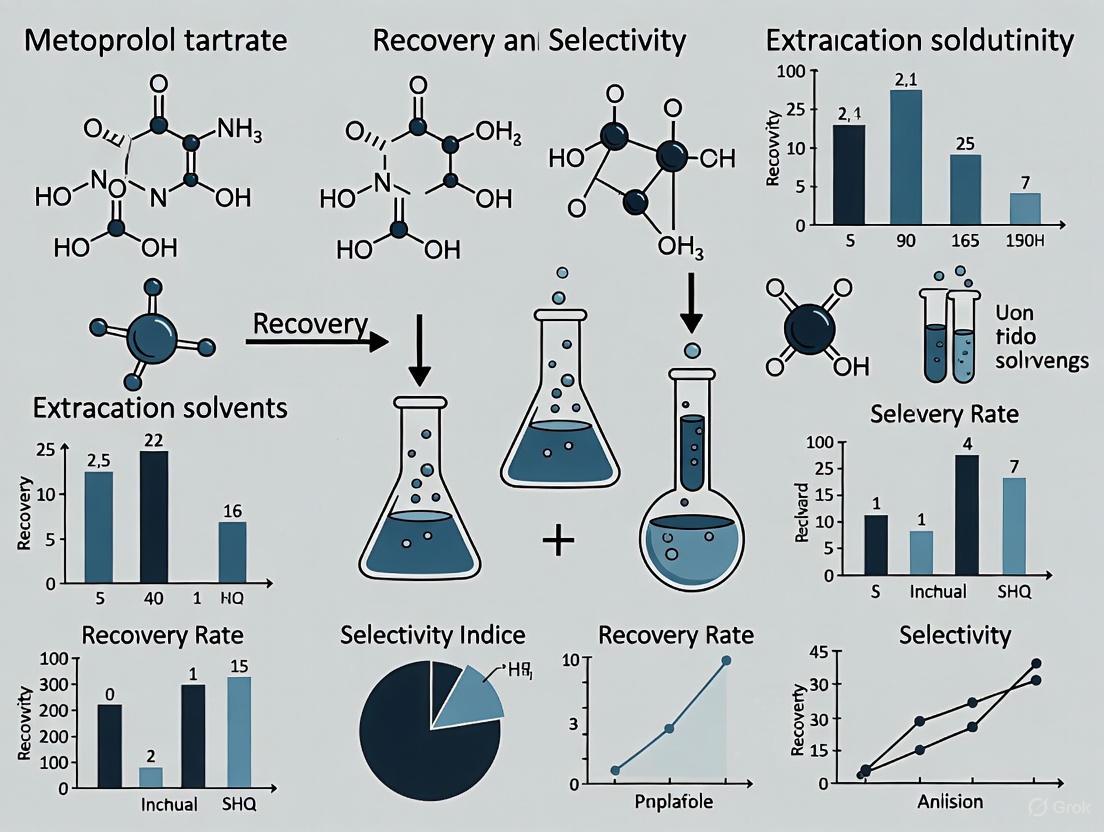
Table 1: Partitioning of Selected Pharmaceuticals in DES-Based ATPS [6]
| Pharmaceutical | DES System (HBA:HBD) | Molar Ratio | Partition Coefficient (K) | Extraction Efficiency (EE%) |
|---|---|---|---|---|
| Metoprolol Tartrate | ChCl: 1,2-Propanediol | 1:3 | 2.50 - 4.10 | 71 - 80% |
| Pregabalin | ChCl: 1,2-Propanediol | 1:3 | 1.20 - 1.80 | 55 - 65% |
| Mesalamine | ChCl: 1,2-Propanediol | 1:3 | 0.30 - 0.50 | 24 - 33% |
Experimental Protocol: The ATPS was formed using ChCl:1,2-propanediol (1:3) DES and K₂HPO₄ salt. Systems with varying DES concentrations (25.58, 29.92, 32.95, and 35.25 wt%) at a constant salt concentration (31.19 wt%) were tested. Partition coefficients (K) were calculated as the ratio of the target compound's concentration in the DES-rich top phase to its concentration in the salt-rich bottom phase. Extraction efficiency (EE%) was determined as the percentage of the total target compound partitioned to the DES-rich phase [6].
The partition behavior demonstrates the system's selectivity, with metoprolol tartrate showing strong affinity for the DES-rich phase, while mesalamine preferentially partitioned to the salt-rich bottom phase [6].
Partitioning of Phenolic Compounds
Phenolic compounds are another important class of bioactive molecules effectively separated using DES-based ATPS.
Table 2: Partitioning of Phenolic Acids in DES-Based ATPS [42]
| Phenolic Compound | Log Kₒw | DES System | Partition Coefficient (K) | Selectivity |
|---|---|---|---|---|
| Caffeic Acid | 1.424 | ChCl:Sucrose + Acetonitrile | >1 (Top Phase) | - |
| Syringic Acid | 1.324 | ChCl:Sucrose + Acetonitrile | <1 (Bottom Phase) | >2 |
| Vanillic Acid | - | ChCl:Sucrose + Acetonitrile | <1 (Bottom Phase) | >2 |
| Ferulic Acid | 1.671 | ChCl:Sucrose + Acetonitrile | >1 (Top Phase) | - |
| Vanillin | - | ChCl:Sucrose + Acetonitrile | 90.09% Recovery (Top Phase) | - |
Experimental Protocol: ATPS were prepared using DES composed of choline chloride and carbohydrates (sucrose, glucose, mannose, arabinose, xylose) in 1:1, 1:2, and 2:1 molar ratios, combined with acetonitrile and water. Binodal curves were determined at 25°C and 0.1 MPa. Phenolic compound partitioning was evaluated, with the top phase being acetonitrile-rich and the bottom phase DES-rich. The system achieved selective separation of ferulic acid and vanillin to the top phase, and syringic, caffeic, and vanillic acids to the bottom phase [42].
The study demonstrated that DES with sucrose as HBD showed the strongest phase-forming ability due to its numerous hydroxyl groups enhancing hydrophilicity and the sugaring-out effect [42].
Comparative Analysis with Alternative Solvents
DES vs. Ionic Liquids in ATPS
DES offer distinct advantages when used in ATPS compared to Ionic Liquids.
Table 3: DES vs. Ionic Liquids for ATPS Applications
| Parameter | Deep Eutectic Solvents | Ionic Liquids |
|---|---|---|
| Cost | Low-cost, readily available components [44] | Expensive, synthetic precursors [44] |
| Synthesis | Simple preparation by mixing and stirring, 100% atom economy [41] | Complex synthesis, requires purification [44] |
| Toxicity | Generally low toxicity, biodegradable [41] [45] | Variable toxicity, poor biodegradability [44] |
| Environmental Impact | Biocompatible, often from natural sources [45] | Questionable "green" credentials [44] |
| Tunability | Highly customizable by varying HBA/HBD [41] | Tunable but with limited biocompatible options |
DES-ATPS vs. Conventional Extraction Methods
DES-based ATPS provide significant advantages over traditional extraction techniques for bioactive compounds.
Table 4: Comparison of Extraction Techniques
| Extraction Technique | Selectivity | Biocompatibility | Environmental Impact | Cost | Operational Complexity |
|---|---|---|---|---|---|
| DES-based ATPS | High | High | Low | Low | Simple |
| Organic Solvent Extraction | Moderate | Low | High (toxic solvents) | Moderate | Simple |
| Chromatography | Very High | High | Moderate | High | Complex |
| Supercritical Fluid Extraction | Moderate | High | Low | High | Complex |
| Polymer-Salt ATPS | Moderate | High | Low | Low | Simple |
DES-based ATPS eliminate or significantly reduce the use of volatile organic solvents like n-hexane, petroleum ether, chloroform, and dichloromethane, which are harmful to environmental and human health [41]. While water is the greenest solvent, it has limitations including high boiling point, energy-intensive removal, and inability to dissolve non-polar compounds [41].
Research Reagent Solutions Toolkit
Table 5: Essential Reagents for DES-Based ATPS Research
| Reagent Category | Specific Examples | Function in DES-ATPS |
|---|---|---|
| Hydrogen Bond Acceptors | Choline Chloride, Betaine, Proline | Forms DES framework, interacts with target compounds [41] [6] |
| Hydrogen Bond Donors | Urea, Glycerol, 1,2-Propanediol, Carbohydrates (Glucose, Sucrose, Xylose), Organic Acids (Malic, Citric) | Modifies DES properties, enables selective partitioning [41] [6] [42] |
| Salts for ATPS | K₂HPO₄, K₃PO₄ | Phase-forming component in polymer-salt or DES-salt ATPS [6] |
| Organic Solvents for ATPS | Acetonitrile, Ethanol | Phase-forming component with DES, creates immiscible aqueous phases [42] |
| Polymers for ATPS | PEG, PVDF, Pebax 1657 | Phase-forming component or support matrix in polymer-DES ATPS [44] |
Experimental Workflow and Molecular Interactions
The following diagrams illustrate the experimental workflow for constructing DES-based ATPS and the molecular interactions responsible for selective partitioning.
Diagram 1: Experimental workflow for developing and characterizing DES-based aqueous two-phase systems.
Diagram 2: Key molecular interactions governing selective partitioning in DES-based ATPS.
DES-based ATPS represent a significant advancement in separation technology, combining the tunability and green credentials of deep eutectic solvents with the biocompatibility and efficiency of aqueous two-phase systems. Experimental evidence demonstrates their effectiveness in selectively partitioning diverse compounds, from pharmaceuticals like metoprolol tartrate to phenolic bioactive compounds. The high selectivity, customizability, and environmental benefits position DES-based ATPS as a powerful tool for researchers and drug development professionals seeking sustainable and efficient separation methods. Future developments will likely focus on expanding DES formulations for targeted applications, integrating computational prediction models, and optimizing industrial-scale implementation.
The selective and efficient recovery of active pharmaceutical ingredients (APIs) from complex matrices is a critical step in drug development and bioanalysis. This guide provides a detailed comparison of two fundamental sample preparation techniques—Solid-Phase Extraction (SPE) and Liquid-Liquid Extraction (LLE)—within the specific context of metoprolol tartrate recovery and selectivity research. Metoprolol tartrate, a widely prescribed beta-blocker with a therapeutic range of 20–100 ng/mL, presents a compelling case study due to its polar nature and the need for precise quantification in biological and environmental samples [46]. The choice between SPE and LLE significantly impacts key analytical parameters such as recovery efficiency, sample purity, operational workflow, and cost-effectiveness. This article objectively compares these methodologies by synthesizing current experimental data and established protocols, providing drug development professionals with a evidence-based framework for selecting the optimal extraction solvent and technique for their specific research requirements.
Theoretical Foundations and Mechanistic Principles
The core distinction between SPE and LLE lies in their fundamental separation mechanisms. SPE operates on a solid-liquid interfacial principle, where analytes partition between a liquid sample and a solid stationary phase. In contrast, LLE is governed by liquid-liquid partitioning, relying on the differential solubility of an analyte between two immiscible liquids.
Solid-Phase Extraction Mechanism
SPE is an adsorption-desorption process. For metoprolol tartrate, which contains amine and ether functional groups, the mechanism often involves reversed-phase chromatography on hydrophobic sorbents like Hydrophilic-Lipophilic Balance (HLB) copolymer [47]. The process involves:
- Conditioning: Preparing the sorbent (e.g., HLB cartridge) with a solvent like methanol to wet the surface and create a conducive environment for interaction.
- Loading: The aqueous sample (e.g., wastewater or biological fluid) is passed through the cartridge. Metoprolol, with its moderate hydrophobicity, is retained on the sorbent via van der Waals forces and hydrophobic interactions.
- Washing: Interfering matrix components with weaker affinity are removed with a mild solvent (e.g., 5-10% methanol in water).
- Elution: A strong organic solvent (e.g., 100% methanol) disrupts the interactions, desorbing and recovering the purified metoprolol [47]. The selectivity can be finely tuned by adjusting sorbent chemistry, sample pH, and solvent strength.
Liquid-Liquid Extraction Mechanism
LLE exploits the Nernst distribution law, where a solute (metoprolol) distributes itself between two immiscible solvents (e.g., an aqueous sample and an organic solvent like ethyl acetate) based on its partition coefficient [48]. The efficiency depends on the analyte's ionization state. Metoprolol, being a basic compound (pKa ~9.7), remains largely unionized at high pH. By adjusting the aqueous sample to a basic pH, the neutral metoprolol molecules favor transfer into the organic phase, separating from water-soluble interferences. Subsequent centrifugation and isolation of the organic layer, followed by evaporation and reconstitution, yields the purified analyte.
Table 1: Core Principles of SPE and LLE
| Feature | Solid-Phase Extraction (SPE) | Liquid-Liquid Extraction (LLE) |
|---|---|---|
| Fundamental Principle | Adsorption chromatography & interfacial solid-liquid affinity | Liquid-liquid partitioning & solubility differential |
| Primary Interactions | Hydrophobic, polar, ionic, affinity-based | Solvent-solute solubility, hydrogen bonding |
| Driving Force | Chemical affinity for solid sorbent & elution strength | Partition coefficient between two immiscible phases |
| Selectivity Control | Sorbent chemistry, pH, elution solvent gradient | pH manipulation, organic solvent type |
| Typical Phase Composition | Solid (sorbent) vs. Liquid (sample/solvent) | Organic solvent vs. Aqueous sample |
Performance Comparison and Experimental Data
Direct comparative studies reveal significant differences in the performance of SPE and LLE for metoprolol and similar pharmaceuticals, particularly in terms of recovery, purity, and operational throughput.
Quantitative Recovery and Efficiency
A direct comparative study of extraction techniques for various pharmaceuticals highlighted SPE's superior performance. The study reported that SPE demonstrated higher recovery rates and better precision compared to LLE for most analytes. Furthermore, optimized SPE methods for metoprolol and other drugs have achieved correlation coefficients (R²) > 0.98 in calibration curves, indicating excellent linearity and reliable quantification [47]. In a specific optimization for metoprolol recovery from aqueous matrices using HLB cartridges, the recovery rates were consistently high under optimal parameters [47].
Table 2: Experimental Performance Data for SPE vs. LLE
| Parameter | Solid-Phase Extraction (SPE) | Liquid-Liquid Extraction (LLE) |
|---|---|---|
| Typical Recovery Efficiency | High, often >80% for metoprolol with HLB [47] | Generally lower and more variable for polar pharmaceuticals [48] |
| Process Precision (RSD) | High (<10% RSD in optimized methods) [47] | Can be moderate to high, depending on emulsion formation |
| Concentration Factor | High (enables significant pre-concentration) [49] | Moderate |
| Limit of Detection (LOD) | Enables lower LODs (e.g., µg/L to ng/L range) [47] | Generally higher LODs compared to SPE |
| Sample Throughput | Amenable to automation and batch processing | Typically more manual and time-consuming |
Selectivity and Matrix Cleanup
SPE provides superior selectivity due to the multiple steps of conditioning, loading, washing, and elution. The washing step is particularly crucial for removing matrix interferences that have different polarities than the target analyte. For example, in wastewater analysis, SPE effectively isolates metoprolol from a complex background of other organic contaminants and inorganic salts [47]. LLE, while effective in separating analytes based on solubility, offers fewer opportunities for intermediate cleanup. It can be less selective when the sample matrix contains compounds with similar partition coefficients to the analyte, potentially leading to co-extraction of impurities.
Detailed Experimental Protocols
Solid-Phase Extraction Protocol for Metoprolol from Aqueous Matrices
This protocol is adapted from methods used for the simultaneous extraction of pharmaceuticals from wastewater, optimized for metoprolol recovery [47].
Research Reagent Solutions:
- Sorbent: 60 mg/3 mL Oasis HLB cartridge or equivalent.
- Solvents: Methanol (HPLC grade), Acetonitrile (HPLC grade), Ultrapure water.
- Acids/Bases: Hydrochloric acid (HCl, 0.1 M), Sodium hydroxide (NaOH, 0.1 M).
- Standards: Metoprolol tartrate certified reference standard.
- Equipment: SPE vacuum manifold, pH meter, nitrogen evaporator, amber vials.
Step-by-Step Workflow:
- Conditioning: Activate the HLB cartridge by passing 5 mL of methanol through it at a flow rate of ~1 mL/min. Equilibrate the sorbent with 5 mL of ultrapure water. Do not let the sorbent run dry.
- Sample Pretreatment: Adjust the pH of the aqueous sample (e.g., 100 mL wastewater or buffered solution) to pH 2.0 using 0.1 M HCl. This pH enhances the retention of many pharmaceuticals on the HLB sorbent.
- Loading: Load the acidified sample onto the conditioned cartridge under vacuum at a controlled flow rate of 1-5 mL/min.
- Washing: Rinse the cartridge with 5 mL of ultrapure water followed by 5 mL of 10% methanol to remove weakly retained matrix components.
- Drying: Apply a strong vacuum (e.g., 10-15 inches Hg) for 5-10 minutes to dry the sorbent completely and remove residual water.
- Elution: Elute the adsorbed metoprolol with 4 mL of 100% methanol. Collect the eluate in a clean tube.
- Post-Processing: Evaporate the eluate to dryness under a gentle stream of nitrogen at 50°C. Reconstitute the dry residue in 1 mL of methanol or a mobile phase compatible with your subsequent analysis (e.g., HPLC). Filter through a 0.22 µm nylon syringe filter prior to injection.
Liquid-Liquid Extraction Protocol for Metoprolol
This generic protocol for basic pharmaceuticals can be adapted for metoprolol recovery from biological or aqueous samples [48].
Research Reagent Solutions:
- Extraction Solvent: Ethyl acetate, Dichloromethane, or a mixture.
- Buffers: Phosphate buffer (e.g., 0.1 M, pH 10-11) or Ammonium hydroxide solution.
- Solvents: Methanol (HPLC grade), Ultrapure water.
- Standards: Metoprolol tartrate certified reference standard.
- Equipment: Centrifuge, vortex mixer, glass separation funnels or conical tubes, nitrogen evaporator.
Step-by-Step Workflow:
- Sample Aliquoting: Transfer 1 mL of the sample (e.g., plasma, urine, or wastewater) into a glass centrifuge tube.
- pH Adjustment: Add 0.5 mL of 0.1 M phosphate buffer (pH 10-11) or a few drops of ammonium hydroxide to make the sample alkaline. This suppresses the ionization of metoprolol, promoting its partitioning into the organic solvent.
- Extraction: Add 3 mL of ethyl acetate to the tube. Cap the tube securely and vortex mix vigorously for 1-2 minutes.
- Phase Separation: Centrifuge the mixture at 3000-4000 rpm for 5-10 minutes to achieve complete phase separation.
- Organic Layer Isolation: Carefully transfer the upper organic layer (ethyl acetate) to a new clean tube using a Pasteur pipette. Avoid transferring any of the aqueous interface.
- Back-Extraction (Optional, for Cleanup): For additional purity, the collected organic phase can be shaken with a small volume of a mild acidic aqueous solution. Metoprolol will transfer back to the aqueous phase due to protonation. The aqueous phase can then be re-basified and extracted again with a fresh organic solvent.
- Evaporation and Reconstitution: Combine the organic extracts (if multiple steps are performed) and evaporate to dryness under a nitrogen stream. Reconstitute the dry residue in an appropriate volume (e.g., 100-200 µL) of mobile phase for analysis.
The following workflow diagram synthesizes the core procedural steps for both SPE and LLE, highlighting their parallel stages and key decision points.
The Scientist's Toolkit: Essential Research Reagents and Materials
Selecting the appropriate reagents is fundamental to the success of any extraction protocol. The following table details key solutions and materials used in the featured experiments for metoprolol recovery.
Table 3: Essential Research Reagent Solutions for Extraction
| Reagent/Material | Function/Description | Application Context |
|---|---|---|
| Oasis HLB Cartridge | A hydrophilic-lipophilic balanced copolymer sorbent that retains a wide range of analytes (acids, bases, neutrals) via multiple mechanisms. | Primary sorbent for SPE of metoprolol from aqueous matrices like wastewater [47]. |
| Methanol (HPLC Grade) | High-purity solvent used for SPE sorbent conditioning and analyte elution, and for reconstitution of dried extracts. | Universal solvent in SPE protocols; used as 100% eluent for metoprolol [47]. |
| Ethyl Acetate | An organic solvent with medium polarity, immiscible with water. Commonly used to extract non-ionized organic compounds. | Common organic solvent in LLE for basic drugs like metoprolol from biological fluids [48]. |
| pH Buffer Solutions | Aqueous solutions (e.g., Acetate, Phosphate) used to adjust and control the ionization state of the analyte, critically impacting recovery. | SPE: Acidic pH (e.g., 2) for retention. LLE: Basic pH (~10-11) for transfer to organic phase [47] [48]. |
| Certified Reference Standard | A highly purified and well-characterized material of the target analyte (e.g., Metoprolol Tartrate) used for method validation and calibration. | Essential for quantifying recovery efficiency and constructing calibration curves in both SPE and LLE [47]. |
The choice between Solid-Phase Extraction and Liquid-Liquid Extraction for metoprolol tartrate recovery is not a matter of one technique being universally superior, but rather dependent on the specific research goals and constraints. SPE is the recommended technique for applications demanding high recovery efficiency, superior sample cleanup, and automated high-throughput analysis, such as in pharmacokinetic studies or environmental monitoring where metoprolol concentrations can be trace-level [46] [47]. The ability to pre-concentrate the analyte and achieve low detection limits is a significant advantage. Conversely, LLE remains a valuable and pragmatic choice for labs with lower sample volumes, limited budget for sorbent cartridges, or when processing samples where its proven simplicity and effectiveness for specific matrices are adequate [48]. Ultimately, the decision should be guided by a balanced consideration of the required data quality, available resources, and the nature of the sample matrix, with SPE generally offering greater control and performance for rigorous analytical applications.
Troubleshooting Common Challenges and Optimizing Extraction Efficiency
Addressing Matrix Effects from Plasma and Formulation Excipients
Matrix effects present a significant challenge in the quantitative bioanalysis of pharmaceuticals like metoprolol tartrate, potentially compromising assay accuracy, sensitivity, and reproducibility. This guide compares strategies to manage these effects, focusing on experimental data for extraction solvent selection to optimize recovery and selectivity.
Understanding Matrix Effects in LC-MS/MS Bioanalysis
In liquid chromatography-tandem mass spectrometry (LC-MS/MS), matrix effects refer to the suppression or enhancement of analyte ionization caused by co-eluting components from the sample matrix. For drug analysis, these interfering substances originate primarily from two sources: the biological matrix (e.g., plasma phospholipids) and the formulation excipients used in drug products [50] [51] [52].
Plasma phospholipids, particularly phosphatidylcholine (PC) and lysophosphatidylcholine (Lyso-PC), are a major source of ion suppression in electrospray ionization (ESI) [50]. Their concentration can vary between individual plasma lots, leading to inconsistent analytical results. Simultaneously, functional excipients—such as surfactants, alkalinizing agents, and complexation agents like cyclodextrins—which constitute up to 90% of a solid oral dosage form, can leach during sample preparation and cause similar interference [53]. The core problem is that these matrix components compete with the analyte for available charge during the ionization process, leading to a loss of signal and potentially erroneous quantification [54] [51].
Experimental Assessment of Matrix Effects
Before developing mitigation strategies, it is crucial to empirically evaluate the presence and extent of matrix effects. The following experimental protocols are standard in the field.
Post-Column Infusion for Qualitative Assessment
This method provides a visual map of ionization suppression or enhancement regions throughout the chromatographic run [54] [51].
Experimental Protocol:
- Configure the LC-MS/MS system with a T-connector between the HPLC column outlet and the MS ion source.
- Infuse a constant, known concentration of the analyte (e.g., metoprolol) or a stable isotope-labeled internal standard directly into the post-column effluent at a low flow rate (e.g., 10 µL/min).
- Inject a blank, prepared sample matrix (e.g., extracted plasma) into the LC system and run the chromatographic method.
- Monitor the signal of the infused analyte. A stable signal indicates no matrix effect, while a dip or rise in the baseline indicates suppression or enhancement, respectively, at that specific retention time [55] [51].
The diagram below illustrates this workflow:
Post-Extraction Spiking for Quantitative Assessment
This method provides a quantitative measure (Matrix Factor, MF) of the matrix effect [51] [56] [52].
Experimental Protocol:
- Prepare three sets of samples:
- Set A (Neat Solution): Analyze the analyte at a specific concentration in a pure, matrix-free solvent.
- Set B (Post-Extraction Spiked): Take several lots of blank matrix (e.g., plasma from different donors), process them through the entire sample preparation procedure, and then spike the analyte into the resulting clean extract at the same concentration as Set A.
- Set C (Monitoring IS): Include a constant concentration of internal standard in all samples.
- Analyze all sets by LC-MS/MS.
- Calculate the Matrix Factor (MF) using the following formula:
Comparison of Mitigation Strategies and Experimental Data
A systematic evaluation of supported liquid extraction (SLE) for 10 model pharmaceutical compounds with diverse properties provides robust, comparative data on extraction efficiency and matrix effect reduction [50]. The table below summarizes key findings relevant to metoprolol tartrate analysis.
Table 1: Comparison of Sample Preparation Techniques for Mitigating Matrix Effects
| Strategy/Technique | Mechanism of Action | Impact on Phospholipid Removal | Reported Recovery for Model Compounds | Key Experimental Findings |
|---|---|---|---|---|
| Supported Liquid Extraction (SLE) [50] | Partitioning of analytes from aqueous sample into a solid support, followed by elution with organic solvent. | Highly effective; removes significantly more phospholipids than protein precipitation (PPT) and many SPE sorbents. | >75% recovery achieved for all 10 model compounds, including those with a wide range of pKa and LogP. | Under optimized conditions, insignificant matrix effect was observed. Methyl tert-butyl ether (MTBE) and dichloromethane (DCM) eluted fewer phospholipids than ethyl acetate. |
| Protein Precipitation (PPT) [50] | Denaturation and removal of proteins using organic solvents. | Least effective; co-precipitates some phospholipids but the supernatant remains rich in them. | High recovery is possible, but the extract is dirty. | Yields the most significant matrix effect compared to SLE and LLE due to poor removal of phospholipids. |
| Liquid-Liquid Extraction (LLE) [50] [33] | Partitioning of analytes between immiscible organic solvent and aqueous sample. | Can provide clean extracts; effectiveness depends on solvent choice. | Generally high, but can be variable. | SLE is considered an alternative to traditional LLE, offering faster extraction and easier automation while providing clean samples. |
| Microextraction Techniques (e.g., DLLME) [33] [57] | Miniaturized, solvent-efficient version of LLE using µL volumes of extraction solvent. | Good cleaning potential, depending on the solvent. | Good recoveries reported for β-blockers like atenolol and propranolol (e.g., 53–102%). | Techniques like Dispersive Liquid-Liquid Microextraction (DLLME) are simple, rapid, and offer high enrichment factors, making them suitable for complex matrices. |
Optimizing Supported Liquid Extraction
The effectiveness of SLE is highly dependent on the loading conditions. Research shows that the pH and organic content of the sample loading buffer critically impact the recovery of both the analyte and phospholipids [50].
Table 2: Impact of SLE Loading Conditions on Phospholipid and Analyte Recovery
| Loading Condition | Impact on Phospholipid Recovery | Recommendation for Optimal Cleanup |
|---|---|---|
| Addition of Acetonitrile (ACN) or Methanol | Increases the recovery of phospholipids from the SLE cartridge. | Avoid using ACN or methanol in the loading buffer if the goal is to minimize matrix effects. |
| Use of Aqueous Buffer at Optimal pH | Minimizes phospholipid elution. | Use an aqueous buffer to reconstitute or dilute the sample before loading onto the SLE cartridge. Adjust pH to ensure the analyte is in its uncharged form for efficient transfer to the organic eluent. |
The following workflow outlines the optimized SLE procedure for clean sample preparation:
The Scientist's Toolkit: Essential Research Reagents and Materials
The following table details key materials required for experiments aimed at addressing matrix effects.
Table 3: Essential Research Reagents and Materials
| Item | Function/Benefit | Application Example |
|---|---|---|
| Blank Plasma Lots | Critical for assessing inter-lot variability of matrix effects during method development and validation [51] [52]. | Used in post-extraction spiking experiments to quantitatively determine the Matrix Factor. |
| Stable Isotope-Labeled Internal Standard (SIL-IS) | Gold standard for compensating matrix effects; co-elutes with analyte, mimicking its behavior during ionization [54] [51]. | Metoprolol-d7 is used as an IS to normalize for ionization suppression/enhancement during quantitation. |
| Supported Liquid Extraction (SLE) Plates/Cartridges | Provides a high-throughput, automated platform for efficient sample clean-up and phospholipid removal [50]. | Used for preparing plasma samples prior to LC-MS/MS analysis of metoprolol to minimize matrix effects. |
| Appropriate Organic Eluents (MTBE, DCM) | Effective for eluting analytes while leaving significant amounts of phospholipids behind on the SLE support [50]. | Used as the elution solvent in the optimized SLE protocol. |
| Phospholipid-Specific MRM Transitions | Enables direct monitoring of phospholipid elution profiles during method development to identify and avoid regions of high interference [50]. | Used to create a phospholipid map of the chromatographic method and confirm the cleanliness of the final extract. |
Optimizing pH and Buffer Conditions for Maximum Recovery and Selectivity
The optimization of pH and buffer conditions is a critical determinant of success in the extraction and analysis of active pharmaceutical ingredients (APIs). For ionizable compounds like metoprolol tartrate, a selective β1-adrenergic blocker, controlling the pH of the extraction environment directly influences ionization state, solubility, and partitioning behavior, thereby dictating the efficiency and selectivity of the recovery process. This guide provides a systematic comparison of extraction solvents and methodologies, focusing specifically on how pH and buffer selection impact the recovery and selectivity of metoprolol tartrate, a drug widely prescribed for cardiovascular diseases [58] [59]. Researchers must navigate a complex landscape of techniques, from traditional liquid-liquid extraction to advanced aqueous two-phase systems (ATPS), each with unique buffer requirements and performance characteristics. By examining experimental data across different platforms, this review offers evidence-based recommendations for optimizing these key parameters, ensuring maximum recovery and purity for pharmaceutical analysis and drug development applications.
Fundamentals of pH-Dependent Extraction
Chemical Properties of Metoprolol Tartrate
Metoprolol tartrate (C34H56N2O12) is a basic drug substance with a pKa of approximately 9.7, existing predominantly in its ionized, water-soluble form at physiological and acidic pH conditions [37]. This protonation state significantly enhances its solubility in aqueous environments but diminishes its partitioning into organic solvents. The molecule contains a secondary amine group in its aryloxypropanolamine side chain that serves as the primary site of protonation. As pH increases towards and beyond its pKa, metoprolol undergoes a critical transition to its neutral form, substantially increasing its hydrophobicity and affinity for organic phases. This pH-dependent speciation forms the fundamental basis for selective extraction protocols, allowing researchers to manipulate recovery and selectivity through careful buffer selection and pH adjustment. Understanding this acid-base behavior is essential for predicting metoprolol's distribution across different solvent systems and designing efficient extraction workflows with appropriate buffer conditions.
Mechanisms of pH Influence on Extraction
The ionization state of metoprolol, governed by the pH of the extraction environment, directly controls its intermolecular interactions and partitioning behavior through several key mechanisms:
Solubility Modulation: In acidic environments (pH < pKa), the protonated, cationic form of metoprolol exhibits high aqueous solubility due to favorable electrostatic interactions with water molecules and ionic constituents in buffer solutions. This charged state significantly reduces extraction efficiency into organic solvents, as indicated by studies showing decreased distribution coefficients at lower pH values [10].
Hydrogen Bonding Capacity: The neutral form of metoprolol predominant at basic pH retains the ability to form hydrogen bonds through its ether oxygen, secondary alcohol, and secondary amine functional groups. This enables specific interactions with hydrogen bond acceptors in extraction solvents, such as the deep eutectic solvents (DES) used in ATPS, which can enhance selectivity relative to other compounds [10].
Phase Partitioning Behavior: The lipophilicity of metoprolol increases substantially in its neutral form, dramatically improving its partitioning into organic solvents and polymer-rich phases. This phenomenon is quantitatively described by the distribution coefficient (Log D), which is highly pH-dependent and directly correlates with extraction recovery across different methodologies [10] [33].
Comparative Analysis of Extraction Methodologies
Aqueous Two-Phase Systems (ATPS)
Deep Eutectic Solvent (DES)-based ATPS represent an advanced extraction platform that leverages the unique properties of eutectic mixtures for selective pharmaceutical separations. In one comprehensive study, researchers developed a DES-based ATPS composed of tetra-n-butylammonium bromide (TBAB) and polyethylene glycol 200 (PEG200) in a 1:3 molar ratio with K₂HPO₄ as the salt phase for separating metoprolol from other pharmaceuticals [10].
Table 1: DES-ATPS Composition and Performance for Metoprolol Extraction
| System Component | Composition/Value | Function in Extraction |
|---|---|---|
| DES (HBA) | Tetra-n-butylammonium bromide (TBAB) | Hydrogen bond acceptor, forms DES structure |
| DES (HBD) | Polyethylene glycol 200 (PEG200) | Hydrogen bond donor, modulates DES properties |
| DES Molar Ratio | TBAB:PEG200 (1:3) | Optimized for drug partitioning |
| Salt Component | K₂HPO₄ | Induces phase separation via salting-out effect |
| Salt Concentration | 18.95–23.75 wt% | Higher concentration decreases drug distribution |
| DES Concentration | 23.95–26.03 wt% | Higher concentration improves drug partitioning |
The investigation revealed that increasing salt concentration from 18.95 to 23.75 wt% caused a noticeable decrease in both partition coefficient and extraction efficiency due to enhanced ion hydration, which favored retention of metoprolol in the salt-rich aqueous phase [10]. Conversely, increasing DES concentration from 23.95 to 26.03 wt% improved drug partitioning into the DES-rich phase. This system achieved extraction efficiencies of 85-95% for metoprolol under optimized buffer and concentration conditions, demonstrating the effectiveness of DES-ATPS for pharmaceutical separations when properly calibrated [10].
Chromatographic Methods with Biological Matrices
For bioanalytical applications, reversed-phase high-performance liquid chromatography (RP-HPLC) represents the gold standard for metoprolol quantification in biological fluids, with buffer pH playing a critical role in both extraction recovery and chromatographic separation. Multiple studies have optimized buffer conditions specifically for metoprolol analysis in complex matrices:
Table 2: Buffer Conditions for Chromatographic Analysis of Metoprolol
| Analytical Method | Buffer Conditions | Metoprolol Recovery/Performance | Application Context |
|---|---|---|---|
| RP-HPLC-FD [11] | 30mM KH₂PO₄, pH 2.5 with OPA | Linear range: 0.003-1.00 µg/mL; Accuracy: ±10% of nominal | Spiked human plasma |
| RP-HPLC-UV [59] | 12.5mM phosphate buffer, pH 7.0 | Linear range: 1.14-50 µg/mL; R² = 0.9994 | Intestinal perfusion studies |
| HPTLC-UV/FL [37] | Mobile phase with formic acid-ammonia | Linearity: 50-900 ng/band (UV); 50-550 ng/band (FL) | Bulk and pharmaceutical dosage |
A particularly robust RP-HPLC method was developed for simultaneous determination of atenolol, metoprolol, and phenol red in intestinal perfusion studies [59]. This method employed a phosphate buffer (pH 7.0, 12.5 mM) in a gradient elution with acetonitrile, demonstrating excellent linearity across the concentration range of 1.14–50 μg/mL for metoprolol. The neutral pH condition was critical for maintaining the physiological relevance of the permeability assay while ensuring optimal chromatographic separation and detection sensitivity [59]. The method was comprehensively validated according to ICH M10 guidelines, confirming that the selected buffer conditions provided the necessary precision, accuracy, and robustness for reliable metoprolol quantification in complex biological samples.
Spectrofluorimetric Methods
Native fluorescence properties of metoprolol have been leveraged for its direct quantification in biological matrices without extensive chromatographic separation. A green spectrofluorimetric method was developed for simultaneous quantification of aspirin, olmesartan, and metoprolol in spiked human plasma, utilizing the intrinsic fluorescence of metoprolol at excitation/emission wavelengths of 230/302 nm [60].
The sample preparation protocol incorporated a protein precipitation step using acetonitrile, followed by reconstitution in ethanol and addition of acetate buffer (pH 5). This slightly acidic pH condition was optimal for stabilizing all three analytes while maximizing fluorescence intensity for detection. The method demonstrated excellent linearity across the concentration range of 100–1400 ng/mL for metoprolol, with precision and accuracy meeting ICH M10 validation criteria [60]. The success of this approach highlights how pH optimization in both extraction and detection phases can enable selective quantification without the need for complex separation systems, provided that the target analyte possesses distinctive fluorescent properties that can be exploited through appropriate buffer selection.
Experimental Protocols for Method Comparison
DES-ATPS Protocol for Metoprolol Extraction
Phase System Preparation:
- DES Synthesis: Combine tetra-n-butylammonium bromide (TBAB) and polyethylene glycol 200 (PEG200) in a 1:3 molar ratio. Heat the mixture at 60°C with continuous stirring at 500 rpm until a clear, homogeneous liquid forms [10].
- Binodal Curve Determination: Prepare a series of systems with varying DES/water/salt ratios. Visually determine the point of phase transition from homogeneous to turbid upon water addition. Fit the binodal data using the Merchuk equation to establish the phase diagram [10].
- Tie-Line Construction: Prepare ATPS with overall compositions located in the two-phase region. Separate the top and bottom phases after equilibration, then determine their compositions through density measurements and refractometry [10].
Drug Partitioning Experiment:
- Sample Preparation: Dissolve metoprolol tartrate in deionized water at 0.15 wt% concentration [10].
- System Setup: Combine the drug solution with predetermined amounts of DES and K₂HPO₄ in centrifuge tubes to match the desired operating points from the phase diagram.
- Equilibration and Separation: Vortex mixtures for 5 minutes, then equilibrate in a water bath at 25°C for 2 hours to ensure complete phase separation. Centrifuge at 3000 rpm for 15 minutes to sharpen the phase boundary [10].
- Analysis: Carefully separate the top and bottom phases. Determine metoprolol concentration in each phase using HPLC-UV at 224 nm or another validated analytical method [10].
- Calculation: Determine the partition coefficient (K) as K = Ctop/Cbottom, where Ctop and Cbottom represent the equilibrium concentrations of metoprolol in the top and bottom phases, respectively.
RP-HPLC Method for Bioanalytical Applications
Mobile Phase Preparation:
- Buffer Solution: Prepare 12.5 mM phosphate buffer by dissolving potassium dihydrogen phosphate in purified water. Adjust pH to 7.0 using sodium hydroxide solution [59].
- Mobile Phase: Combine phosphate buffer with HPLC-grade acetonitrile in gradient elution program starting from 10% acetonitrile, increasing linearly to 35% over 15 minutes. Filter through 0.45 μm membrane and degas by sonication for 10 minutes before use [59].
Sample Preparation and Chromatography:
- Standard Solutions: Prepare metoprolol stock solution at 1 mg/mL in methanol. Dilute with mobile phase to construct calibration standards in the range of 1.14–50 μg/mL [59].
- Chromatographic Conditions: Use an InertSustain C18 column (250 × 4.6 mm, 5 μm) maintained at 35°C. Set flow rate to 1 mL/min and injection volume to 20 μL. Detect metoprolol at 224 nm [59].
- Validation: Assess method selectivity, linearity, accuracy, precision, and recovery according to ICH M10 guidelines. Calculate peak area ratios of metoprolol to internal standard (if used) for quantification [59].
Figure 1: Experimental workflow comparison for three primary extraction and analysis methods for metoprolol tartrate
Research Reagent Solutions Toolkit
Table 3: Essential Reagents for Metoprolol Extraction and Analysis
| Reagent/Chemical | Function/Purpose | Application Examples |
|---|---|---|
| Tetra-n-butylammonium bromide (TBAB) | Hydrogen bond acceptor in DES formation | DES-ATPS with PEG200 [10] |
| Polyethylene glycol 200 (PEG200) | Hydrogen bond donor in DES formation | DES-ATPS with TBAB [10] |
| Potassium dihydrogen phosphate (KH₂PO₄) | Buffer component; salting-out agent | ATPS; Mobile phase for HPLC [10] [11] |
| Acetonitrile (HPLC grade) | Organic modifier; protein precipitant | HPLC mobile phase; sample cleanup [59] [60] |
| Ammonium sulfate | Salting-out agent in LLME | Enhances extraction efficiency [33] |
| Acetate buffer (pH 5) | pH control in fluorescence methods | Optimizes fluorescence detection [60] |
| 1-Dodecanol | Extraction solvent in LLME | Low toxicity organic solvent for microextraction [33] |
| Formic acid | Mobile phase modifier | Improves chromatographic peak shape [37] |
The optimization of pH and buffer conditions emerges as a fundamental consideration for maximizing recovery and selectivity in metoprolol tartrate extraction across all methodologies examined. For ATPS, higher DES concentrations (23.95–26.03 wt%) in combination with appropriate salt concentrations significantly enhance metoprolol partitioning, while extreme pH conditions generally reduce extraction efficiency by altering the drug's ionization state [10]. In chromatographic applications, neutral pH conditions (pH 7.0 phosphate buffer) provide optimal separation and detection for bioanalytical methods, while slightly acidic conditions (pH 2.5-5.0) enhance sensitivity in spectrofluorimetric and certain HPLC techniques [11] [59] [60].
The selection of an appropriate extraction and analysis methodology must consider the specific research objectives, matrix complexity, and required sensitivity. DES-ATPS offers environmentally friendly alternatives with tunable properties for selective pharmaceutical separations, while RP-HPLC provides robust, validated approaches for bioanalytical applications requiring high specificity. Spectrofluorimetric methods present green, cost-effective alternatives for direct quantification without extensive separation needs. In all cases, careful optimization of buffer composition, ionic strength, and pH remains essential for achieving maximum recovery and selectivity in metoprolol tartrate extraction and analysis.
Developing sustained-release formulations for highly soluble drugs presents a unique challenge in pharmaceutical technology. While low solubility drugs naturally resist dissolution, water-soluble drugs exhibit rapid release profiles that are difficult to prolong. Their high aqueous solubility and poor partitioning into insoluble polymers make long-term release particularly challenging, especially when clinical applications require high dosing due to low intrinsic drug potency or the desire for sustained prevention over days or weeks [61].
The core problem lies in overcoming the inherent properties of hydrophilic small molecule drugs, which readily dissolve in gastrointestinal fluids, often leading to a rapid burst release rather than controlled, sustained delivery. This review compares advanced strategies and technologies designed to overcome these challenges, with specific attention to experimental data and material systems that enable high drug loading while maintaining controlled release profiles essential for therapeutic efficacy.
Comparative Analysis of Sustained-Release Platform Technologies
Various advanced formulation strategies have been developed to control the release of highly soluble drugs. The table below compares the performance characteristics of four major technological approaches.
Table 1: Comparison of Sustained-Release Technologies for Soluble Drugs
| Technology Platform | Typical Polymer Components | Maximum Drug Loading | Release Duration | Key Release Mechanisms |
|---|---|---|---|---|
| Electrospun Fibers | PLGA, PLA, PCL, PU | Up to 60% [61] | Several days to weeks [61] | Diffusion, polymer degradation [61] |
| Hydrophilic Matrices | HPMC, sodium alginate, xanthan gum | Varies by drug solubility | Hours to days [62] | Diffusion, erosion, gel layer formation [62] |
| 3D Printed Systems | PEG-based polymers, hydrogels | High dose achievable [63] | Tunable, sustained release [63] | Diffusion, matrix erosion, designed geometry [63] |
| Polymeric Micelles/Nanoparticles | PLGA, PEG-diacrylate, smart polymers | Varies by system | Hours to days [64] | Diffusion, polymer swelling, environmental triggers [64] |
Each technology offers distinct advantages for specific application requirements. Electrospun fibers demonstrate exceptional versatility with high drug loading capacity, while hydrophilic matrix systems provide the simplest formulation approach. Emerging technologies like material extrusion 3D printing enable unprecedented control over geometry and release profiles through core-shell structures and complex architectures [63].
Table 2: Experimental Drug Release Performance in Various Polymer Systems
| Drug Formulation | Drug Loading (wt.%) | Aqueous Solubility (mg/mL) | % Release at 24h | % Release at 7 days |
|---|---|---|---|---|
| Metronidazole in PLA [61] | 40% | 29 | 25% | 45% |
| Cefoxitin sodium in PLGA [61] | 5% | 0.15 | 72% | 80% |
| Metoclopramide HCl in PCL:PVA [61] | 1% | 0.24 | 55% | 65% |
| Amoxicillin in PEG:cellulose acetate [61] | 3.70% | 0.58 | 100% | - |
Critical Factors Governing Drug Release Kinetics
Material Composition and Selection
The choice of polymer system fundamentally controls release kinetics. Biodegradable polyesters like PLGA, PLA, and PCL are most prevalent in sustained release fibers, while hydrophilic polymers such as HPMC and polyethylene oxide dominate matrix tablet formulations [61] [62]. For highly soluble drugs, non-swellable polymers force the drug to diffuse through a solid polymer matrix, while swellable systems create a gel layer through which drugs must diffuse [61] [62].
Material selection must balance multiple factors:
- Polymer molecular weight: Higher molecular weights generally produce more viscous gel layers and slower release [62]
- Polymer composition: Varying molecular weight and concentration of components like PEGDA affects swelling behavior and drug release profiles [64]
- Drug-polymer interactions: Electrostatic interactions can be leveraged, as demonstrated with positively charged peptide hydrogels for antibiotic attachment [65]
Formulation Design Parameters
Beyond polymer selection, specific formulation parameters critically impact release performance:
- Drug loading percentage: Higher loading often increases release rates but is necessary for low-potency drugs [61]
- Particle size: Smaller drug particles dissolve more rapidly, while polymer particle size affects gel layer formation [62]
- Excipient selection: Incorporation of viscosity enhancers, solubility modifiers, and pore formers fine-tunes release profiles [65] [62]
For hydrophilic matrix systems, the polymer disentanglement concentration is a critical threshold determining whether the matrix maintains integrity or undergoes erosion-mediated release [62]. The drug solubility directly influences which release mechanism dominates, with soluble drugs primarily following diffusion-controlled release through the gel layer [62].
Advanced Experimental Approaches and Analytical Methods
Fabrication Technologies and Workflows
Advanced manufacturing technologies enable precise control over drug delivery system architecture:
Diagram 1: Experimental Workflow for Sustained-Release Formulation Development
The diagram illustrates the systematic approach to developing sustained-release formulations. Coaxial electrospinning creates core-shell fibers where the shell acts as a diffusion barrier, significantly prolonging release compared to uniaxial fibers [61]. Material extrusion 3D printing enables fabrication of complex geometries including core-shell systems that provide high drug loading with sustained release profiles [63].
Analytical and Monitoring Methodologies
Advanced analytical techniques are essential for characterizing release mechanisms:
- FRET technology: Enables real-time monitoring of drug release from PLGA microspheres by correlating fluorescence changes with release profiles [64]
- NMR and MRI techniques: Provide non-invasive analysis of polymer mobilization, water penetration, and drug diffusion during matrix swelling [62]
- Spectrophotometric methods: Utilize complexation reactions (e.g., with Cu(II) ions) for drug quantification in release media [1]
For partitioning studies of drugs like metoprolol tartrate, deep eutectic solvent (DES)-based aqueous two-phase systems (ATPS) offer environmentally friendly separation with tunable properties. These systems enable determination of distribution coefficients critical for understanding drug-polymer interactions [10].
Research Reagents and Materials Toolkit
Table 3: Essential Research Reagents for Sustained-Release Formulation Development
| Reagent/Material | Function in Formulation | Example Applications |
|---|---|---|
| PLGA/PLA/PCL | Biodegradable polymer matrix for controlled release | Electrospun fibers, injectable depots [61] [64] |
| HPMC (Hydroxypropyl methylcellulose) | Hydrophilic matrix former | Swellable tablet matrices [62] |
| PEG derivatives | Porogen, plasticizer, hydrogel component | Modifying release kinetics, 3D printing [63] [64] |
| Deep Eutectic Solvents | Green separation media for partitioning studies | Drug recovery and selectivity studies [10] |
| Cu(II) chloride | Complexation agent for drug quantification | Spectrophotometric determination of drug concentration [1] |
The development of sustained-release formulations for highly soluble drugs requires strategic integration of material science, processing technology, and analytical methodology. Electrospun fiber systems offer superior drug loading capacity for applications requiring high dosing, while hydrophilic matrices provide the most straightforward formulation path for conventional oral dosage forms. Emerging technologies like material extrusion 3D printing enable unprecedented control over release profiles through geometric design and complex architectures.
The selection of optimal technology must consider multiple factors: required drug loading, target release duration, manufacturing scalability, and clinical application route. Successful formulation development hinges on understanding the fundamental release mechanisms—diffusion, erosion, and swelling—and how material properties and processing parameters influence these mechanisms. As the field advances, the integration of "smart" polymers responsive to physiological stimuli and the application of quality by design (QbD) principles will further enhance our ability to precisely control the release of highly soluble drugs, addressing significant unmet needs in chronic disease treatment where sustained therapeutic levels are critical for efficacy.
Dealing with Solvent Incompatibility and Evaporation Losses in High-Throughput Environments
High-Throughput Screening (HTS) has become a standard method in drug discovery, enabling the rapid testing of hundreds of thousands of compounds against biological targets. A fundamental challenge in this automated environment involves managing the chemical and physical properties of the solvents used, primarily dimethyl sulfoxide (DMSO), which is the nearly universal vehicle for compound libraries. As HTS evolves toward miniaturization and higher-density microplate formats (from 96-well to 1536-well and beyond), the issues of solvent incompatibility and evaporation losses become critically magnified, directly impacting assay performance, data quality, and the validity of screening results [66] [67] [68].
This guide objectively compares the performance of strategies and technologies designed to mitigate these challenges, with a specific focus on their application in research concerning the recovery and selectivity of compounds like metoprolol tartrate. The ability to manage solvents effectively is not merely a technical detail but a prerequisite for generating reliable, reproducible data in high-throughput environments.
Core Challenges: Incompatibility and Evaporation
Solvent Incompatibility
DMSO can exert multiple adverse effects on an assay system. At high final concentrations, it can denature proteins, disrupt cell membranes, and generally interfere with the biological system under investigation, leading to false positives or false negatives [69]. The Assay Guidance Manual stipulates that for cell-based assays, the final DMSO concentration should typically be kept below 1%, unless specific experiments demonstrate that higher concentrations are tolerable [69]. The process of validating an assay must therefore include rigorous DMSO compatibility tests, where the assay is run in the presence of a range of DMSO concentrations (e.g., 0% to 10%) to establish a tolerable threshold that will be used during the actual screen [69].
Evaporation Losses
Evaporation is a direct consequence of miniaturization. As assay volumes decrease (e.g., to 5 μL in a 1536-well plate or even 1-2 μL in a 3456-well plate), the surface-to-volume ratio increases, accelerating solvent loss [66] [68]. This leads to several critical problems:
- Concentration Drift: Evaporation increases the effective concentration of both the compound and assay reagents, skewing dose-response relationships and making IC₅₀ or EC₅₀ values unreliable.
- Edge Effects: Wells at the periphery of a microplate, particularly in higher-density formats, are more susceptible to evaporation, causing a systematic gradient in signal strength across the plate [68].
- Precipitation: The loss of DMSO can cause dissolved compounds to precipitate out of solution, rendering them unavailable for interaction with the biological target.
Comparative Analysis of Mitigation Strategies
The following sections compare the primary strategies employed to combat solvent-related issues in HTS. The table below provides a high-level summary of their performance, advantages, and limitations.
Table 1: Comparison of Solvent Challenge Mitigation Strategies
| Strategy | Key Mechanism | Impact on Evaporation | Impact on Incompatibility | Key Limitations |
|---|---|---|---|---|
| Environmental Control | Controls humidity and temperature around plates | High | Low | Does not address DMSO's chemical effects; requires integrated hardware |
| Assay Miniaturization | Reduces absolute volume of solvent used | Lower (but increases risk) | Lower | Amplifies volumetric errors; requires high-precision instrumentation [68] |
| Liquid Handling Automation | Enables precise, low-volume dispensing of solvents | Medium | Medium | High capital investment; requires rigorous maintenance |
| Microplate Selection | Uses plates with seals and optimal surface chemistry | High | Medium | Seal efficacy varies; plate material can cause compound binding |
| DMSO Tolerance Testing | Empirically determines a safe solvent concentration | Low | High | Foundational step, but does not physically solve the problem [69] |
Technological and Workflow Solutions
Microplate Technology and Sealing
The choice of microplate and the use of effective seals are a first line of defense. While 96-well plates are still used, the industry standard for HTS has moved to 384-well plates, with 1536-well plates used for ultra-high-throughput screening (uHTS) [66] [68]. The material (e.g., polystyrene, polypropylene) and surface treatment (e.g., non-binding) of the plate must be selected to minimize non-specific binding of compounds or proteins [68]. Using fitted lids and adhesive sealant films is critical to creating a physical barrier against evaporation, directly addressing "edge effects" [68].
Automated Liquid Handling and Environmental Control
Integrated automated systems are essential for modern HTS. High-precision liquid handlers, including acoustic dispensers, are required to accurately transfer the nanoliter to microliter volumes of DMSO stock solutions, ensuring the final solvent concentration is consistent and within the tolerated range [68]. Integrating these systems with humidified incubators and environmental chambers helps maintain a saturated atmosphere around the microplates, drastically reducing the driving force for evaporation [68]. A fully optimized HTS workflow uses scheduling software to coordinate plate movement, minimizing the time plates spend outside of controlled environments.
Assay Design and Validation
A robust assay design incorporates strategies to detect and correct for solvent effects. The Assay Guidance Manual outlines a comprehensive validation process, including a Plate Uniformity assessment [69]. This involves running plates with "Max," "Min," and "Mid" control signals to establish a stable signal window and identify systematic errors like drift or edge effects. Furthermore, reagent stability must be tested under storage and assay conditions, including stability in the presence of the intended DMSO concentration [69]. This foundational work is non-negotiable for a successful screen.
Experimental Protocols for Validation
To ensure an HTS assay is robust against solvent issues, the following validation protocols should be implemented.
Table 2: Key Experimental Protocols for Solvent-Related Assay Validation
| Protocol | Primary Objective | Key Steps | Data Output & Success Criteria |
|---|---|---|---|
| DMSO Compatibility Test [69] | To determine the maximum tolerable final DMSO concentration for the assay. | 1. Run the assay in the absence of test compounds.2. Include a dilution series of DMSO (e.g., 0%, 0.5%, 1%, 2%, 5%, 10%).3. Use controls for Max and Min signal. | A DMSO concentration is selected where the assay signal (e.g., Z'-factor) is not statistically different from the 0% DMSO control. |
| Plate Uniformity & Edge Effect Test [69] [68] | To assess signal variability across the plate and identify evaporation-driven edge effects. | 1. Use an "Interleaved-Signal" plate layout with Max, Min, and Mid controls distributed across the entire plate.2. Run multiple plates over several days.3. Analyze data for spatial patterns. | A consistent Z'-factor > 0.5 across all plates and no significant signal gradient from the center to the edge wells. |
| Reagent Stability Test [69] | To establish the shelf-life of reagents under assay conditions and in the presence of DMSO. | 1. Prepare assay reagent mixtures.2. Subject them to multiple freeze-thaw cycles or store at assay temperature for extended periods.3. Test performance against a fresh control. | The reagent performance remains within acceptable statistical variance (e.g., < 10% signal loss) over the defined storage period. |
Case Study: Metoprolol Tartrate Analysis in Biological Matrices
Research on metoprolol provides a relevant case study for the importance of solvent and sample management in analytical science, which shares many challenges with HTS. A 2024 cross-sectional study quantified metoprolol in various biological samples (plasma, urine, and exhaled breath condensate (EBC)) using liquid chromatography-mass spectrometry (LC-MS/MS) [29].
- Sample Preparation and Solvents: The sample preparation protocols differed significantly based on the matrix. EBC samples, having a simpler matrix, were analyzed directly without pretreatment, minimizing solvent use and handling. In contrast, plasma samples required protein precipitation using trichloroacetic acid and methanol followed by centrifugation to remove interfering components and prepare a clean sample compatible with the LC-MS/MS system [29].
- Evaporation and Sensitivity: The need for high sensitivity in detecting low drug concentrations (in the µg·L⁻¹ range) often necessitates a pre-concentration step, which can involve evaporating solvents and reconstituting the sample in a different solvent [29]. This process must be meticulously controlled to prevent compound loss (including metoprolol) due to incomplete evaporation or precipitation. The use of internal standards is critical here to correct for any recovery variations.
This exemplifies how managing the sample matrix and solvents is crucial for achieving accurate, quantifiable results in drug recovery studies, directly parallel to ensuring compound integrity and consistent concentration in HTS.
The Scientist's Toolkit: Essential Research Reagents & Materials
Table 3: Key Research Reagent Solutions for HTS Solvent Management
| Item | Function in Solvent Management |
|---|---|
| DMSO (High-Purity, Anhydrous) | The standard solvent for compound libraries. Its purity is critical to prevent compound degradation and assay interference. |
| Low-Volume Microplates (384-/1536-well) | The physical platform for miniaturized assays, designed to hold small volumes (2.5-10 µL) [66] [68]. |
| Adhesive Sealant Films | Create a vapor-tight seal on microplates to prevent evaporation during incubation and storage. |
| Non-Binding Surface Plates | Microplates with a specialized surface coating that minimizes non-specific binding of compounds and proteins, ensuring consistent concentration. |
| Automated Liquid Handlers | Robotics capable of precision dispensing in the nL-µL range to ensure accurate DMSO and compound transfer. |
| Humidified Incubators | Maintain a high-humidity environment for microplates, reducing the rate of solvent evaporation during extended incubation steps. |
| LC-MS/MS System | Used for analytical quantification in recovery studies (e.g., metoprolol) and for characterizing HTS hit compounds, reliant on robust solvent-based separation. |
Workflow and Pathway Visualization
The following diagram illustrates the logical workflow for developing a robust HTS assay that accounts for solvent challenges, from initial design to full-scale screening.
Diagram 1: HTS assay robustness workflow.
Managing solvent incompatibility and evaporation is a multi-faceted challenge in high-throughput environments that requires an integrated approach. There is no single solution; success depends on the synergistic combination of rigorous assay validation, appropriate microplate technology and sealing, precision automation, and controlled micro-environments. As evidenced by analytical research on drugs like metoprolol, the principles of careful solvent and sample handling are universally critical for obtaining reliable quantitative data.
The comparison presented in this guide demonstrates that while technological solutions like acoustic liquid handling and humidified incubators are powerful, they are most effective when built upon a foundation of thorough biochemical validation, as outlined in standardized protocols. For researchers, investing time in the initial DMSO tolerance and plate uniformity studies is the most cost-effective strategy to ensure that a high-throughput screen yields high-quality, actionable results.
Method Validation, Green Metric Assessment, and Comparative Solvent Performance
This guide provides an objective comparison of extraction solvents and methodologies, focusing on their performance in the recovery and selectivity of metoprolol tartrate. The evaluation is framed within the critical context of analytical method validation, ensuring that the data generated is reliable, reproducible, and fit for purpose. For researchers in drug development, validating an analytical method is a regulatory necessity, providing documented evidence that the method accurately measures what it is intended to [70]. The parameters of Linearity, Limit of Detection (LOD), Limit of Quantitation (LOQ), Accuracy, and Precision form the cornerstone of this process, confirming that an extraction and analysis method can deliver trustworthy results from complex biological matrices [71].
Core Validation Parameters: Definitions and Experimental Protocols
The following section details the fundamental parameters tested to ensure any extraction method for metoprolol tartrate is robust and reliable.
Linearity and Range
Linearity is the ability of a method to produce test results that are directly, or through a well-defined mathematical transformation, proportional to the concentration of the analyte [70] [71].
- Experimental Protocol: To establish linearity, prepare a minimum of five standard solutions of metoprolol tartrate across a specified range (e.g., 50-150% of the expected target concentration) and analyze each in triplicate [72]. A calibration curve is constructed by plotting the analytical response (e.g., peak area in HPLC) against the known concentration.
- Data Evaluation: The data is typically evaluated using linear regression. The correlation coefficient (r²) should exceed 0.995 [72]. However, a high r² value alone is not sufficient; the residual plot (the difference between the observed and predicted values) should be examined for random scatter around zero, indicating no systematic bias [72].
Limit of Detection (LOD) and Limit of Quantitation (LOQ)
These parameters define the sensitivity of an analytical method.
- LOD is the lowest concentration of an analyte that can be detected, but not necessarily quantified, under the stated experimental conditions. It represents a limit where detection is feasible [73] [70].
LOQ is the lowest concentration that can be quantified with acceptable precision and accuracy [73] [70]. It is the level at which the analyte can be reliably measured.
Experimental Protocol (Calibration Curve Method): A common and scientifically satisfying approach for instrumental techniques (e.g., HPLC) is based on the calibration curve [74]. After running a linearity study, the LOD and LOQ can be calculated as:
- LOD = 3.3 × σ / S
- LOQ = 10 × σ / S
Where
σis the standard deviation of the response (often taken as the standard error of the regression) andSis the slope of the calibration curve [75] [74]. These calculated values must be validated by preparing and analyzing multiple samples (e.g., n=6) at the LOD and LOQ concentrations to confirm performance [74].
Accuracy
Accuracy expresses the closeness of agreement between a measured value and a value accepted as either a conventional true value or a known reference value [70] [71]. It is often reported as percent recovery.
- Experimental Protocol: Accuracy is determined by analyzing samples (e.g., blank biological matrix spiked with known quantities of metoprolol tartrate) at a minimum of three concentration levels (low, medium, high) across the method's range, with at least three replicates per level [70]. The recovery percentage is calculated by comparing the measured concentration to the spiked concentration.
Precision
Precision describes the closeness of agreement between a series of measurements obtained from multiple sampling of the same homogeneous sample under prescribed conditions [70]. It is usually expressed as the relative standard deviation (%RSD).
- Experimental Protocol:
- Repeatability (Intra-assay): Assessed by analyzing a minimum of six determinations at 100% of the test concentration or nine determinations across the specified range (three concentrations, three replicates each) under the same operating conditions over a short time [70].
- Intermediate Precision: Investigates the impact of random variations within a laboratory, such as different days, different analysts, or different equipment. An experimental design is used where, for example, two analysts prepare and analyze replicates using their own standards and instruments [70].
Comparative Experimental Data: Extraction Methods for Beta-Blockers
The following tables summarize experimental data from published studies on the extraction and analysis of beta-blockers, including metoprolol, from biological fluids. This provides a practical comparison of different methodologies.
Table 1: Comparison of Microextraction Techniques for Beta-Blockers in Biological Fluids
| Analyte(s) | Sample Matrix | Extraction Technique & Solvent | Analytical Technique | LOD/LOQ (ng/mL) | Recovery (%) | Key Findings |
|---|---|---|---|---|---|---|
| Atenolol, Metoprolol, Propranolol [33] | Human Plasma | DLLME / 1-butyl-3-methyl imidazolium hexafluoro phosphate | HPLC-DAD | 2.6–3.0 / 8.9–9.9 | 99.37–100.21 | High accuracy and precision achieved using an ionic liquid as the extraction solvent. |
| Carvedilol, Metoprolol, Propranolol [33] | Human Plasma | DLLME / Dichloromethane | HPLC-UV | Info missing / 2.0–1000 (linear range) | >80 for all | Method optimized at pH 6; used for quantifying drugs in patient samples. |
| Dorzolamide, Timolol [33] | Blood | Salting-out-assisted LLME / Acetonitrile | HPLC-UV | Info missing / 8.75 and 10.32 | 98.5–101.1 | Microextraction performed in a 1 mL syringe; optimized with experimental design. |
Table 2: Method Validation Data from a Specific RP-HPLC Study
| Validation Parameter | Experimental Results for the Developed RP-HPLC Method | Comments |
|---|---|---|
| Application | Simultaneous determination of Atenolol, Metoprolol Tartrate, and Phenol Red in rat intestinal perfusion studies [59]. | Method developed for in-situ permeability studies. |
| Specificity | Successful separation of all three compounds was achieved using a gradient elution method [59]. | Confirms the method can measure the analyte without interference. |
| Validation Status | The method was validated according to ICH guidance for bioanalytical methods [59]. | Indicates that all relevant parameters (accuracy, precision, etc.) were assessed. |
Visualization of Method Validation Workflow
The following diagram illustrates the logical sequence and relationships between the key steps in the method validation process for an extraction method.
The Scientist's Toolkit: Key Reagents and Materials
The table below lists essential materials and reagents commonly used in developing and validating extraction methods for compounds like metoprolol tartrate, based on the cited research.
Table 3: Essential Research Reagent Solutions and Materials
| Item | Function / Purpose | Example from Research |
|---|---|---|
| Molecularly Imprinted Polymers (MIPs) | Sorbents designed for highly selective extraction of a target molecule, improving specificity and recovery [33]. | Used as adsorbents in modern microextraction techniques for beta-blockers [33]. |
| Primary Secondary Amine (PSA) | A clean-up material used in dispersive solid-phase extraction (d-SPE) to remove fatty acids and other polar impurities from samples [76]. | Part of the QuEChERS methodology for avermectins in fish; applicable to various matrices [76]. |
| Octadecyl (C18) Sorbent | A reversed-phase sorbent used for cleanup to remove non-polar interferences from complex sample matrices [76] [33]. | Used in d-SPE for the analysis of avermectins and in various microextraction techniques [76] [33]. |
| Ionic Liquids | Used as green, efficient extraction solvents in techniques like DLLME, offering low volatility and tunable selectivity [33]. | 1-butyl-3-methyl imidazolium hexafluoro phosphate used for extracting atenolol, metoprolol, and propranolol from plasma [33]. |
| Internal Standard (e.g., Selamectin) | A compound added in a constant amount to samples and standards to correct for variability in sample preparation and instrument response [76]. | Used in the LC-MS/MS analysis of avermectins to improve the precision and accuracy of quantification [76]. |
The rigorous validation of extraction methods using the parameters of linearity, LOD, LOQ, accuracy, and precision is non-negotiable in pharmaceutical research. The comparative data presented demonstrates that modern microextraction techniques, such as DLLME with innovative solvents like ionic liquids, can achieve excellent recovery, selectivity, and sensitivity for beta-blockers like metoprolol tartrate. By adhering to established ICH protocols and systematically testing each validation parameter, researchers can ensure their analytical methods are capable of generating high-quality data that supports robust and reliable conclusions in drug development and bioanalysis.
The selection of an appropriate extraction solvent is a critical determinant of success in pharmaceutical research and development, directly influencing the yield, purity, and environmental footprint of the process. This guide provides a objective comparison between traditional organic solvents and emerging green solvents, with a specific focus on the recovery and selectivity of metoprolol tartrate, a cardioselective β-adrenergic blocking agent. As regulatory pressure increases and the industry shifts toward sustainable science, understanding the performance metrics of alternative solvents becomes paramount for researchers, scientists, and drug development professionals [77] [78]. This analysis synthesizes current experimental data and methodologies to inform solvent selection within the broader context of modern, environmentally conscious analytical chemistry.
The drive for green alternatives is fueled by the significant ecological and health challenges posed by traditional petroleum-based solvents, which are often volatile, toxic, and persistent in the environment [79] [80]. Green solvents, derived from renewable resources or designed for minimal environmental impact, present a promising sustainable solution. This comparison evaluates these solvent classes not only on their environmental merits but, crucially, on their technical performance in terms of recovery rates—the efficiency of extracting the target compound—and selectivity—the ability to isolate the target compound from a complex matrix with minimal co-extraction of impurities [77].
Comparative Analysis of Solvent Properties
The fundamental properties of a solvent dictate its performance in extraction processes. The table below provides a comparative overview of key characteristics for traditional organic solvents and their green counterparts.
Table 1: Property Comparison of Organic and Green Solvents
| Property | Traditional Organic Solvents | Green Solvents |
|---|---|---|
| Vapor Pressure | High, leading to significant VOC emissions [80] | Negligible (e.g., ILs, DESs) to low [79] [77] |
| Toxicity | Often high (e.g., neurotoxicity, carcinogenicity) [80] [81] | Generally designed for low toxicity and biodegradability [77] [78] |
| Feedstock Source | Petroleum-based, non-renewable [79] | Renewable biomass (e.g., plants, agricultural waste) [77] [78] |
| Flammability | Often high (e.g., hexane, diethyl ether) [80] | Typically non-flammable or have reduced flammability [77] |
| Tunability | Fixed properties | Highly tunable (e.g., ILs, DESs) for specific applications [79] [77] |
| Example Solvents | Chloroform, hexane, benzene [82] | Bio-based ethanol, ILs, DESs, Supercritical CO₂ [79] [77] |
The "green" nature of a solvent is not intrinsic but must be assessed through a full life-cycle analysis, considering its production, use, and disposal [80]. For instance, while Ionic Liquids (ILs) have negligible vapor pressure, their synthesis can be energy-intensive and involve toxic precursors, which may offset their use-phase benefits [77]. Similarly, the excellent performance of supercritical fluids must be weighed against the high energy cost of maintaining critical pressure and temperature [77].
Performance Data: Recovery and Selectivity
Quantitative data from extraction studies reveals how solvent properties translate to practical performance. The following table summarizes key performance indicators for various solvent types.
Table 2: Performance Comparison in Extraction Applications
| Solvent Class | Example Solvent | Target Compound/Application | Recovery/Efficiency | Selectivity Notes |
|---|---|---|---|---|
| Traditional Organic | Chloroform [82] | Metoprolol dithiocarbamate complex | Effective for extraction | Used in documented spectrophotometric/AAS methods [82] |
| Traditional Organic | Ethanol [83] | Organic pollutants from industrial waste salt | ~80% removal efficiency | Efficiency is solvent volume-dependent [83] |
| Green: Bio-based | Ethanol [77] | Cannabidiol from industrial hemp | Effective with DES coupling | High enrichment using macroporous resin [79] |
| Green: DES | Various (Choline Chloride-based) [79] | Cannabidiol, various phytochemicals | High efficiency demonstrated | Tunable polarity allows for targeted extraction [79] |
| Green: ILs | Functionalized ILs [79] | Aflatoxin B1 and precursors from grain | Effective extraction | High selectivity due to designed interactions [79] |
| Green: Switchable Solvents | CO₂-triggered [79] | General purification | High recovery rates | Enables purification without additional steps [79] |
Analysis of Metoprolol Tartrate Extraction
For the specific recovery of metoprolol tartrate, established analytical methods rely on traditional organic solvents. A documented protocol involves forming a dithiocarbamate derivative using carbon disulfide in an ammonia medium. This derivative is then complexed with copper(II) ions, and the resulting copper-bis(dithiocarbamate) complex is extracted into chloroform for subsequent spectrophotometric or atomic absorption spectrometric (AAS) determination [82]. This method confirms that organic solvents like chloroform can be effectively used for the extraction and analysis of metoprolol.
However, this underscores a significant gap in the current literature: a direct, head-to-head experimental comparison of this established chloroform-based method against modern green solvent alternatives for the extraction of metoprolol tartrate is not readily available. The performance data for green solvents in Table 2, while promising for other compounds, cannot be directly extrapolated to predict their efficacy and selectivity for metoprolol without further dedicated research.
Experimental Protocols for Solvent Evaluation
To facilitate rigorous comparison, researchers can adopt standardized experimental protocols. The following workflow outlines a general methodology for evaluating solvent performance for a target analyte like metoprolol tartrate.
Detailed Methodology
Sample Preparation: A simulated pharmaceutical mixture or a real production side-stream should be spiked with a known, precise quantity of metoprolol tartrate standard. This establishes a baseline for calculating recovery rates.
Extraction Procedure:
- Solvent Application: The test solvent (e.g., chloroform as a control vs. a DES, IL, or bio-based solvent) is applied to the sample. A consistent solvent-to-sample ratio must be maintained across all tests.
- Parameter Optimization: Key extraction parameters must be optimized for each solvent. This includes:
- Time: Varying the extraction contact time.
- Temperature: Testing the effect of temperature on yield and selectivity.
- pH: For solvents like switchable solvents or pH-responsive DES, the pH can be a critical parameter to trigger phase separation or enhance selectivity [79]. For metoprolol-specific methods, the required ammonia concentration for derivatization must be considered [82].
Analysis: The extracted material is analyzed using high-performance liquid chromatography (HPLC) with UV or mass spectrometry detection. This allows for the precise quantification of metoprolol tartrate and the identification of any co-extracted impurities, enabling the calculation of both recovery and selectivity.
Data Calculation:
- Recovery Rate:
(Amount of metoprolol recovered / Amount of metoprolol spiked) * 100% - Selectivity: Assessed by the purity of the metoprolol peak in the chromatogram relative to other peaks, or by the ability of the solvent to exclude known matrix components.
- Recovery Rate:
The Researcher's Toolkit for Solvent Evaluation
A well-equipped laboratory requires specific reagents and instruments to conduct a fair and accurate comparison of solvent performance. The following table details essential items for such a study.
Table 3: Essential Research Reagents and Equipment
| Item Name | Function/Application | Relevance to Comparison |
|---|---|---|
| Metoprolol Tartrate Standard | High-purity reference material for spiking and calibration curve generation. | Essential for quantifying recovery rates accurately. |
| Deep Eutectic Solvent (DES) Kits | Pre-mixed or component chemicals (e.g., choline chloride, various HBDs like urea, lactic acid). | Allows for the testing of tunable, low-cost green solvents with customizable properties [79] [77]. |
| Ionic Liquids (ILs) | Such as imidazolium or pyridinium-based salts. | Enables evaluation of solvents with high thermal stability and tunable solvation power [79] [78]. |
| Switchable Solvent Systems | Solvents like CO₂-triggered amines. | Used to test solvents that can be switched between hydrophilic and hydrophobic states for easy recovery and recycling [79]. |
| Hydrogen Bond Donors (HBDs) & Acceptors (HBAs) | Raw materials for custom DES formulation (e.g., glycerol, organic acids, sugars). | Provides flexibility to design solvents tailored to the polarity of metoprolol tartrate. |
| Subcritical Water Extraction System | Equipment for using water at high temperatures and pressures. | For evaluating the greenest solvent (water) under conditions that enhance its extraction power for a wider range of compounds [77]. |
The comparative analysis indicates a nuanced landscape. Traditional organic solvents, such as the chloroform used in established metoprolol assays, provide proven efficacy and high recovery rates for specific applications [82]. However, they carry significant environmental, health, and safety liabilities that conflict with modern sustainability goals [80] [81].
Green solvents—including DESs, ILs, and bio-based solvents—demonstrate highly promising performance in terms of both recovery and tunable selectivity for a wide array of compounds, as evidenced by their successful application in extracting cannabinoids, flavonoids, and other bioactive ingredients [79]. Their principal advantages lie in their low volatility, reduced toxicity, and the ability to tailor their chemical structure for specific separation tasks.
The critical research gap is the lack of direct, systematic studies applying these green solvents to the extraction of metoprolol tartrate. The available data suggests that green solvents are capable of matching or potentially surpassing traditional solvents, but this must be validated through targeted experimentation as outlined in this guide. The future of solvent selection in pharmaceutical development will likely be driven by a combination of performance optimization and comprehensive environmental life-cycle assessment, pushing the industry toward greener, safer, and equally efficient alternatives.
Application of Green Analytical Chemistry Metrics (AGREE, GAPI) for Environmental Impact Assessment
The imperative for sustainable practices in analytical laboratories has catalyzed the development of Green Analytical Chemistry (GAC), a discipline focused on mitigating the adverse effects of analytical activities on human health and the environment [84]. GAC has evolved from a conceptual framework into a practical approach with standardized assessment tools that enable researchers to quantify, compare, and improve the environmental footprint of their methodologies [85]. The transition from traditional analytical procedures to greener alternatives necessitates robust evaluation metrics that can provide comprehensive environmental impact assessments across the entire analytical workflow [86].
The foundational principles of GAC, derived from the broader green chemistry movement, provide the theoretical basis for these assessment tools [84]. Traditional metrics like E-Factor and Atom Economy proved inadequate for evaluating analytical methods, leading to the development of specialized GAC metrics [85]. These tools have become increasingly sophisticated, progressing from basic checklists to multi-dimensional assessment frameworks that offer both visual and quantitative outputs [85] [86]. This evolution reflects a growing commitment to integrating environmental responsibility as a core parameter in analytical science alongside traditional figures of merit such as accuracy, precision, and sensitivity [85].
Within the specific context of pharmaceutical analysis, particularly for compounds like metoprolol tartrate, the application of GAC metrics provides critical insights for developing sustainable extraction and quantification methods [32] [29]. This review systematically compares the predominant GAC metrics, detailing their application frameworks, relative strengths, and limitations, with a specific focus on their utility in assessing extraction solvents for metoprolol tartrate recovery and selectivity research.
Foundational and Emerging Assessment Tools
Table 1: Key Green Analytical Chemistry Metrics and Their Characteristics
| Metric Name | Assessment Type | Output Format | Key Strengths | Primary Limitations |
|---|---|---|---|---|
| NEMI (National Environmental Methods Index) | Qualitative | Pictogram (Pass/Fail) | Simple, user-friendly, accessible | Binary assessment lacks granularity; limited scope [85] |
| Analytical Eco-Scale | Semi-quantitative | Numerical score (0-100) | Facilitates direct method comparison; transparent scoring | Relies on expert judgment; lacks visual component [85] |
| GAPI (Green Analytical Procedure Index) | Semi-quantitative | Color-coded pictogram (5 parts) | Visualizes entire analytical process; identifies high-impact stages | No overall score; some subjectivity in color assignment [85] |
| AGREE (Analytical GREENness) | Quantitative | Pictogram + numerical score (0-1) | Based on 12 GAC principles; comprehensive; user-friendly interface | Does not fully account for pre-analytical processes [85] |
| AGREEprep | Quantitative | Pictogram + numerical score (0-1) | Focuses specifically on sample preparation | Must be used with broader tools for full method evaluation [85] |
| GEMAM (Greenness Evaluation Metric for Analytical Methods) | Quantitative | Pictogram (7 hexagons) + score (0-10) | Comprehensive (21 criteria); flexible weighting; simple interpretation | Newer metric with less established track record [86] |
| AGSA (Analytical Green Star Analysis) | Quantitative | Star-shaped diagram + score | Intuitive visualization; integrated scoring system | Limited adoption in current literature [85] |
Detailed Examination of Leading Metrics
AGREE (Analytical Greenness) represents a significant advancement in GAC metrics by incorporating all 12 principles of GAC into its evaluation framework [85]. The tool employs a circular pictogram divided into 12 sections, each corresponding to one GAC principle, with colors ranging from green (ideal) to red (undesirable) [85]. A key innovation of AGREE is its provision of a unified numerical score between 0 and 1, which enables direct comparison between methods and enhances interpretability [85]. The methodology involves assessing criteria such as energy consumption, waste generation, reagent toxicity, and operator safety, then synthesizing these evaluations into both visual and quantitative outputs [85]. However, a notable limitation is its insufficient accounting for pre-analytical processes, including reagent synthesis and probe preparation [85].
GAPI (Green Analytical Procedure Index) offers a more comprehensive visual assessment through its five-part color-coded pictogram that encompasses the entire analytical process from sample collection to final detection [85]. Each section of the GAPI pictogram evaluates different stages of the analytical workflow, allowing users to quickly identify which specific aspects of a method contribute most significantly to its environmental impact [85]. The strength of GAPI lies in its ability to provide a detailed breakdown of environmental performance across the methodological pipeline, though it lacks an overall numerical score, making direct comparisons between methods somewhat more subjective [85]. The recent introduction of ComplexGAPI extends this assessment to include preliminary steps, making it particularly valuable for material-based testing where processes before chemical analysis represent substantial environmental impacts [84].
Emerging metrics continue to refine these assessment approaches. The GEMAM (Greenness Evaluation Metric for Analytical Methods) tool, introduced in 2025, employs a hexagonal pictogram assessing six key dimensions: sample, reagent, instrument, method, waste, and operator [86]. Its evaluation is based on 21 criteria derived from both the 12 principles of GAC and the 10 factors of green sample preparation, providing one of the most comprehensive assessment frameworks currently available [86]. Similarly, AGSA (Analytical Green Star Analysis) utilizes a star-shaped diagram to represent performance across multiple green criteria, with the total area of the star offering direct visual comparison between methods [85].
Experimental Applications in Metoprolol Analysis
Extraction and Analysis of Metoprolol Tartrate
Metoprolol tartrate, a selective β1-adrenergic receptor blocker widely used in cardiovascular therapy, presents particular challenges for analytical determination in biological matrices [29] [33]. Therapeutic monitoring typically requires detection in plasma at concentrations ranging from 5-80 μg·L⁻¹ after a 20 mg dose to 14-212 μg·L⁻¹ after a 50 mg dose, necessitating highly sensitive and selective extraction and analysis methods [29]. Traditional sample preparation approaches for metoprolol analysis in biological fluids have included protein precipitation, liquid-liquid extraction (LLE), and solid-phase extraction (SPE), often requiring substantial volumes of organic solvents and generating significant waste [29] [33].
Recent research has explored more sustainable alternatives, including Deep Eutectic Solvent (DES)-based Aqueous Two-Phase Systems (ATPS) for metoprolol extraction [32]. One experimental protocol utilized a DES composed of tetra-n-butylammonium bromide (TBAB) as hydrogen bond acceptor and polyethylene glycol 200 (PEG200) as hydrogen bond donor in a 1:3 molar ratio [32]. The DES was synthesized by heating the components at 60°C until a homogeneous transparent liquid formed, then combined with dipotassium hydrogen phosphate (K₂HPO₄) and water to form the ATPS [32]. For partitioning studies, aqueous solutions of metoprolol tartrate (0.1-0.15 wt%) were prepared and added to the ATPS, which was then centrifuged and allowed to reach equilibrium at 25°C [32]. Results demonstrated that increasing DES concentration enhanced the partition coefficient of metoprolol, while higher salt concentrations decreased it, with the Non-Random Two-Liquid (NRTL) model effectively predicting the partitioning behavior [32]. This system achieved high extraction yields of 85-95% while utilizing more environmentally benign solvents compared to traditional organic solvents [32].
Alternative extraction approaches for beta-blockers include microextraction techniques that significantly reduce solvent consumption [33]. Methods such as dispersive liquid-liquid microextraction (DLLME) have been successfully applied to metoprolol analysis in plasma, utilizing minimal volumes of extraction solvents (e.g., 90 μL of acetonitrile) while maintaining good recovery rates of 99.37-100.21% [33]. Another study employed vortex-assisted LLME with dichloromethane as the extraction solvent followed by LC-MS/MS analysis, achieving linearity in the range of 2-1000 ng/mL with adequate precision for therapeutic monitoring [33].
Table 2: Comparison of Extraction Methods for Metoprolol Analysis
| Extraction Method | Solvents/Reagents Used | Extraction Efficiency | Key Advantages | Limitations |
|---|---|---|---|---|
| DES-ATPS (TBAB:PEG200) | TBAB, PEG200, K₂HPO₄ | 85-95% | High selectivity; low toxicity solvents; tunable properties | Lengthy phase separation; optimization complexity [32] |
| DLLME | 1-butyl-3-methyl imidazolium hexafluorophosphate or dichloromethane | 99.37-100.21% | Minimal solvent volume; rapid extraction; high enrichment | Limited to specific analyte types; requires optimization [33] |
| Salting-Out Assisted LLE | Acetonitrile, (NH₄)₂SO₄ | >98% | Simple procedure; effective for biological samples | Uses traditional organic solvents; moderate greenness [33] |
| Conventional LLE | Dichloromethane or chloroform, NaCl | ~62% | Well-established; high reproducibility | High organic solvent consumption; environmental concerns [87] |
| ATPS (Alcohol-Salt) | Ethanol, K₂HPO₄ | 75.64% | No organic solvents; cost-effective; environmentally friendly | Lengthy extraction time (2.8 h) [87] |
Analytical Determination Techniques
The quantitative analysis of metoprolol typically employs chromatographic techniques coupled with various detection systems. Liquid chromatography-tandem mass spectrometry (LC-MS/MS) has emerged as a gold standard due to its high sensitivity and selectivity [29]. One detailed methodology utilized a Zorbax RR Eclipse C18 column (100 mm × 4.6 mm, 3.5 μm) maintained at 30°C, with a mobile phase consisting of methanol and 0.1% formic acid (65:35, v/v) at a flow rate of 0.6 mL·min⁻¹ [29]. Detection was performed using multiple reaction monitoring (MRM) with transitions from m/z 268.1 to 116.2 for metoprolol [29]. This method demonstrated excellent linearity (R² = 0.9963 for urine samples) with detection limits of 0.12-0.21 μg·L⁻¹ across different biological matrices [29].
Alternative approaches include high-performance liquid chromatography with ultraviolet detection (HPLC-UV) and gas chromatography with mass spectrometry (GC-MS), though these generally offer lower sensitivity compared to LC-MS/MS [87] [33]. The selection of analytical technique significantly influences the overall greenness of the method through its energy consumption, waste generation, and required sample preparation steps [85] [86].
Comparative Assessment Using GAC Metrics
Case Study: Applying Multiple Metrics to an Analytical Method
A comparative evaluation of a Sugaring-Out Liquid-Liquid Microextraction (SULLME) method for antiviral compounds provides valuable insights into how different GAC metrics assess the same analytical procedure [85]. When evaluated using multiple metrics, this method demonstrated varying greenness scores:
- MoGAPI (Modified GAPI): Score of 60/100, indicating moderate greenness. Positive aspects included microextraction (<10 mL solvent consumption) and green solvents, while negatives included specific storage requirements, moderately toxic substances, vapor emissions, and waste generation exceeding 10 mL without treatment [85].
- AGREE: Score of 56/100, reflecting a balanced profile. Strengths included miniaturization, semi-automation, absence of derivatization, and small sample volume (1 mL). Limitations included toxic and flammable solvents, low throughput (2 samples/hour), and moderate waste generation [85].
- AGSA: Score of 58.33/100. Strengths included semi-miniaturization and avoided derivatization. Weaknesses included manual sample handling, multiple pretreatment steps, absence of integrated processes, numerous hazard pictograms, and lack of waste management [85].
- CaFRI (Carbon Footprint Reduction Index): Score of 60/100. Positive aspects were low energy consumption (0.1-1.5 kWh/sample); negatives included no renewable energy, no CO₂ tracking, long-distance transportation, and >10 mL organic solvents per sample [85].
This multi-metric assessment demonstrates how complementary tools provide a multidimensional view of sustainability, highlighting both strengths in miniaturization and weaknesses in waste management and reagent safety [85].
Greenness Assessment of Metoprolol Extraction Methods
Applying GAC metrics to metoprolol extraction methods reveals significant differences in their environmental profiles. DES-ATPS extraction [32] would likely achieve favorable scores across multiple metrics due to its use of low-toxicity solvents (TBAB and PEG200), high extraction efficiency (85-95%), and minimal waste generation. The AGREE metric would particularly reward its avoidance of hazardous organic solvents and energy-efficient operation at ambient temperature [85]. Similarly, GAPI would assign favorable ratings to most process steps, with potential deductions depending on the specific waste management practices [85].
In contrast, traditional LLE methods using dichloromethane or chloroform [87] would receive considerably lower greenness scores. NEMI would likely show failure in the "persistent, bioaccumulative toxic" category for chlorinated solvents [85]. AGREE would penalize these methods for hazardous reagents, substantial waste generation, and potential operator safety issues [85]. The Analytical Eco-Scale would assign penalty points for toxic solvents and high waste volumes, resulting in a significantly lower final score compared to greener alternatives [85].
Microextraction techniques [33] represent a middle ground, with AGREE and GAPI highlighting their advantages in solvent reduction and miniaturization, while potentially noting limitations in throughput or automation. The AGREEprep metric, specifically designed for sample preparation, would particularly favor these methods for their minimal reagent consumption and waste generation [85].
Essential Research Reagents and Materials
Table 3: Key Research Reagents for Green Extraction of Metoprolol
| Reagent/Material | Function in Analysis | Greenness Considerations | Application Example |
|---|---|---|---|
| Deep Eutectic Solvents (e.g., TBAB:PEG200) | Extraction solvent in ATPS | Low toxicity; biodegradable; renewable sources | Primary solvent in DES-ATPS for metoprolol partitioning [32] |
| Ionic Liquids (e.g., 1-butyl-3-methyl imidazolium hexafluorophosphate) | Extraction solvent in DLLME | Low vapor pressure; tunable properties | Extraction solvent for metoprolol from plasma in DLLME [33] |
| Polyethylene Glycol (PEG200) | Hydrogen bond donor in DES; phase-forming component | Biocompatible; low toxicity; biodegradable | Component of DES for ATPS [32] |
| Biobased Alcohols (ethanol, 1-dodecanol) | Extraction solvent | Renewable sources; lower toxicity than traditional solvents | ATPS with ethanol for ethyl carbamate extraction [87]; Solidified floating organic droplet DLLME [33] |
| Salting-Out Agents ((NH₄)₂SO₄, K₂HPO₄) | Enhance partitioning to organic phase | Generally low environmental impact; can be recycled | Salt in ATPS [32] [87]; salting-out assisted LLE [33] |
The systematic application of Green Analytical Chemistry metrics provides an objective framework for evaluating the environmental impact of analytical methods, particularly in the context of metoprolol tartrate analysis. The comparative assessment presented in this review demonstrates that AGREE, GAPI, and newer metrics like GEMAM offer complementary approaches, each with distinct strengths in assessing different aspects of method greenness [85] [86]. The evolution of these tools from basic checklists to comprehensive, multi-dimensional assessment frameworks reflects the growing sophistication of green chemistry principles in analytical science [84] [85].
For metoprolol extraction specifically, the data indicate that DES-ATPS and microextraction techniques generally outperform traditional LLE in greenness assessments across multiple metrics [32] [33]. These methods achieve comparable or superior extraction efficiencies (85-100%) while significantly reducing environmental impacts through minimized solvent consumption, utilization of less hazardous materials, and reduced waste generation [32] [87] [33]. The case study applying multiple metrics to a SULLME method further illustrates how complementary assessment tools can provide a more holistic understanding of environmental performance [85].
Future developments in GAC metrics will likely continue toward more comprehensive lifecycle assessments that incorporate factors such as carbon footprint, energy source sustainability, and full supply chain impacts [85] [86]. The recent introduction of specialized metrics like AGREEprep for sample preparation and CaFRI for carbon emissions demonstrates this trend toward greater specificity and comprehensiveness [85]. As these tools evolve, they will provide increasingly sophisticated guidance for researchers seeking to optimize analytical methods for both performance and environmental sustainability, ultimately supporting the transition of analytical chemistry toward more sustainable practices without compromising analytical quality.
Correlating Extraction Efficiency with Analytical Technique Performance (HPLC-UV vs. HPLC-MS/MS)
The accurate quantification of active pharmaceutical ingredients (APIs) in biological matrices and pharmaceutical formulations is a cornerstone of drug development and therapeutic drug monitoring. The analytical workflow primarily consists of two critical stages: the initial sample preparation and extraction, followed by the chromatographic separation and detection. Extraction efficiency—the effectiveness of recovering the analyte from a complex sample matrix—is a fundamental parameter that directly influences the sensitivity, accuracy, and overall reliability of the final analytical result. However, the performance of an extraction protocol cannot be evaluated in isolation; it is intrinsically linked to the selectivity and sensitivity of the subsequent detection technique.
This guide provides a comparative analysis of two prominent chromatographic techniques: High-Performance Liquid Chromatography with Ultraviolet detection (HPLC-UV) and High-Performance Liquid Chromatography with Tandem Mass Spectrometry (HPLC-MS/MS). The comparison is framed within a research context focusing on the recovery and selectivity of metoprolol tartrate, a widely prescribed beta-1 adrenergic receptor antagonist used in managing cardiovascular diseases [59] [29]. The objective is to delineate how the choice of detection technology impacts the required stringency of sample preparation and the quality of the resulting analytical data, thereby serving as a definitive resource for researchers and drug development professionals.
Fundamental Principles and Capabilities
HPLC-UV is a workhorse in analytical laboratories, prized for its robustness, ease of use, and cost-effectiveness. It separates compounds based on their interaction with a stationary phase and a mobile phase, with detection relying on the analyte's ability to absorb ultraviolet light at a specific wavelength. Its excellent precision makes it indispensable for quality control of pharmaceutical formulations [88]. However, its main limitation is the lack of a universal detector; it is primarily suitable for chromophoric compounds and can suffer from selectivity issues in complex matrices where co-eluting compounds may obscure the target analyte [88].
HPLC-MS/MS couples the separation power of HPLC with the exceptional selectivity and sensitivity of a tandem mass spectrometer. The mass spectrometer acts as a detector that identifies compounds based on their mass-to-charge ratio (m/z). The tandem (MS/MS) configuration allows for a two-stage mass analysis: the first stage selects the precursor ion (the intact molecule), which is then fragmented, and the second stage analyzes the resulting product ions. This process provides a unique "fingerprint" for the analyte, making HPLC-MS/MS exceptionally specific, even in the presence of significant matrix interferences [89] [90]. It is rapidly becoming the standard for bioanalytical testing, trace analysis, and life science research due to its unmatched specificity and sensitivity [88] [91].
Direct Performance Comparison
Table 1: Core Performance Characteristics of HPLC-UV and HPLC-MS/MS
| Feature | HPLC-UV | HPLC-MS/MS |
|---|---|---|
| Detection Principle | Ultraviolet light absorption | Mass-to-charge ratio (m/z) and fragmentation pattern |
| Selectivity | Moderate; can be affected by co-eluting compounds with similar UV spectra | Very High; based on molecular mass and unique fragmentation pattern |
| Sensitivity | Good (ng to µg levels) | Excellent (pg to fg levels) [91] |
| Therapeutic Range Applicability | Suitable for higher concentration analyses (e.g., formulation assays) | Essential for low-concentration analyses (e.g., biological fluids, toxicology) [91] [89] |
| Sample Throughput | Good | Can be higher with multiplexing and UHPLC [90] |
| Capital & Operational Cost | Lower | Higher |
| Ease of Use | More accessible for novices | Requires significant expertise [88] |
| Key Advantage | Robustness, precision, cost-effectiveness for QC | Unmatched sensitivity and specificity for complex matrices |
Experimental Data: Extraction and Analysis of Metoprolol
The interplay between extraction efficiency and analytical technique is clearly demonstrated in studies quantifying metoprolol.
Sample Preparation and Extraction Protocols
The choice of extraction method is critical for isolating the analyte from the sample matrix. For metoprolol in biological samples, the following protocols are representative:
- Protein Precipitation for Plasma (HPLC-MS/MS): A 0.4 mL plasma sample is mixed with 0.225 mL of methanol and 0.2 mL of a 25% (w/v) trichloroacetic acid solution. The mixture is sonicated for 2 minutes and then centrifuged at 13,000 rpm for 10 minutes. The clear supernatant is injected into the LC-MS/MS system [29]. This method is simple and rapid but may be less selective.
- Liquid-Liquid Extraction (LLE) for Blood (HPLC-MS/MS): A validated LLE method using ethyl acetate at pH 9 has been successfully applied to extract metoprolol and 17 other β-blockers from human blood. This method offers high selectivity and very good recovery (80.0–119.6%) with a clean matrix effect [91].
- Direct Injection for Simple Matrices (HPLC-MS/MS): For less complex matrices like Exhaled Breath Condensate (EBC), samples can be analyzed directly without any pre-treatment, leveraging the high selectivity of MS/MS to avoid interferences [29].
Quantitative Performance Data
The following table summarizes key validation parameters for metoprolol assays using HPLC-MS/MS, highlighting the technique's performance in different biological matrices.
Table 2: HPLC-MS/MS Assay Performance for Metoprolol in Biological Matrices
| Matrix | Linear Range (µg·L⁻¹) | Limit of Quantification (LOQ) (µg·L⁻¹) | Recovery (%) | Intra-day Precision (RSD%) | Inter-day Precision (RSD%) | Application Context |
|---|---|---|---|---|---|---|
| Human Blood [91] | Not Specified | 0.1 - 0.5 (for 18 β-blockers) | 80.0 - 119.6 | 1.7 - 12.3 | Within ±14.4% (Accuracy RE%) | Forensic toxicology, postmortem analysis |
| Plasma [29] | 0.4 - 500 | 0.4 | Not Specified | 5.2 - 6.1 | 3.3 - 4.6 | Therapeutic Drug Monitoring (TDM) |
| Exhaled Breath Condensate (EBC) [29] | 0.6 - 500 | 0.6 | Not Specified | 5.2 - 6.1 | 3.3 - 4.6 | Non-invasive TDM research |
| Urine [29] | 0.7 - 10,000 | 0.7 | Not Specified | 5.2 - 6.1 | 3.3 - 4.6 | Excretion and metabolism studies |
The Scientist's Toolkit: Essential Research Reagents and Materials
Table 3: Key Reagents and Materials for Metoprolol Analysis
| Item | Function / Description | Example from Research |
|---|---|---|
| Analytical Standard | High-purity reference compound for calibration and quantification | Metoprolol tartrate analytical standard [29] |
| Deuterated Internal Standards | Isotopically labeled analogs of the analyte to correct for sample loss and matrix effects | Atenolol-d7, Metoprolol-d7, Propranolol-d7 [91] |
| HPLC-MS/MS Grade Solvents | High-purity solvents (e.g., methanol, acetonitrile) for mobile phase preparation to minimize background noise | HPLC-grade methanol and acetonitrile [59] [29] |
| Buffers and Additives | Mobile phase modifiers to control pH and improve ionization efficiency | Ammonium formate, formic acid [59] [91] |
| Extraction Solvents | Solvents for isolating the analyte from the sample matrix | Ethyl acetate (for LLE) [91], Trichloroacetic acid (for protein precipitation) [29] |
| Solid-Phase Extraction (SPE) Cartridges | An alternative to LLE for selective and efficient sample clean-up and pre-concentration | Not explicitly detailed for metoprolol, but widely used in bioanalysis [89] |
| Chromatography Column | The core component for separations; typically a reversed-phase C18 column | Zorbax RR Eclipse C18 column [29]; ACE Excel 2 C18 column [59] |
Workflow and Decision Pathway
The following diagrams illustrate the general analytical workflow and the logical process for selecting the appropriate technique.
Diagram 1: General workflow for sample analysis, showing the two detection routes.
Diagram 2: A decision pathway for selecting between HPLC-UV and HPLC-MS/MS.
The correlation between extraction efficiency and analytical technique performance is unequivocal. HPLC-UV remains a powerful, robust, and cost-effective solution for the analysis of metoprolol and similar APIs in relatively clean matrices or where concentrations are high, such as in quality control of finished pharmaceutical products. Its reliance on effective sample preparation to ensure selectivity is a key consideration.
In contrast, HPLC-MS/MS is the unequivocal leader for applications demanding the highest levels of sensitivity and specificity, particularly in complex biological matrices like blood, plasma, or urine. Its superior detection capabilities can, in some cases, simplify sample preparation protocols (e.g., direct injection of EBC) and can compensate for less-than-perfect extraction efficiency through the use of deuterated internal standards, ensuring accurate quantification [91] [29] [89]. For researchers focused on metoprolol recovery and selectivity in challenging environments such as forensic toxicology or therapeutic drug monitoring, HPLC-MS/MS provides the data quality and reliability necessary for confident decision-making.
Conclusion
The systematic comparison of extraction solvents for metoprolol tartrate underscores that solvent choice is a critical determinant of recovery, selectivity, and environmental footprint. While traditional organic solvents like methanol and chloroform-methanol mixtures offer robust performance in techniques like HPLC-MS/MS and HPTLC, emerging green solvents, particularly Deep Eutectic Solvents (DES) in Aqueous Two-Phase Systems (ATPS), present a sustainable alternative with promising selectivity. The integration of rigorous method validation with green chemistry principles provides a comprehensive framework for developing efficient, reliable, and environmentally conscious analytical methods. Future directions should focus on automating green extraction workflows, exploring novel biodegradable solvents, and applying these optimized protocols to the analysis of complex biological samples and novel drug delivery systems, thereby accelerating pharmaceutical development and ensuring analytical sustainability.
References
- 1. Spectrophotometric Determination of Metoprolol Tartrate in ... [https://pmc.ncbi.nlm.nih.gov/articles/PMC4058672/]
- 2. Spectrophotometric Determination of Metoprolol Tartrate in ... [https://www.mdpi.com/1424-8247/4/7/964]
- 3. Metoprolol tartrate | 56392-17-7 [https://www.chemicalbook.com/ChemicalProductProperty_EN_CB7436617.htm]
- 4. A stability-indicating potentiometric platform for assaying ... [https://bmcchem.biomedcentral.com/articles/10.1186/s13065-025-01435-z]
- 5. Measurements, Thermodynamic Modeling, and a ... [https://pmc.ncbi.nlm.nih.gov/articles/PMC6222524/]
- 6. Study on drug separation in Two-Phase aqueous systems ... [https://www.sciencedirect.com/science/article/abs/pii/S0167732223024091]
- 7. SolECOs : a data-driven platform for sustainable and ... [https://pubs.rsc.org/en/content/articlehtml/2025/gc/d5gc04176g]
- 8. Solvent Selection from the Green Perspective [https://www.chromatographyonline.com/view/solvent-selection-from-the-green-perspective]
- 9. Ion-pair RP-HPLC strategy for resolving polarity ... [https://www.sciencedirect.com/org/science/article/pii/S0428029625001775]
- 10. Partitioning of metoprolol tartrate and mebeverine drugs ... [https://www.sciencedirect.com/science/article/abs/pii/S0167732225014710]
- 11. An eco-friendly bioanalytical RP-HPLC method coupled ... [https://pmc.ncbi.nlm.nih.gov/articles/PMC12102954/]
- 12. Concepts for a New Rapid and Simple HPLC Method ... [https://www.mdpi.com/2218-0532/90/4/65]
- 13. Development of a sustained-release microcapsule ... [https://www.spandidos-publications.com/10.3892/etm.2017.4247]
- 14. Synthesis and applications of BEH particles in liquid ... [https://www.chromatographyonline.com/view/synthesis-and-applications-beh-particles-liquid-chromatography]
- 15. Spectroscopy and Spectrophotometry: Principles and ... [https://www.intechopen.com/chapters/79874]
- 16. Spectrophotometry: Uses, Advantages & Applications [https://lifesciences.danaher.com/us/en/library/spectrophotometry.html]
- 17. Principles of Spectrophotometry: Seeing Science Through ... [https://www.msesupplies.com/blogs/news/principles-of-spectrophotometry-seeing-science-through-light?srsltid=AfmBOor-psK07fO70xw8lErUOg5dBadZI3qQIpNieLmTCn4ut2ycp1wx]
- 18. and R-metoprolol in human plasma [https://www.sciencedirect.com/science/article/abs/pii/S1570023208001232]
- 19. Aqueous two-phase system (ATPS): from basic science to ... [https://pmc.ncbi.nlm.nih.gov/articles/PMC11934030/]
- 20. Aqueous Two-phase Systems: Pros and Cons [https://www.news-medical.net/life-sciences/Aqueous-Two-phase-Systems-Pros-and-Cons.aspx]
- 21. Deep Eutectic Solvents as a Green Tool for the Extraction of ... [https://pmc.ncbi.nlm.nih.gov/articles/PMC9572341/]
- 22. Application of deep eutectic solvent-based aqueous two ... [https://pmc.ncbi.nlm.nih.gov/articles/PMC11514413/]
- 23. Aqueous Two-Phase Systems Based on Ionic Liquids and ... [https://www.mdpi.com/2227-9717/11/1/31]
- 24. Review of recent applications and modifications ... [https://www.sciencedirect.com/science/article/abs/pii/S0141813024046610]
- 25. Comparison of different serum sample extraction methods ... [https://www.sciencedirect.com/science/article/pii/S1319016415000365]
- 26. Comparison between 5 extractions methods in either ... [https://pmc.ncbi.nlm.nih.gov/articles/PMC10200056/]
- 27. LC–MS-MS Total Drug Analysis of Biological Samples ... [https://www.chromatographyonline.com/view/lc-ms-ms-total-drug-analysis-biological-samples-using-high-throughput-protein-precipitation-method]
- 28. Solvents in Sample Preparation for Chromatography and ... [https://www.organomation.com/solvents-in-sample-preparation-for-chromatography-and-mass-spectrometry]
- 29. A cross-sectional study on metoprolol concentrations in ... [https://pmc.ncbi.nlm.nih.gov/articles/PMC11311911/]
- 30. Development and validation of a rapid HPLC-MS ... [https://pubmed.ncbi.nlm.nih.gov/39530138/]
- 31. The development and validation of an HPLC-MS ... [https://pubs.rsc.org/en/content/articlelanding/2020/ra/c9ra10925k]
- 32. Partitioning of metoprolol tartrate and mebeverine drugs ... [https://www.sciencedirect.com/science/article/pii/S0167732225014710?dgcid=rss_sd_all]
- 33. Microextraction-Based Techniques for the Determination of ... [https://www.mdpi.com/2297-8739/12/1/14]
- 34. Liquid Liquid Extraction - an overview [https://www.sciencedirect.com/topics/nursing-and-health-professions/liquid-liquid-extraction]
- 35. Liquid-Liquid Extraction Techniques Principles and ... [https://www.elementlabsolutions.com/uk/chromatography-blog/post/liquid-liquid-extraction-techniques-principles-and-optimisation]
- 36. Heterogeneous extractive batch distillation of chloroform– ... [https://www.sciencedirect.com/science/article/abs/pii/S0009250907007245]
- 37. study for simultaneous determination of... Comparative HPTLC [https://bmcchem.biomedcentral.com/articles/10.1186/s13065-023-01025-x]
- 38. Biological and pharmaceutical analysis [https://pubmed.ncbi.nlm.nih.gov/29753282/]
- 39. Comparison of liquid - liquid - extraction ... thin layer chromatography [https://pmc.ncbi.nlm.nih.gov/articles/PMC3596733/]
- 40. Evaluation of chloroform/methanol extraction to facilitate ... [https://pmc.ncbi.nlm.nih.gov/articles/PMC2840667/]
- 41. A Comprehensive Review on Deep Eutectic Solvents and Its ... [https://pmc.ncbi.nlm.nih.gov/articles/PMC10820243/]
- 42. Deep Eutectic Solvent-Based Aqueous Two-Phase ... [https://pmc.ncbi.nlm.nih.gov/articles/PMC11434047/]
- 43. Navigating the Deep Eutectic Solvent Landscape [https://www.mdpi.com/1422-0067/26/20/10099]
- 44. Experimental and predictive analysis of deep eutectic ... [https://www.nature.com/articles/s41598-025-14520-z]
- 45. Natural Deep Eutectic Solvents as a Green Alternative for ... [https://link.springer.com/article/10.1007/s41664-025-00383-w]
- 46. Comparative bioavailability of metoprolol tartrate after oral ... [https://pubmed.ncbi.nlm.nih.gov/18020541/]
- 47. Optimization of solid phase extraction for simultaneous ... [https://www.frontiersin.org/journals/environmental-science/articles/10.3389/fenvs.2025.1539932/full]
- 48. A Comparative Study of the Extraction Methods for ... [https://doaj.org/article/c4adba6cb86f4314a4095f5890802a3e]
- 49. Monolithic vs. particle-based solid-phase extraction for ... [https://pubs.rsc.org/en/content/articlehtml/2025/va/d5va00057b]
- 50. Systematic evaluation of supported liquid extraction in ... [https://www.sciencedirect.com/science/article/abs/pii/S1570023212001213]
- 51. Compensate for or Minimize Matrix Effects? Strategies ... [https://pmc.ncbi.nlm.nih.gov/articles/PMC7412464/]
- 52. Challenges and strategies of matrix effects using ... [https://www.sciencedirect.com/science/article/abs/pii/S0165993620302971]
- 53. The Role of Functional Excipients in Solid Oral Dosage ... [https://pmc.ncbi.nlm.nih.gov/articles/PMC7284856/]
- 54. Strategies for the Detection and Elimination of Matrix ... [https://www.chromatographyonline.com/view/strategies-detection-and-elimination-matrix-effects-quantitative-lc-ms-analysis]
- 55. Matrix Effects on Quantitation in Liquid Chromatography [https://www.chromatographyonline.com/view/matrix-effects-on-quantitation-in-liquid-chromatography-sources-and-solutions]
- 56. What is matrix effect and how is it quantified? [https://sciex.com/support/knowledge-base-articles/what-is-matrix-effect-how-to-quantify-it_en_us]
- 57. The Effectiveness of Liquid-Phase Microextraction of Beta ... [https://pmc.ncbi.nlm.nih.gov/articles/PMC11901778/]
- 58. Analysis of Beta-Blockers in Environment - A Review [https://www.jscimedcentral.com/jounal-article-info/JSM-Environmental-Science-and-Ecology/Analysis-of-Beta-Blockers-in-Environment-A-Review-12203]
- 59. Development of a RP-HPLC method for simultaneous ... [https://www.sciencedirect.com/science/article/abs/pii/S1570023224001685]
- 60. Green spectrofluorimetric quantification of aspirin, ... [https://www.nature.com/articles/s41598-023-46042-x]
- 61. Current strategies for sustaining drug release from ... [https://pmc.ncbi.nlm.nih.gov/articles/PMC5235363/]
- 62. Critical factors in the release of drugs from sustained ... [https://www.sciencedirect.com/science/article/abs/pii/S0168365911002124]
- 63. Material extrusion 3D-printing technology: A new strategy ... [https://www.sciencedirect.com/science/article/pii/S2590006424002126]
- 64. Recent Advances in Polymeric Delivery Vehicles for ... [https://pmc.ncbi.nlm.nih.gov/articles/PMC11435192/]
- 65. Sustained-release drug delivery systems | Eye [https://www.nature.com/articles/s41433-024-03134-w]
- 66. Adaptation of High-Throughput Screening in Drug Discovery ... [https://pmc.ncbi.nlm.nih.gov/articles/PMC3269696/]
- 67. High Throughput Screening - an overview [https://www.sciencedirect.com/topics/chemistry/high-throughput-screening]
- 68. Designing High-Throughput Experiments Using Microplate ... [https://www.labx.com/resources/designing-high-throughput-experiments-using-microplate-technology/5576]
- 69. HTS Assay Validation - NCBI [https://www.ncbi.nlm.nih.gov/books/NBK83783/]
- 70. Analytical Method Validation: Back to Basics, Part II [https://www.chromatographyonline.com/view/analytical-method-validation-back-basics-part-ii]
- 71. The 6 Key Aspects of Analytical Method Validation [https://www.elementlabsolutions.com/uk/chromatography-blog/post/6-key-aspects-of-analytical-method-validation]
- 72. Ensuring Linearity in Method Validation - A Step-by- ... [https://altabrisagroup.com/linearity-in-method-validation/]
- 73. Limit of Blank, Limit of Detection and Limit of Quantitation [https://pmc.ncbi.nlm.nih.gov/articles/PMC2556583/]
- 74. Determining LOD and LOQ Based on the Calibration Curve [https://www.sepscience.com/hplc-solutions-126-chromatographic-measurements-part-5-determining-lod-and-loq-based-on-the-calibration-curve-6959]
- 75. What is meant by the limit of detection and quantification (LOD ... [https://mpl.loesungsfabrik.de/en/english-blog/method-validation/limit-of-detection-quantifcation]
- 76. Linearity study on detection and quantification limits for ... [https://pmc.ncbi.nlm.nih.gov/articles/PMC9355011/]
- 77. Green solvents and their role in modern sample ... [https://link.springer.com/article/10.1007/s44371-025-00325-6]
- 78. Green Analytical Approaches and Eco-Friendly Solvents [https://www.orientjchem.org/vol41no2/green-analytical-approaches-and-eco-friendly-solvents-advancing-industrial-applications-and-environmental-sustainability-a-comprehensive-review/]
- 79. A review of green solvents for the extraction and separation ... [https://www.sciencedirect.com/science/article/abs/pii/S0308814625009549]
- 80. The green solvent: a critical perspective - PMC [https://pmc.ncbi.nlm.nih.gov/articles/PMC8482956/]
- 81. Biden-Harris Administration Announces Latest Actions ... [https://www.epa.gov/newsreleases/biden-harris-administration-announces-latest-actions-under-nations-chemical-safety-law]
- 82. AAS and spectrophotometric methods for the determination ... [https://www.sciencedirect.com/science/article/pii/S1386142599001213]
- 83. Comparative organic pollutants removal efficiency and life ... [https://www.sciencedirect.com/science/article/abs/pii/S0301479724032043]
- 84. Green analytical chemistry metrics for evaluating the ... [https://www.sciencedirect.com/science/article/pii/S2095177924001102]
- 85. Assessing the Environmental Impact of Analytical Methods [https://www.chromatographyonline.com/view/the-green-component-assessing-the-environmental-impact-of-analytical-methods]
- 86. Greenness evaluation metric for analytical methods and ... [https://pmc.ncbi.nlm.nih.gov/articles/PMC12318332/]
- 87. Comparison of Extraction Methods and ... - PubMed Central [https://pmc.ncbi.nlm.nih.gov/articles/PMC9309073/]
- 88. The Essence of Modern HPLC: Advantages, Limitations ... [https://www.chromatographyonline.com/view/essence-modern-hplc-advantages-limitations-fundamentals-and-opportunities]
- 89. Utility, promise, and limitations of liquid chromatography‐mass ... [https://pmc.ncbi.nlm.nih.gov/articles/PMC8597589/]
- 90. Liquid Chromatography Coupled with Tandem Mass ... [https://www.spectroscopyonline.com/view/liquid-chromatography-coupled-tandem-mass-spectrometry-clinical-applications]
- 91. Application of UHPLC-QqQ-MS/MS Method for Quantification ... [https://pmc.ncbi.nlm.nih.gov/articles/PMC11477679/]