Extractive Freezing Centrifugation: Principles, Optimization, and Advanced Applications in Biomedical Sample Preparation
This article provides a comprehensive analysis of extractive freezing centrifugation, an emerging sample preparation technique that combines solvent extraction, freezing, and centrifugal force to isolate and concentrate analytes.
Extractive Freezing Centrifugation: Principles, Optimization, and Advanced Applications in Biomedical Sample Preparation
Abstract
This article provides a comprehensive analysis of extractive freezing centrifugation, an emerging sample preparation technique that combines solvent extraction, freezing, and centrifugal force to isolate and concentrate analytes. Tailored for researchers, scientists, and drug development professionals, it explores the foundational principles of low-temperature partitioning, details methodological protocols for biological matrices, and offers troubleshooting guidance for optimization. The content validates the technique's efficacy through comparative performance data with traditional methods, highlighting its superior recovery of thermosensitive bioactive compounds, enhanced purification from complex matrices, and significant implications for analytical accuracy in pharmaceutical and clinical research.
Unlocking the Science: How Extractive Freezing Centrifugation Revolutionizes Sample Prep
The combination of solvent extraction, freezing, and centrifugal force represents a powerful, synergistic approach in advanced sample preparation. This methodology leverages the unique advantages of each technique to achieve superior analyte isolation, concentration, and purification, particularly for challenging samples in pharmaceutical and biological research. Solvent extraction selectively isolates target compounds based on solubility, freezing steps can concentrate analytes by phase separation or protect sample integrity, and centrifugation efficiently partitions components based on density and physical state. When integrated, these techniques enable researchers to process complex matrices with enhanced recovery rates, minimal analyte degradation, and improved reproducibility, making them indispensable for modern drug development and diagnostic applications [1].
This application note details the core principles, provides quantitative experimental data, and outlines robust protocols for implementing this integrated methodology, with a specific focus on the technique of extractive freezing centrifugation.
Core Principles and Synergistic Effects
The efficacy of this integrated approach stems from the fundamental principles of each component and their synergistic interactions.
Solvent Extraction serves as the selective front-end step. It exploits the differential solubility of analytes and interfering matrix components between two immiscible liquid phases (typically aqueous and organic). This selectivity is governed by the chemical nature of the solvents and the partition coefficient of the target analyte, allowing for the initial cleanup and transfer of the analyte into a solvent amenable to subsequent freezing steps [1].
Freezing acts as a concentrating and stabilizing mechanism. In techniques like freeze concentration, the controlled freezing of an aqueous solution causes the formation of pure ice crystals, thereby excluding dissolved solutes and particles into a progressively smaller volume of unfrozen, concentrated solution. This process simultaneously concentrates the analyte and, when performed at low temperatures, preserves labile compounds from thermal degradation—a significant advantage over evaporative concentration. Furthermore, flash-freezing with liquid nitrogen is a standard method for stabilizing biological samples prior to storage or further processing [2] [3].
Centrifugal Force provides the driving force for separation. It dramatically enhances the separation efficiency of immiscible liquid phases after solvent extraction and accelerates the removal of the concentrated liquid fraction from the ice matrix in freeze concentration. By applying a force much greater than gravity, centrifugation ensures rapid, complete, and reproducible phase separations that are critical for high recovery and throughput [2] [4].
The synergy is most evident in extractive freezing centrifugation, where a solute is first extracted into a solvent, the mixture is frozen, and then centrifugal force is used to separate the concentrated solute fraction from the frozen solvent. This combination can achieve higher concentrations and purity levels than any single method alone.
Quantitative Data and Performance Metrics
The optimization of integrated protocols requires careful attention to key parameters. The following tables summarize critical quantitative data from model studies, providing a framework for experimental design.
Table 1: Influence of Centrifugation Parameters on Freeze Concentration Efficiency (Blueberry Juice Model) [2]
| Temperature (°C) | Time (min) | Percentage of Concentrate (%) | Efficiency (%) | Solutes Recovered (Yield) |
|---|---|---|---|---|
| 5 | 10 | 32.5 | 75.2 | 0.45 |
| 5 | 15 | 35.1 | 76.8 | 0.48 |
| 5 | 20 | 38.9 | 78.5 | 0.52 |
| 10 | 10 | 40.1 | 79.1 | 0.53 |
| 10 | 15 | 45.5 | 82.3 | 0.58 |
| 10 | 20 | 48.2 | 84.6 | 0.61 |
| 15 | 10 | 51.8 | 86.6 | 0.65 |
| 15 | 15 | 55.4 | 89.2 | 0.68 |
| 15 | 20 | 58.3 | 91.5 | 0.71 |
| 20 | 10 | 53.1 | 87.1 | 0.66 |
| 20 | 15 | 56.2 | 89.8 | 0.69 |
| 20 | 20 | 57.5 | 90.4 | 0.70 |
Table 2: Impact of Storage Conditions on Extracellular Vesicle (EV) Integrity [3]
| Condition | Particle Concentration | Size Distribution & Morphology | RNA Content | Bioactivity |
|---|---|---|---|---|
| -80°C (Long-term) | Minimal loss | Uniform size, maintained integrity | Preserved | Maintained |
| -20°C | Significant loss | Increased aggregation and size | Reduced | Impaired |
| Multiple Freeze-Thaw Cycles | Marked decrease | Vesicle enlargement, fusion, membrane deformation | Significant loss | Severely impaired |
| With Trehalose (Stabilizer) | Improved preservation | Reduced aggregation, maintained structure | Enhanced protection | Better maintained |
Detailed Experimental Protocols
Protocol 1: Centrifugation-Assisted Freeze Concentration for Liquid Samples
This protocol is adapted from studies on fruit juice cryoconcentration and is applicable to various aqueous solutions [2].
Aim: To concentrate solutes from a liquid sample using a combination of block freezing and centrifugal force.
Materials:
- Refrigerated centrifuge (capable of maintaining temperatures up to 20°C)
- Static freezer (-20°C)
- Plastic centrifuge tubes
- Thermal insulation material (e.g., foamed polystyrene)
- Refractometer or other suitable analytical instrument
Procedure:
- Sample Preparation: Transfer 45 mL of the liquid sample (e.g., fruit juice, aqueous extract) into a plastic centrifuge tube.
- Insulation: Wrap the external surface of the tube with thermal insulation to promote unidirectional heat transfer.
- Freezing: Place the tube in a static freezer at -20°C for 12 hours, or until completely frozen.
- Centrifugation Setup: Pre-equilibrate the refrigerated centrifuge to the desired temperature (e.g., 15°C).
- Separation: Transfer the frozen sample directly from the freezer to the centrifuge. Centrifuge at 4000 RPM (approx. 1878 RCF) for 20 minutes.
- Collection: After centrifugation, immediately collect the concentrated solution released from the ice matrix.
- Analysis: Weigh the collected concentrate and the remaining ice. Determine the solute concentration (e.g., in °Brix) of both the initial sample and the final concentrate using a refractometer.
- Calculation: Calculate the efficiency, percentage of concentrate, and solute yield using the equations provided in Section 3.
Protocol 2: Isolation of Protein Aggregates via Centrifugation-Filtration
This protocol, based on a method for isolating alpha-synuclein species, demonstrates the integration of centrifugation for separating soluble and insoluble fractions, a form of analytical separation [4].
Aim: To separate and quantify monomeric, oligomeric, and fibrillar protein species from a heterogeneous mixture.
Materials:
- High-speed centrifuge
- Centrifugal filtration devices (with appropriate molecular weight cut-offs, e.g., 100 kDa)
- Protein sample (e.g., pre-formed fibrils mixed with monomers)
Procedure:
- Preparation: Dilute or prepare the protein sample in a suitable buffer (e.g., PBS or TBS).
- Initial Separation: Centrifuge the sample at high speed (e.g., 100,000 x g) for 1 hour at 4°C. This pellets large fibrils and aggregates.
- Fraction Collection: The supernatant contains monomers and smaller oligomers. Decant and retain this supernatant.
- Filtration: Apply the supernatant to a centrifugal filter device with a molecular weight cut-off designed to retain mid-size oligomers (e.g., 100 kDa).
- Concentration and Separation: Centrifuge the filter device according to the manufacturer's instructions. The retentate will be enriched in oligomers, while the filtrate will contain monomers.
- Quantification: Quantify the protein content in the initial sample, the pellet (fibrils), the filter retentate (oligomers), and the filtrate (monomers) using a method like bicinchoninic acid (BCA) assay.
- Assessment: Analyze the distribution of species using techniques like SDS-PAGE, electron microscopy, or atomic force microscopy.
The Scientist's Toolkit: Research Reagent Solutions
Table 3: Essential Materials for Integrated Sample Preparation
| Item | Function & Application Notes |
|---|---|
| Refrigerated Centrifuge | Essential for temperature-controlled separations; maintains sample integrity during force-driven partitioning. [2] |
| Thermal Insulation (Polystyrene) | Promotes unidirectional freezing during block freeze concentration, leading to a more efficient separation. [2] |
| Cryoprotectants (Trehalose, DMSO) | Stabilizes biological structures (e.g., proteins, EVs) during freezing cycles, reducing aggregation and cargo loss. [3] |
| Protease Inhibitor Cocktail | Prevents proteolytic degradation of protein targets during extraction and processing of biological samples. [5] |
| Centrifugal Filtration Devices | Allows for rapid size-based separation and concentration of oligomeric species or buffer exchange. [4] |
| HPLC-Grade Solvents | High-purity solvents for extraction ensure minimal interference during analysis and consistent partitioning. [1] |
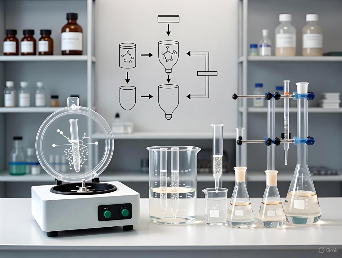
Workflow Visualization
The following diagram illustrates the decision pathway and procedural steps for implementing the core integrated principles.
Integrated Separation Workflow
The strategic integration of solvent extraction, freezing, and centrifugal force provides a versatile and powerful framework for modern sample preparation. The quantitative data and standardized protocols provided herein offer researchers a foundation for developing robust, reproducible methods for concentrating, purifying, and isolating analytes from complex matrices. Adherence to optimized parameters—such as centrifugation at 15°C for 20 minutes in freeze concentration—and the use of appropriate stabilizers during cryopreservation are critical for success. This synergistic approach addresses key challenges in drug development and biomedical research, enabling higher sensitivity in analysis and more reliable downstream results.
The Role of Low-Temperature Partitioning (LTP) in Purifying Complex Matrices
Low-temperature partitioning (LTP) is a powerful sample preparation technique that significantly enhances traditional solvent extraction methods by incorporating a freezing step to purify complex matrices. Originally conceptualized in the 1960s for purifying extracts from fatty matrices, LTP has evolved into a robust approach for extracting organic compounds from diverse samples, often without requiring additional cleanup steps [6] [7]. This technique leverages the differential solubility and partitioning behavior of analytes between the sample medium and a water-miscible organic solvent when subjected to freezing temperatures, typically around -20°C [6] [8].
In an analytical procedure, the sample preparation step is decisive in obtaining accurate and reliable results about the presence and concentration of analytes in complex matrices. LTP transforms these complex matrices (water, fruits, vegetables, soil, urine, etc.) into solutions suitable for analysis using instrumental techniques [6]. The most detachable characteristic of LTP is its ability to use the extractor solvent as a unique chemical, eliminating the need for multiple solvents and reducing the environmental impact of analytical procedures [6]. By freezing the sample, LTP induces phase separation while preserving the liquid organic phase, effectively immobilizing matrix components and particles that could interfere with subsequent analysis [6] [7].
Fundamental Principles and Mechanism
The underlying mechanism of LTP relies on the physical and chemical changes that occur when a sample-solvent mixture is subjected to low temperatures. When the mixture is cooled to temperatures around -20°C, the aqueous portion and solid components of the matrix precipitate and solidify, enabling clean phase separation [8]. This process allows target compounds to be retained in the liquid organic layer while minimizing co-extraction of matrix interferences [8].
The efficiency of LTP is governed by several key factors:
- Extractor solvent selection: Water-miscible organic solvents (primarily acetonitrile) are essential for proper phase separation [6]
- Freezing temperature and duration: Typically -20°C for 4-12 hours, depending on the matrix [9]
- Sample-to-solvent ratio: Affects extraction efficiency and phase separation [9]
- Vortexing and centrifugation parameters: Influence the initial extraction and subsequent phase separation [9]
The LTP approach enables the use of less toxic solvents than the chlorinated ones used in conventional extraction techniques, while also avoiding the formation of emulsions—a common problem in traditional liquid-liquid extraction [6].
Applications Across Sample Matrices
LTP techniques have demonstrated remarkable versatility across various complex matrices. The following table summarizes key applications and their demonstrated efficacy:
Table 1: Applications of LTP in Purifying Complex Matrices
| Matrix Category | Specific Matrices | Target Analytes | Key Findings | Citation |
|---|---|---|---|---|
| Food & Feed | Lettuce, bell pepper, beans, pineapple, tea | Pesticides (deltamethrin, thiamethoxam, triadimenol, flutriafol) | High recovery rates (81-111%) with minimal matrix effects; effective for monitoring MRLs | [6] [10] |
| Environmental | Water, soil, sewage sludge | Pesticides, pharmaceuticals, POPs, OCPs, PCBs | Efficient extraction from complex environmental samples; average recovery of 61% for POPs in caiman eggs | [6] [9] |
| Biological | Fish fillet, bovine liver, blood serum, urine | Veterinary drugs (levamisole), pharmaceuticals, cocaine | LOQ of 0.2 μg kg⁻¹ for levamisole in fish when combined with DLLME; effective for biological monitoring | [8] [6] |
| High-fat Foods | Edible oils, milk powder, eggs | Macrocyclic lactones, abamectin, ivermectin, chloramphenicol | Efficient extraction without additional cleanup; successful monitoring of veterinary drug residues | [6] |
Food and Feed Applications
The quantification and monitoring of organic compounds harmful to animal or human health in food and feed matrices represent a significant application area for LTP techniques. For instance, SLE-LTP has been successfully optimized and validated for determining pesticide residues in dehydrated green tea leaves, achieving limits of detection and quantification of 0.015 and 0.050 mg kg⁻¹ respectively, with recovery rates between 81-111% [10]. The method proved effective for extracting pesticides from green tea samples and was successfully applied to 14 different tea varieties sampled in Brazil [10].
Similarly, LTP methods have been applied to determine pesticides in lettuce, bell pepper, and other vegetables with high recovery rates and minimal matrix effects [6]. The technique has also been used for monitoring veterinary drug residues in foods of animal origin, including the determination of abamectin and ivermectin in edible oils, and chloramphenicol in milk powder [6].
Environmental Applications
Environmental monitoring presents unique challenges due to the complexity of matrices and the low concentrations of target analytes. LTP has demonstrated excellent performance in this domain. A notable application involves the determination of persistent organic pollutants (POPs), including organochlorinated pesticides and PCBs, in eggs of the Pantanal caiman (Caiman yacare) [9]. Using SLE-LTP with chemometric optimization, researchers achieved adequate recovery for environmental investigation, enabling monitoring of these contaminants in sensitive ecosystems [9].
LTP methods have also been successfully applied to various environmental samples, including water, soil, and sewage sludge, for extracting pesticides, pharmaceuticals, and other organic contaminants [6] [8]. The simplicity and efficiency of LTP make it particularly valuable for environmental monitoring in resource-limited settings.
Biological and Pharmaceutical Applications
The analysis of biological samples presents particular challenges due to their complex composition and the presence of interfering compounds such as proteins and lipids. LTP has shown great promise in this area. Recent research has demonstrated the first application of LTP for veterinary drug determination in fish fillet, specifically for levamisole detection [8]. When combined with dispersive liquid-liquid microextraction (DLLME), the method achieved an impressive limit of quantification of 0.2 μg kg⁻¹, well below the maximum residue limits established by regulatory agencies [8].
Other biological applications include the determination of cocaine in postmortem human liver tissue exposed to overdose, where LTP provided an efficient extraction and cleanup procedure [6]. The technique has also been applied to blood serum and urine samples for pharmaceutical analysis, demonstrating its versatility across different biological matrices [6].
Experimental Protocols
Standard SLE-LTP Protocol for Plant Materials
Principle: This protocol describes the determination of pesticide residues in dehydrated green tea leaves using SLE-LTP followed by GC-MS analysis [10].
Reagents and Materials:
- Dehydrated green tea leaves
- Acetonitrile (HPLC grade)
- Primary Secondary Amine (PSA) and Octadecylsilane (C18) sorbents
- Anhydrous magnesium sulfate
- Target pesticide standards (pirimiphos-methyl, flutriafol, cyproconazole, bifenthrin)
Procedure:
- Sample Preparation: Homogenize 2.0 g of dehydrated green tea leaves with 10 mL of acetonitrile in a 50 mL centrifuge tube.
- Extraction: Vortex the mixture for 5 minutes, then add 1.5 g of anhydrous magnesium sulfate and vortex for an additional 1 minute.
- Centrifugation: Centrifuge at 4000 rpm for 5 minutes to separate the solid residue.
- Low-Temperature Partitioning: Transfer the supernatant to a freezer at -20°C for 12 hours to complete phase separation.
- Clean-up: After LTP, subject the acetonitrile extract to a clean-up step using PSA and C18 as sorbents for chlorophyll removal.
- Analysis: Analyze by GC-MS using appropriate instrumental parameters.
Validation Parameters:
- Limit of Detection: 0.015 mg kg⁻¹
- Limit of Quantification: 0.050 mg kg⁻¹
- Recovery: 81-111%
- Coefficient of variation: <16%
Chemometrically Optimized SLE-LTP Protocol for Biological Tissues
Principle: This protocol describes the optimization of SLE-LTP for determining persistent organic pollutants in caiman eggs using a chemometric approach [9].
Reagents and Materials:
- Caiman egg homogenate
- Acetonitrile (HPLC grade)
- Organochlorine pesticide and PCB standards
- Anhydrous sodium sulfate
Procedure:
- Experimental Design: Implement a 2⁴ factorial design with triplicate central points considering extractor solvent volume (8-10 mL), vortexing time (1-5 min), centrifugation time (5-15 min), and freezing time (4-12 h).
- Sample Preparation: Homogenize 1.0 g of egg sample with the optimized solvent volume (12 mL) of acetonitrile.
- Extraction: Vortex for the optimized time (5 min) to ensure proper extraction.
- Centrifugation: Centrifuge at the optimized time (5 min) to separate phases.
- Low-Temperature Partitioning: Subject to freezing at -20°C for the optimized duration (12 h).
- Analysis: Analyze by GC-ECD or GC-MS.
Optimized Conditions:
- Extractor solvent volume: 12 mL
- Vortex time: 5 min
- Centrifugation time: 5 min
- Freezing time: 12 h
- Average recovery: 61% with RSD of 15% for samples with 22.5 ng g⁻¹
Advanced LTP-DLLME Protocol for Trace Analysis
Principle: This protocol combines LTP with dispersive liquid-liquid microextraction for sensitive determination of levamisole in fish fillets [8].
Reagents and Materials:
- Fish fillet (muscle plus skin in natural proportion)
- Levamisole hydrochloride analytical standard (>99.9% purity)
- Acetonitrile (HPLC grade)
- Internal standard: Levamisole-d5 Hydrochloride
- Extraction solvent for DLLME (chloroform)
- Disperser solvent (methanol)
Procedure:
- LTP Extraction:
- Homogenize 2.0 g of fish fillet with 10 mL of acetonitrile
- Vortex for 3 minutes, then centrifuge at 5000 rpm for 10 minutes
- Transfer supernatant to a freezer at -20°C for 12 hours
DLLME Concentration:
- Collect the organic phase after LTP
- Use as disperser solvent in DLLME with chloroform as extraction solvent
- Vortex mixture and centrifuge to separate phases
Analysis:
- Analyze by LC-MS/MS using multiple reaction monitoring (MRM)
- Use a C18 column (100 × 2.1 mm, 2.6 μm) with gradient elution
- Mobile phase: water and methanol both with 0.1% formic acid
Performance Characteristics:
- Limit of Quantification: 0.2 μg kg⁻¹
- Recovery: >85%
- Precision: RSD <15%
Workflow Visualization
LTP Experimental Workflow Diagram: This diagram illustrates the two main pathways (SLE-LTP and LLE-LTP) for purifying complex matrices using low-temperature partitioning techniques.
Research Reagent Solutions
Table 2: Essential Reagents and Materials for LTP Experiments
| Reagent/Material | Specifications | Function in LTP | Application Examples |
|---|---|---|---|
| Acetonitrile | HPLC grade, low water content | Primary extraction solvent, completely miscible with water, forms separate phase at low temperature | Universal solvent for most LTP applications [6] [10] |
| Primary Secondary Amine (PSA) | 40-60 μm particle size | Removal of fatty acids, sugars, and other polar organic acids from extracts | Clean-up step in tea and vegetable analysis [10] |
| C18 Sorbent | Octadecylsilane-bonded silica | Retention of non-polar interferents like chlorophyll and lipids | Pigment removal from plant extracts [10] |
| Anhydrous MgSO₄ | Analytical grade, finely powdered | Water removal from organic phase, prevents ice crystal formation during LTP | Standard in QuEChERS-based LTP methods [10] |
| Chloroform | HPLC grade | Extraction solvent in DLLME combined with LTP | Pre-concentration for trace analysis [8] |
| Centrifugal Filter Tubes | Amicon Ultra-15, polypropylene | Facilitate separation of concentrated solute from frozen matrix | Cryoconcentration of bioactive compounds [11] |
Low-temperature partitioning techniques represent a significant advancement in the purification of complex matrices for analytical purposes. The simplicity, efficiency, and environmental benefits of LTP make it particularly valuable for modern analytical laboratories, especially those with limited resources. The ability to use less toxic solvents, avoid emulsion formation, and achieve effective extract purification with minimal steps positions LTP as a technique aligned with green chemistry principles [6].
Future perspectives for LTP include further integration with other extraction and microextraction techniques to enhance sensitivity and selectivity, particularly for trace analysis [8]. The combination of LTP with techniques like DLLME has already demonstrated remarkable improvements in detection limits, as evidenced by the achievement of 0.2 μg kg⁻¹ LOQ for levamisole in fish [8]. Additionally, the ongoing optimization of LTP parameters through chemometric approaches will continue to expand its applications to new matrices and analyte classes [9].
As analytical science continues to emphasize sustainability and efficiency, LTP techniques are poised to play an increasingly important role in sample preparation across diverse fields including environmental monitoring, food safety, and pharmaceutical analysis. The demonstrated versatility, reliability, and practicality of LTP ensure its continued relevance in the analytical chemist's toolkit for the foreseeable future.
Sample preparation is a critical preliminary step in the analytical process, determining the accuracy, reproducibility, and sensitivity of final results [1]. Traditional methods often involve thermal processes or harsh chemical conditions that can compromise analyte integrity. This application note details how extractive freezing centrifugation addresses two pervasive challenges in sample preparation: analyte degradation and emulsification.
By leveraging cryogenic temperatures and centrifugal force, this technique significantly outperforms conventional approaches for processing thermosensitive and complex biological matrices, enabling researchers to obtain more reliable and representative analytical data.
Comparative Data: Advantages of Freeze-Based Techniques
The following tables summarize quantitative evidence from recent studies, demonstrating the superior performance of freeze-based concentration and preparation methods compared to traditional thermal techniques.
Table 1: Performance Comparison of Concentration Techniques for Bioactive Compounds in Maqui Extract
| Performance Metric | Cryoconcentration (Centrifugation-Filtration) | Evaporation at 50°C | Evaporation at 70°C | Evaporation at 80°C |
|---|---|---|---|---|
| Increase in Total Polyphenols | +280% | Data Not Provided | Data Not Provided | Data Not Provided |
| Increase in Total Anthocyanins | +573% | Data Not Provided | Data Not Provided | Data Not Provided |
| Increase in Antioxidant Capacity | +226% | Data Not Provided | Data Not Provided | Data Not Provided |
| Solute Separation Efficiency | >95% | Not Applicable | Not Applicable | Not Applicable |
| Cyanidin 3,5-diglucoside | Maintained | Maintained | Degraded | Degraded |
Source: Adapted from [11].
Table 2: Optimization of Freeze-Drying Conditions for Kashmiri Saffron Bioactives
| Freeze-Drying Temperature | Crocin Content (mg/g) | Picrocrocin Content (mg/g) | Safranal Content (mg/g) | Antioxidant Capacity (%) | Total Phenolic Content (mg GAE/g) |
|---|---|---|---|---|---|
| -50°C | Lower than -80°C | Lower than -80°C | Lower than -80°C | Lower than -80°C | Lower than -80°C |
| -60°C | Lower than -80°C | Lower than -80°C | Lower than -80°C | Lower than -80°C | Lower than -80°C |
| -70°C | Lower than -80°C | Lower than -80°C | Lower than -80°C | Lower than -80°C | Lower than -80°C |
| -80°C | 90.14 | 9.48 | 1.80 | 69.63 | 72.41 |
Source: Data compiled from [12]. Note: The study reported non-significant differences in the absolute values of bioactives across temperatures but identified -80°C as the optimal condition for maximal retention.
Detailed Experimental Protocols
Protocol 1: Cryoconcentration by Centrifugation-Filtration for Thermosensitive Berry Extracts
This protocol, adapted from methods developed for maqui berry, efficiently concentrates thermolabile bioactive compounds while avoiding thermal degradation [11].
Materials and Equipment
- Sample Material: Aqueous fruit extract (e.g., from maqui, blueberry).
- Centrifugal Filter Tubes: Amicon Ultra-15 centrifugal filter devices (or equivalent), with the nanofilter cellulose membrane removed [11].
- Laboratory Freezer: Capable of maintaining -30°C.
- Refrigerated Centrifuge: Capable of housing the filter tubes.
- Analytical Balance, Vortex Mixer, Refractometer.
Step-by-Step Procedure
- Preparation: Place 15 mL of the aqueous maqui extract into a prepared Amicon Ultra-15 filter tube [11].
- Freezing: Transfer the tubes to a freezer and freeze the sample completely at -30°C [11].
- Partial Thawing: Remove samples from the freezer and let them stand at ambient temperature for exactly 5 minutes [11].
- Centrifugation: Immediately place the tubes in a pre-balanced centrifuge. Spin at 4000 RPM for 10 minutes at 20°C. The centrifugal force will separate the concentrated solute from the frozen ice fraction [11].
- Collection: After centrifugation, collect the liquid concentrate and the remaining frozen matrix separately.
- Analysis: Determine the concentration of soluble solids (e.g., °Brix) in both fractions using a refractometer. Analyze bioactive content as required (e.g., total polyphenols, anthocyanins) [11].
Protocol 2: Freeze-Pour Sample Preparation for GC Analysis of Semivolatiles
This protocol is designed for complex, viscous matrices like e-liquids, where traditional dilution leads to matrix interference in GC systems. It effectively minimizes emulsification and protects semivolatile flavorings [13].
Materials and Equipment
- Sample: Complex liquid sample (e.g., e-liquid, flavored solution).
- Extraction Solvent: HPLC-grade hexane.
- Cooling Bath: Dry ice dissolved in acetone (-78°C).
- Glass Vials (20 mL) with Teflon-lined seals.
- Vortex Mixer, Sonication Bath, Refrigerated Centrifuge.
- GC-MS System with appropriate column and detector.
Step-by-Step Procedure
- Weighing: Accurately weigh 0.3 g of the sample into a 20 mL glass vial [13].
- Liquid-Liquid Extraction (LLE): Add 3 mL of hexane to the vial, cap it, and vortex for 10 seconds. Subsequently, sonicate the mixture for 3 minutes at 50°C [13].
- Freeze-Out: Transfer the vial to a cooling bath of dry ice and acetone (-78°C) for 2 minutes. This solidifies the viscous aqueous matrix (e.g., propylene glycol and glycerol) while the organic extract containing the analytes remains liquid [13].
- Centrifugation: Quickly transfer the vial to a centrifuge and spin at 860 g for 1 minute to compact the frozen matrix [13].
- Pour-Off: Decant approximately three-quarters of the hexane supernatant into a clean collection vial [13].
- Second Extraction: Repeat steps 2-5 with a fresh 3 mL of hexane and combine the supernatants [13].
- Analysis: Transfer an aliquot (e.g., 300 µL) of the combined extract to a GC vial for analysis [13].
Workflow and Performance Visualization
The following diagram illustrates the core logical relationship and procedural advantage of the extractive freezing centrifugation method over traditional pathways, highlighting how it avoids the primary pitfalls of degradation and emulsification.
Figure 1: Comparative Workflow Analysis. The traditional pathway (red) leads to analyte degradation or emulsification, while the extractive freezing centrifugation pathway (green) preserves analyte integrity for high-quality results.
The Scientist's Toolkit: Essential Research Reagent Solutions
Successful implementation of these protocols requires specific materials. The following table lists key reagents and their critical functions in the process.
Table 3: Essential Materials for Extractive Freezing Centrifugation Protocols
| Material/Reagent | Function/Application | Protocol |
|---|---|---|
| Amicon Ultra-15 Centrifugal Filter Tubes | Acts as a support to mechanically separate the concentrated solute from the frozen ice matrix during centrifugation, drastically improving efficiency [11]. | 3.1 |
| Polyethylene Glycol 6000 (PEG6000) | A crowding polymer used to precipitate nanovesicles and bioactive compounds from solution in a detergent-free manner, preserving their native structure [14]. | - |
| HPLC-Grade Hexane | Organic solvent used for liquid-liquid extraction of semivolatile analytes from complex matrices. Its low miscibility with water and volatility make it ideal for subsequent GC analysis [13]. | 3.2 |
| Dimethyl Sulfoxide (DMSO) | A cryoprotective agent used in cell freezing protocols to reduce ice crystal formation, preventing cellular damage and preserving viability during freezing [15]. | - |
| Dry Ice (Solid CO₂) | Used to create an ultra-low temperature bath (-78°C with acetone) for the "freeze-out" step, rapidly solidifying the matrix to facilitate separation [13]. | 3.2 |
| Synth-a-Freeze Cryopreservation Medium | A chemically defined, protein-free freezing medium, often containing DMSO, optimized for cryopreserving sensitive primary and stem cells [15]. | - |
Key Solvent Properties for Efficient Freezing-Out and Phase Separation
Phase separation is a fundamental thermodynamic process where a homogeneous mixture splits into two or more distinct phases, a principle leveraged in numerous chemical and biological sample preparation techniques. In the context of extractive freezing centrifugation for sample preparation, controlling this separation is paramount for achieving high purity and yield of the target analyte. The "freezing-out" phenomenon specifically refers to the process where a solute precipitates or is excluded from a solution phase as the temperature is lowered, often facilitated by the careful selection of a solvent system. The efficiency of this process is critically dependent on the thermodynamic and physical properties of the solvent used, which govern the phase equilibrium and the final morphology of the separated phases [16] [17]. This application note details the key solvent properties and provides a standardized protocol for researchers aiming to implement this technique.
Key Solvent Properties for Efficient Freezing-Out
The selection of an appropriate solvent is the most critical factor in designing an efficient freezing-out and phase separation process. The following properties determine a solvent's suitability.
Table 1: Key Solvent Properties for Freezing-Out and Phase Separation
| Property | Description | Impact on Process Efficiency |
|---|---|---|
| Distribution Coefficient (K) | Ratio of solute concentration in extract phase to its concentration in the raffinate phase ((K = y/x)) [18]. | A higher K value indicates greater solute transfer into the desired phase, improving yield and reducing solvent volume or number of stages needed [18]. |
| Selectivity (β) | Measure of the solvent's ability to preferentially dissolve the desired solute over other components ((β = KA / KB)) [18]. | High selectivity is crucial for achieving pure separations, minimizing the co-extraction of impurities [18]. |
| Solubility & Miscibility | The solubility of the target solute in the solvent and the miscibility of the solvent with the feed and anti-solvent [18] [16]. | Ensures sufficient solute capacity; controls the rate of phase separation (demixing) which impacts the final structure (e.g., sponge-like vs. macrovoids) [16]. |
| Physical Properties | Includes viscosity, density difference between phases, and surface tension [18] [16]. | A low viscosity and a high density difference promote faster phase disengagement. Surface tension affects droplet coalescence and interface stability [18]. |
| Chemical Stability | Inertness towards the solute, feed components, and equipment under process conditions [18]. | Prevents unwanted chemical reactions, degradation of the solute, or corrosion of equipment, ensuring product integrity and operational safety [18]. |
| Ease of Recovery | Governed by properties like boiling point relative to the solute and miscibility gap with the raffinate [18]. | A solvent that is easily separated and recycled (e.g., via distillation) significantly reduces operating costs and environmental impact [18]. |
| Safety & Environmental Impact | Considers toxicity, flammability, and biodegradability [18] [19]. | Moving towards greener, halogen-free solvents (e.g., o-xylene) minimizes health risks and environmental footprint [19] [20]. |
Thermodynamic Fundamentals
The phase behavior of a multi-component system is best understood using a ternary phase diagram. For a polymer/solvent/non-solvent system, this diagram is divided by a binodal curve into a stable single-phase region and a metastable two-phase region [16]. When the composition path of a polymer solution, during precipitation, crosses the binodal curve, liquid-liquid demixing occurs, leading to a polymer-rich phase and a polymer-lean phase. The tie-lines within the two-phase region connect the equilibrium compositions of these coexisting phases [16]. The fundamental driving force is the saturation concentration ((c{sat})). For a binary mixture, when the solute concentration exceeds (c{sat}), the system gains thermodynamic stability by separating into a solute-rich dense phase and a solute-poor dilute phase [17]. The interaction strength between components is often quantified by the Flory-Huggins interaction parameter (χ); a positive χ indicates unfavorable solvent-solute interactions, promoting phase separation [17].
Diagram 1: Phase Separation Process
Experimental Protocol: Extractive Freezing Centrifugation for Tissue Samples
This protocol outlines the steps for enriching membrane proteins from animal tissue using a freezing-out step and centrifugation, adapting principles from lysate and membrane fraction preparation [21].
Research Reagent Solutions
Table 2: Essential Materials and Reagents
| Item | Function / Description |
|---|---|
| Lysis Buffer A | 4 mM HEPES (pH 7.4), 320 mM Sucrose, 5 mM EDTA. Provides an isotonic, stabilizing environment for cellular components during homogenization [21]. |
| Lysis Buffer C | 50 mM Tris (pH 7.4), 1% Triton X-100, 5 mM EDTA. A detergent-based buffer for total protein extraction by solubilizing membranes [21]. |
| Protease Inhibitor Cocktail | Prevents proteolytic degradation of the target protein during the extraction process [21]. |
| Liquid Nitrogen | For flash-freezing tissue to preserve protein integrity and to potentially induce a pre-solidification step, limiting excessive phase separation [21] [20]. |
| Polytron Homogenizer | For efficient mechanical disruption of tissue to create a homogeneous lysate [21]. |
| Ultracentrifuge | For high-g-force separations required to pellet membrane fractions or to separate phases after freezing-out [21]. |
Step-by-Step Procedure
Tissue Harvesting and Flash-Freezing:
- Excise the tissue of interest and immediately submerge it in liquid nitrogen to flash-freeze. Store the tissue at -80°C until use [21].
- Rationale: Freezing rapidly halts metabolic activity and proteolysis, preserving the native state of proteins. This step can also initiate the controlled precipitation of components.
Preparation of Homogenate:
- Place the frozen tissue in 5 volumes of ice-cold Lysis Buffer A (for membrane enrichment) or Lysis Buffer C (for total lysate) supplemented with a protease inhibitor cocktail [21].
- Homogenize the tissue thoroughly using a polytron homogenizer until a uniform mixture is achieved.
- Rationale:
Initial Clarification and Freezing-Out Cycle:
- Centrifuge the homogenate at 2,000 x g for 10 minutes at 4°C. Discard the pellet containing large cellular debris and nuclei [21].
- Freezing-Out Step: Subject the clarified supernatant to a freeze-thaw cycle (e.g., flash-freeze in liquid nitrogen or at -80°C, then thaw slowly on ice). Alternatively, introduce a controlled non-solvent to induce phase separation.
- Rationale: This step promotes the aggregation and precipitation of less soluble components, including large macromolecular complexes and target aggregates, via a shift in thermodynamic equilibrium [17] [20].
Enrichment of Target Fraction by Ultracentrifugation:
- Transfer the supernatant from the previous step to a clean ultracentrifuge tube.
- Centrifuge at 100,000 x g for 1 hour at 4°C [21].
- Rationale: The high g-force pellets the membrane fragments and precipitated protein aggregates. The freezing-out step enhances this separation by increasing the size and density of the target aggregates.
Solubilization and Protein Quantification:
- Carefully discard the supernatant. Resuspend the pellet (containing the enriched membranes/target) in an appropriate volume of Lysis Buffer or a suitable extraction buffer [21].
- Briefly homogenize with the polytron to ensure a uniform suspension.
- Determine the protein concentration using the Bradford assay and adjust the concentration to the desired level (e.g., 4 mg/ml) [21].
- Aliquot and store the final protein samples at -80°C.
Diagram 2: Sample Preparation Workflow
Troubleshooting and Optimization
Challenge: Inefficient phase separation or low yield of target analyte.
- Solution: Optimize solvent properties, particularly the distribution coefficient and selectivity, by testing different solvent/anti-solvent combinations. Ensure a significant density difference between phases [18] [16]. The freezing-out temperature and rate can be adjusted to control crystal size and purity.
Challenge: Excessive or overly rapid phase separation leading to large, impure phases.
- Solution: Utilize strategies like active layer pre-solidification with liquid nitrogen. This rapid freezing accelerates molecular precipitation and crystallization, suppressing excessive phase separation and leading to a more favorable morphology for efficient extraction [20]. Switching to a greener, halogen-free solvent like ortho-xylene can also offer more controlled aggregation kinetics compared to harsh halogenated solvents [19] [20].
Challenge: Poor subcellular fractionation or co-precipitation of contaminants.
- Solution: Carefully tailor the lysis buffer composition (e.g., using sucrose for isotonic conditions) and centrifugation speed to isolate specific organelles. The inclusion of specific detergents (like Triton X-100) in the buffer can help solubilize desired components while leaving others in the pellet [21].
Historical Development and Evolution of the Technique in Analytical Chemistry
Sample preparation is a critical preliminary step in the analytical process, ensuring that raw samples are converted into a form suitable for accurate and reliable analysis. [1] Within this domain, techniques utilizing centrifugal force have revolutionized the ability to separate and purify analytes from complex matrices. This article explores the historical development and evolution of centrifugal techniques, with a specific focus on the principles and applications of extractive freezing centrifugation. This method integrates liquid-liquid extraction with a freeze-out step and centrifugal separation, offering a powerful approach for isolating target compounds from challenging sample matrices such as e-liquids, biological fluids, and environmental samples. [13]
Framed within a broader thesis on advanced sample preparation, this application note provides a detailed protocol for researchers, scientists, and drug development professionals seeking to implement this robust methodology.
Historical Development of Centrifugation
The genesis of centrifugal techniques dates back several centuries, with key innovations paving the way for modern applications.
Early Concepts and Industrial Adoption
The fundamental concept of centrifugal force was first formally described by the Dutch mathematician and scientist Christiaan Huygens in 1659. [22] The first significant industrial application came in 1878 with Gustaf de Laval's invention of the continuous cream separator in Sweden, which revolutionized the dairy industry by efficiently separating cream from milk. [23] [24] This established the centrifuge as a vital preparative tool.
The Rise of Analytical and Ultra-High-Speed Centrifugation
The early 20th century marked a shift from purely preparative to analytical applications. A pivotal figure was Theodor Svedberg, a Swedish chemist who invented the analytical ultracentrifuge in the 1920s. [23] His machine, capable of reaching speeds of up to 900,000 x g, enabled the study of macromolecular subunit structures and weights, for which he was awarded the Nobel Prize in Chemistry in 1926. [23] Concurrently, in the 1930s, Albert Claude pioneered the process of cell fractionation, using centrifugation as an essential step to separate cellular components based on mass, which unlocked new frontiers in cell biology. [22]
Modern Innovations and Miniaturization
The latter half of the 20th century saw the development of direct-drive motors by companies like Beckman, simplifying instrument design. [24] A significant milestone was the invention of the microcentrifuge in 1962, which, due to its compact size, became a ubiquitous tool in modern laboratories for routine sample processing, including nucleic acid extraction and protein precipitation. [23] Contemporary centrifuges now feature advanced capabilities such as intelligent control systems, automatic rotor identification, and precise temperature control, enhancing safety, reproducibility, and efficiency. [24]
Table 1: Key Historical Milestones in Centrifuge Development
| Time Period | Key Innovator/Developer | Contribution | Impact on Sample Preparation |
|---|---|---|---|
| 1659 | Christiaan Huygens | Formalized the term "centrifugal force". | Established the theoretical foundation for the technique. |
| 1878 | Gustaf de Laval | Invented the first continuous cream separator. | Introduced centrifugation for industrial-scale preparative separation. |
| 1869 / 1920s | Friedrich Miescher / Theodor Svedberg | First isolation of nucleic acids via centrifugation; invented the analytical ultracentrifuge. | Enabled the study of macromolecules and advanced biochemical research. |
| 1930s | Albert Claude | Discovered the process of cell fractionation. | Opened new avenues for cellular biology by separating organelles. |
| 1962 | Netheler & Hinz Medizintechnik | Invented the first microcentrifuge. | Made centrifugation accessible and routine for countless lab applications. |
| 2000s - Present | Various Manufacturers | Integrated smart controls, auto-balancing, and precise refrigeration. | Improved safety, reproducibility, and throughput in sample preparation. |
Evolution Towards Extractive Freezing Centrifugation
The evolution of sample preparation has been characterized by a drive towards miniaturization, automation, and the reduction of hazardous solvent use in line with Green Analytical Chemistry (GAC) principles. [25] While simple dilute-and-shoot approaches are common, complex matrices often require more sophisticated techniques to isolate analytes and remove interfering components. [13]
The extractive freezing centrifugation method represents an advanced evolution that combines multiple principles:
- Liquid-Liquid Extraction (LLE): Separates compounds based on differential solubility in two immiscible solvents. [1]
- Freeze-Out ("Freeze-Pour") Step: Uses ultra-low temperatures to solidify the aqueous or matrix-rich phase, allowing for the facile decanting of the organic extract. [13]
- Centrifugal Force: Enhances the phase separation and ensures a clean recovery of the supernatant. [13]
This synergistic approach effectively overcomes challenges such as the co-extraction of abundant, interfering matrix components (e.g., propylene glycol and glycerol in e-liquids), which can compromise chromatographic analysis and detector performance. [13]
Application Note: Analysis of Semivolatile Flavouring Chemicals in E-Liquids
The following protocol, adapted from a published method for the GC-analysis of semivolatile flavourings, details the application of extractive freezing centrifugation. [13]
Principle
This method uses liquid-liquid extraction with hexane to transfer target semivolatile analytes from a propylene glycol/glycerol-based e-liquid matrix into an organic solvent compatible with GC analysis. A subsequent freeze-out step at -78°C solidifies the viscous e-liquid matrix, allowing for the clean separation and collection of the hexane extract via centrifugation and decanting. This process effectively removes matrix interferents and concentrates the analytes.
Research Reagent Solutions
Table 2: Essential Materials and Reagents
| Item | Function / Specification |
|---|---|
| Hexane (ACS grade or higher) | Extraction solvent; immiscible with the e-liquid matrix, selectively dissolves target organic analytes. |
| Propylene Glycol (PG) / Glycerol (GL) | Matrix simulation; prepare a 60:40 (w/w) PG/GL mixture for method validation. [13] |
| Analytical Reference Standards | High-purity target analytes (e.g., 2-furyl methyl ketone, pulegone, estragole, cinnamaldehyde). [13] |
| Internal Standard Solution | e.g., o-methyl anisole or 2-chlorobenzaldehyde in hexane (10 mg/mL). Corrects for procedural variability. [13] |
| Dry Ice / Acetone Cooling Bath | Provides the ultra-low temperature (-78°C) required for the freeze-out step. |
| Refrigerated Benchtop Centrifuge | Capable of accommodating extraction vials and achieving ~860 x g. |
| Glass Vials (20 mL) with Teflon-lined Caps | For extraction; chemically resistant and suitable for vortexing and sonication. |
| Gas Chromatograph-Mass Spectrometer (GC-MS) | Equipped with a suitable capillary column (e.g., 5% phenyl–95% methylpolysiloxane, 60 m x 0.25 mm, 0.25 µm). [13] |
Detailed Experimental Protocol
Sample Preparation Workflow
The following diagram illustrates the complete experimental workflow from sample weighing to instrumental analysis:
Step-by-Step Procedure
- Sample Weighing: Precisely weigh 0.5 g of the e-liquid sample into a 20 mL glass vial. For validation, spike the appropriate amount of analytical standard into a 60:40 PG/GL mixture. [13]
- First Extraction:
- Add 3 mL of hexane and the internal standard solution (to a final concentration of 10 µg/mL after extraction) to the vial. [13]
- Cap the vial securely with a Teflon-lined seal.
- Vortex the mixture vigorously for 10 seconds to ensure thorough mixing. [13]
- Sonicate the vial in a water bath at 50°C for 3 minutes to enhance extraction efficiency. [13]
- Freeze-Out and Collection:
- Transfer the vial to a cooling bath of dry ice dissolved in acetone (-78°C) for exactly 2 minutes. The aqueous/matrix phase will solidify. [13]
- Immediately centrifuge the vial at 860 x g for 1 minute to ensure complete separation. [13]
- Carefully decant and collect approximately three-quarters of the organic (hexane) supernatant into a clean glass vial. [13]
- Second Extraction Cycle:
- Add another 3 mL of fresh hexane to the original sample vial containing the frozen matrix pellet.
- Repeat steps 2 and 3 (Vortex, Sonicate, Freeze-Out, Centrifuge, and collect the supernatant).
- Combine this second supernatant with the first. [13]
- Analysis:
Method Validation Data
When validated according to ICH guidelines for four flavourings, this methodology has demonstrated robust performance, as summarized below. [13]
Table 3: Example Validation Parameters for a Selected Analyte
| Validation Parameter | Result (e.g., Cinnamaldehyde) | Acceptance Criteria |
|---|---|---|
| Linear Range | 1 - 100 µg/mL | R² > 0.995 |
| Accuracy (% Recovery) | 95 - 105% | 85 - 115% |
| Precision (% RSD) | < 5% (Intra-day) | ≤ 10% |
| Limit of Quantification (LOQ) | 1 µg/mL | S/N ≥ 10 |
Integration with Modern Trends
The extractive freezing centrifugation protocol aligns with major trends in analytical chemistry:
- Green Chemistry: The method minimizes matrix waste and can be adapted to use smaller solvent volumes, reducing environmental impact. [25]
- Automation: Steps like vortexing, solvent addition, and even supernatant transfer can be automated using robotic liquid handlers, enhancing throughput and reproducibility. [26]
- Hyphenated Techniques: The clean extract produced is ideally suited for direct analysis by advanced hyphenated systems like GC-MS/MS, providing high sensitivity and definitive identification. [13] [26]
The historical journey of centrifugation, from Huygens' theoretical concept to Svedberg's analytical ultracentrifuge and today's smart microcentrifuges, underscores its indispensable role in science. The extractive freezing centrifugation method exemplifies the continued evolution of these principles into a sophisticated, robust, and effective sample preparation technique. By successfully isolating semivolatile analytes from complex and challenging matrices, it addresses a critical need in analytical chemistry for drug development, environmental monitoring, and product safety assessment. This protocol provides a validated foundation that researchers can adapt and optimize for their specific application needs.
Step-by-Step Protocols and Real-World Applications for Biomedical Research
Standardized Protocol for Liquid-Liquid Extractive Freezing Centrifugation (LLE-LTP)
Liquid-Liquid Extractive Freezing Centrifugation (LLE-LTP) is an advanced sample preparation technique that synergistically combines the principles of liquid-liquid extraction (LLE) with the purification and concentration capabilities of freeze-centrifugation. This protocol is framed within a broader thesis on extractive freezing centrifugation, which posits that the integration of selective solvent extraction with physical separation under centrifugal force and freezing temperatures can significantly enhance the recovery and purity of target analytes from complex matrices.
The core principle leverages the differential solubility of a target compound between two immiscible liquid phases, followed by a rapid freezing step that separates the solute-enriched solvent from the aqueous phase. The subsequent application of centrifugal force efficiently partitions the now-immiscible phases—the frozen aqueous matrix and the liquid solvent concentrate—leading to a high-yield, high-purity extract. This method is particularly advantageous for concentrating thermosensitive bioactive compounds, such as those found in natural product extracts, where traditional thermal evaporation can lead to degradation [11]. The process can achieve solute separation efficiencies exceeding 95% from the frozen fraction [11] [27].
Applications
This LLE-LTP protocol is designed for researchers and drug development professionals requiring highly concentrated and purified extracts. Its specific applications include:
- Harvesting Bioactive Compounds from Natural Extracts: Efficiently concentrating thermolabile molecules like polyphenols and anthocyanins from berry extracts (e.g., maqui) without thermal degradation [11].
- Isolation of Marine Lipophilic Toxins: Extracting compounds such as Gymnodimine-A (GYM-A) from large-volume microalgal cultures with high recovery rates [28].
- Pre-concentration of Organic Acids: Enhancing the extraction efficiency of monocarboxylic acids from aqueous solutions using immiscible organic solvents like acetonitrile under centrifugal force [27].
- Sample Pre-treatment for Analytical Chemistry: Preparing samples for high-performance liquid chromatography (HPLC) or mass spectrometry (MS) by isolating and concentrating analytes from complex biological or environmental samples [1].
Equipment & Reagents
Research Reagent Solutions
| Item | Specification / Function |
|---|---|
| Extraction Solvent | Select based on target analyte solubility and immiscibility with water (e.g., Dichloromethane for lipophilic toxins [28]; Acetonitrile for organic acids [27]). |
| Aqueous Sample | The solution containing the target analyte(s). pH may require adjustment to stabilize compounds [28]. |
| Salting-Out Agents | (Optional) Salts like NaCl or MgSO4 to improve partitioning of analytes into the organic solvent. |
| Internal Standards | (Optional) For quantitative analysis, to correct for variability in extraction efficiency. |
| pH Buffers | To adjust and maintain the sample at an optimal pH for extraction and analyte stability. |
Laboratory Equipment
- Centrifuge: Capable of maintaining 4,000 rpm and 20°C, with a rotor compatible with 15-50 mL tubes [11].
- Centrifugal Filter Tubes: Polypropylene tubes with a filter support, such as Amicon Ultra-15 centrifugal filter devices (the nanofilter cellulose membrane may be removed to function solely as a support) [11].
- Freezer: Capable of maintaining -30°C.
- Analytical Balances: High-precision for accurate weighing.
- Vortex Mixer: For thorough mixing of sample and solvent.
- Volumetric Pipettes: For accurate measurement and transfer of liquids.
- Refractometer: For determining the concentration of soluble solids (°Brix) to calculate process efficiency [11].
Step-by-Step Protocol
The following diagram illustrates the complete LLE-LTP experimental workflow:
Detailed Experimental Procedure
Step 1: Solvent Addition and Mixing
- Measure a defined volume of the aqueous sample (e.g., 15 mL) [11].
- Add a precisely measured volume of the selected organic extraction solvent. The ratio is critical; for example, a ratio of 55 mL of dichloromethane per liter of sample (5.5%, v/v) has been used for toxin extraction [28].
- Mix the biphasic system thoroughly using a vortex mixer for several minutes to ensure maximum contact between the phases and partitioning of the analyte into the solvent.
Step 2: Primary Incubation
- Allow the mixture to settle in a separation funnel or a capped tube for a predetermined time to facilitate phase separation. This step can be performed at room temperature or under controlled conditions based on analyte stability.
Step 3: Transfer to Centrifuge Tube
- Transfer the entire mixture, or the solvent-rich layer if visible, into a polypropylene centrifugal filter tube. The filter acts as a support to separate the concentrated liquid phase from the frozen matrix later in the process [11].
Step 4: Freezing
- Place the loaded centrifugal filter tubes in a freezer and freeze completely at -30°C [11]. Ensure the tubes are upright and stable.
Step 5: Centrifugation
- Remove the samples from the freezer and let them stand at ambient temperature for a brief period (e.g., 5 minutes) to slightly temper the frozen matrix [11].
- Load the tubes into the centrifuge and spin at a defined force and time (e.g., 4000 rpm for 10 minutes at 20°C). The centrifugal force will push the solute-enriched liquid solvent through and out of the frozen aqueous matrix, which is retained by the filter support [11].
Step 6: Collect Concentrate
- Carefully decant or pipette the liquid concentrate from the collection vessel of the centrifugal filter unit.
- The frozen aqueous fraction, now largely depleted of the target solute, can be discarded.
Step 7: Multi-Cycle Concentration (Optional)
- For higher concentration factors, the concentrate from one cycle can be used as the "aqueous sample" input for a subsequent LLE-LTP cycle [11]. This cascading approach can dramatically increase the final concentration of the analyte.
Data Analysis & Calculations
Key Performance Metrics
The efficiency of the LLE-LTP process should be quantified using the following metrics, derived from the concentration of soluble solids measured by a refractometer [11].
Concentration Efficiency (η): This is the primary metric for evaluating the process's performance in separating solute from the frozen aqueous phase.
η (%) = [(C_s - C_f) / C_s] × 100
C_s= Concentration of solids (°Brix) in the concentrated solution.C_f= Concentration of solids (°Brix) in the frozen fraction.
Analyte Recovery (%): For specific target compounds, this is calculated by comparing the total amount of analyte in the final concentrate to the known amount in the initial sample, typically using HPLC or GC-MS analysis. Recovery rates of ~88% have been reported for specific toxins using related methods [28].
Concentration Factor (CF): The fold-increase in the concentration of the target analyte or total soluble solids.
CF = C_final / C_initial
Quantitative Data from Related Studies
The following table summarizes performance data from studies utilizing related cryoconcentration and LLE techniques, which this protocol aims to integrate.
| Application | Method | Key Performance Outcome | Reference |
|---|---|---|---|
| Aqueous Maqui Extract | Cryoconcentration by Centrifugation-Filtration (3 cycles) | Efficiency (η): >95% Total Polyphenols: +280% Total Anthocyanins: +573% Antioxidant Capacity: +226% | [11] |
| Gymnodimine-A from Microalgae | Liquid-Liquid Extraction (Dichloromethane) | Recovery: 88% from 2L culture | [28] |
| Monocarboxylic Acids from Water | Freeze-Out Extraction with Centrifugation | Partition Coefficient: Linear growth with molecule length; process efficiency increased under centrifugal force. | [27] |
Troubleshooting
| Problem | Potential Cause | Solution |
|---|---|---|
| Low Concentration Efficiency | Incomplete freezing; insufficient centrifugal force or time; incorrect solvent-to-sample ratio. | Ensure complete freezing at -30°C; optimize centrifuge speed and duration; re-evaluate solvent ratio. |
| Poor Analyte Recovery | Incorrect solvent choice for the target analyte; pH instability of the analyte; analyte degradation. | Select a solvent with a higher partition coefficient for the analyte; adjust sample pH to stabilize the analyte (e.g., pH 5.0 for GYM-A [28]). |
| Emulsion Formation | Presence of surfactants or emulsifying agents in the sample. | Use salting-out agents, perform a brief centrifugation before the freezing step, or gently stir instead of vortexing. |
| Incomplete Phase Separation | Solvent and sample are not sufficiently immiscible; freezing is incomplete/uneven. | Confirm the immiscibility of the solvent-water system. Ensure uniform and complete freezing. |
Protocol for Solid-Liquid Extractive Freezing Centrifugation (SLE-LTP) from Tissues and Plants
Solid-Liquid Extractive Freezing Centrifugation (SLE-LTP) is an advanced sample preparation technique designed for the efficient extraction of analytes from complex biological matrices such as plant and tissue samples. This method integrates solid-liquid extraction (SLE) with a low-temperature partitioning (LTP) clean-up step, enabling high recovery rates of target compounds while effectively removing interfering substances like pigments, lipids, and sugars [29] [30]. Within the broader context of extractive freezing centrifugation research, SLE-LTP represents a significant advancement for preparing samples for sophisticated analytical techniques including liquid chromatography-mass spectrometry (LC-MS) and gas chromatography-mass spectrometry (GC-MS), particularly in pharmaceutical and environmental analysis [1].
The fundamental principle of SLE-LTP involves using a water-miscible organic solvent for initial extraction, followed by the induction of phase separation through the addition of salts and exposure to freezing temperatures. This process simultaneously concentrates analytes and removes water-soluble interferences, making it especially valuable for multi-residue analysis of pesticides, pharmaceuticals, and other organic compounds in challenging matrices [29].
Principles and Mechanisms
SLE-LTP operates on several interconnected physicochemical principles that govern its efficiency. The initial extraction phase utilizes solvents with high affinity for target analytes, facilitating their diffusion from the solid matrix into the liquid phase. The subsequent low-temperature partitioning step capitalizes on the differential solubility of compounds and the phenomenon of freezing point depression induced by salt addition [30].
When salts such as sodium chloride or magnesium sulfate are added to the aqueous-organic mixture, they reduce the solubility of organic compounds in the aqueous phase via salting-out effects. Concurrently, the mixture's freezing point is depressed, allowing for phase separation and analyte concentration at temperatures above the normal freezing point of water. This process effectively partitions non-polar analytes into the organic phase while leaving polar interferents in the aqueous phase, which solidifies upon cooling [29] [30].
The theoretical foundation of this technique aligns with the broader objectives of extractive freezing centrifugation, which aims to integrate extraction, clean-up, and concentration into a single streamlined process. This integration minimizes sample handling, reduces potential sources of contamination, and enhances overall methodological efficiency—critical considerations in modern analytical chemistry [1].
Materials and Equipment
Research Reagent Solutions
Table 1: Essential reagents for SLE-LTP protocols
| Reagent/Material | Function | Example Specifications |
|---|---|---|
| Acetonitrile (with acid modifier) | Primary extraction solvent | HPLC grade, with 0.1-1% acetic or formic acid [29] |
| Anhydrous Magnesium Sulfate (MgSO₄) | Water removal, induces exothermic reaction | Analytical grade, ensures complete dehydration [29] |
| Sodium Chloride (NaCl) | Salting-out agent, phase separation | Analytical grade, enhances organic/aqueous partition [29] [30] |
| Primary Secondary Amine (PSA) | Clean-up sorbent for polar interferences | 40-60 μm particle size, removes fatty acids & sugars [29] |
| Octadecylsilane (C18) | Clean-up sorbent for non-polar interferences | 50 μm particle size, removes lipids & pigments [29] |
| Water (Deionized) | Hydration of samples, solvent component | Ultrapure (18.2 MΩ·cm) [29] |
Required Equipment
- Polypropylene centrifuge tubes (50 mL capacity, screw cap)
- Analytical balance (±0.1 mg precision)
- Vortex mixer
- Ultrasonic bath (with temperature control, 40°C)
- Refrigerated centrifuge (capable of 4,500 rpm and 12,000 rpm)
- Freezer (-20°C to -80°C capability)
- pH meter
- Syringe filters (0.22 μm, nylon or PTFE)
- Autosampler vials (compatible with HPLC/UPLC systems) [29]
Step-by-Step Experimental Protocol
Sample Preparation and Pre-Treatment
Proper sample preparation is critical for ensuring representative analysis and maximizing extraction efficiency.
- Homogenization: Fresh tissue or plant samples should be finely chopped and homogenized using a blender or food processor. For lyophilized samples, grind to a fine powder using a mortar and pestle or mechanical grinder [1].
- Sieving: Pass the homogenized material through a sieve (0.25-2 mm mesh) to ensure particle size uniformity [31].
- Storage: Store processed samples at -18°C or below if not extracted immediately to prevent analyte degradation [31].
SLE-LTP Extraction Procedure
Table 2: Step-by-step SLE-LTP protocol for plant and tissue samples
| Step | Procedure | Parameters | Critical Notes |
|---|---|---|---|
| 1. Weighing | Accurately weigh 10.0 ± 0.1 g of homogenized sample into a 50 mL polypropylene centrifuge tube. | Sample mass: 10.0 ± 0.1 g | Use analytical balance; record exact weight [29] |
| 2. Hydration | Add 10 mL deionized water to the sample. | Water volume: 10 mL | Essential for dried samples to rehydrate matrix [29] |
| 3. Solvent Addition | Add 10 mL acetonitrile containing 0.1% acetic acid. | Solvent volume: 10 mL; Acid modifier: 0.1% acetic acid | Acid improves recovery of acidic compounds [29] |
| 4. Vortex Mixing | Secure tube cap and vortex vigorously for 3 minutes. | Time: 3 minutes | Ensure complete sample-solvent contact [29] |
| 5. Ultrasonic Extraction | Place tube in ultrasonic bath maintained at 40°C for 5 minutes. | Time: 5 min; Temperature: 40°C | Enhances extraction efficiency; controlled temperature prevents degradation [29] |
| 6. Salt Addition | Add approximately 2 g sodium chloride (NaCl). | Mass: ~2 g NaCl | Induces phase separation via salting-out effect [29] [30] |
| 7. Initial Centrifugation | Centrifuge at 4500 rpm for 5 minutes. | Speed: 4500 rpm; Time: 5 min | Clear phase separation; organic phase should be distinct [29] |
| 8. Extract Collection | Transfer 1.0 mL of supernatant (organic layer) to a 2 mL centrifuge tube containing 100 mg MgSO₄, 25 mg PSA, and 25 mg C18. | Volume: 1.0 mL; Sorbents: MgSO₄, PSA, C18 | Precise volume measurement critical for reproducibility [29] |
| 9. Clean-up | Vortex the mixture for 1 minute. | Time: 1 minute | Ensures proper interaction with clean-up sorbents [29] |
| 10. Final Centrifugation | Centrifuge at 12,000 rpm for 5 minutes. | Speed: 12,000 rpm; Time: 5 min | Pelletizes sorbents and removed interferents [29] |
| 11. Filtration | Filter supernatant through 0.22 μm syringe filter into autosampler vial. | Filter pore size: 0.22 μm | Removes particulate matter; prevents instrument clogging [29] |
Process Flow Visualization
Method Validation and Quality Control
Rigorous validation ensures the reliability, accuracy, and precision of the SLE-LTP method for analytical applications.
Quantitative Performance Parameters
Table 3: Method validation parameters and typical acceptance criteria
| Validation Parameter | Experimental Procedure | Acceptance Criteria | Reported Performance |
|---|---|---|---|
| Recovery | Compare analyte response in spiked matrix vs. pure solvent | 70-120% with RSD < 20% | 67-87% for fungicides in bell peppers [30] |
| Precision (Repeatability) | Analyze multiple replicates (n≥5) on same day | RSD ≤ 20% | Satisfactory per ICH & EC criteria [30] |
| Linearity | Analyze calibration standards across working range | R² ≥ 0.990 | Established for multi-herbicide analysis [29] |
| Limit of Detection (LOD) | Signal-to-noise ratio of 3:1 | Compound-dependent | Verified for azoxystrobin, chlorothalonil, difenoconazole [30] |
| Limit of Quantification (LOQ) | Signal-to-noise ratio of 10:1 | Compound-dependent | Verified for azoxystrobin, chlorothalonil, difenoconazole [30] |
| Selectivity/Specificity | Analyze blank matrix; check interference at retention times | No significant interference at target RT | Confirmed via chromatographic comparison [30] |
Quality Control Measures
- Procedure Blanks: Analyze solvent blanks with each batch to monitor contamination.
- Matrix Spikes: Include spiked samples with each batch to verify recovery performance.
- Internal Standards: Use deuterated or structural analog internal standards when available to correct for matrix effects and volume variations.
- Calibration Standards: Prepare matrix-matched calibration standards to compensate for matrix effects [29] [30].
Applications and Case Studies
SLE-LTP has demonstrated particular effectiveness in several application areas:
Multi-Residue Pesticide Analysis in Agricultural Soils
In a comprehensive study of long-lasting herbicides in agricultural soils across Henan Province, China, researchers employed a SLE-LTP approach to analyze nine herbicides (including atrazine, imazethapyr, fomesafen, and pyroxasulfone) in 365 soil samples. The method successfully detected target compounds with varying physicochemical properties, with detection rates ranging from 1.2% for mesosulfuron-methyl to 33.9% for atrazine [29].
Fungicide Residue Analysis in Bell Peppers
SLE-LTP has been validated for determining systemic (azoxystrobin, difenoconazole) and contact (chlorothalonil) fungicides in bell peppers. The method demonstrated satisfactory performance according to International Council for Harmonisation (ICH) and European Commission criteria, enabling the evaluation of washing strategies for pesticide residue removal. The study found that ozone treatment effectively reduced fungicide levels by 67-87% [30].
Emerging Pollutant Monitoring
While not directly applied in the cited studies, the principles of SLE-LTP align with extraction techniques used for emerging pollutants (EPs) in complex matrices. The method's efficiency with acetonitrile-based extraction and clean-up makes it suitable for pharmaceuticals, personal care products, flame retardants, and plasticizers in environmental samples [31].
Troubleshooting and Optimization
Common Issues and Solutions
Table 4: Troubleshooting guide for SLE-LTP protocols
| Problem | Potential Causes | Solutions |
|---|---|---|
| Poor Recovery | Incomplete extraction; analyte degradation; insufficient clean-up | Optimize solvent composition; reduce extraction temperature; extend ultrasonic time; adjust sorbent ratios |
| Inconsistent Results | Inhomogeneous sample; variable water content; improper phase separation | Improve homogenization; standardize hydration; ensure consistent salt addition; verify centrifugation parameters |
| Matrix Interferences | Inadequate clean-up; co-extracted compounds | Increase sorbent amounts; adjust sorbent combinations (e.g., add GCB for pigments); implement additional freezing step |
| Low Precision | Inconsistent sample weights; variable solvent volumes; temperature fluctuations | Calibrate balances and pipettes; standardize laboratory conditions; use internal standards |
| Phase Separation Issues | Insufficient salt; incomplete mixing; incorrect centrifugation | Verify salt quality and quantity; ensure proper vortexing; optimize centrifugation speed and time |
Method Optimization Strategies
- Solvent Selection: Acetonitrile is generally preferred for polar to medium-polarity compounds. For more non-polar analytes, acetone or ethyl acetate may provide better recovery.
- Acid/Base Modification: Adjust pH with acetic acid (for acidic compounds) or ammonium hydroxide (for basic compounds) to enhance extraction efficiency.
- Sorbent Combinations: Optimize PSA/C18 ratios based on matrix composition. For pigmented samples, consider adding graphitized carbon black (GCB).
- Freezing Temperature and Duration: Experiment with freezing temperatures (-20°C to -80°C) and durations (1-12 hours) to maximize interference removal while maintaining analyte recovery [29] [30].
The SLE-LTP protocol represents a robust, efficient, and versatile sample preparation methodology particularly suited for the extraction of organic compounds from complex plant and tissue matrices. Its integration of extraction, clean-up, and concentration into a single workflow reduces analysis time, minimizes solvent consumption, and enhances laboratory efficiency.
Within the broader context of extractive freezing centrifugation research, SLE-LTP demonstrates how controlled temperature manipulation combined with solvent-salt systems can achieve superior sample clean-up compared to traditional approaches. The method's effectiveness has been validated across multiple applications, from pesticide residue analysis in agricultural products to environmental monitoring of organic contaminants.
Future developments in SLE-LTP may focus on further automation, adaptation to novel sorbent materials, and expansion to additional analyte classes. The continued refinement of this technique will undoubtedly contribute to its growing adoption in analytical laboratories specializing in food safety, environmental monitoring, and pharmaceutical research.
The demand for health-promoting foods and pharmaceuticals has intensified the search for robust methods to concentrate bioactive compounds from natural sources. Thermosensitive phytochemicals, such as polyphenols and anthocyanins, present a particular processing challenge as conventional thermal concentration methods often degrade these valuable compounds, reducing their bioactivity and nutritional value [11]. Cryoconcentration has emerged as a superior alternative for processing thermolabile substances, utilizing low temperatures to preserve compound integrity. This application note details an innovative cryoconcentration by centrifugation–filtration method, which simultaneously separates and concentrates thermosensitive bioactives from aqueous natural extracts with remarkable efficiency [11]. Within the broader thesis research on extractive freezing centrifugation for sample preparation, this protocol demonstrates how combining freezing with centrifugal filtration can achieve solute separation efficiencies exceeding 95% while maintaining the structural and functional integrity of even the most heat-labile compounds.
Key Principles and Advantages of Cryoconcentration
Cryoconcentration operates on the fundamental principle of separating concentrated solutes from a frozen matrix through controlled ice crystal formation. Unlike evaporation technologies that apply heat and risk degrading thermosensitive compounds, cryoconcentration processes occur at low temperatures, minimizing the loss of volatile aromas and the degradation of heat-labile nutrients [11]. This method offers significant advantages for producing high-quality concentrates from fruit extracts and other natural products containing valuable phytochemicals.
Traditional single-stage cryoconcentration systems often rely on gravitational separation, which typically results in lower process efficiency. The integration of centrifugation–filtration technology represents a substantial advancement, dramatically improving the separation of the concentrated phase from the frozen fraction [11]. This hybrid approach maintains the temperature-sensitive nature of the cryoconcentration process while overcoming the efficiency limitations that have previously restricted its widespread industrial adoption. The method is particularly suited for concentrating berry extracts and other natural products where preserving anthocyanin content and antioxidant capacity is paramount for end-product quality.
Experimental Protocol: Cryoconcentration by Centrifugation–Filtration
Materials and Equipment
Research Reagent Solutions
| Item | Function/Specification |
|---|---|
| Fresh Maqui Fruits | Source of anthocyanins and polyphenols [11]. |
| Amicon Ultra-15 Centrifugal Filter Tubes | Polypropylene tubes for centrifugation-filtration; nanofilter cellulose membrane removed [11]. |
| Refractometer | Measurement of soluble solids concentration (°Brix) with 0.1° precision [11]. |
| Rotary Evaporator | For comparative thermal concentration at 50°, 70°, and 80°C [11]. |
| Folin-Ciocalteu Reagent | Spectrophotometric determination of total polyphenol content [11]. |
| DPPH (2,2-Diphenyl-1-picrylhydrazyl) | Assessment of antioxidant capacity via radical scavenging assay [11]. |
| Orbital Shaker | Extraction of soluble compounds from seeds and skin (570 rpm for 50 min) [11]. |
| Freezing Chamber | Sample freezing at -30°C [11]. |
| Centrifuge | Sample separation at 4,000 rpm for 10 minutes at 20°C [11]. |
Sample Preparation: Aqueous Maqui Extract
- Fruit Preparation: Clean and prepare fresh maqui fruits (Aristotelia chilensis (Mol.) Stuntz). Refrigerate at 4°C until processing [11].
- Pulping: Process fruits in a pulper to separate the pulp from the seeds and skin [11].
- Aqueous Extraction: Combine the separated seeds and skin with water at a 1:1 weight/volume ratio. Agitate the mixture at 570 rpm for 50 minutes using an orbital shaker to extract soluble compounds [11].
- Homogenization and Filtration: Mix the freshly obtained maqui juice with the aqueous extract. Vacuum filter the combined liquid, then store the final aqueous extract in amber bottles at -30°C until analysis [11].
Cryoconcentration Procedure
- Sample Loading: Transfer a 15 mL aliquot of the aqueous maqui extract into an Amicon Ultra-15 centrifugal filter tube (with the nanofilter membrane removed) [11].
- Freezing: Place the loaded tube in a freezing chamber and maintain at -30°C until completely frozen [11].
- Conditioning: Remove the sample from the freezer and let it stand at ambient temperature for exactly 5 minutes [11].
- Centrifugation: Load the tube into a centrifuge and process at 4,000 rpm for 10 minutes at 20°C. This step mechanically separates the concentrated solute from the frozen water matrix [11].
- Multi-Cycle Processing: For higher concentration factors, use the concentrate from one cycle (C1) as the starting material for the next cycle (C2). Repeat this process sequentially (e.g., for a third cycle, C3) to achieve the desired solute concentration [11].
- Concentration Measurement: Use a refractometer to determine the soluble solids concentration (°Brix) in both the collected concentrate and the remaining frozen fraction at ambient temperature [11].
Efficiency Calculation
The efficiency ((n)) of each cryoconcentration cycle is calculated as the percentage increase in solute concentration in the concentrate, accounting for solids remaining in the ice fraction [11]. The formula is: [ n(\%) = \frac{(Cs - Cf)}{C_s} \times 100 ] Where:
- (C_s) = concentration of solids (°Brix) in the concentrated solution
- (C_f) = concentration of solids (°Brix) in the frozen fraction
Results and Performance Data
Concentration Efficiency and Bioactive Compound Recovery
Cryoconcentration by centrifugation–filtration demonstrated exceptional performance in separating solutes from aqueous maqui extract. The process achieved a separation efficiency exceeding 95%, significantly higher than the approximately 60% efficiency reported for other centrifugation-assisted cryoconcentration techniques [11]. This high efficiency is attributed to the filter tube design, which acts as a support to maintain physical separation between the concentrated and frozen phases throughout the process.
Table 1: Comparison of Bioactive Compound Recovery Between Cryoconcentration and Thermal Evaporation
| Concentration Method | Total Polyphenols Increase (%) | Total Anthocyanins Increase (%) | Antioxidant Capacity Increase (%) | Key Observations |
|---|---|---|---|---|
| Cryoconcentration | 280% | 573% | 226% | Preserved all anthocyanin compounds; highest bioactivity retention. |
| Evaporation at 50°C | Lower than cryoconcentrate | Lower than cryoconcentrate | Lower than cryoconcentrate | Moderate compound degradation. |
| Evaporation at 70°C | Lower than cryoconcentrate | Lower than cryoconcentrate | Lower than cryoconcentrate | Degradation of cyanidin 3,5-diglucoside observed. |
| Evaporation at 80°C | Lower than cryoconcentrate | Lower than cryoconcentrate | Lower than cryoconcentrate | Significant degradation of cyanidin 3,5-diglucoside. |
The data reveals that while thermal evaporation increased bioactive compound concentrations, it did so to a lesser extent than cryoconcentration. More importantly, higher evaporation temperatures led to the degradation of specific anthocyanins, such as cyanidin 3,5-diglucoside, which was preserved in the cryoconcentrate [11].
Process Workflow and Comparative Analysis
The following workflow diagram illustrates the sequential steps involved in the cryoconcentration process and its comparative advantage over thermal methods.
Discussion
The experimental results confirm that cryoconcentration by centrifugation–filtration is a superior method for concentrating thermosensitive bioactive compounds from natural extracts. The dramatic increases in total polyphenols (280%), anthocyanins (573%), and antioxidant capacity (226%) are significantly higher than those achieved through thermal evaporation and are attributable to two key factors: the low-temperature process environment and the highly efficient separation mechanism [11].
The preservation of cyanidin 3,5-diglucoside in the cryoconcentrate, contrasted with its degradation at higher evaporation temperatures, underscores the critical importance of method selection for heat-labile compounds. This finding has substantial implications for pharmaceutical and nutraceutical applications where specific anthocyanin profiles are essential for bioactivity and therapeutic efficacy.
From a process engineering perspective, the integration of filtration support within the centrifugation system addresses a fundamental limitation of conventional cryoconcentration. By preventing re-mixing of the concentrate with the frozen phase, the system achieves unprecedented separation efficiencies above 95%, making this approach commercially viable for industrial-scale applications [11]. The multi-cycle capability further enhances its utility, allowing researchers to achieve target concentration factors through sequential processing.
Cryoconcentration by centrifugation–filtration represents a significant advancement in the concentration of thermosensitive bioactive compounds. This method successfully combines the compound-preserving benefits of low-temperature processing with the high efficiency of mechanical separation, yielding concentrates with superior retention of polyphenols, anthocyanins, and antioxidant capacity compared to thermal methods. The protocol detailed in this application note provides researchers with a robust, reproducible methodology for preparing high-quality natural extract concentrates, supporting ongoing investigations in extractive freezing centrifugation for pharmaceutical and nutraceutical applications. Future method development should focus on scaling this technology for industrial production while adapting it to a broader range of natural matrices beyond berry extracts.
The purification of pharmaceuticals and metabolites from complex biological fluids is a critical step in drug development, bioanalysis, and metabolomics studies. Efficient sample preparation is paramount for achieving accurate quantification, ensuring reliable results, and maintaining the integrity of target analytes. Extractive freezing centrifugation represents an emerging approach that integrates liquid-liquid extraction with freezing and centrifugal force to achieve rapid and efficient purification of target compounds from aqueous biological matrices. This methodology is particularly valuable for isolating sensitive pharmaceutical compounds and diverse metabolite classes from challenging samples like plasma, urine, and microbial cell cultures, enabling more accurate downstream analysis in pharmaceutical research and development.
Current Methodologies and Comparative Data
Recent research has optimized various purification strategies for biological samples, focusing on improving recovery rates, preserving structural integrity, and enhancing analytical sensitivity. The table below summarizes key performance data from recent studies on different purification techniques.
Table 1: Quantitative Performance of Recent Purification Techniques
| Methodology | Application | Key Performance Metrics | Reference |
|---|---|---|---|
| Freeze-Pressure Regulated Extraction (FE) | Herbal medicine extraction (Gui Zhi) | Increased cinnamaldehyde content from 348.53 to 370.20 μg/g; lower pH (4.74); higher zeta potential (−13.93 mV); smaller average particle size (304.57 nm) [32]. | |
| Optimized Continuous Sucrose Density Gradient Centrifugation | Baculovirus Budded Virion (BV) purification | Increased proportion of virions with intact envelopes from 36% to 81%; preserved prefusion conformation of envelope protein GP64 [33] [34]. | |
| Freeze-Pour (LLE with Freeze-Out) | E-liquid flavour analysis | Successful validation for 4 flavourings; extraction with hexane; freeze-out at -78°C for 2 min; centrifugation at 860 g for 1 min [13]. | |
| 50% Methanol Extraction with Sonication | P. aeruginosa metabolite extraction | Two-fold increase in signal intensities for approximately half of metabolites identified via 1H NMR; minimal intracellular metabolite leakage with PBS wash [35]. | |
| Centrifugation-Forced Ice-Concentrate Separation | High salinity wastewater desalination | Conductivity dropped from 137.1 mS/cm to 72.9 mS/cm; dewatering rate enhanced from 17.6% to 40.1% (at 500 rpm vs. 100 rpm) [36]. |
Detailed Experimental Protocols
Protocol: Freeze-Pour Method for Semivolatile Compounds
This protocol, adapted for biological fluids, uses liquid-liquid extraction (LLE) followed by a freeze-out step to remove interfering matrix components [13].
- Step 1: Sample Preparation. Homogenize the biological fluid (e.g., plasma, urine). Accurately weigh 0.3 g of the sample into a 20 mL glass vial.
- Step 2: Liquid-Liquid Extraction. Add 3 mL of organic solvent (e.g., hexane, ethyl acetate) to the vial. Cap with a Teflon seal. Vortex mix for 10 seconds, then sonicate in a water bath at 50°C for 3 minutes.
- Step 3: Freeze-Out. Transfer the vial to a cooling bath of dry ice dissolved in acetone (-78°C) for 2 minutes. This freezes the aqueous phase and residual matrix components.
- Step 4: Centrifugation. Immediately centrifuge the vial at 860 g for 1 minute to compact the frozen aqueous phase and separate the clarified organic supernatant [13].
- Step 5: Collection. Carefully decant or pipette three-quarters of the organic supernatant into a clean glass vial.
- Step 6: Second Extraction. Repeat steps 2-5 for a second extraction cycle to maximize recovery.
- Step 7: Analysis. Combine the supernatants from both extractions. A 300 μL aliquot can be transferred to a GC vial for downstream analysis [13].
Protocol: Intracellular Metabolite Extraction from Microbial Cells
This optimized protocol is for extracting intracellular metabolites from microbial pellets, such as P. aeruginosa, and is applicable to other cell types [35].
- Step 1: Harvesting and Washing. Pellet microbial cells from culture via centrifugation at 5,000 rpm for 10 minutes at 25°C. Critical: Resuspend and wash the cell pellet twice with 1 mL of ice-cold 1X Phosphate-Buffered Saline (PBS). Centrifuge after each wash. PBS is the preferred wash solution as it causes minimal leakage of intracellular metabolites [35].
- Step 2: Extraction. Resuspend the final washed pellet in 1.5 mL of 50% methanol (v/v in water). Place the suspension at -80°C for a minimum of 2 hours to freeze.
- Step 3: Freeze-Thaw and Sonication. Thaw the frozen sample in a circulating cold water bath. Transfer to a glass tube and sonicate using a ultrasonic homogenizer at 30% power for 30 seconds. Repeat this freeze-thaw and sonication cycle two additional times for a total of three cycles [35].
- Step 4: Clarification and Processing. Centrifuge the lysate at 14,000 g for 5 minutes at 4°C. Collect the supernatant. Resuspend the remaining cell debris in 1 mL of deionized water, centrifuge again, and pool this supernatant with the first.
- Step 5: Clean-up (Optional). For further purification, add 1 mL of chloroform to the pooled supernatant, vortex briefly, and centrifuge at 5,000 rpm for 5 minutes to achieve phase separation.
- Step 6: Sample Preparation for NMR. Collect the top aqueous phase, dry under vacuum at room temperature, and store at -20°C. For NMR analysis, resuspend the dried sample in 700 μL of NMR buffer [35].
Workflow Visualization
The following diagram illustrates the logical workflow for the freeze-pour protocol, highlighting the key decision points and procedural steps.
The Scientist's Toolkit: Research Reagent Solutions
The table below lists essential reagents and materials required for implementing the described purification protocols.
Table 2: Essential Reagents and Materials for Purification Protocols
| Item | Application / Function | Protocol Example |
|---|---|---|
| Hexane | Organic solvent for liquid-liquid extraction of semivolatile analytes [13]. | Freeze-Pour |
| Methanol (50% & 100%) | Extraction solvent for intracellular metabolites; causes protein precipitation and cell membrane disruption [35]. | Metabolite Extraction |
| Phosphate-Buffered Saline (PBS) | Isotonic wash solution for cell pellets; minimizes leakage of intracellular metabolites [35]. | Metabolite Extraction |
| Chloroform | Used in liquid-liquid partitioning to remove lipids and non-polar contaminants from aqueous extracts [35]. | Metabolite Extraction |
| Sucrose Solutions (10%-60%) | Medium for density gradient centrifugation; provides a gentle, osmotically stable environment for purifying enveloped viruses [33]. | Sucrose Gradient Centrifugation |
| Dry Ice / Acetone Coolant | Creates a -78°C freezing bath for the "freeze-out" step, solidifying the aqueous matrix [13]. | Freeze-Pour |
| Sonication Homogenizer | Applies ultrasonic energy to disrupt cell walls and facilitate the release of intracellular content [35]. | Metabolite Extraction |
| Preparative HPLC | High-resolution chromatographic technique for final purification and polishing of compounds [37]. | Downstream Processing |
Within the framework of developing novel sample preparation techniques like extractive freezing centrifugation, the isolation of intact extracellular vesicles (EVs) from organ dissociates presents a significant challenge and opportunity. EVs are nanoscale, membrane-bound particles released by cells that play crucial roles in intercellular communication, making them valuable targets for therapeutic and diagnostic applications [38] [39]. Unlike EVs from cell cultures or biofluids, isolating EVs from dissociated organ tissues is complex due to the presence of abundant intracellular organelles, membrane fragments, and protein aggregates released during the dissociation process [40]. This application note details a standardized protocol for isolating intact EVs from such complex matrices, providing a critical sample preparation step for downstream analysis in drug development and biomedical research.
Background and Biological Significance
Extracellular Vesicle Heterogeneity
EVs constitute a heterogeneous population of particles classified based on their biogenesis and size. The main subtypes include:
- Exosomes (30-150 nm): Formed within the endosomal system through the inward budding of multivesicular bodies (MVBs), which subsequently fuse with the plasma membrane for release [38] [39].
- Microvesicles (MVs, 50-1000 nm): Generated by the direct outward budding and pinching of the plasma membrane [38].
- Apoptotic Bodies (100-5000 nm): Produced during programmed cell death and contain cellular debris [3].
For organ dissociates, which may contain all these vesicle types, the general term "Extracellular Vesicles (EVs)" is recommended unless specific subpopulations are isolated and characterized [40].
Functional Role and Therapeutic Potential
EVs are natural carriers of bioactive molecules—including proteins, lipids, DNA, and various RNA species like miRNA and mRNA—which they can transfer to recipient cells to modulate function [38] [39]. Their innate stability, biocompatibility, and targeting capabilities make them promising intrinsic therapeutics and drug delivery vehicles [38]. Mesenchymal stem cell-derived EVs (MSC-EVs), for instance, have shown therapeutic potential in treating tissue injury, inflammatory diseases, and degenerative conditions by modulating immune responses and promoting tissue repair [38]. Isolating intact EVs from organs allows researchers to study these native mechanisms and develop organ-specific therapeutic strategies.
Comprehensive Isolation Workflow
This protocol for isolating pulmonary-specific EVs from mouse bronchoalveolar lavage fluid (BALF) and lung tissue dissociates exemplifies a approach that can be adapted for other organs [41]. The core steps involve tissue dissociation, differential centrifugation, and purification via density gradient centrifugation or polyethylene glycol (PEG)-based precipitation.
Workflow Visualization
Critical Procedural Details
A. Preparation of Organ Dissociate
- Animal Model: Use C57BL/6 mice (male, 6-8 weeks). For lung-specific EVs, an acute lung injury model may be induced using lipopolysaccharide (LPS) or particulate matter to enhance EV yield [41].
- Tissue Processing: Perfuse organs with cold phosphate-buffered saline (PBS) to remove blood-derived contaminants. Mince tissue finely and dissociate using a combination of mechanical disruption and enzymatic digestion (e.g., collagenase/DNase mix) in PBS.
- Bronchoalveolar Lavage (BAL): For lung EVs, cannulate the trachea and instill/withdraw ice-cold PBS slowly. Centrifuge BALF at 500 × g for 5 min at 4°C to remove cells. Use the supernatant for EV isolation [41].
B. Differential Centrifugation
This series of steps progressively removes larger particles [41]:
- 300-500 × g for 10 min (4°C): Pellet cells and large debris.
- 2,000 × g for 20 min (4°C): Removes apoptotic bodies and larger organelles.
- 10,000 × g for 30 min (4°C): Pellets larger microvesicles and organelles. The resulting supernatant contains the mixture of exosomes and smaller microvesicles.
- 100,000 × g for 70 min (4°C): Ultracentrifugation step pellets the EVs of interest. Carefully discard the supernatant.
C. Purification and Concentration
- PEG-based Precipitation: As an alternative to ultracentrifugation, incubate the 10,000 × g supernatant with PEG solution (e.g., 8-10% final concentration) overnight at 4°C. Centrifuge at ~10,000 × g to pellet the precipitated EVs [41].
- Density Gradient Centrifugation: For higher purity, resuspend the ultracentrifugation pellet and layer onto a sucrose or iodixanol density gradient (1.06-1.21 g/ml). Centrifuge at 100,000 × g for 70 min. EVs will band at their characteristic buoyant density [41].
Characterization and Quality Control
Rigorous characterization is essential to confirm the identity, purity, and integrity of isolated EVs. The following table summarizes the key techniques and expected outcomes.
Table 1: Standard Techniques for EV Characterization and Quality Control
| Analysis Method | Parameter Measured | Typical Result for Intact EVs | Implication for Sample Quality |
|---|---|---|---|
| Nanoparticle Tracking Analysis (NTA) | Size distribution & concentration [41] | Peak mode: 50-150 nm [38] [39] | Confirms isolation of nano-sized particles; indicates aggregation if size is increased [3]. |
| Transmission Electron Microscopy (TEM) | Morphology & ultrastructure [41] | Cup-shaped, spherical vesicles with intact membrane [41] | Visual confirmation of EV structure; reveals membrane damage or impurities. |
| Flow Cytometry | Surface protein markers [41] | Positive for tetraspanins (CD9, CD63, CD81) [38] [39]; positive for tissue-specific markers (e.g., Podoplanin for lung) [41] | Verifies EV identity and cellular origin; assesses sample heterogeneity. |
| Western Blot | Protein marker presence/absence [41] | Positive for EV markers (CD63, CD81, TSG101, Alix); Negative for contaminants (e.g., Calnexin, GM130) [39] | Assesses purity; absence of organelle-specific proteins indicates minimal co-isolation. |
| RNA Analysis (Bioanalyzer) | RNA content & profile [38] | Enriched in small RNAs (<200 nucleotides) [38] | Functional quality check; degradation suggests vesicle lysis or improper storage. |
The Scientist's Toolkit: Essential Research Reagents
Successful isolation and analysis depend on key reagents and instruments. The following table details the core components required for this protocol.
Table 2: Key Research Reagent Solutions for EV Isolation from Organ Dissociates
| Reagent / Instrument | Function / Application | Specific Examples & Notes |
|---|---|---|
| Ultracentrifuge | High-g-force pelleting of EVs from large volume samples [42] [41] | Critical for final concentration step; requires fixed-angle or swinging-bucket rotors. |
| Polyethylene Glycol (PEG) | Polymer that precipitates EVs out of solution [41] | A common alternative to ultracentrifugation; suitable for low-resource labs. |
| DPBS (Calcium/Magnesium-free) | Washing buffer for tissues and EVs [41] | Maintains physiological pH and osmolarity; prevents EV aggregation. |
| Protease/Phosphatase Inhibitors | Protects EV cargo from degradation during processing [40] | Added to all buffers during isolation to preserve protein and phosphoprotein integrity. |
| Tetraspanin Antibodies | Detection of common EV surface markers for characterization [41] | Anti-CD63, Anti-CD9, Anti-CD81; used in flow cytometry and Western blot [39]. |
| Tissue-Specific Antibodies | Detection of EVs derived from target organ cells [41] | e.g., Anti-Podoplanin for lung epithelium-derived EVs [41]. |
| Density Gradient Medium | Purification of EVs based on buoyant density [41] | Sucrose or iodixanol gradients; used to separate EVs from non-vesicular contaminants. |
| Trehalose | Cryoprotectant for EV storage [3] | Helps maintain EV integrity and prevent aggregation during freezing at -80°C [3]. |
Storage and Stability Considerations
Maintaining EV integrity post-isolation is critical for reliable research data.
- Optimal Temperature: For long-term storage, -80°C is recommended. EVs stored at -20°C show significant particle aggregation and size increase compared to those stored at -80°C [3].
- Freeze-Thaw Cycles: Avoid multiple freeze-thaw cycles, as they decrease particle concentration, impair RNA content and bioactivity, and increase EV size and aggregation [3].
- Cryoprotectants: The use of stabilizers like trehalose can help EVs maintain their integrity during freezing [3].
- Aliquoting: Aliquot purified EV samples into single-use volumes to avoid repeated freezing and thawing.
Troubleshooting and Technical Notes
- Low Yield: Ensure complete tissue dissociation and consider using a higher starting tissue mass. Verify ultracentrifugation rotor efficiency.
- Protein Contamination: Include additional filtration steps (e.g., 0.22 µm filtering post 10,000 × g spin) or optimize density gradient centrifugation conditions [40].
- EV Aggregation: Resuspend pellets gently and thoroughly. Avoid vortexing. Use PBS containing trehalose or human serum albumin as a stabilizer [3].
- Inconsistent Results: Adhere strictly to standardized protocols for dissociation and centrifugation. Record all processing times and conditions meticulously [40].
Plant-derived polyphenols and anthocyanins are prominent bioactive compounds with applications spanning nutraceuticals, pharmaceuticals, and functional foods due to their potent antioxidant, anti-inflammatory, and health-promoting properties [43] [44]. However, their recovery and preservation in extracts are challenging, as these compounds are often thermolabile and susceptible to degradation during processing [45] [46]. The efficiency of recovery is critically dependent on the selection of extraction and post-extraction drying techniques. This case study, situated within a broader thesis investigating extractive freezing centrifugation, evaluates and details optimized protocols for maximizing the yield and stability of these valuable compounds from fruit matrices. We integrate green extraction solvents, optimized drying parameters, and encapsulation technologies to provide a comprehensive guide for researchers and industrial practitioners.
Comparative Analysis of Extraction & Drying Techniques
The selection of extraction and drying methods directly influences the yield, stability, and bioactivity of recovered polyphenols and anthocyanins. The following data summarizes key findings from recent investigations.
Table 1: Quantitative Comparison of Bioactive Compound Recovery under Different Drying Conditions
| Fruit Source | Drying Method | Key Parameters | Total Phenolic Content (TPC) | Total Anthocyanin Content (TAC) | Antioxidant Activity | Reference |
|---|---|---|---|---|---|---|
| Saffron Stigmas | Freeze-Drying (FD) | -80°C for 44 h | N/A | Optimal retention of crocin (90.14 mg/g) | 69.63% (Antioxidant Capacity) | [12] |
| Loquat Flowers | Freeze-Drying (FD) | -50°C for 48 h | Significantly higher flavonoid retention | Cyanidin increased 6.62-fold vs HD | 608.83 μg TE/g (FDP) | [46] |
| Loquat Flowers | Heat-Drying (HD) | 60°C for 6 h | Selective flavonoid enhancement | Delphinidin 3-O-beta-D-sambubioside surged 49.85-fold in FD | Lower than FD | [46] |
| Chenpi Extract | Spray-Drying (SD) | Inlet: 160°C, Outlet: 120°C | 90.35% encapsulation | N/A | Higher than FDMCs | [45] |
| Chenpi Extract | Freeze-Drying (FD) | -58°C for 48 h | 93.45% flavonoid encapsulation | N/A | High | [45] |
| Barhi Dates | Freeze-Drying + Ascorbic Acid | Pre-treatment | Highest TPC retention | N/A | Most potent activity | [47] |
| Papaya Puree | Freeze-Drying (no formulation) | N/A | 1.72 mg GAE/g DM | N/A | 359.31 mM TE/100g (FRAP) | [48] |
Table 2: Impact of Extraction Solvent on Metabolite Recovery from Plant Matrices
| Plant Material | Extraction Solvent | Key Findings | Implication for Metabolite Preservation | Reference |
|---|---|---|---|---|
| Elderberry | 40.9% Ethanol (GRAS) | Optimal for Anthocyanins (21.0 mg/g) | Safe, economical, high yield | [43] |
| Grape Pomace Peels | ChCl:Lactic Acid NADES (25% water) | High anthocyanin & flavonol recovery | Green solvent, enhances stability & antimicrobial activity | [44] |
| Arthrocnemum macrostachyum | 50% Aqueous Ethanol | Only 3% feature loss with oven-drying | Ethanol stabilizes metabolites against thermal degradation | [49] |
| Arthrocnemum macrostachyum | Water | 27% metabolite loss with oven-drying | High thermal degradation; not recommended for oven-drying | [49] |
Detailed Experimental Protocols
Protocol 1: Pressurized Liquid Extraction (PLE) from Onion Solid Waste
This protocol outlines the optimized PLE procedure for recovering polyphenols from onion solid waste (OSW), as detailed by [50].
- Step 1: Sample Preparation. OSW (skins and trimmings) are freeze-dried and ground to a median particle size of 144 μm using an electric mill and sieved. The powder is stored at -40°C until use.
- Step 2: Experimental Design. A Response Surface Methodology (RSM) approach is employed to optimize four key variables:
- Solvent: Aqueous ethanol concentration (% v/v)
- Liquid-to-Solid Ratio: (mL/g)
- Temperature: (°C)
- Time: (min)
- Step 3: Extraction. A Pressurized Liquid Extraction system is used. Extractions are performed at a constant pressure of 1700 psi. The ground OSW is loaded into the extraction cell, and the solvent is pumped through the cell under the optimized conditions of temperature and time.
- Step 4: Collection. The extract is collected from the outlet stream, centrifuged to separate any particulate matter, and the supernatant is used for analysis or subsequent encapsulation.
Protocol 2: Freeze-Drying for Optimal Bioactive Preservation
This protocol describes the freeze-drying process optimized for saffron stigmas [12] and adapted for loquat flowers [46].
- Step 1: Sample Preparation. Fresh plant material (e.g., saffron stigmas, loquat flowers) is cleaned and sorted.
- Step 2: Freezing. The samples are placed in Petri dishes or on freeze-dryer shelves and frozen at -20°C to -80°C for a minimum of 24 hours to ensure complete solidification.
- Step 3: Primary Drying (Sublimation). The frozen samples are transferred to a pre-cooled freeze-dryer shelf. The chamber pressure is reduced to 200 mTorr (26.6 Pa) or lower. The shelf temperature is set to the optimal level (e.g., -80°C for saffron) for primary drying, where ice sublimates. This phase can take 44-48 hours.
- Step 4: Secondary Drying (Desorption). After ice sublimation, the shelf temperature may be gradually increased to ambient temperature under continued vacuum to remove bound water. The process is complete when the product appears dry and has a stable, low moisture content (<12%).
- Step 5: Post-Processing. The freeze-dried material is ground into a fine powder using an agate mortar and pestle or a ball mill, sieved (e.g., through a 40-mesh screen), and stored in a desiccator under light-protected, vacuum conditions.
Protocol 3: Encapsulation via Spray Drying
This protocol for encapsulating Chenpi extract (CPE) using corn peptide (CT) as a wall material is based on [45].
- Step 1: Feed Solution Preparation. The CPE is added to CT powder at a core-to-wall ratio of 1:200 (m/v). The mixture is stirred with a magnetic stirrer for 2 hours and then hydrated overnight.
- Step 2: Clarification. The solution is subjected to vacuum filtration to obtain a clarified feed solution for spray drying.
- Step 3: Spray Drying. The feed solution is processed using a spray dryer with the following operational parameters:
- Feed Rate: 8 mL/min
- Atomization Pressure: 5.0 bar
- Inlet Air Temperature: 160°C
- Outlet Temperature: 120°C
- Step 4: Collection and Storage. The resulting powder is collected from the cyclone separator. The grinding and storage procedures are identical to those described in Protocol 2 to ensure methodological consistency.
Workflow Visualization
Experimental Workflow for Bioactive Recovery
The Scientist's Toolkit: Essential Research Reagents & Materials
Table 3: Key Reagent Solutions for Extraction and Analysis
| Reagent/Material | Function/Application | Example Use Case |
|---|---|---|
| GRAS Solvents (Ethanol, Acetone) | Green extraction of polyphenols and anthocyanins; Ethanol stabilizes metabolites during thermal processing [43] [49]. | Optimal extraction of anthocyanins from elderberry using 40.9% ethanol [43]. |
| Natural Deep Eutectic Solvents (NADES) | Green, tunable solvents for enhanced and selective recovery of specific phenolic compounds [44]. | Choline Chloride:Lactic Acid for co-extraction of anthocyanins and flavonols from grape pomace [44]. |
| Gum Arabic / Maltodextrin | Wall materials for spray-dry encapsulation, protecting bioactives and improving stability [50] [45]. | Encapsulation of onion waste extract for application in mayonnaise to prevent oxidation [50]. |
| Corn Peptide (CT) | Novel, bioactive wall material for encapsulation with high absorption, solubility, and thermal stability [45]. | Microencapsulation of Chenpi extract, resulting in high flavonoid and polyphenol encapsulation efficiency [45]. |
| Folin-Ciocalteu Reagent | Colorimetric assay for quantifying total phenolic content (TPC) in extracts [50] [43] [48]. | Standard method for TPC determination, expressed as mg Gallic Acid Equivalents (GAE) per gram [43]. |
| DPPH / ABTS / FRAP | Standard assays for evaluating the free radical scavenging capacity and antioxidant power of extracts [50] [45] [48]. | Correlating increased polyphenol retention with higher antioxidant activity in freeze-dried samples [48] [46]. |
This case study demonstrates that a synergistic approach, combining optimized extraction solvents like GRAS ethanol or NADES with gentle preservation techniques such as low-temperature freeze-drying and protective encapsulation, is paramount for maximizing the recovery and stability of polyphenols and anthocyanins from fruit extracts. The integration of these detailed protocols into the framework of advanced sample preparation research, including extractive freezing centrifugation, provides a robust foundation for producing high-quality, bioactive-rich ingredients for the nutraceutical and pharmaceutical industries. Future work will focus on integrating these extraction and preservation techniques directly with novel centrifugation methods to further streamline the process.
Maximizing Yield and Purity: A Practical Guide to Parameter Optimization
Centrifugation is a foundational technique in scientific research, employing centrifugal force to separate particles in a sample based on their density, size, and molecular weight [51]. In the context of advanced sample preparation techniques like extractive freezing centrifugation, the optimization of centrifugation parameters is not merely a preparatory step but is integral to the efficacy of the entire process [52]. This protocol details the critical parameters—speed, time, temperature, and rotor selection—to achieve high-purity separations for demanding applications in pharmaceutical and bioanalytical research.
The principles of centrifugation rely on applying a centrifugal force that causes denser particles to migrate outward more rapidly than less dense ones, enabling the separation of various components in a biological mixture [51]. The effectiveness of this separation is governed by a set of controllable factors, and their precise optimization is crucial for the success of subsequent analytical procedures, ensuring accuracy, reproducibility, and sensitivity [1].
Core Centrifugation Parameters and Their Optimization
The following parameters form the foundation of an optimized centrifugation protocol. Their interrelationships must be considered for efficient separation.
Relative Centrifugal Force (RCF/Speed)
The most critical parameter is the Relative Centrifugal Force (RCF), expressed in g-force (×g), rather than revolutions per minute (RPM). Using RCF ensures methodological reproducibility across different centrifuge models [51].
- Low Speed (e.g., 300–1,000 ×g): Suitable for separating whole cells from suspensions, such as pelleting blood cells from plasma [51].
- High Speed (e.g., 10,000–100,000 ×g): Required for pelleting cellular debris, organelles, and precipitating proteins [53].
- Ultra-High Speed (up to 900,000 ×g): Used in ultracentrifugation for isolating subcellular structures, macromolecules, and viruses [53].
Temperature
Controlling temperature is essential for preserving sample integrity, particularly with heat-sensitive biological analytes.
- Refrigerated Centrifuges: Standard for most biological samples to maintain stability at typically 2–8°C [51].
- Pre-freezing Considerations: In extractive freeze-concentration, the pre-freezing temperature significantly impacts outcomes. Studies show optimal survival rates for different Lactobacillus plantarum strains were achieved at specific pre-freezing temperatures (-196°C, -40°C, -20°C), an effect that was also protectant-dependent [54].
Time
Centrifugation time must be sufficient for the target particles to form a compact pellet or achieve equilibrium in a density gradient.
- Short Runs (1–5 minutes): Sufficient for quick spins to gather liquid at the tube bottom.
- Extended Runs (30 minutes to several hours): Necessary for density gradient separations, pelleting small particles, or protocols like counter-flow centrifugal elutriation (CCE) for separating mixed cell populations [53].
Rotor Selection
The rotor type directly impacts separation efficiency, resolution, and throughput. Selection should be based on application-specific needs.
Table 1: Guide to Centrifuge Rotor Selection
| Rotor Type | Principle of Operation | Best For | Considerations |
|---|---|---|---|
| Fixed-Angle | Holds tubes at a fixed angle (25–45°); particles pellet along the tube side [55]. | Rapid pelleting; DNA/RNA extraction; subcellular fractionation [55]. | High forces and speed; requires careful balancing [55]. |
| Swinging-Bucket | Buckets swing out to a horizontal position during spin; particles pellet at the tube bottom [55]. | Density gradient centrifugation; blood separation; large-volume processing [55]. | Excellent resolution; lower speed capacity; more moving parts [55]. |
| Vertical | Tubes are held vertically; particles travel the shortest distance [55]. | Ultracentrifugation; isopycnic separations; nucleic acid purification [55]. | Extremely fast separation times; limited sample capacity [55]. |
| Continuous Flow | Enables continuous feeding of sample during centrifugation [55]. | Large-scale cell harvesting; industrial bioprocessing; protein purification [55]. | High-throughput processing of large volumes; specialized equipment [55]. ``` |
Experimental Protocols for Extractive Freezing Centrifugation
This protocol adapts traditional centrifugation for extractive freezing applications, drawing from innovations in centrifugal-assisted block freeze concentration (BFC) [52].
Protocol: Centrifugal-Percolation for Bioactive Compound Concentration
Application: Concentration of bioactive compounds from a peppermint infusion using a modified centrifugal-percolation (CP-BFC) technique [52].
Materials and Equipment
Table 2: Research Reagent Solutions and Essential Materials
| Item | Function/Application |
|---|---|
| Dried Peppermint Leaves (Mentha piperita L.) | Source of bioactive compounds for concentration [52]. |
| Glass Centrifuge Tubes | Withstand thermal stress during freezing and centrifugation [52]. |
| Modified Centrifuge Tubes | Tubes with holes in walls to enable percolation during centrifugation, separating cryoconcentrate from ice fraction [52]. |
| Refrigerated Centrifuge | Equipped with a swinging-bucket rotor and precise temperature control (capable of maintaining 4°C) [52]. |
| Freezer | Capable of maintaining -20°C for initial sample freezing [52]. |
| Analytical Balances & Pipettes | For accurate sample preparation and reagent measurement [1]. |
Step-by-Step Procedure
- Sample Preparation: Prepare a peppermint infusion. Dispense a uniform volume into standard and modified centrifuge tubes [52].
- Initial Freezing: Place samples in a freezer at -20°C until completely frozen [52].
- Thawing and Separation: Thaw samples at 4°C for 15 minutes to initiate separation of the cryoconcentrated liquid (CCf) from the ice fraction (ICf) [52].
- Centrifugal-Percolation: Load tubes into a refrigerated centrifuge with a swinging-bucket rotor. Centrifuge at 2200 ×g for 15 minutes at 4°C [52].
- In modified tubes, centrifugal force expels CCf through the holes, while ICf is retained.
- In standard tubes, traditional separation occurs, with CCf moving to the top.
- Fraction Collection: Carefully collect the CCf from both tube types for comparative analysis.
- Process Repetition: For multi-stage concentration, subject the ICf to additional freeze-thaw-centrifugation cycles [52].
- Analysis: Evaluate process efficiency by measuring:
- Concentration Index (CI): Solute concentration in CCf vs. initial solution.
- Solute Yield (%): Percentage of total solutes recovered in CCf.
- Bioactive Compound Retention: Use HPLC to quantify specific compounds like catechin, gallic acid, and caffeic acid [52].
Protocol: Ultracentrifugation for Lipoprotein or Virus Isolation
Application: Isolation of subcellular particles, lipoproteins, or viruses using density gradient ultracentrifugation [53].
Materials and Equipment
- Ultracentrifuge: Capable of generating forces up to 250,000 ×g or higher [53].
- Fixed-Angle or Vertical Rotor: Optimal for density gradient separation [55].
- Density Gradient Medium: e.g., Sucrose, iodixanol, or cesium chloride solutions.
- Ultracentrifuge Tubes: Compatible with rotor and capable of withstanding extreme forces.
Step-by-Step Procedure
- Gradient Preparation: Create a discontinuous or continuous density gradient in an ultracentrifuge tube. For example, layer solutions of 20%, 15%, and 10% sucrose.
- Sample Layering: Carefully layer the pre-cleared sample (e.g., plasma or cell lysate) on top of the gradient.
- Ultracentrifugation:
- Rotor: Fixed-angle or vertical rotor.
- Speed: 250,000 ×g.
- Temperature: 4°C.
- Time: 4–6 hours (time is particle-dependent).
- Fraction Collection: After centrifugation, distinct bands will be visible. Carefully collect these fractions by pipetting or tube puncture.
- Analysis: Analyze fractions for target particles using appropriate biochemical or molecular biology techniques.
Troubleshooting and Best Practices
- Rotor Balancing: Always balance tubes by mass. Use a balance tube filled with water if processing an odd number of samples. An unbalanced rotor causes excessive vibration, wear, and potential failure [55] [51].
- Tube Selection: Use tubes appropriate for the centrifuge, rotor, and applied g-force. Using incorrect tubes may lead to breakage [51].
- Speed Limits: Never exceed the maximum speed rating for the rotor or tubes, as this can cause catastrophic failure [55].
- Temperature Homogeneity: Be aware that sample temperature can be influenced by radiation from chamber walls, especially at low pressures. Using vial holders can reduce vial-to-vial temperature deviations [56].
By systematically applying these optimized parameters and protocols, researchers can significantly enhance the efficiency and reliability of centrifugation in extractive freezing and other advanced sample preparation workflows.
In sample preparation for pharmaceutical and biological research, the integrity of the starting material is paramount. Extractive freezing centrifugation has emerged as a powerful technique that integrates controlled freezing with centrifugal force to separate and concentrate analytes from complex matrices. This process leverages the physical phenomena of ice crystallization, which excludes solutes and particulate matter, thereby concentrating them into a reduced liquid phase. The efficacy of this technique, however, is critically dependent on three fundamental parameters: freezing temperature, process duration, and sample volume. Optimizing these factors is essential for achieving high yield, purity, and structural preservation of sensitive biological molecules, directly impacting the success of downstream analyses in drug development.
Quantitative Data on Freezing Parameters
The following tables consolidate key quantitative findings from experimental studies on freezing processes, highlighting the interdependent effects of temperature, duration, and sample geometry.
Table 1: Impact of Freezing Parameters on Freezing Rate and Crystal Size
| Freezing Temperature (°C) | Sample Type & Volume | Freezing Rate (cm/h) | Ice Crystal Size | Key Findings |
|---|---|---|---|---|
| -20 [57] | Pork loin (1.5 cm thickness) | 0.26 - 0.72 | Largest | Slow freezing produces large, damaging ice crystals. |
| -30 [57] | Pork loin (1.5 cm thickness) | 0.63 - 1.04 | Intermediate | Increased air flow significantly enhances freezing rate. |
| -40 [57] | Pork loin (1.5 cm thickness) | 0.96 - 1.42 | Smallest | A rate of >0.96 cm/h is suggested as a minimum for "rapid freezing". |
| -20 [58] | Gelatin gel (50 mm³ cube) | N/A | Large at center | Significant quality gradient from surface to center in larger samples. |
| -60 [58] | Gelatin gel (50 mm³ cube) | N/A | Intermediate | Reduced freezing time and improved quality over -20°C. |
| -100 [58] | Gelatin gel (50 mm³ cube) | N/A | Smallest | Shortest freezing time and finest ice crystals. |
| -196 (Liquid Nitrogen) [59] | Coal samples (cylindrical) | N/A | N/A | Extreme temperature differences induce pore structure transformation. |
Table 2: Effect of Freezing and Centrifugation on Process Efficiency
| Parameter | Conditions | Impact on Efficiency / Quality | Citation |
|---|---|---|---|
| Centrifugation Force (RCF) | 4000 RPM (1878 RCF) for blueberry juice [2] | Effective for separating concentrate from ice matrix. | |
| Centrifugation Time | 2-10 minutes for high-salinity wastewater [36] | Longer time improves dewatering and desalination rates. | |
| Centrifugation Temperature | 5-25°C for blueberry juice [2] | 15°C found to be optimal for solute recovery. | |
| Sample Volume / Ice Layer Thickness | 7.5-22.5 cm ice layer thickness [36] | Thicker layers lead to higher flow resistance and poorer ice purification. | |
| Freeze-Thaw Cycles | 9 cycles on coal samples [59] | Increased number of meso- and macropores, enhancing permeability. |
Experimental Protocols
Protocol 1: Model System Freezing for Method Development
This protocol uses a gelatin gel model to standardize freezing parameters, minimizing variability inherent in biological tissues [58].
Materials:
- Gelatin powder (16% w/w) [58]
- Sucrose (4% w/w) [58]
- Distilled water
- Liquid Nitrogen Spray Freezing (LNSF) apparatus or ultra-low temperature freezer
- Temperature data logger with thermocouples
Method:
- Sample Preparation: Add gelatin powder and sucrose to distilled water. Stir the mixture for 30 minutes at 20°C. Heat the solution to 60°C for 20 minutes to dissolve completely. Pour into molds (e.g., 50 mm³ cubes) and refrigerate at 4°C for 12 hours to set [58].
- Freezing: Insert a thermocouple into the geometric center of the sample. Subject the sample to freezing at the desired temperature (e.g., -20°C, -60°C, -100°C) using an LNSF system or immersion freezer. Monitor and record the temperature gradient until the core reaches the target temperature [58].
- Analysis:
- Freezing Characteristics: Calculate the total freezing time from the temperature profile [58].
- Microstructure: Analyze the thawed gel using texture analysis or microscopy to compare ice crystal damage at different depths (surface vs. center) [58].
- Mechanical Properties: Measure gel strength and hardness to quantify the impact of freezing parameters [58].
Protocol 2: Block Freeze Concentration with Centrifugation Assist
This protocol details the separation of a concentrated solute from a frozen sample via centrifugation, applicable for concentrating proteins or other solutes from a solution [2].
Materials:
- Sample solution (e.g., fruit juice, tissue homogenate)
- Refrigerated centrifuge
- Plastic centrifuge tubes
- Static freezer (-20°C)
Method:
- Freezing: Pipette a defined volume of sample (e.g., 45 mL) into centrifuge tubes. For unidirectional heat transfer, the tube's external surface can be insulated. Place tubes in a static freezer at -20°C for 12 hours or until completely frozen [2].
- Centrifugation-Assisted Separation: Immediately transfer the frozen sample tubes to a pre-cooled centrifuge. Centrifuge at a defined force and temperature (e.g., 1878 RCF at 15°C for 20 minutes). The centrifugal force will expel the concentrated liquid from the ice matrix [2].
- Collection and Analysis: Collect the separated concentrate. Thaw the remaining ice block and analyze the solute concentration in both the concentrate and the melted ice fractions using an appropriate method (e.g., refractometer for sugars, spectrophotometer for proteins) [2].
- Calculation of Efficiency:
- Concentration Efficiency (ɳ): ( η = \frac{Cs - Cf}{Cs} \times 100 ) where ( Cs ) is the solute concentration in the concentrated solution and ( Cf ) is the concentration in the melted ice fraction [2].
- Percentage of Concentrate (PC): ( PC (\%) = \frac{W{i0} - W{it}}{W{i0}} \times 100 ) where ( W{i0} ) and ( W{it} ) are the initial and final weights of the frozen fraction, respectively [2].
- Recovered Solute (Y): ( Y = \frac{ms}{m0} ) where ( ms ) is the mass of solute in the concentrated solution and ( m0 ) is the initial mass of solute [2].
The Scientist's Toolkit: Research Reagent Solutions
Table 3: Essential Materials for Extractive Freezing Centrifugation Protocols
| Item | Function/Application | Example & Notes |
|---|---|---|
| Model Food System | Mimics thermal properties of biological tissues; reduces experimental variability [58]. | 16% Gelatin, 4% Sucrose gel [58]. |
| Cryoprotectant | Mitigates ice crystal damage to cellular structures and macromolecules. | Sucrose [58]. Other common agents: trehalose, DMSO, glycerol. |
| Protease Inhibitor Cocktail | Prevents proteolytic degradation during sample preparation. | Added to lysis buffers (e.g., Complete EDTA-free from Roche) [60]. |
| Lysis Buffers | Facilitates the release of intracellular components while maintaining target analyte integrity. | HEPES- or Tris-based buffers, often with detergents (Triton X-100, NP-40) [60]. |
| Liquid Nitrogen | Enables ultra-rapid freezing for vitrification or creating extreme temperature gradients. | Used for flash-freezing tissues [60] and in spray/immersion freezing [58] [59]. |
Workflow and Process Optimization Diagrams
Diagram 1: Extractive Freezing Centrifugation Workflow. This diagram outlines the core sequence of steps, highlighting the points where the three critical parameters exert their primary influence on the process.
Diagram 2: Parameter Effects on Output. This diagram visualizes the cause-and-effect relationships between the key parameters and the final outcomes of the extractive freezing centrifugation process, guiding researchers in their optimization efforts.
The selection of an appropriate solvent system is a critical foundational step in the development of efficient and reproducible sample preparation protocols, particularly within the specialized context of extractive freezing centrifugation. This technique, which integrates extraction with a freezing step and subsequent centrifugation, demands careful consideration of solvent properties to ensure optimal recovery of analytes while maintaining the integrity of heat-sensitive compounds. The strategic balancing of solvent polarity, miscibility, and toxicity directly influences key outcomes including extraction efficiency, phase separation behavior during freezing, analyte stability, and alignment with green chemistry principles. For researchers and drug development professionals, a systematic approach to solvent selection is not merely a preliminary step but a central component of methodological rigor that supports both analytical excellence and workplace safety.
The challenge intensifies when working with complex biological matrices or natural products containing diverse phytochemicals, where a single solvent rarely achieves optimal recovery of all target compounds. Recent comparative studies on phytochemical extraction reveal that solvent polarity must be matched to the chemical nature of target analytes, while solvent miscibility governs phase behavior in multi-solvent systems and during freeze-concentration steps. Furthermore, growing regulatory pressures and safety priorities demand careful assessment of toxicity profiles alongside technical performance. This application note provides a structured framework for solvent selection, integrating quantitative property data, experimental protocols, and practical decision tools specifically tailored for applications in extractive freezing centrifugation and related advanced sample preparation techniques.
Fundamental Solvent Properties and Selection Criteria
Polarity and Solvation Power
Solvent polarity represents the primary consideration for matching extraction solvents to target analytes based on the principle of "like dissolves like." Polarity determines a solvent's ability to dissolve specific classes of compounds through mechanisms including hydrogen bonding, dipole-dipole interactions, and dispersion forces. The polarity index provides a quantitative scale for ranking solvents, with water (polarity index: 10.2) and alkanes like hexane (polarity index: 0.1) representing the extremes of the spectrum. In practice, mixtures of solvents often provide optimal extraction efficiency for complex samples. For instance, research on phytochemical extraction from Matthiola ovatifolia demonstrates that ethanol-water mixtures achieve superior yields of phenolic compounds compared to pure solvents, as the binary system accommodates both polar flavonoids and moderately polar glycosides [61].
When selecting solvents for extractive freezing protocols, consideration must extend beyond initial extraction efficiency to include behavior during freezing and subsequent processing. The dielectric constant of a solvent influences its freezing point and crystallization behavior, which directly impacts the efficiency of cryoconcentration. Solvents with high dielectric constants (e.g., water, DMSO) typically exhibit greater freezing point depression and form different crystal structures compared to low-dielectric solvents (e.g., chloroform, hexane). This property becomes particularly important in techniques like cryoconcentration by centrifugation-filtration, where the formation of a well-defined ice crystal matrix facilitates efficient separation of concentrated solute [11].
Miscibility and Phase Behavior
Solvent miscibility governs the formation and stability of homogeneous mixtures, a critical factor in multi-solvent extraction systems and during temperature-induced phase separation in extractive freezing methods. Miscibility is determined by the balance of intermolecular forces between solvent molecules, with Hildebrand solubility parameters providing a quantitative framework for predicting miscibility behavior. Complete miscibility generally occurs when the solubility parameters of two solvents differ by less than 5 MPa¹/², while greater differences lead to partial or complete immiscibility.
In extractive freezing centrifugation, controlled immiscibility can be leveraged for fractionation of compounds based on their partitioning between coexisting liquid phases or between liquid and solid phases during freezing. For example, in cryoconcentration processes, aqueous solutions form ice crystals that exclude dissolved solutes, effectively concentrating them in the remaining liquid phase. The efficiency of this separation exceeds 95% in optimized systems, significantly increasing bioactive compound concentrations—by 280% for total polyphenols and 573% for anthocyanins in maqui berry extract [11]. The miscibility of extraction solvents with water directly influences their suitability for such cryoconcentration techniques, with completely water-miscible solvents (e.g., ethanol, acetone, DMSO) offering different crystallization behaviors compared to partially miscible or immiscible solvents (e.g., ethyl acetate, chloroform).
Toxicity and Environmental, Health & Safety (EHS) Considerations
Solvent toxicity and environmental impact represent critical selection criteria, particularly in regulated industries like pharmaceutical development where residual solvent levels are strictly controlled. Regulatory bodies classify solvents into three categories based on toxicity: Class 1 solvents (known human carcinogens, strongly suspected human carcinogens, and environmental hazards) should be avoided in manufacturing; Class 2 solvents (nongenotoxic animal carcinogens, reproductive toxins) should be limited; and Class 3 solvents (low toxic potential) have permitted daily exposures of 50 mg or less per day [62].
The CHEM21 Solvent Selection Guide provides a comprehensive framework for evaluating solvents based on environmental, health, and safety criteria, aligning with the Globally Harmonized System of Classification and Labelling of Chemicals (GHS) [63]. This guide scores solvents according to safety parameters (flash point, peroxide formation potential), health considerations (carcinogenicity, reproductive toxicity), and environmental impact (aquatic toxicity, persistence). For example, while dimethyl sulfoxide (DMSO) exhibits excellent solvation power for many pharmaceutical compounds, its high boiling point (189°C) and potential skin penetration enhancement present specific handling challenges that must be addressed through engineering controls and personal protective equipment [64].
Table 1: Quantitative Properties of Common Laboratory Solvents
| Solvent | Polarity Index | Dielectric Constant | Boiling Point (°C) | Flash Point (°C) | CHEM21 Recommendation | Water Miscibility |
|---|---|---|---|---|---|---|
| n-Hexane | 0.1 | 1.9 | 69 | -22 | Problematic | Immiscible |
| Toluene | 2.4 | 2.4 | 111 | 4 | Problematic | Immiscible |
| Diethyl Ether | 2.9 | 4.3 | 35 | -45 | Hazardous | Slightly |
| Ethyl Acetate | 4.3 | 6.0 | 77 | -4 | Recommended | Partial |
| Dichloromethane | 3.4 | 8.9 | 40 | None | Hazardous | Immiscible |
| Acetone | 5.4 | 21 | 56 | -17 | Recommended | Complete |
| Ethanol | 5.2 | 25 | 78 | 13 | Recommended | Complete |
| Methanol | 5.1 | 33 | 65 | 11 | Problematic | Complete |
| DMSO | 7.2 | 47 | 189 | 95 | Recommended | Complete |
| Water | 10.2 | 80 | 100 | None | Recommended | - |
Experimental Protocols for Solvent Evaluation
Protocol for Systematic Solvent Screening in Phytochemical Extraction
This protocol provides a standardized approach for evaluating solvent systems for the extraction of bioactive compounds from plant materials, with specific application to extractive freezing centrifugation.
Materials and Equipment:
- Freeze-dried plant material (e.g., Matthiola ovatifolia aerial parts)
- Solvents of varying polarity (water, ethanol, acetone, DMSO, ethyl acetate)
- Liquid nitrogen or -80°C freezer
- Centrifuge with refrigeration capability
- Rotary evaporator or cryovap system
- Analytical balance
- Vortex mixer
- Amicon Ultra-15 centrifugal filter devices (for cryoconcentration)
- Spectrophotometer for phytochemical quantification
Procedure:
- Sample Preparation: Reduce plant material to fine powder using an electric grinder. Store in airtight containers at -20°C until use.
- Extraction Setup: Weigh 1.0 g ± 0.01 g of plant material into separate extraction vessels for each solvent system to be tested.
- Solvent Addition: Add 30 mL of each solvent to achieve a consistent material-to-liquid ratio of 1:30 (w/v).
- Extraction: For conventional solvent extraction, mix using magnetic stirring in the dark for 1 hour at 25°C.
- Phase Separation: Centrifuge at 10,000 × g for 10 minutes at 4°C to separate solid particulates.
- Cryoconcentration: Transfer 15 mL of supernatant to Amicon Ultra-15 centrifugal filter devices. Freeze at -30°C for 24 hours, then centrifuge at 4,000 rpm for 10 minutes at 20°C to separate concentrate from frozen matrix.
- Solvent Removal: Concentrate extracts at 40°C using a rotary evaporator or ambient-temperature cryovap system [64].
- Analysis: Quantify total phenolic content using the Folin-Ciocalteu method, total flavonoids via aluminum chloride method, and antioxidant capacity using DPPH or ORAC assays.
Evaluation Criteria:
- Extraction yield (mg extract/g dry weight)
- Specific phytochemical content (e.g., mg GAE/g for phenolics)
- Antioxidant activity (IC50 values)
- Process efficiency (recovery percentage)
This protocol was applied in comparative studies of Matthiola ovatifolia extraction, demonstrating that ethanol using microwave-assisted extraction yielded the highest total phenolics (69.6 mg GAE/g), flavonoids (44.5 mg QE/g), and antioxidant activity compared to other solvent systems [61].
Protocol for Residual Solvent Analysis
Accurate quantification of residual solvents is essential for compliance with regulatory standards in pharmaceutical applications and quality control in natural products.
Materials and Equipment:
- Gas chromatograph with headspace sampler and flame ionization detector (GC-FID)
- DB-624 or equivalent capillary column (6% cyanopropylphenyl, 94% dimethylpolysiloxane)
- Reference standards of target solvents
- Airtight headspace vials
- Appropriate internal standards (e.g., butanol for Class 1 solvents)
Procedure:
- Sample Preparation: Weigh 100 mg ± 1 mg of sample into a headspace vial and seal immediately with a PTFE-lined septum cap.
- Standard Preparation: Prepare calibration standards in dimethylformamide or water covering the concentration range of interest.
- Equilibration: Heat vials at 80-100°C for 30-60 minutes in the headspace sampler to achieve gas-liquid equilibrium.
- Chromatographic Conditions:
- Injector temperature: 200°C
- Detector temperature: 250°C
- Column temperature program: 40°C for 20 minutes, then ramp at 10°C/min to 240°C
- Carrier gas: Helium at 1.0 mL/min constant flow
- Injection: Inject 1 mL of headspace gas with split ratio 1:5 to 1:10.
- Quantification: Calculate residual solvent concentrations using external or internal standard calibration methods.
Regulatory Considerations:
- Class 1 solvents must be avoided in final products whenever possible
- Class 2 solvents have strict concentration limits (e.g., 720 ppm for hexane)
- Class 3 solvents have higher permitted daily exposures (≤5000 ppm) [62]
Table 2: Residual Solvent Classifications and Limits According to Regulatory Guidelines
| Solvent | USP <467> Classification | Permitted Daily Exposure (PDE) | Concentration Limit (ppm) |
|---|---|---|---|
| Benzene | Class 1 | Avoid | 2 |
| Chloroform | Class 2 | 0.6 mg/day | 60 |
| Hexane | Class 2 | 2.9 mg/day | 290 |
| Methanol | Class 2 | 30.0 mg/day | 3000 |
| Toluene | Class 2 | 8.9 mg/day | 890 |
| Acetone | Class 3 | 50.0 mg/day | 5000 |
| Ethanol | Class 3 | 50.0 mg/day | 5000 |
| Ethyl Acetate | Class 3 | 50.0 mg/day | 5000 |
Advanced and Hybrid Extraction Techniques
The integration of advanced extraction technologies with solvent optimization represents the current state-of-the-art in sample preparation. These approaches often enhance extraction efficiency while reducing solvent consumption and environmental impact.
Microwave-Assisted Extraction (MAE) utilizes microwave energy to rapidly heat the solvent and sample matrix, increasing extraction kinetics and improving yields. MAE has demonstrated superior performance for thermostable compounds, with ethanol extraction of Matthiola ovatifolia showing 15-20% higher phenolic yields compared to conventional solvent extraction [61]. The technique is particularly effective with solvents having high dielectric constants, which efficiently absorb microwave energy.
Ultrasound-Assisted Extraction (UAE) employs acoustic cavitation to disrupt cell walls and enhance mass transfer. The mechanical effects of ultrasound cavitation improve solvent penetration into the sample matrix, potentially reducing extraction times by up to 50% compared to conventional methods [65]. UAE operates at lower temperatures than MAE, making it suitable for thermolabile compounds.
Hybrid techniques such as Ultrasound-Microwave-Assisted Extraction (UMAE) combine the advantages of both technologies, achieving synergistic effects that further improve extraction efficiency. In UMAE, ultrasound disrupts cell structures while microwave energy provides rapid, uniform heating throughout the sample [61]. These advanced methods align with green chemistry principles by typically reducing solvent consumption by 30-50% compared to traditional extraction protocols.
For temperature-sensitive compounds, cryoconcentration by centrifugation-filtration offers a promising alternative to thermal concentration methods. This technique separates concentrated solute from frozen aqueous extracts with efficiency exceeding 95%, significantly increasing bioactive compound concentrations while avoiding thermal degradation [11]. The method has demonstrated particular effectiveness for preserving anthocyanins, which showed degradation at evaporation temperatures of 70°C and above.
The Scientist's Toolkit: Research Reagent Solutions
Table 3: Essential Materials for Solvent-Based Extraction and Cryoconcentration
| Reagent/Equipment | Function/Application | Technical Considerations |
|---|---|---|
| Amicon Ultra-15 Centrifugal Filters | Cryoconcentration by centrifugation-filtration | Polypropylene tubes with nanofilter cellulose membrane; enables efficient solute separation from frozen matrix |
| Rotary Evaporator (BÜCHI R-215) | Solvent removal under reduced pressure | Enables gentle concentration at controlled temperatures (e.g., 40°C); compatible with volatile solvents |
| Cryovap System | Ambient-temperature solvent removal | Ideal for high-boiling solvents (DMSO, NMP); uses pressure gradient in evacuated closed system |
| Millipore Microcon Centrifugal Filters | Small-volume concentration | MWCO 3-5 kD pore size; suitable for DNA, proteins, and HPLC sample preparation |
| Liquid Nitrogen Dewar | Cryogen for freezing steps | Enables rapid freezing at -196°C; preserves thermolabile compounds |
| Refrigerated Centrifuge | Phase separation at controlled temperatures | Maintains 4°C during processing; prevents degradation of labile compounds |
| CHEM21 Solvent Selection Guide | Green solvent assessment tool | Evaluates solvents based on environmental, health, and safety criteria |
Workflow and Decision Pathways
The following workflow diagrams illustrate systematic approaches for solvent selection and extractive freezing centrifugation protocols.
Solvent Selection Decision Pathway
Extractive Freezing Centrifugation Workflow
Strategic solvent selection represents a critical multidimensional decision process in modern sample preparation, particularly within the evolving methodology of extractive freezing centrifugation. The optimal solvent system must simultaneously address multiple constraints including extraction efficiency, phase behavior during freezing, compound stability, and compliance with increasingly stringent safety and environmental standards. This application note provides a structured framework for navigating these complex considerations through quantitative property data, standardized evaluation protocols, and practical decision-support tools.
Future developments in solvent selection will likely emphasize further integration of green chemistry principles, with increased utilization of bio-based solvents, deep eutectic solvents, and solvent-free extraction techniques. The ongoing refinement of hybrid extraction methods combining physical assistance mechanisms (ultrasound, microwave, pressure) with optimized solvent systems promises enhanced selectivity and reduced environmental impact. For researchers implementing extractive freezing centrifugation protocols, the systematic approach outlined here provides both a practical workflow for immediate application and a conceptual foundation for adapting to emerging solvent technologies and methodologies.
In sample preparation for pharmaceutical research and bioanalysis, achieving complete phase separation while minimizing the loss of target analytes represents a significant challenge. Inefficient separation and analyte adsorption to consumables can drastically reduce recovery rates, compromising the accuracy and reproducibility of downstream analytical results such as LC-MS [66] [67]. This application note examines these challenges within the context of advanced techniques like extractive freezing centrifugation, providing detailed protocols and data-driven strategies to enhance methodological robustness for researchers and drug development professionals.
Core Challenges and Quantitative Impact
The primary obstacles in sample preparation often lead to quantifiable deficits in data quality. The table below summarizes the impact of common issues.
Table 1: Common Challenges and Their Impact on Sample Preparation
| Challenge | Impact on Sample Preparation | Quantitative/Functional Consequence |
|---|---|---|
| Incomplete Phase Separation | Compromised purity of the analyte fraction [67]. | Introduction of matrix effects; reduced sensitivity and accuracy in LC-MS analysis [66]. |
| Analyte Loss from Adsorption | Non-specific binding of peptides to container surfaces [66]. | Lower recovery rates, particularly critical at the Limit of Quantitation (LOQ) [66]. |
| Suboptimal Freezing Protocols | Vesicle rupture, cargo loss, and aggregation of sensitive nanoparticles like EVs [68]. | Decreased particle concentration, impaired bioactivity, and increased particle size [68]. |
| Multiple Freeze-Thaw Cycles | Degradation of labile biomolecules [68] [69]. | Reduced RNA content in EVs; increased particle aggregation [68]. |
Essential Research Reagent Solutions
Selecting appropriate reagents is critical for mitigating analyte loss and ensuring efficient separation. The following table lists key solutions and their functions.
Table 2: Key Research Reagent Solutions for Sample Preparation
| Reagent/Material | Function in Protocol |
|---|---|
| EDTA-containing Buffer | Enhances dissolution of precipitated proteins and reduces missed cleavages during trypsin digestion, improving protein identification in LC-MS/MS [69]. |
| Methanol/Chloroform (M/C) | Provides high-efficiency protein precipitation, achieving around 80% protein recovery rate and high purity for proteomics [69]. |
| Trehalose | Acts as a stabilizer during freezing, helping to maintain the structural and functional integrity of extracellular vesicles (EVs) [68]. |
| Tris-HCl Buffer | Aids in eliminating staining contaminants from precipitated urine proteins when combined with M/C precipitation, improving sample purity [69]. |
| Appropriate Diluents (pH-optimized) | Minimizes ionic interaction and adsorption of peptide molecules to glass/plastic consumables, improving recovery rates [66]. |
Detailed Experimental Protocols
Protocol: High-Recovery Protein Precipitation using Methanol/Chloroform
This protocol is optimized for protein preparation from solutions like urine, achieving high recovery and purity for subsequent proteomic analysis [69].
- Sample Input: Start with 250 µL of clarified sample (e.g., urine centrifuged at 1,000 × g for 10 min).
- Precipitation:
- Add 250 µL of 100% methanol and 62.5 µL of chloroform to the sample.
- Mix the solution thoroughly for 5 minutes.
- Centrifuge at 12,000 × g for 15 minutes at room temperature. After centrifugation, proteins will form a pellet at the interface.
- Supernatant Removal:
- Carefully remove and discard the upper supernatant without disturbing the protein interface layer.
- Pellet Wash:
- Add 250 µL of 100% methanol to the tube.
- Mix the contents gently for 5 minutes.
- Centrifuge again at 12,000 × g at 25°C for 15 minutes.
- Discard the supernatant completely.
- Drying: Air-dry the protein pellet to remove residual solvent.
- Solubilization: Dissolve the dehydrated pellet in 100 µL of 8 M urea/50 mM Tris-HCl (pH 8.0) containing 50 mM EDTA. The EDTA is critical for improving pellet solubility and subsequent digestion efficiency [69].
Protocol: Optimized Freezing for Biomolecule Integrity
This protocol outlines best practices for freezing and storing sensitive biological samples like extracellular vesicles (EVs) or protein solutions to prevent analyte loss and maintain functionality [68] [69].
- Aliquot Preparation: Divide the sample into single-use aliquots to avoid repeated freeze-thaw cycles [69].
- Freezing Method: Employ rapid freezing at a constant temperature of -80 °C. This approach is superior to slower freezing for preserving EV quantity, cargo, and bioactivity [68].
- Stabilizer Addition: For critical samples like EVs, consider adding cryoprotectants like trehalose to the solution before freezing to help maintain vesicle integrity [68].
- Thawing: When needed, thaw aliquots rapidly in a 37°C water bath for approximately 10 minutes [69]. Use the aliquot immediately after thawing; do not re-freeze.
Protocol: Mitigating Analyte Adsorption in LC-MS Analysis
This protocol provides strategies to minimize peptide loss due to adsorption during sample preparation for chromatographic analysis [66].
- Diluent Selection:
- Determine the isoelectric point (pI) of your target peptide.
- Optimize the pH of the diluent buffer. Using a pH lower than the pI can give the molecule a net positive charge, potentially minimizing ionic interaction with surfaces.
- Consider the HPLC index during diluent selection.
- Sample Processing:
- Use low-protein-binding tubes and tips throughout the process.
- Follow the optimized protein precipitation (Protocol 4.1) and solubilization steps to keep the analyte in solution.
Workflow Visualization and Best Practices
The following diagram illustrates a consolidated workflow integrating the protocols above to effectively address phase separation and analyte loss challenges.
Sample Preparation Workflow
Centrifugation Techniques for Improved Separation
Centrifugation is a cornerstone of phase separation. Modern practices include [70]:
- Adaptive Rotor Designs: Use rotors that accommodate varying sample volumes and types to maximize efficiency and recovery.
- Temperature Control: Utilize centrifuges with precise temperature control to maintain the stability of thermosensitive genomic and protein samples during processing.
- High-Speed and Automation: Leverage high-speed centrifugation for rapid processing and automated systems to minimize handling errors and variability, which is crucial for high-throughput applications.
The challenges of incomplete phase separation and analyte loss are manageable. By implementing the detailed protocols outlined here—particularly the high-recovery methanol/chloroform precipitation, the optimized freezing and storage conditions, and the strategies to minimize surface adsorption—researchers can significantly improve the reliability of their sample preparation. The integration of these methods into a streamlined workflow, supported by appropriate reagent selection and modern centrifugation techniques, ensures the production of high-quality samples. This robustness is fundamental for generating accurate, reproducible data in critical downstream applications like LC-MS analysis, ultimately accelerating drug development and biopharmaceutical research.
Sample pre-treatment represents a critical initial step in analytical workflows, profoundly influencing the accuracy, efficiency, and reproducibility of downstream analyses. The methods employed for drying and particle size reduction directly affect the preservation of bioactive compounds, extraction efficiency, and overall analytical outcome. Within the context of advanced techniques like extractive freezing centrifugation, optimized pre-treatment is not merely beneficial but essential for achieving precise fractionation and high recovery rates of target analytes. This Application Notes and Protocols document synthesizes current research to provide evidence-based guidelines for researchers, scientists, and drug development professionals seeking to standardize and optimize these fundamental procedures. The protocols outlined herein are designed to integrate seamlessly with modern extractive methodologies, ensuring that sample integrity is maintained from preparation to analysis.
The selection of drying technique and particle size parameters significantly influences the yield and quality of extracted compounds. The following tables summarize key quantitative findings from recent studies.
Table 1: Impact of Drying Method and Particle Size on Bioactive Compound Recovery from Plant Materials
| Material | Drying Method | Particle Size (µm) | Total Phenolic Content | Key Compound Recovery | Antioxidant Activity (FRAP) | Reference |
|---|---|---|---|---|---|---|
| Spent Sour Cherry Pomace | Freeze-Drying | ≤100 | 224.32 ± 3.13 mg GAE/L | Cyanidin-3-rutinoside: 9.81 ± 0.06 mg/L | 487 ± 3.58 mg TE/L | [71] |
| Spent Sour Cherry Pomace | Oven-Drying | ≤100 | Not Reported | Epicatechin: Significantly Lower | Significantly Lower | [71] |
| Cistus creticus L. | Spray Drying | Not Specified | ~78.5 mg/g dry matter | Punicalagin isomers, Flavonoids | Comparable to Freeze-Drying with Inulin | [72] |
| Cistus creticus L. | Freeze Drying | Not Specified | ~73.2 mg/g dry matter | Punicalagin isomers, Flavonoids | Comparable to Spray Drying | [72] |
| Strawberry Puree | Foam-Mat Freeze-Drying | N/A | High Retention | Anthocyanin Retention Enhanced at 40°C | Not Reported | [73] |
Table 2: Functional and Nutritional Properties of Abalone Peptides as Influenced by Drying Method
| Drying Method | Protein Solubility (%) | Fat Absorption Capacity (g/g) | Emulsifying Activity Index (cm²/g) | Foam Capacity (%) | Protein Content (g/100g) | Reference |
|---|---|---|---|---|---|---|
| Spray Drying | 82.81 | 0.90 (est.) | ~140 (est.) | ~40 (est.) | Lowest (~91.5) | [74] |
| Spray Freeze-Drying | 82.77 | 0.94 | 165.32 | 42.75 | ~95 (est.) | [74] |
| Vacuum Freeze-Drying | ~78 (est.) | 0.88 (est.) | ~150 (est.) | ~39 (est.) | 97.3 | [74] |
| Hot Air Drying | 67.83 | 0.69 | 135.88 | Lowest (~30 est.) | ~93 (est.) | [74] |
Detailed Experimental Protocols
Protocol 1: Freeze-Drying of Plant Materials for Optimal Polyphenol Recovery
This protocol is optimized for maximizing the recovery of heat-sensitive bioactive compounds, such as polyphenols and anthocyanins, from plant matrices prior to extraction and centrifugation.
3.1.1 Materials and Reagents
- Fresh biological material (e.g., fruit pomace, herbs, plant leaves)
- Liquid Nitrogen
- Freeze Dryer (e.g., Labconco FreeZone series)
- Polytron or high-speed homogenizer
- Stainless steel sieves (100 µm, 200 µm mesh)
- 5% (w/w) Carrier agents: Maltodextrin or Inulin (e.g., Beneo-Orafti) [72]
3.1.2 Step-by-Step Procedure
- Sample Preparation: Homogenize the fresh starting material into a uniform puree or coarse powder. For high-moisture materials, a carrier agent like maltodextrin or inulin (5% w/w) should be dissolved into the sample to improve powder stability and process efficiency [72].
- Freezing: Spread the prepared sample evenly in Petri dishes and flash-freeze rapidly at -80°C for 24 hours or by immersion in liquid nitrogen. This step is critical for the formation of small ice crystals, which preserves cellular structure and facilitates sublimation [73].
- Primary Drying (Sublimation): Transfer the frozen samples to a pre-cooled freeze-dryer shelf. Set the chamber pressure to 65-200 mTorr and the condenser temperature to -80°C. The shelf temperature should be ramped to and maintained between 20-40°C for the duration of primary drying, which typically takes 24-48 hours depending on sample thickness [72] [73].
- Secondary Drying (Desorption): After ice sublimation is complete, the shelf temperature may be maintained or slightly increased to remove bound water molecules, achieving a final moisture content below 5%.
- Particle Size Reduction: Remove the freeze-dried cake and grind it gently using a mortar and pestle or a mill designed for brittle materials. Pass the resulting powder through a series of stainless steel sieves (e.g., ≤100 µm, ≤200 µm) to obtain a uniform particle size. Note: Smaller particle sizes (≤100 µm) have been shown to significantly increase surface area and improve extraction efficiency [71].
- Storage: Pack the finished powder in airtight, light-proof containers under an inert atmosphere if possible. Store at -20°C until further use to prevent degradation of bioactive compounds.
Protocol 2: Cryogenic Tissue Preparation for Protein and Membrane Analysis
This protocol describes the preparation of tissue samples for proteomic studies or membrane protein isolation, ensuring minimal protein degradation or denaturation.
3.2.1 Materials and Reagents
- Tissue of interest (e.g., heart, liver, brain)
- 4 mM HEPES Lysis Buffer (pH 7.4) containing 320 mM Sucrose, 5 mM EDTA [75]
- 1X Protease Inhibitor Cocktail (e.g., Complete EDTA-free, Roche)
- Liquid Nitrogen
- Polytron homogenizer
- Pre-cooled centrifuge and ultracentrifuge
- -80°C Freezer
3.2.2 Step-by-Step Procedure
- Tissue Harvest and Cryopreservation: Immediately upon harvest, flash-freeze the tissue by immersion in liquid nitrogen. Store the frozen tissue at -80°C until processing to preserve protein integrity [75].
- Homogenization: Weigh the frozen tissue and add 5 volumes of ice-cold Lysis Buffer supplemented with protease inhibitors. Homogenize the tissue thoroughly on ice using a Polytron homogenizer until a uniform suspension is achieved [75].
- Differential Centrifugation for Subcellular Fractionation:
- Centrifuge the homogenate at 2,000 x g for 10 minutes at 4°C to pellet nuclei and large cellular debris. Transfer the supernatant (S1) to a clean tube.
- Resuspend the pellet in 2 volumes of Lysis Buffer, re-homogenize, and repeat the centrifugation. Combine this supernatant with S1.
- Centrifuge the combined supernatants at 100,000 x g for 1 hour at 4°C using an ultracentrifuge. The resulting pellet contains the enriched membrane fraction [75].
- Pellet Solubilization: Carefully discard the supernatant. Resuspend the membrane pellet in 2 volumes of an appropriate extraction or lysis buffer (e.g., RIPA buffer) and homogenize briefly.
- Protein Quantification and Storage: Determine the protein concentration using the Bradford assay. Adjust the concentration to the desired level (e.g., 4 mg/ml) with lysis buffer, aliquot, and store at -80°C [75].
Workflow and Pathway Visualizations
The following diagram illustrates the logical decision-making process for selecting an appropriate sample pre-treatment workflow based on the nature of the analyte and the desired analytical outcome.
The Scientist's Toolkit: Essential Research Reagent Solutions
The following table details key reagents, their specific functions in sample pre-treatment protocols, and illustrative examples from the literature.
Table 3: Essential Reagents for Sample Pre-Treatment Protocols
| Reagent/Carrier | Primary Function in Pre-Treatment | Application Example | Impact on Downstream Analysis |
|---|---|---|---|
| Inulin | Prebiotic carrier in spray/freeze-drying; improves powder stability and encapsulation efficiency. | Used as a 5% (w/w) carrier for Cistus creticus L. extract drying [72]. | Provides a viable, functional alternative to maltodextrin, potentially enhancing bioavailability via gut microbiota interaction. |
| Maltodextrin | Common drying carrier; provides a protective matrix around bioactive compounds, reduces hygroscopicity. | Standard carrier in spray drying and freeze-drying of plant extracts [72]. | Neutral taste and high solubility prevent oxidative degradation, improving powder stability and compound retention. |
| HEPES-Sucrose Buffer | Isotonic lysis buffer for subcellular fractionation; maintains organelle integrity during homogenization. | Used in enriched membrane fraction preparation from animal tissues [75]. | Enables effective separation of membrane-bound proteins from cytosolic components for proteomic studies. |
| Protease Inhibitor Cocktail | Prevents proteolytic degradation of proteins during tissue disruption and extraction. | Added to all lysis buffers during tissue processing for western blot [75]. | Crucial for maintaining the native state and full-length integrity of protein targets, ensuring accurate analysis. |
| Egg White Powder | Effective foaming agent for foam-mat drying; incorporates air to increase surface area. | Used at 0.75% (w/w) in strawberry puree to create stable foam for freeze-drying [73]. | Significantly reduces total drying time (up to 44.55%) and can enhance retention of heat-sensitive compounds like anthocyanins. |
The integration of optimized drying methods and controlled particle size reduction forms the foundation of robust and reproducible sample preparation. As demonstrated, freeze-drying excels in preserving heat-labile bioactive compounds, while spray-drying offers a scalable alternative with specific carrier agents mitigating quality loss. Concurrently, reducing particle size to ≤100 µm systematically enhances extraction yield by increasing surface area. In the context of extractive freezing centrifugation, these pre-treatment protocols ensure that samples are in an ideal physical and chemical state for efficient fractionation, maximizing recovery and purity of target analytes. The standardized protocols and quantitative data provided herein serve as a critical resource for advancing methodological rigor in pharmaceutical and bioanalytical research.
Strategies for Maintaining the Integrity of Thermolabile Compounds
Thermolabile compounds, such as flavonoids, anthocyanins, and vitamins, are bioactive substances highly susceptible to degradation when exposed to heat, light, or oxygen. These compounds are of significant interest to researchers and drug development professionals for their health-promoting properties, including antioxidant, anti-inflammatory, and cardioprotective effects. Maintaining their structural and functional integrity during sample preparation is paramount for ensuring the efficacy and reliability of research outcomes and final nutraceutical or pharmaceutical products. Within the context of advanced sample preparation research, extractive freezing centrifugation emerges as a synergistic technique that combines the principles of cryopreservation with mechanical separation to maximize the recovery and stability of these delicate molecules.
The challenge of preserving thermolabile compounds is evident across numerous studies. For instance, conventional thermal drying methods often lead to the degradation of heat-sensitive metabolites. Research on loquat flowers demonstrated that heat-drying significantly degraded many flavonoids, while freeze-drying preserved thermolabile compounds, with cyanidin showing a 6.62-fold increase and delphinidin 3-O-beta-D-sambubioside surging 49.85-fold in freeze-dried samples compared to heat-dried ones [46]. Similarly, a study on Hemerocallis citrina Baroni cultivars found that freeze-drying (FD) preserved thermolabile compounds more effectively than hot-air drying (HD) or traditional sun drying (SD), maintaining significantly higher levels of essential nutrients and bioactive compounds [76].
Comparative Analysis of Preservation Techniques
Quantitative Comparison of Drying Methods
Table 1: Impact of Drying Techniques on Bioactive Compound Retention
| Drying Method | Total Polyphenols | Total Flavonoids | Antioxidant Activity | Key Findings |
|---|---|---|---|---|
| Freeze-Drying | 7.15 mg/g (XHH) [76] | 2.37 mg/g (XHH) [76] | 84.60% ABTS radical scavenging (XHH) [76] | Optimal for thermolabile compounds; preserves structural integrity |
| Heat-Drying | 4.80 mg/g (MLHH) [76] | 1.28 mg/g (MLHH) [76] | 43.18% ABTS radical scavenging (XHH) [76] | Causes thermal degradation but may enhance select heat-stable compounds |
| Sun Drying | 3.10 mg/g (MLHH) [76] | 0.49 mg/g (MLHH) [76] | 76.44% ABTS radical scavenging (XHH) [76] | Superior flavor development but variable compound retention |
Table 2: Preservation Efficiency for Different Plant Materials
| Plant Material | Preservation Method | Bioactive Compound Metrics | Experimental Outcomes |
|---|---|---|---|
| Black Chokeberry | Freeze-drying | TPC: 79.99 ± 0.32 mg GAE/g dw [77] | Color most similar to fresh fruit; significant increase in antioxidant activity |
| Black Chokeberry | Freezing | Decreased TPC vs. fresh [77] | Decrease in total phenolic content |
| Black Chokeberry | Convective Drying | Decreased TPC vs. fresh [77] | Decrease in total phenolic content |
| Blackthorn | Freezing | TPC: 13.99 ± 0.04 mg GAE/g [78] | Highest antioxidant activity (91.78 ± 0.80% DPPH) |
| Blackthorn | Air-drying | TPC: 7.97 ± 0.04 mg GAE/g [78] | Reduced phenolic content and antioxidant activity |
| Blackthorn | Freeze-drying | TPC: 7.39 ± 0.08 mg GAE/g [78] | Reduced phenolic content and antioxidant activity |
| Maqui Extract | Cryoconcentration | 280% increase in polyphenols, 573% in anthocyanins [11] | 226% increase in antioxidant capacity; superior to thermal evaporation |
Advanced Concentration Technologies
Beyond conventional drying methods, advanced concentration technologies offer promising alternatives for thermolabile compound preservation. Cryoconcentration by centrifugation–filtration has emerged as a highly efficient simultaneous process for concentrating aqueous extracts while maintaining bioactive integrity. In a study on maqui berry extract, this method achieved a separation efficiency of over 95%, significantly increasing total polyphenols (280%), total anthocyanins (573%), and antioxidant capacity (226%) [11].
This technique offers substantial advantages over thermal evaporation. When maqui extract was concentrated at 50, 70, and 80°C, the increases in bioactive compounds were lower than in the cryoconcentrate. Furthermore, cyanidin 3,5-diglucoside was degraded at 70 and 80°C, demonstrating the thermal vulnerability of specific anthocyanins [11]. The cryoconcentration process operates at low temperatures, minimizing thermal degradation and volatile compound loss, making it particularly suitable for thermosensitive natural berry extracts.
Detailed Experimental Protocols
Protocol 1: Freeze-Drying for Optimal Flavonoid Retention
Principle: Lyophilization removes water by sublimation under vacuum after freezing, minimizing thermal stress on bioactive compounds.
Materials:
- Freeze-dryer (e.g., Scientz-100F, Christ Alpha)
- Ultra-low temperature freezer (-80°C preferred)
- Ball mill (e.g., MM 400, Retsch) or mortar and pestle
- Vacuum desiccator
- Analytical balance
Procedure:
- Sample Preparation: Fresh plant material should be cleaned, trimmed, and sliced into uniform pieces (2-3 mm thickness) to ensure consistent drying.
- Flash Freezing: Immerse samples in liquid nitrogen or freeze at -80°C for at least 2 hours to form small ice crystals that minimize cell wall damage.
- Primary Drying: Transfer samples to pre-cooled freeze-dryer shelves. Maintain shelf temperature at -50°C and chamber pressure below 0.1 mbar for 48-72 hours until complete sublimation of ice.
- Secondary Drying: Gradually increase shelf temperature to 25°C over 8 hours while maintaining vacuum to remove bound water.
- Post-Processing: Grind lyophilized material using a ball mill at 30 Hz for 1.5 minutes [46] and store in vacuum-sealed containers with desiccant at -20°C.
Quality Control:
- Determine residual moisture content (should be <5%)
- Analyze bioactive compound retention using UPLC-MS/MS
- Measure antioxidant activity via DPPH or ORAC assays
Protocol 2: Cryoconcentration by Centrifugation–Filtration
Principle: Combines freezing with centrifugal force to separate concentrated solutes from ice crystals through a filtration support.
Materials:
- Centrifuge with rotor (e.g., Rotofix 32 A Universal 320, HETTICH)
- Amicon Ultra-15 centrifugal filter tubes (Merck)
- Ultra-low temperature freezer (-30°C)
- Refractometer
Procedure:
- Extract Preparation: Prepare aqueous plant extract (e.g., maqui berry) using a 1:1 (w/v) ratio of plant material to water at 570 rpm for 50 minutes in an orbital shaker [11].
- Vacuum Filtration: Filter the extract through Whatman No. 1 filter paper to remove particulate matter.
- Loading: Transfer 15 mL of extract to Amicon Ultra-15 centrifugal filter tubes (with nanofilter cellulose membrane removed) [11].
- Freezing: Place loaded tubes in a freezer at -30°C until completely solid.
- Centrifugation: Remove samples from freezer, allow to temper at ambient temperature for 5 minutes, then centrifuge at 4000 rpm for 10 minutes at 20°C [11].
- Collection: Separate the concentrated solute from the frozen matrix.
- Multi-Stage Concentration: Use concentrate from first cycle (C1) as raw material for subsequent cycles (C2, C3) to achieve higher solute concentration.
Efficiency Calculation: Calculate concentration efficiency using the formula: [ n(\%) = \frac{(Cs - Cf)}{Cs} \times 100 ] where (Cs) and (C_f) are the concentrations of solids (°Brix) in the concentrated solution and frozen fraction, respectively [11].
Protocol 3: Ultra-Performance Liquid Chromatography–Mass Spectrometry (UPLC–MS/MS) for Metabolite Profiling
Principle: High-resolution separation coupled with tandem mass spectrometry enables precise identification and quantification of thermolabile compounds.
Materials:
- UPLC-ESI-MS/MS system (e.g., ExionLC AD with QTRAP-MS)
- Analytical column (e.g., Agilent SB-C18, 1.8 μm, 2.1 mm × 100 mm)
- Pre-cooled 70% methanol–water solution with internal standards
- 0.22 μm membrane filters
- Ball mill for sample homogenization
Procedure:
- Sample Preparation: Grind 30 mg of freeze-dried sample using a ball mill at 30 Hz for 1.5 minutes [46].
- Metabolite Extraction: Add 1,500 μL of pre-cooled (-20°C) 70% methanol–water solution containing internal standards (2-chlorophenylalanine at 1 mg/L concentration) to the powdered sample.
- Vortex and Centrifuge: Agitate samples periodically (30 seconds at 30-minute intervals) for six cycles, then centrifuge at 12,000 rpm for 3 minutes.
- Filtration: Pass supernatant through 0.22 μm membrane filters into autosampler vials.
- Chromatographic Separation:
- Mobile Phase: Solvent A (ultrapure water with 0.1% formic acid), Solvent B (acetonitrile with 0.1% formic acid)
- Gradient: 95% A to 5% A over 9 minutes, hold for 1 minute, return to initial conditions in 1.1 minutes
- Flow rate: 0.35 mL/min, column temperature: 40°C, injection volume: 2 μL [46]
- Mass Spectrometric Detection:
- Ion source temperature: 500°C
- Electrospray voltages: +5,500 V (positive), -4,500 V (negative)
- Gas pressures: GS1 (50 psi), GS2 (60 psi), curtain gas (25 psi)
Workflow Visualization
Diagram 1: Comprehensive Workflow for Thermolabile Compound Preservation
The Scientist's Toolkit: Essential Research Reagents and Materials
Table 3: Essential Materials for Thermolabile Compound Research
| Item | Function | Application Example |
|---|---|---|
| Amicon Ultra-15 Centrifugal Filter Tubes | Support matrix for separating concentrated phase from frozen phase during cryoconcentration | Cryoconcentration of aqueous maqui extract [11] |
| Liquid Nitrogen | Flash freezing for crystal structure preservation | Immediate immobilization of enzymatic activity in fresh samples |
| Methanol with 0.1% Formic Acid | Mass spectrometry-compatible extraction solvent | Metabolite isolation for UPLC-MS/MS analysis [46] |
| Folin-Ciocalteu Reagent | Quantification of total phenolic content | Antioxidant capacity assessment in blackthorn extracts [78] |
| DPPH (2,2-diphenyl-1-picrylhydrazyl) | Free radical for antioxidant activity determination | Evaluation of radical scavenging capacity in plant extracts [77] |
| Density Gradient Medium | Separation of cells/subcellular components based on buoyant density | Isolation of specific cell types or exosomes [79] |
| C18 Analytical Columns | Reverse-phase chromatographic separation | UPLC-MS/MS analysis of flavonoid profiles [46] |
| Cryoprotectants (e.g., Trehalose) | Stabilization of biomolecules during freezing | Long-term storage of sensitive compounds |
The integrity of thermolabile compounds during sample preparation is best maintained through integrated approaches that minimize thermal degradation while maximizing compound stability. Freeze-drying emerges as the superior drying technique for most applications, particularly for flavonoid-rich materials, while cryoconcentration by centrifugation–filtration offers an innovative and highly efficient alternative for liquid extracts. These methods, combined with appropriate analytical techniques like UPLC-MS/MS, provide researchers with robust tools for reliable quantification and characterization of heat-sensitive bioactive compounds. The strategic implementation of these protocols supports the growing demand for standardized, bioactive-rich natural products in nutraceutical and pharmaceutical development, ensuring that the therapeutic potential of these valuable compounds is preserved from sample preparation to final product.
Proof of Performance: Validating Efficacy Against Established Methods
Efficiency, yield, and solute concentration are fundamental quantitative metrics in sample preparation, particularly for advanced techniques like extractive freezing centrifugation. This methodology, which combines principles of cryoconcentration with mechanical separation, has demonstrated significant advantages for processing thermosensitive biological compounds found in pharmaceutical extracts and natural products. Precise quantification of these parameters enables researchers to optimize protocols for maximum recovery of bioactive compounds while maintaining structural integrity and biological activity. The growing demand for standardized evaluation frameworks stems from increased application of these techniques in drug development pipelines where reproducible yield and potency are critical. This document provides a detailed framework for evaluating these essential metrics, supported by experimental data and standardized protocols relevant to research on extractive freezing centrifugation.
Performance Metrics and Quantitative Comparisons
Table 1: Quantitative Performance Metrics for Extractive Freezing Centrifugation
| Sample Type | Processing Technique | Efficiency (%) | Solute Concentration Increase | Bioactive Compound Enhancement | Reference |
|---|---|---|---|---|---|
| Aqueous Maqui Extract | Cryoconcentration by centrifugation–filtration (3 cycles) | >95% | N/A | Total polyphenols: 280%Total anthocyanins: 573%Antioxidant capacity: 226% | [11] |
| Aqueous Maqui Extract | Single-cycle cryoconcentration with centrifugation-filtration | >95% | N/A | Total polyphenols: Significant increaseTotal anthocyanins: Significant increaseAntioxidant capacity: Significant increase | [80] |
| Calafate Extract | Single-cycle cryoconcentration with centrifugation-filtration | >95% | N/A | Total polyphenols: Significant increaseTotal anthocyanins: Significant increaseAntioxidant capacity: Significant increase | [80] |
| Aqueous Maqui Extract | Evaporation concentration (50°C) | N/A | N/A | Bioactive compounds: Increased (lower percentage than cryoconcentrate) | [11] |
| Aqueous Maqui Extract | Evaporation concentration (70-80°C) | N/A | N/A | Cyanidin 3,5-diglucoside: Degraded | [11] |
Table 2: Comparative Analysis of Concentration Techniques
| Parameter | Cryoconcentration with Centrifugation-Filtration | Traditional Evaporation | Ultrafiltration |
|---|---|---|---|
| Thermosensitive Compound Preservation | Excellent (prevents degradation of anthocyanins and polyphenols) | Poor (causes degradation at elevated temperatures) | Variable (depends on membrane properties) |
| Process Efficiency | High (>95% solute separation efficiency) | High | Moderate to High |
| Energy Consumption | Moderate (requires freezing and centrifugation) | High (requires sustained heating) | Low to Moderate |
| Equipment Complexity | Moderate (requires specialized centrifugation equipment) | Low | Moderate to High |
| Scalability | Demonstrated at laboratory scale | Highly scalable | Highly scalable |
The quantitative data demonstrates that cryoconcentration assisted by centrifugation-filtration achieves exceptional process efficiency exceeding 95% for both maqui and calafate extracts [80]. This technique significantly outperforms thermal evaporation methods in preserving thermosensitive bioactive compounds, particularly anthocyanins which degrade at temperatures between 70-80°C [11]. The enhancement of bioactive compounds is substantial, with one study reporting increases of 280% for total polyphenols and 573% for total anthocyanins after three cryoconcentration cycles [11]. These metrics are crucial for researchers and drug development professionals seeking to maximize recovery of labile pharmaceutical compounds while minimizing degradation during sample preparation.
Detailed Experimental Protocols
Cryoconcentration by Centrifugation-Filtration for Bioactive Compound Enhancement
This protocol describes the process for concentrating thermosensitive bioactive compounds from berry extracts using cryoconcentration assisted by centrifugation-filtration, adapted from established methodologies [11] [80].
Materials and Equipment
- Fresh maqui or calafate fruits
- Liquid nitrogen
- -80°C freezer
- Centrifuge (capable of maintaining 4°C)
- Amicon Ultra-15 centrifugal filter tubes (with nanofilter cellulose membrane removed) [11]
- Polytron homogenizer
- Refractometer (for °Brix measurement)
- HEPES, sucrose, EDTA, protease inhibitor cocktail
Extraction Procedure
- Fruit Preparation: Wash and sanitize fresh fruits. Refrigerate at 4°C until processing.
- Pulping: Place fruits in a pulper to separate pulp from seeds and skin.
- Aqueous Extraction: Mix seeds and skin with distilled water at a 1:1.5 w/v ratio.
- Homogenization: Shake the mixture at 500 rpm using an orbital shaker to obtain aqueous extract.
- Combination: Mix the juice and aqueous extracts followed by vacuum filtration to produce the final extract.
Cryoconcentration Process
- Sample Loading: Place 15 mL of extract into modified Amicon Ultra-15 centrifugal filter tubes.
- Freezing: Freeze samples at -30°C until completely solid.
- Partial Thawing: Remove samples from freezing chamber and leave at ambient temperature for 5 minutes.
- Centrifugation: Centrifuge at 4000 rpm for 10 minutes at 20°C to separate solute from the frozen fraction.
- Collection: Collect the concentrate while the matrix remains partially frozen.
- Multi-Cycle Processing: Use concentrate from first cryoconcentration cycle (C1) as raw material for second cycle (C2), and C2 concentrate for third cycle (C3) to achieve higher concentration.
Efficiency Calculation
Calculate cryoconcentration efficiency using the following equation [11]:
[ \eta(\%) = \frac{(Cs - Cf)}{C_s} \times 100 ]
Where:
- (\eta) = process efficiency (%)
- (C_s) = concentration of solids (°Brix) in the concentrated solution
- (C_f) = concentration of solids (°Brix) in the frozen fraction
Tissue Extraction and Pre-processing for Pharmaceutical Applications
This protocol describes the preparation of tissue extracts for downstream analysis, relevant to drug discovery and development research.
Materials and Equipment
- Tissue of interest
- Liquid nitrogen
- -80°C freezer
- Centrifuge (capable of maintaining 4°C)
- Complete extraction buffer (100 mM Tris pH 7.4, 150 mM NaCl, 1 mM EGTA, 1 mM EDTA, 1% Triton X-100, 0.5% Sodium deoxycholate)
- Phosphatase inhibitor cocktail
- Protease inhibitor cocktail
- PMSF (Phenylmethylsulfonyl fluoride)
- Electric homogenizer
Extraction Procedure
- Tissue Dissection: Dissect tissue of interest with clean tools on ice as quickly as possible to prevent protease degradation.
- Snap Freezing: Place tissue in round bottom microfuge tubes and immerse in liquid nitrogen to "snap freeze." Store samples at -80°C for later use or keep on ice for immediate homogenization.
- Homogenization: For a ~5 mg piece of tissue, add ~300 µL complete extraction buffer to the tube and homogenize with an electric homogenizer.
- Rinse and Extract: Rinse the blade twice using 300 µL complete extraction buffer for each rinse, then maintain constant agitation for 2 hr at 4°C (e.g., on an orbital shaker in a cold room).
- Clarification: Centrifuge for 20 min at 15,000-17,000 x g at 4°C.
- Collection: Place on ice, aliquot supernatant (soluble protein extract) to a fresh, chilled tube, and store samples at -80°C.
Quality Control
- Determine protein concentration using Bradford method or similar
- Adjust protein concentration to 4 mg/mL with appropriate buffer [81]
- Minimize freeze-thaw cycles to maintain protein integrity
- Prior to use after thawing, centrifuge samples at 10,000 x g for 5 minutes at 4°C to remove any precipitate
Workflow Visualization
Figure 1: Extractive Freezing Centrifugation Workflow
The Scientist's Toolkit: Essential Research Reagents
Table 3: Essential Reagents for Extractive Freezing Centrifugation
| Reagent/Buffer | Composition | Function | Application Notes |
|---|---|---|---|
| HEPES Buffer | 4 mM HEPES (pH 7.4), 320 mM sucrose, 5 mM EDTA | Maintains pH stability during extraction; sucrose provides osmotic balance | Particularly useful for membrane protein isolation [81] |
| Complete Extraction Buffer | 100 mM Tris (pH 7.4), 150 mM NaCl, 1 mM EGTA, 1 mM EDTA, 1% Triton X-100, 0.5% sodium deoxycholate | Cell lysis and protein extraction; Triton X-100 solubilizes membrane proteins | Add PMSF and protease inhibitors immediately before use [82] [83] |
| Protease Inhibitor Cocktail | Various protease inhibitors in combination | Prevents protein degradation by endogenous proteases | Essential for maintaining protein integrity during extraction [83] |
| PMSF (Phenylmethylsulfonyl fluoride) | 0.3 M stock in DMSO, used at 1 mM final concentration | Serine protease inhibitor | Very unstable in aqueous solutions; add immediately before use [83] |
| Extraction Buffer for Brain Synaptosomes | 50 mM Tris (pH 9), 150 mM NaCl, 1% NP-40, 0.5% sodium deoxycholate, 1 mM PMSF | Isolation of synapse-enriched proteins ("P2" protocol) | Specifically designed for neural tissue applications [81] |
The selection of appropriate buffers and reagents is critical for successful extractive freezing centrifugation. The composition of extraction buffers should be tailored to the specific sample type and target compounds. For instance, HEPES-based buffers with sucrose are particularly effective for membrane protein isolation from tissues [81], while complete extraction buffers containing Triton X-100 and sodium deoxycholate provide comprehensive cell lysis for general protein extraction [82]. The addition of protease inhibitors, particularly unstable compounds like PMSF which must be added immediately before use, is essential for maintaining the integrity of protein targets during the extraction and concentration process [83]. These reagents collectively ensure that the structural and functional properties of target analytes are preserved throughout the concentration process.
This application note provides a comparative analysis of common evaporation techniques used in the preparation of biological samples for the analysis of labile bioactive compounds. The focus is on their impact on compound integrity within the broader research context of extractive freezing centrifugation. We provide a detailed, side-by-side comparison of technical parameters and data from a model study on salivary cortisol to guide researchers in selecting the optimal methodology for their drug development workflow.
Core Comparative Findings: A foundational study comparing freeze drying, centrifugal concentration, and nitrogen flow evaporation for preparing salivary cortisol samples for Liquid Chromatography-Electrospray Tandem Mass Spectrometry (LC-MS/MS) found no significant difference in the final detected cortisol concentrations between the techniques [84]. The critical differentiating factors were processing time and automation potential, with freeze drying and centrifugal concentration offering reduced overall processing time for high-throughput analysis [84].
Comparative Data Analysis
The following table summarizes the key quantitative and qualitative findings from the comparative study of evaporation techniques.
Table 1: Comparative Analysis of Evaporation Techniques for Bioactive Sample Preparation
| Parameter | Freeze Drying / Centrifugal Concentration | Nitrogen Flow Evaporation |
|---|---|---|
| Analytical Recovery (Cortisol) | No significant difference from other methods [84] | No significant difference from other methods [84] |
| Throughput & Automation | Automated process; suitable for numerous samples [84] | Manual process; less suited for high-throughput |
| Overall Processing Time | Reduced [84] | Higher |
| Key Application | Preparation of salivary cortisol for LC-MS/MS analysis [84] | Preparation of salivary cortisol for LC-MS/MS analysis [84] |
Detailed Experimental Protocols
Protocol: Comparative Analysis of Evaporation Techniques
This protocol is adapted from a clinical methodology study comparing evaporation techniques for the analysis of salivary cortisol [84].
Objective: To compare the efficiency of different evaporation techniques (freeze drying, centrifugal concentration, and nitrogen flow) in the sample work-up procedure for the analysis of cortisol via LC-MS/MS.
Materials:
- Samples: Saliva specimens.
- Equipment:
- Freeze dryer
- Centrifugal concentrator (e.g., SpeedVac)
- Nitrogen evaporation system
- Liquid chromatography-tandem mass spectrometry (LC-MS/MS) system
Methodology:
- Sample Allocation: Divide each saliva sample into three equal aliquots.
- Parallel Evaporation:
- Aliquot 1 (Freeze Drying): Place the aliquot in a freeze dryer and lyophilize to complete dryness according to the manufacturer's instructions.
- Aliquot 2 (Centrifugal Concentration): Place the aliquot in a centrifugal concentrator and evaporate the solvent under vacuum and mild heat until dry.
- Aliquot 3 (Nitrogen Flow): Place the aliquot in a tube under a stream of nitrogen gas, typically with gentle heating, until the solvent is fully evaporated.
- Reconstitution: Reconstitute the dried residue from each evaporation technique in a fixed volume of mobile phase compatible with the subsequent LC-MS/MS analysis.
- Analysis: Analyze all reconstituted samples using the previously validated LC-MS/MS method [84].
- Data Comparison: Compare the detected cortisol concentrations, processing time, and ease of use across the three techniques.
Protocol: Extractive Freezing Centrifugation for Tissue Samples
This protocol details the initial steps for preparing tissue samples, which can be combined with the evaporation techniques above for analyte concentration. It is based on standard lysate preparation methods [85].
Objective: To homogenize and extract bioactive compounds from tissue samples while preserving their integrity through freezing and centrifugation.
Materials:
- Reagents:
- Liquid Nitrogen
- Lysis Buffer C: 50 mM Tris (pH 7.4), 1% Triton X-100, 5 mM EDTA (pH 8), supplemented with a complete EDTA-free protease inhibitor cocktail [85].
- Equipment:
- Polytron homogenizer
- Pre-cooled centrifuge and ultracentrifuge
- Cryovials
Methodology:
- Tissue Harvesting and Freezing: Excise the tissue of interest and immediately flash-freeze it in liquid nitrogen. Store at -80°C until use [85].
- Homogenization: Weigh the frozen tissue and suspend it in 5 volumes of ice-cold Lysis Buffer C. Homogenize thoroughly on ice using a polytron homogenizer [85].
- Extraction: Rotate the homogenate for 30 minutes at 4°C to facilitate complete extraction [85].
- Clarification: Centrifuge the homogenate at 100,000 x g for 1 hour at 4°C to remove insoluble debris [85].
- Collection: Carefully transfer the resulting supernatant (the clarified lysate) to a clean tube [85].
- Concentration (Optional): This lysate can now be subjected to a chosen evaporation technique (see Protocol 3.1) to concentrate the analytes of interest prior to analysis.
- Storage: Aliquot the lysate or concentrated sample, snap-freeze, and store at -80°C [85].
Workflow and Relationship Visualizations
Experimental Decision Pathway
Technique Comparison Logic
Research Reagent Solutions
Table 2: Essential Materials for Sample Preparation Protocols
| Item | Function / Application |
|---|---|
| Lysis Buffer C [85] | A detergent-based buffer for total protein extraction from tissues; contains Tris for pH stability and Triton X-100 to solubilize membranes and proteins [85]. |
| Lysis Buffer A [85] | An isotonic sucrose-based buffer for preparing enriched membrane fractions from tissues; maintains organelle integrity during homogenization and differential centrifugation [85]. |
| Complete EDTA-free Protease Inhibitor Cocktail [85] | Added to lysis buffers to prevent proteolytic degradation of target proteins and bioactive compounds during sample preparation [85]. |
| Polyton Homogenizer | Mechanical homogenizer used to efficiently disrupt tough tissue structures and create a uniform homogenate [85]. |
| Cryovials | For secure, long-term storage of processed samples, lysates, and isolated fractions at ultra-low temperatures (e.g., -80°C) [85]. |
| Dried Blood Spot (DBS) Cards (Whatman 903) [86] | Filter paper cards for simple collection, storage, and shipment of blood samples at ambient temperatures, eliminating the need for a cold chain [86]. |
| Desiccant Packs & Humidity Indicator Cards [86] | Used in conjunction with DBS cards and other samples to control moisture and ensure a dry storage environment, preserving sample stability [86]. |
Sample preparation is a critical foundation of scientific research, particularly in fields such as drug development, biomarker discovery, and diagnostics. The centrifugation process, long considered the gold standard for separating biological components based on size, density, and shape, now faces challenges in meeting the escalating demands for higher purity, yield, and efficiency in modern laboratories. While conventional centrifugation techniques have provided reliable service for decades, emerging technologies and methodologies are demonstrating remarkable potential to overcome inherent limitations in traditional approaches, including sample disruption, prolonged processing times, and co-isolation of contaminants. This application note provides a comprehensive comparative analysis of innovative separation strategies against conventional centrifugation, with specific focus on enhancing purity and yield across diverse sample types. We present structured quantitative data, detailed experimental protocols, and analytical visualizations to guide researchers, scientists, and drug development professionals in selecting and implementing optimized sample preparation workflows that align with their specific research objectives and technical requirements, ultimately supporting the advancement of precision medicine and therapeutic innovation.
Comparative Performance Analysis of Separation Techniques
Quantitative Comparison of Key Methodologies
Table 1: Comprehensive performance comparison of separation techniques for various biological samples
| Separation Technique | Typical Application | Relative Yield | Relative Purity | Processing Time | Key Advantages | Primary Limitations |
|---|---|---|---|---|---|---|
| Ultracentrifugation (UC) | sEV Isolation | Baseline | Moderate | ~4-5 hours | Wide accessibility, established protocol | Low yield, potential vesicle damage, protein contamination [87] |
| Tangential Flow Filtration + SEC | sEV Isolation | Significantly Higher | High | ~2-3 hours | High scalability, preserved vesicle integrity, superior reproducibility [87] | Requires specialized equipment |
| Size-Exclusion Chromatography (SEC) | Plasma EV Isolation | Moderate | Highest | ~1-2 hours | Excellent contaminant removal, superior proteomic compatibility [88] | Sample dilution, lower yield |
| Precipitation-Based Kits | EV Isolation | High | Low to Moderate | ~1 hour | Technical simplicity, minimal equipment needs | Significant co-precipitation of contaminants [88] |
| Pressure and Immiscibility-Based EXtraction (PIBEX) | Cell-free DNA Extraction | Comparable to Gold Standards | Comparable to Gold Standards | <30 minutes | No centrifugation, automation-compatible, minimal sample loss [89] | Requires specialized vacuum system |
| Double-Spin Centrifugation | Platelet-Rich Plasma | 86-99% recovery | 6.4× concentration | ~35 minutes | High platelet recovery, maintained viability [90] | Optimized for specific application |
Impact of Centrifugation Parameters on Solubility Measurements
Recent investigations have systematically evaluated how centrifugation parameters directly influence analytical outcomes, particularly in pharmaceutical solubility studies. Research demonstrates that centrifugation speed and duration significantly impact measured solubility values for active pharmaceutical ingredients (APIs). In saturation shake-flask method experiments, samples centrifuged at lower forces (5,000 rpm for 5 minutes) yielded results closest to sedimentation-only reference values, while higher speeds (10,000 rpm) and extended durations (20 minutes) caused overestimation of solubility by 60-70% for certain compounds like papaverine hydrochloride [91]. These findings underscore the critical importance of parameter optimization in centrifugation protocols to ensure data accuracy, with researchers recommending incorporation of pre-sedimentation steps (6 hours stirring followed by 18 hours sedimentation) before centrifugation to minimize equilibrium disruption and produce more reliable measurements [91].
Detailed Experimental Protocols
High-Yield Small Extracellular Vesicle Isolation Using Tangential Flow Filtration with Size-Exclusion Chromatography
Principle and Rationale
This protocol combines Tangential Flow Filtration (TFF) and Size-Exclusion Chromatography (SEC) to isolate small extracellular vesicles (sEVs) from serum-containing cell culture media. TFF employs cross-flow filtration where media moves parallel to the filter membrane, minimizing clogging and enabling gentle concentration of sEVs. Subsequent SEC purification separates sEVs from contaminating proteins based on hydrodynamic size, resulting in high-purity vesicles with preserved integrity and biological activity [87].
Materials and Equipment
- Cell Culture: Appropriate cell line (e.g., HeLa, MDA-MB-231), DMEM with 5% EV-depleted FBS, 150mm cell culture dishes
- TFF System: TFF device with appropriate molecular weight cutoff (typically 100-500kDa), peristaltic pump, reservoir
- SEC Columns: Commercial qEV columns (Izon Science) or equivalent packed with Sepharose CL-2B
- Buffers: Phosphate-buffered saline (PBS), pH 7.4, sterile-filtered
- Centrifuge and Rotors: Capable of 2,000 × g and 10,000 × g
- Filtration: 0.22μm vacuum filters and syringe filters
Step-by-Step Procedure
Cell Culture and Conditioned Media Collection:
- Culture cells in DMEM supplemented with 5% EV-depleted FBS to 70-80% confluence in 150mm dishes.
- Replace medium with fresh DMEM containing 5% EV-depleted FBS (15mL/dish).
- Incubate for 48 hours at 37°C with 5% CO₂.
- Collect conditioned media and remove cells and debris by centrifugation at 500 × g for 10 minutes at 4°C.
- Filter supernatant through 0.22μm filters to remove larger particles and contaminants [87].
Tangential Flow Filtration Concentration:
- Prime TFF system with PBS according to manufacturer's instructions.
- Circulate clarified conditioned media through TFF system with appropriate molecular weight cutoff membrane.
- Maintain cross-flow rate and transmembrane pressure within manufacturer's recommended ranges.
- Concentrate sample volume to approximately 10-20x.
- Recover concentrated sEV sample in final volume of 1-2mL [87].
Size-Exclusion Chromatography Purification:
- Equilibrate SEC column with 2-3 column volumes of PBS.
- Apply concentrated sEV sample to column (typically 1-2mL per column).
- Elute with PBS and collect sequential fractions (usually 0.5-1mL each).
- Monitor elution profile by absorbance at 280nm; sEVs typically elute in early fractions (fractions 7-10 for qEV columns).
- Pool sEV-containing fractions based on nanoparticle tracking analysis or Western blotting for EV markers [87] [88].
Characterization and Storage:
- Concentrate pooled SEC fractions if necessary using centrifugal concentrators (100kDa cutoff).
- Analyze sEV size distribution by nanoparticle tracking analysis (typical yield: 1-5×10¹⁰ particles from 50mL conditioned media).
- Verify purity by Western blotting for EV markers (CD63, CD81, TSG101) and absence of apolipoproteins.
- Aliquot purified sEVs and store at -80°C for downstream applications.
Centrifugation-Free Cell-Free DNA Extraction Using PIBEX System
Principle and Rationale
The Pressure and Immiscibility-Based EXtraction (PIBEX) system eliminates the need for high-speed centrifugation in nucleic acid extraction by utilizing immiscible solvents and controlled vacuum pressure. This approach overcomes surface tension forces in silica membranes that traditionally require extreme centrifugal forces (>20,000 × g), enabling efficient cfDNA extraction while minimizing sample loss and processing time [89].
Materials and Equipment
- Vacuum System: Programmable vacuum source capable of maintaining 0.7-4kPa
- PIBEX Cartridge: Custom device with silica membrane and immiscible solvent compartments
- Reagents: Lysis buffer, wash buffers, elution buffer, immiscible solvent (e.g., perfluorocarbon)
- Sample: Plasma or serum samples (1-3mL)
- Analysis Equipment: Qubit fluorometer, Bioanalyzer, qPCR system
Step-by-Step Procedure
System Preparation:
- Pre-load PIBEX cartridge with immiscible solvent in appropriate chambers.
- Activate silica membrane by pre-washing with conditioning buffer.
- Connect vacuum lines to cartridge ports and ensure secure seals.
Sample Processing:
- Mix plasma sample with lysis/binding buffer containing guanidine hydrochloride and detergent.
- Apply sample mixture to PIBEX cartridge through sample inlet port.
- Apply low vacuum pressure (0.7-3.3kPa) to draw sample through silica membrane, capturing cfDNA.
- Maintain vacuum until all sample has passed through membrane.
Wash and Elution:
- Apply wash buffer 1 (high-salt) through sample port with vacuum assistance.
- Apply wash buffer 2 (low-salt/ethanol) to remove residual salts and contaminants.
- Apply immiscible solvent to displace residual aqueous solution from membrane pores.
- Apply elution buffer (TE or nuclease-free water) and collect eluate containing purified cfDNA.
Quality Control and Storage:
- Quantify cfDNA using Qubit dsDNA HS assay.
- Assess fragment size distribution using Bioanalyzer or TapeStation.
- Verify extraction efficiency via spike-in controls or qPCR for reference genes.
- Store cfDNA at -20°C or -80°C for long-term preservation.
Workflow Visualization and Decision Pathways
Comparative Separation Technique Selection Algorithm
Research Reagent Solutions and Essential Materials
Table 2: Essential research reagents and materials for advanced separation techniques
| Category | Specific Product/Kit | Primary Function | Application Context |
|---|---|---|---|
| Chromatography Media | Sepharose CL-2B, qEV columns (Izon) | Size-based separation of nanoparticles | SEC purification of EVs from contaminants [87] [88] |
| Filtration Systems | Tangential Flow Filtration cartridges | Gentle concentration without membrane fouling | Large-volume sEV processing from conditioned media [87] |
| Extraction Kits | QIAamp circulating nucleic acid kit | Standardized nucleic acid isolation | Reference method for cfDNA extraction efficiency [89] |
| Precipitation Reagents | ExoQuick, Total Exosome Isolation | Polymer-based EV precipitation | Rapid EV isolation with moderate purity [88] |
| Specialized Buffers | Halt Protease Inhibitor Cocktail, PMSF | Inhibition of proteolytic degradation | Preservation of protein integrity during extraction [92] [83] |
| Centrifugation Equipment | Ultracentrifuges with fixed-angle rotors | High-g-force particle pelleting | Conventional EV isolation via differential centrifugation [87] |
The evolving landscape of sample preparation technologies demonstrates a clear trend toward specialized solutions that optimize for specific performance metrics—whether purity, yield, scalability, or throughput. While conventional centrifugation remains a valuable tool in specific contexts, innovative approaches such as Tangential Flow Filtration with SEC and centrifugation-free extraction systems offer compelling alternatives that address critical limitations of traditional methods. The data and protocols presented in this application note provide researchers with evidence-based guidance for selecting and implementing separation techniques that enhance analytical accuracy, experimental reproducibility, and operational efficiency. As research requirements continue to advance in complexity and precision, the strategic adoption of these optimized methodologies will play an increasingly vital role in accelerating scientific discovery and therapeutic development across biomedical disciplines.
Within modern pharmaceutical research, the demand for robust, sensitive, and specific analytical techniques for drug analysis is paramount. This is particularly true when complex sample preparation techniques, such as extractive freezing centrifugation, are employed to isolate analytes from challenging matrices. This application note details the validation and application of High-Performance Liquid Chromatography-Mass Spectrometry (HPLC-MS) and Gas Chromatography-Mass Spectrometry (GC-MS) for the confirmation of analytes following advanced sample preparation. Framed within a broader thesis on extractive freezing centrifugation, the content herein provides detailed protocols and validation data, serving as a definitive guide for researchers, scientists, and drug development professionals aiming to ensure data integrity and regulatory compliance [93] [94].
Principles of Chromatography-Mass Spectrometry
Chromatography-MS is an integrated analytical technique that combines the superior separation capabilities of chromatography with the molecular identification and quantification power of mass spectrometry.
- Chromatographic Separation: This step resolves complex mixtures from processed samples. In HPLC and UHPLC, a liquid mobile phase carries the sample through a column with a stationary phase, separating components based on their differential affinities. GC performs the same function for volatile compounds, using an inert gas as the mobile phase [93].
- Mass Spectrometric Detection: Following separation, components are ionized. Common techniques include Electrospray Ionization (ESI), ideal for polar and ionic compounds, and Atmospheric Pressure Chemical Ionization (APCI), suited for less polar molecules. The resulting ions are separated based on their mass-to-charge ratio (m/z) in a mass analyzer (e.g., Quadrupole, Time-of-Flight, Orbitrap) and detected, providing detailed molecular mass and structural information [93] [94].
The synergy of these techniques is indispensable for understanding critical aspects of drug behavior, including pharmacokinetics, metabolism, and impurity profiling [93].
Integration with Sample Preparation: Extractive Freezing Centrifugation
Extractive freezing centrifugation is an emerging sample preparation technique that enriches target analytes and purifies samples from complex biological matrices. The process typically involves the solidification of an aqueous sample through freezing, during which solutes are excluded from the forming ice crystals and become concentrated in a residual liquid phase. This phase is then separated via centrifugation [95].
The efficacy of this preparatory step is fully realized when coupled with advanced analytical confirmation. HPLC-MS and GC-MS serve as the orthogonal detection methods that validate the extraction efficiency, identify the isolated compounds, and quantify the results with high specificity and sensitivity. The following workflow illustrates the logical integration of this sample preparation method with subsequent analytical confirmation:
Experimental Protocols
Protocol A: Sample Preparation via Enriched Membrane Fraction Isolation
This protocol is designed for tissues and aligns with principles of extractive concentration [96].
- Homogenization: Place the frozen tissue in 5 volumes of ice-cold Lysis Buffer A (4 mM HEPES pH 7.4, 320 mM Sucrose, 5 mM EDTA, plus protease inhibitors). Homogenize the tissue thoroughly using a polytron homogenizer.
- Initial Clarification: Centrifuge the homogenate for 10 minutes at 2,000 × g at 4°C. Discard the pellet containing large debris and intact cells.
- Membrane Pellet Formation: Transfer the supernatant to a clean tube and centrifuge for 1 hour at 100,000 × g at 4°C.
- Resuspension: Carefully discard the supernatant. Resuspend the pellet (containing the enriched tissue membranes) in 2 volumes of Lysis Buffer A and homogenize briefly.
- Storage: Measure the protein concentration using the Bradford method, adjust to a concentration of 4 mg/ml with Lysis Buffer A, and store protein samples at -80°C until analysis [96].
Protocol B: GC-MS/MS Analysis of Volatile Compounds
This method, adapted for the determination of volatile impurities, demonstrates high sensitivity and selectivity [97].
- Sample Derivatization (if required): For non-volatile or reactive analytes, perform derivatization. For instance, react with trimethylsilyl diazomethane (TMSD) to form a volatile derivative that is safer and easier to store [97].
- Instrument Setup:
- Column: TG-1MS capillary column (30.0 m × 0.25 mm i.d., 0.25 μm).
- Carrier Gas: Helium at a constant flow rate of 1.0 mL/min.
- Injection: Splitless mode at 280°C, injection volume of 1.0 μL.
- Oven Program: Initial temperature 100°C held for 1 min, ramped at 30°C/min to 220°C for 1 min, then ramped at the same rate to 280°C for 5 min.
- MS Detection: Electron Impact (EI) ionization; Tandem Mass Spectrometry (MS/MS) in Selected Reaction Monitoring (SRM) mode for enhanced specificity [97].
- Data Analysis: Identify and quantify analytes based on retention time and characteristic ion transitions.
Protocol C: HPLC-MS Analysis for Drug Metabolites and Impurities
This protocol provides a general framework for the analysis of non-volatile drugs and their metabolites, a common application in drug development [93] [94].
- Sample Reconstitution: Reconstitute the dried extract from the preparatory centrifugation step in a mobile phase-compatible solvent (e.g., a mixture of water and acetonitrile).
- Instrument Setup:
- Column: Reversed-phase UHPLC column (e.g., C18, 1.7-1.8 μm particle size).
- Mobile Phase: (A) Water with 0.1% Formic Acid and (B) Acetonitrile with 0.1% Formic Acid.
- Gradient: 5% B to 95% B over 10-15 minutes.
- Flow Rate: 0.3 - 0.4 mL/min.
- MS Detection: Electrospray Ionization (ESI) in positive or negative mode; High-Resolution Mass Spectrometry (HRMS) with a Time-of-Flight (TOF) or Orbitrap mass analyzer for accurate mass measurement [93] [94].
- Data Analysis: Use accurate mass data to confirm elemental compositions and perform tandem MS/MS for structural elucidation of impurities and metabolites.
Validation Data and Results
Robust method validation is required to ensure that the analytical methods are fit for their intended purpose, in compliance with ICH Q2(R1) guidelines [98]. The following parameters are typically assessed.
Table 1: Key Analytical Performance Characteristics and Validation Criteria [98]
| Performance Characteristic | Definition | Acceptance Criteria Example |
|---|---|---|
| Accuracy | Closeness of agreement between the accepted reference value and the value found. | Recovery of 80-120% for impurities; 98-102% for assay. |
| Precision | Closeness of agreement between a series of measurements. | RSD ≤ 5% for repeatability; ≤ 10% for intermediate precision. |
| Specificity | Ability to assess unequivocally the analyte in the presence of other components. | Baseline resolution (Resolution > 1.5); Peak purity confirmed by PDA or MS. |
| Linearity | Ability to obtain test results directly proportional to analyte concentration. | Coefficient of determination (R²) ≥ 0.999. |
| Range | Interval between the upper and lower concentrations with suitable precision, accuracy, and linearity. | Typically 50-150% of the target concentration. |
| LOD/LOQ | Limit of Detection/Quantitation. Lowest amount of analyte that can be detected/quantitated. | LOD: S/N ≥ 3. LOQ: S/N ≥ 10, with precision RSD ≤ 5% and accuracy 80-120%. |
| Robustness | Capacity of the method to remain unaffected by small, deliberate variations in method parameters. | System suitability criteria are met despite variations. |
Exemplary validation data for a GC-MS/MS method, such as one developed for penicillin G, demonstrates the high sensitivity attainable: Linearity (R² ≥ 0.9994), Accuracy (Recovery of 80.31–94.50%), and Precision (Intra-day RSD 2.13–4.82%), with LOQs in the low µg/kg range [97]. Similarly, for volatile mutagenic impurities, GC-MS methods can achieve detection limits in the parts-per-trillion (ppt) range, far below the safety-based limits stipulated in ICH M7 guidelines [99].
The Scientist's Toolkit: Essential Research Reagent Solutions
The following materials and reagents are critical for the successful execution of the protocols described in this application note.
Table 2: Essential Materials and Reagents for Sample Preparation and Analysis
| Item | Function / Application |
|---|---|
| HEPES Buffer | A buffering agent used in lysis buffers to maintain stable pH during tissue homogenization and fraction preparation [96]. |
| Protease Inhibitor Cocktail | Added to lysis buffers to prevent proteolytic degradation of target proteins and analytes during sample preparation [96]. |
| Sucrose | Used to create an isotonic environment in homogenization buffers, helping to preserve organelle integrity during initial centrifugation steps [96]. |
| Trimethylsilyl Diazomethane (TMSD) | A derivatizing agent used in GC-MS to convert non-volatile acids (e.g., penicillin G) into volatile methyl esters, enabling their analysis [97]. |
| Oasis HLB Cartridges | A solid-phase extraction (SPE) sorbent used for the purification and concentration of analytes from complex samples prior to chromatographic analysis [97]. |
| DMSO (Dimethyl Sulfoxide) | A high-boiling-point solvent used in headspace GC-MS to dissolve samples, minimizing solvent interference and enhancing the activity coefficient of volatile analytes [99]. |
| UHPLC-grade Acetonitrile & Water | High-purity solvents used as mobile phase components in HPLC-MS to ensure minimal background noise and optimal chromatographic performance [93] [97]. |
| Formic Acid | A mobile phase additive in HPLC-MS that promotes protonation of analytes in positive-ion ESI mode, thereby enhancing ionization efficiency and sensitivity [94]. |
| Cryoprotective Agent (e.g., DMSO) | Used for the cryopreservation of cell stocks, ensuring high viability upon thawing for subsequent experiments [15]. |
| Controlled-Rate Freezing Apparatus | Equipment used to freeze cell or tissue samples at an optimal, slow rate (approx. -1°C/min) to maintain viability and structural integrity for biobanking [15]. |
The combination of sophisticated sample preparation techniques like extractive freezing centrifugation with the confirmatory power of HPLC-MS and GC-MS creates a powerful pipeline for modern pharmaceutical analysis. The detailed protocols and validation frameworks provided in this application note ensure that researchers can generate reliable, accurate, and defensible data. As the industry moves towards increasingly complex therapeutics and stricter regulatory requirements, the role of these validated advanced analytics will only grow in importance, solidifying their status as cornerstones of drug research and development [93] [94] [99].
The isolation of extracellular vesicles (EVs) from whole organs presents a significant challenge in bioanalytical science, with extraction efficiency and EV characteristics demonstrating considerable organ-to-organ variability. This application note systematically examines the impact of different tissue dissociation strategies—enzymatic digestion and automated tissue dissociation (ATD)—on EV yield and purity from murine heart, kidney, and lung tissue. Our analysis, framed within broader research on extractive freezing centrifugation, reveals that optimal EV recovery requires tissue-specific protocol optimization rather than a universal approach. We provide detailed, validated protocols and reagent solutions to enable researchers to overcome these variability challenges and achieve reproducible, high-quality organ-derived EV isolates for downstream diagnostic and therapeutic applications.
Extracellular vesicles (EVs) isolated directly from tissues, known as tissue-derived EVs (Ti-EVs), offer a more physiologically representative snapshot of intercellular communication within complex tissue microenvironments compared to EVs from biofluids or cell cultures [100]. However, the efficient extraction of intact EVs from whole organs is complicated by inherent tissue-specific characteristics and the methodological challenges of tissue dissociation [101]. The selection of an appropriate dissociation technique is critical, as it directly influences cell viability, artificial vesicle release, and ultimately, the yield and molecular composition of the isolated EV population [101]. This application note delineates the comparative effectiveness of enzymatic and non-enzymatic automated methods for EV isolation from major organs, providing a structured framework for navigating tissue-specific variability within extractive freezing centrifugation workflows.
Quantitative Analysis of Tissue-Specific EV Yields
The efficiency of EV extraction is highly dependent on both the organ source and the dissociation methodology employed. A comparative assessment of two dissociation techniques across mouse organs revealed significant variability in particle yields.
Table 1: EV Yield and Characteristics from Different Murine Organs Using Two Dissociation Methods
| Organ | Dissociation Method | Key Findings on EV Yield & Characteristics |
|---|---|---|
| Kidney | Enzymatic Digestion | Highest EV yield obtained across all organs tested [101]. |
| Kidney | Automated Tissue Dissociation (ATD) | Lower EV yield compared to enzymatic digestion [101]. |
| Heart | Enzymatic vs. ATD | Distinct differences in overall cell and particle yields [101]. |
| Lung | Enzymatic vs. ATD | Distinct differences in overall cell and particle yields [101]. |
| All Tested Organs | Enzymatic Digestion | Results in higher EV yield overall compared to ATD [101]. |
| All Tested Organs | Both Methods | EV characteristics vary across organs and isolation protocols [101]. |
The data underscores that while enzymatic digestion generally provides superior yields, the optimal isolation strategy must account for the unique cellular composition and structural properties of each target organ.
Detailed Experimental Protocols
Organ Pre-Processing and EV Isolation Protocol
This protocol is adapted from established methodologies for isolating EVs from whole organs [101].
I. Materials and Reagents
- KRN T cell receptor transgenic mice (C57BL/6J background) or other appropriate model
- EV-free, sterile, double 0.22-µm filtered Ca²⁺/Mg²⁺ free PBS (PBS⁻/⁻)
- Digestion media: HBSS + Ca/Mg, 0.2 mg/mL Liberase TH, 60 U/mL DNase I
- EV-free fetal bovine serum (FBS)
- TissueGrinder machine (Fast Forward Discoveries) with custom grinding inserts
- 70 µm cell strainer
- Refrigerated centrifuge
II. Step-by-Step Procedure
Organ Collection and Perfusion:
- Euthanize mouse via exsanguination by cardiac puncture under isoflurane anesthesia.
- Flush the vasculature with 20 mL of room-temperature PBS⁻/⁻ via the left ventricle to remove intravascular blood components [101].
- Excise the target organs (heart, kidneys, lungs), weigh them, and cut into equal halves.
Tissue Dissociation (Two Methods):
- A. Enzymatic Digestion:
- Mince the organ with a scalpel into small pieces (approx. 2 x 2 x 2 mm) on a culture dish [101].
- Transfer the tissue pieces into 2.5 mL of pre-warmed digestion media.
- Incubate for 30 minutes at 37°C with agitation (200 rpm).
- At the 15-minute mark, triturate the tissue using an 18G needle.
- After incubation, triturate again and pass the suspension through a 70 µm cell strainer.
- Quench the enzymatic reaction by adding 200 µL of EV-free FBS.
- B. Automated Tissue Dissociation (ATD):
- Cut the organ into large pieces (>5 mm) and place them into a custom grinding insert containing a 70 µm cell strainer.
- Add 1 mL of PBS⁻/⁻.
- Run the pre-programmed protocol on the TissueGrinder machine according to the manufacturer's recommendations for the specific organ (e.g., Kidney: 3 minutes total, 7 steps at 12-25 rpm) [101].
- After grinding, bring the volume up to 2.5 mL with PBS⁻/⁻ and add 200 µL of EV-free FBS.
- A. Enzymatic Digestion:
Cell and Debris Removal:
- Centrifuge the dissociated tissue suspension from either method at 1200 × g for 15 minutes at 4°C to pellet cells [101].
- Transfer the supernatant to a new tube.
- Centrifuge the supernatant at 4400 × g for 15 minutes at 4°C to pellet any remaining cells and large debris.
EV Isolation via Ultracentrifugation:
- Transfer the resulting supernatant to ultracentrifugation tubes.
- Pellet EVs by ultracentrifugation at 100,000 × g for 70 minutes at 4°C [102].
- Resuspend the final EV pellet in a suitable buffer (e.g., pre-filtered PBS) for downstream analysis.
Protocol Workflow Visualization
The Scientist's Toolkit: Essential Research Reagents
Table 2: Key Reagent Solutions for Organ-Derived EV Isolation
| Reagent / Solution | Function & Application in Protocol |
|---|---|
| Liberase TH | Enzyme blend for gentle and effective enzymatic tissue dissociation, critical for maximizing cell viability and EV yield [101]. |
| DNase I | Degrades extracellular DNA released during tissue processing, reducing viscosity and preventing aggregate formation that can co-pellet with EVs [101]. |
| EV-free Fetal Bovine Serum (FBS) | Used to quench enzymatic activity post-digestion; must be EV-depleted to prevent contamination of the sample with exogenous vesicles [101]. |
| PBS⁻/⁻ (Ca²⁺/Mg²⁺ free) | Used for organ perfusion and as a base for solutions; the absence of divalent cations helps prevent cell adhesion and aggregation [101]. |
| Sodium Citrate | Calcium chelator that can be used to dissociate casein micelles in milk EVs; illustrative of additives used to clear specific contaminants from EV preps [102]. |
Tissue-Specific Marker Identification Strategy
Isolating tissue-specific EV subpopulations from complex mixtures like blood requires a targeted strategy based on surface markers. The following workflow outlines the logical process for selecting and validating markers for immunoaffinity-based isolation.
This strategy relies on immunoaffinity capture (IAC), which uses antibodies or other molecular probes immobilized on solid surfaces to selectively pull down particles expressing specific surface markers from a complex mixture like blood [103]. This approach is essential for enriching rare EV subpopulations whose diagnostic signals would otherwise be obscured by the bulk of circulating particles.
Concluding Remarks
The journey to reliable isolation of organ-derived EVs is fraught with tissue-specific challenges. This application note establishes that a one-size-fits-all approach is ineffective; success hinges on tailoring the dissociation and isolation strategy to the unique physicochemical properties of the target organ. While enzymatic digestion currently offers higher yields, non-enzymatic methods provide a valuable alternative for applications where enzyme introduction is undesirable. As the field advances, the integration of refined dissociation techniques with highly selective purification methods like immunoaffinity capture will be paramount for unlocking the full diagnostic and therapeutic potential of Ti-EVs, ultimately enabling minimally invasive monitoring of organ function and disease.
Assessment of Antioxidant Capacity and Biofunctional Activity in Final Extracts
Within the framework of a broader thesis investigating extractive freezing centrifugation for sample preparation, the accurate assessment of antioxidant capacity and biofunctional activity in final extracts represents a critical validation step. This protocol provides detailed methodologies and application notes for the standardized evaluation of these parameters, essential for researchers and drug development professionals working with bioactive plant, fruit, and herbal extracts. The selection of appropriate assessment techniques directly impacts the reliability of data regarding the therapeutic potential of extracted compounds, particularly when correlating processing parameters from novel extraction techniques with final product quality [104] [105].
Multiple studies have demonstrated that sample preparation and drying methods significantly influence the measurable antioxidant activity of biological extracts. For instance, comparative evaluations of freeze-drying versus spray-drying for Chenpi extracts revealed that spray-dried microcapsules exhibited enhanced antioxidant and hypoglycemic activities alongside superior storage stability [45]. Similarly, investigations on loquat flowers established that freeze-drying significantly preserved thermolabile flavonoid compounds and associated antioxidant capacity compared to heat-drying methods [46]. These processing effects underscore the necessity for standardized assessment protocols to enable valid cross-study comparisons and accurate characterization of extract bioactivity.
This document presents comprehensively detailed experimental workflows for quantifying major antioxidant classes and determining biofunctional activities through standardized in vitro assays. The integrated approach facilitates reliable assessment of extracts prepared via extractive freezing centrifugation and other advanced sample preparation techniques, supporting quality control and efficacy validation in nutraceutical and pharmaceutical development.
Quantitative Analysis of Antioxidant Compounds in Various Plant Extracts
Table 1: Retention of Bioactive Compounds in Plant Materials Under Different Drying Conditions
| Plant Material | Processing Method | Total Phenolic Content | Ascorbic Acid | β-Carotene | Key Flavonoids | Reference |
|---|---|---|---|---|---|---|
| Tropical Fruits (Starfruit, Mango, Papaya, Muskmelon, Watermelon) | Freeze-Drying | Significant reduction in starfruit (181.71 to 137.95 mg GAE/100g), mango (99.69 to 76.57 mg GAE/100g), papaya (67.76 to 40.84 mg GAE/100g) | No significant change | No significant change except mango & watermelon | - | [104] |
| Immature Citrus sinensis Fruits | Freeze-Drying | Higher retention | - | - | Hesperidin (22.38% in 10mm), Narirutin (1.34% in 8mm), Diosmin (5.29% in 8mm) | [105] |
| Immature Citrus sinensis Fruits | Hot-Air Drying | Lower retention | - | - | Hesperidin (18.38% in 10mm), Narirutin (1.04% in 8mm), Diosmin (3.95% in 8mm) | [105] |
| Loquat Flowers | Freeze-Drying | - | - | - | Cyanidin (6.62-fold increase), Delphinidin (49.85-fold increase) | [46] |
| Loquat Flowers | Heat-Drying | - | - | - | 6-Hydroxyluteolin (27.36-fold increase) | [46] |
| Kashmiri Saffron | Freeze-Drying at -80°C | 72.41 mg GAE/g | - | - | Crocin (90.14 mg/g), Picrocrocin (9.48 mg/g), Safranal (1.80 mg/g) | [12] |
| Bee Honey | Freeze-Drying | ~2-fold increase | - | - | - | [106] |
Table 2: Antioxidant Capacity Measurements Across Different Plant Extracts
| Plant Material | Processing Method | DPPH Assay | FRAP Assay | ABTS Assay | Other Methods | Reference |
|---|---|---|---|---|---|---|
| Tropical Fruits | Freeze-Drying | Variation between fruits | Variation between fruits | - | Reducing power assay, Linoleic acid peroxidation inhibition | [104] |
| Immature Citrus sinensis Fruits | Freeze-Drying | 8.164-14.710 mmol L⁻¹ Trolox | 4.008-5.863 mmol L⁻¹ Trolox | 7.548-11.643 mmol L⁻¹ Trolox | - | [105] |
| Loquat Flowers | Freeze-Drying | - | - | - | 608.83 μg TE/g (Composite antioxidant activity) | [46] |
| Chenpi Extract | Spray-Drying | Higher activity | Higher activity | - | Enhanced hypoglycemic activity | [45] |
| Oregano Waste | Freeze-Drying vs. Spray-Drying | No significant difference between methods | No significant difference between methods | - | Effective in ground beef food system | [107] |
| Kashmiri Saffron | Freeze-Drying at -80°C | - | - | - | 69.63% Radical scavenging activity | [12] |
| Bee Honey | Freeze-Drying | Significant increase | Significant increase | Significant increase | ORAC: Significant increase | [106] |
Experimental Protocols for Antioxidant Assessment
Sample Preparation Protocol
Extractive Freezing Centrifugation Pre-Treatment
- Sample Homogenization: Homogenize plant material in appropriate solvent (typically 70% methanol-water or aqueous buffer) at a 1:20 ratio (w/v) using a high-speed blender [46].
- Extractive Freezing: Rapidly freeze homogenate at -80°C for 24 hours in sealed containers compatible with centrifugation [46].
- Centrifugation Processing: Thaw samples at 4°C and centrifuge at 12,000 rpm for 15 minutes at 4°C to separate soluble fractions [46].
- Clarification: Filter supernatant through 0.22 μm membrane filters prior to analysis [46].
- Drying Considerations: For powder production, pre-freeze extracts at -80°C for 24 hours followed by lyophilization at -50°C for 48 hours [45] [46].
Total Phenolic Content (TPC) Determination
Folin-Ciocalteu Assay Protocol
- Reagent Preparation:
Assay Procedure:
Calculation:
DPPH Radical Scavenging Assay
Antioxidant Activity Assessment
- Reagent Preparation:
Assay Procedure:
Calculation:
FRAP (Ferric Reducing Antioxidant Power) Assay
Reducing Capacity Assessment
- FRAP Reagent Preparation:
Assay Procedure:
Calculation:
ABTS Radical Cation Scavenging Assay
Alternative Antioxidant Assessment
- ABTS⁺ Solution Preparation:
Assay Procedure:
Calculation:
Hypoglycemic Activity Assessment
Alpha-Glucosidase Inhibition Assay
- Reagent Preparation:
- Prepare 0.1 M phosphate buffer (pH 6.8)
- Prepare alpha-glucosidase solution (0.5 U/mL in buffer)
- Prepare p-nitrophenyl-β-D-glucopyranoside (p-NPG) solution (5 mM in buffer) [45]
Assay Procedure:
- Mix 50 μL of appropriately diluted extract with 50 μL of alpha-glucosidase solution
- Incubate at 37°C for 10 minutes
- Add 50 μL of p-NPG solution and incubate at 37°C for 30 minutes
- Stop reaction with 100 μL of Na₂CO₃ solution (0.2 M)
- Measure absorbance at 405 nm
- Include control without extract and blank without enzyme [45]
Calculation:
- Calculate percentage inhibition: % Inhibition = [(Acontrol - Asample)/A_control] × 100
- Express IC₅₀ values for potent inhibitors [45]
Workflow Visualization: Antioxidant Assessment Methodology
Antioxidant Assessment Workflow for Final Extracts
Biofunctional Pathway Analysis
Biofunctional Pathways of Antioxidant Extracts
The Scientist's Toolkit: Essential Research Reagents and Materials
Table 3: Key Research Reagent Solutions for Antioxidant Assessment
| Reagent/Chemical | Function in Analysis | Example Application | Technical Considerations |
|---|---|---|---|
| Folin-Ciocalteu Reagent | Oxidation-reduction indicator for phenolics | Total Phenolic Content assay [104] [105] | Light-sensitive; prepare fresh dilutions |
| DPPH (2,2-diphenyl-1-picrylhydrazyl) | Stable free radical for scavenging assays | Antioxidant capacity measurement [104] [105] | Store in dark; prepare fresh daily |
| ABTS (2,2'-azino-bis(3-ethylbenzothiazoline-6-sulfonic acid)) | Radical cation generator for antioxidant assay | Alternative antioxidant capacity method [105] [107] | Requires 12-16 hours for radical formation |
| TPTZ (2,4,6-tripyridyl-s-triazine) | Chromogenic complexing agent | FRAP assay for reducing power [105] [107] | Unstable in light; prepare immediately before use |
| Trolox (6-hydroxy-2,5,7,8-tetramethylchroman-2-carboxylic acid) | Water-soluble vitamin E analog | Standard reference for antioxidant assays [105] [107] | Prepare fresh standard solutions |
| Alpha-Glucosidase Enzyme | Hydrolase for bioactivity screening | Hypoglycemic activity assessment [45] | Maintain enzyme activity with proper storage |
| p-NPG (p-nitrophenyl-β-D-glucopyranoside) | Synthetic substrate for enzyme assays | Alpha-glucosidase inhibition studies [45] | Colorimetric detection at 405 nm |
| HPLC/UPLC-MS Grade Solvents (Acetonitrile, Methanol) | Mobile phase for compound separation | Flavonoid profiling and quantification [46] [105] | Use HPLC-grade with 0.22 μm filtration |
| Reference Standards (Gallic acid, Hesperidin, Rutin) | Quantitative calibration standards | Compound identification and quantification [46] [105] | Purity >95%; prepare concentration series |
Quality Assurance and Data Validation
Method Validation Parameters
Linearity and Range:
Precision and Accuracy:
- Perform intra-day (repeatability) and inter-day (intermediate precision) assays
- Include spike recovery experiments (85-115% acceptable range) [105]
Specificity and Selectivity:
- Verify compound identification through retention time matching and spectral analysis
- Use UPLC-MS/MS for confirmation of flavonoid compounds [46]
Statistical Analysis
Experimental Design:
Data Correlation:
This comprehensive protocol for assessing antioxidant capacity and biofunctional activity in final extracts provides researchers with standardized methodologies essential for validating extracts prepared through novel techniques like extractive freezing centrifugation. The integrated approach encompassing quantitative analysis of antioxidant compounds, multiple antioxidant capacity assays, and evaluation of biofunctional activities such as hypoglycemic potential enables thorough characterization of extract quality and efficacy. Implementation of these protocols with attention to quality assurance measures ensures generation of reliable, reproducible data supporting the development of evidence-based nutraceutical and pharmaceutical products.
Conclusion
Extractive freezing centrifugation establishes itself as a powerful, versatile sample preparation methodology that critically addresses the need for efficient, gentle isolation of analytes from complex biological matrices. By synergistically combining low-temperature partitioning with centrifugal force, this technique outperforms traditional methods in preserving the integrity of thermosensitive compounds, achieving superior purification, and enhancing overall analytical accuracy. The validated protocols and optimization strategies provide a robust framework for biomedical researchers to improve reproducibility in pharmaceutical analysis, biomarker discovery, and natural product research. Future directions should focus on automating the process, developing standardized kits for high-throughput applications, and expanding its use in novel clinical diagnostics and nanomedicine, solidifying its role as an indispensable tool in modern bioanalytical pipelines.
References
- 1. Sample Preparation: A Comprehensive Guide [https://www.organomation.com/sample-preparation-a-comprehensive-guide]
- 2. Evaluation of the Temperature and Time in Centrifugation- ... [https://www.mdpi.com/2076-3417/10/24/9130]
- 3. Different storage and freezing protocols for extracellular vesicles [https://stemcellres.biomedcentral.com/articles/10.1186/s13287-024-04005-7]
- 4. A simple, versatile and robust centrifugation‐based filtration ... [https://pmc.ncbi.nlm.nih.gov/articles/PMC7155127/]
- 5. Sample Preparation Protocols for Tissues [https://www.alomone.com/info/sample-preparation-protocols-for-tissues?srsltid=AfmBOoqg2XU3GZje6wo8O71C3-jopf5WQDcim0HjiKN9rhrG1rhW-R5A]
- 6. Liquid-liquid and solid-liquid extractions with low- ... [https://www.sciencedirect.com/science/article/abs/pii/S0003267024005968]
- 7. Liquid-liquid and solid-liquid extractions with low- ... [https://pubmed.ncbi.nlm.nih.gov/38969398/]
- 8. Novel LC-MS/MS strategies combining low-temperature ... [https://www.sciencedirect.com/science/article/abs/pii/S0308814625040774]
- 9. Chemometric optimization of solid–liquid extraction with ... [https://www.sciencedirect.com/science/article/pii/S0026265X14000137]
- 10. Solid-Liquid Extraction with Low-Temperature Partitioning ... [https://www.ivysci.com/en/articles/3980550__SolidLiquid_Extraction_with_LowTemperature_Partitioning_as_an_Alternative_to_Determinate_Pesticide_R]
- 11. Cryoconcentration by Centrifugation–Filtration [https://www.mdpi.com/2227-9717/10/1/25]
- 12. Advancing freeze drying as an innovative technique for ... [https://www.nature.com/articles/s41598-025-07713-z]
- 13. Development of a “Freeze-Pour” Sample Preparation ... [https://www.chromatographyonline.com/view/development-of-a-freeze-pour-sample-preparation-method-for-the-gc-analysis-of-semivolatile-flavouring-chemicals-present-in-e-cigarette-refill-liquids]
- 14. Optimization and Characterization of PEG Extraction ... [https://pmc.ncbi.nlm.nih.gov/articles/PMC11353963/]
- 15. Cell Freezing Protocols | Thermo Fisher Scientific - US [https://www.thermofisher.com/us/en/home/references/gibco-cell-culture-basics/cell-culture-protocols/freezing-cells.html]
- 16. Phase separation analysis of Extem/solvent/non- ... [https://www.sciencedirect.com/science/article/pii/S0376738816320944]
- 17. A conceptual framework for understanding phase separation ... [https://pmc.ncbi.nlm.nih.gov/articles/PMC9233049/]
- 18. selection and Solvent equilibria | phase ... | Fiveable Separation [https://fiveable.me/separation-processes/unit-6/solvent-selection-phase-equilibria/study-guide/bzPKo3eiBA3b5wXm]
- 19. Limiting phase via halogen-free separation slot-die... solvent [https://pubs.rsc.org/en/content/articlelanding/2023/ta/d2ta08572k]
- 20. Green Solvent -Processed Organic Solar Cells Approaching... [https://pubmed.ncbi.nlm.nih.gov/40444348/]
- 21. Sample Preparation Protocols for Tissues [https://www.alomone.com/info/sample-preparation-protocols-for-tissues?srsltid=AfmBOoqh-x_BWszMpq9KTWtoRFoY7toqWLo5vj0y1Q6NfwIX2AfcNWS5]
- 22. A Short History of Centrifugation [https://www.eppendorf.com/us-en/lab-academy/life-science/cell-biology/a-short-history-of-centrifugation/]
- 23. History of Centrifuges [https://biofargo.com/blogs/news/history-of-centrifuge?srsltid=AfmBOooZMRoLq9VUcwe0KWD7M6JPY-UytLgah-ttWy8CmWQujkAwY0Tx]
- 24. The Development History of Centrifuges [https://www.haiermedical.com/news/blog/The-Development-History-of-Centrifuges.html]
- 25. Green Approaches to Sample Preparation Based on ... [https://pmc.ncbi.nlm.nih.gov/articles/PMC7180442/]
- 26. Top 10 Analytical Chemistry Breakthroughs of the Last ... [https://www.labmanager.com/top-10-analytical-chemistry-breakthroughs-of-the-last-decade-34225]
- 27. Freeze-out extraction of monocarboxylic acids from water ... [https://link.springer.com/article/10.1134/S0036024416100046]
- 28. Development of an Efficient Extraction Method for ... [https://pmc.ncbi.nlm.nih.gov/articles/PMC8621799/]
- 29. A Case Study of Henan Province, a Major Agricultural ... [https://www.mdpi.com/2077-0472/15/9/996]
- 30. The efficacy of washing strategies in the elimination ... [https://www.sciencedirect.com/science/article/abs/pii/S0963996921004786]
- 31. Extraction Methods of Emerging Pollutants in Sewage Sludge [https://pmc.ncbi.nlm.nih.gov/articles/PMC12390028/]
- 32. Effect of freeze-pressure regulated extraction technology on ... [https://pmc.ncbi.nlm.nih.gov/articles/PMC12061963/]
- 33. Centrifugation-Based Purification Protocol Optimization ... [https://www.mdpi.com/2075-4450/16/4/424]
- 34. Centrifugation-Based Purification Protocol Optimization ... [https://pubmed.ncbi.nlm.nih.gov/40332984/]
- 35. Optimization of Metabolite Extraction Protocols for the ... [https://pmc.ncbi.nlm.nih.gov/articles/PMC8152816/]
- 36. Centrifugation forced ice-concentrate separation to ... [https://www.sciencedirect.com/science/article/abs/pii/S0011916423005519]
- 37. Fractionation, purification and downstream processing [https://www.europeanpharmaceuticalreview.com/article/217866/fractionation-purification-and-downstream-processing-the-path-to-commercialisation/]
- 38. Extracellular vesicle-based drug overview [https://www.nature.com/articles/s41392-025-02312-w]
- 39. Extracellular vesicles for targeted drug delivery [https://pmc.ncbi.nlm.nih.gov/articles/PMC12486671/]
- 40. Standardization of sample collection, isolation and analysis ... [https://pmc.ncbi.nlm.nih.gov/articles/PMC3760646/]
- 41. Protocol to isolate and characterize pulmonary-specific ... [https://pmc.ncbi.nlm.nih.gov/articles/PMC11345560/]
- 42. Protocol for isolating and characterizing extracellular ... [https://www.sciencedirect.com/science/article/pii/S2666166725005040]
- 43. Optimization of Major Extraction Variables to Improve ... [https://www.mdpi.com/2227-9717/11/1/72]
- 44. Sustainable valorization of grape pomace peels using ... [https://link.springer.com/article/10.1007/s00216-025-06164-9]
- 45. Comparative Evaluation of Spray-Drying Versus Freeze ... [https://pmc.ncbi.nlm.nih.gov/articles/PMC12110876/]
- 46. comparative metabolomic analysis of dried loquat ... [https://pmc.ncbi.nlm.nih.gov/articles/PMC12354605/]
- 47. Evaluating the nutritional, physicochemical, and quality ... [https://www.frontiersin.org/journals/sustainable-food-systems/articles/10.3389/fsufs.2025.1673255/full]
- 48. Effect of freeze drying on selective bioactive compounds ... [https://www.sciencedirect.com/science/article/pii/S2405844025020122]
- 49. A metabolomics approach to evaluate the effect of ... [https://www.nature.com/articles/s41598-021-02158-6]
- 50. Recovery of Natural Antioxidants from Onion Solid Waste via ... [https://pmc.ncbi.nlm.nih.gov/articles/PMC12564815/]
- 51. Centrifugation: Lab Techniques, Examples and Types [https://www.corning.com/worldwide/en/products/life-sciences/resources/stories/at-the-bench/unveiling-centrifugation-techniques-theory-and-applications.html]
- 52. Exploring centrifugal-percolation as an innovative external ... [https://www.sciencedirect.com/science/article/abs/pii/S0963996925018885]
- 53. Centrifugal-assisted sample preparation techniques [https://www.sciencedirect.com/science/article/abs/pii/S0165993624003923]
- 54. Influence of freezing temperature before freeze-drying on ... [https://www.sciencedirect.com/science/article/pii/S0022030220300965]
- 55. Types of Centrifuge Rotors [https://newlifescientific.com/blogs/new-life-scientific-blog/centrifuge-rotor-types?srsltid=AfmBOoqucGjEESFtd68S1GyXNtMmjDTrSLnBR5rKCB-rH__f_iiLdnww]
- 56. Temperature and Heat Transfer Control During Freeze ... [https://pmc.ncbi.nlm.nih.gov/articles/PMC9556401/]
- 57. Evaluation of the Relationship between Freezing Rate and ... [https://pmc.ncbi.nlm.nih.gov/articles/PMC8564323/]
- 58. Effect of sample volume on the variation of freezing ... [https://www.sciencedirect.com/science/article/abs/pii/S0963996925016424]
- 59. Research on the Effect of Freeze–Thaw Cycles at Different ... [https://pmc.ncbi.nlm.nih.gov/articles/PMC9366978/]
- 60. Sample Preparation Protocols for Tissues [https://www.alomone.com/info/sample-preparation-protocols-for-tissues?srsltid=AfmBOoqEd0fgWdAI4tzZHadXpXm9hBHucH0xftZo7pu309DzOFqwYbA_]
- 61. Comparative analysis of solvent and advanced extraction ... [https://www.nature.com/articles/s41598-025-23565-z]
- 62. Considerations in Testing for Residual Solvents [https://www.eurofinsus.com/food-testing/resources/considerations-in-testing-for-residual-solvents/]
- 63. Solvent Selection from the Green Perspective [https://www.chromatographyonline.com/view/solvent-selection-from-the-green-perspective]
- 64. A Versatile Method of Ambient-Temperature Solvent ... [https://pmc.ncbi.nlm.nih.gov/articles/PMC8174813/]
- 65. Current developments and trends in hybrid extraction ... [https://www.sciencedirect.com/science/article/abs/pii/S1570023225000959]
- 66. Analytical Challenges and Emerging Strategies for GLP-1 ... [https://www.chromatographyonline.com/view/analytical-challenges-and-emerging-strategies-for-glp-1-analysis]
- 67. Current developments of bioanalytical sample preparation ... [https://www.sciencedirect.com/science/article/pii/S2095177922000193]
- 68. Different storage and freezing protocols for extracellular ... [https://pmc.ncbi.nlm.nih.gov/articles/PMC11600612/]
- 69. The Optimized Workflow for Sample Preparation in LC-MS ... [https://pmc.ncbi.nlm.nih.gov/articles/PMC6632171/]
- 70. Optimized Sample Preparation Techniques for Genomics ... [https://www.drawellanalytical.com/optimized-sample-preparation-techniques-for-genomics-using-centrifuges/]
- 71. Unveiling the effects of sieving and drying on ultrasound ... [https://www.sciencedirect.com/science/article/pii/S1350417725001543]
- 72. Influence of Freeze Drying and Spray Drying on the ... [https://pmc.ncbi.nlm.nih.gov/articles/PMC11898460/]
- 73. Efficient freeze-drying of foamed strawberry puree: a study on ... [https://pubs.rsc.org/en/content/articlehtml/2025/fb/d4fb00227j]
- 74. Effects of Different Drying Methods on Physicochemical ... [https://www.mdpi.com/1420-3049/30/7/1516]
- 75. Sample Preparation Protocols for Tissues [https://www.alomone.com/info/sample-preparation-protocols-for-tissues?srsltid=AfmBOoqXaEy4Fs9Tgn7mvo-Qmlba7elTkY5E-co8dyGMEmorBZa6j2BN]
- 76. A comparative evaluation of freeze-drying, hot-air ... [https://www.sciencedirect.com/science/article/pii/S002364382500194X]
- 77. Effects of Preservation Methods on the Volatile Compound ... [https://www.mdpi.com/2076-3417/15/19/10470]
- 78. Influence of Various Fruit Preservation Methods on the ... [https://pmc.ncbi.nlm.nih.gov/articles/PMC12349259/]
- 79. Centrifuge Applications | Thermo Fisher Scientific - ES [https://www.thermofisher.com/us/en/home/life-science/lab-equipment/lab-equipment-learning-center/centrifuges/centrifuge-applications.html]
- 80. Effect of Cryoconcentration Assisted by Centrifugation- ... [https://www.mdpi.com/2227-9717/9/4/692]
- 81. Sample Preparation Protocols for Tissues [https://www.alomone.com/info/sample-preparation-protocols-for-tissues?srsltid=AfmBOopXcj3ScEcGee_gdYTPDpSP4IgmVysoZCORsi1mpi_OvT7h9uOb]
- 82. ELISA sample preparation [https://www.abcam.com/en-us/technical-resources/guides/elisa-guide/sample-preparation-for-elisa]
- 83. Cell and Nuclear Extraction Protocols [https://www.thermofisher.com/us/en/home/references/protocols/cell-and-tissue-analysis/elisa-protocol/elisa-sample-preparation-protocols/cell-nuclear-extraction-protocols.html]
- 84. of Comparison techniques for the preparation of salivary... evaporation [https://pubmed.ncbi.nlm.nih.gov/18789315/]
- 85. Sample Preparation Protocols for Tissues [https://www.alomone.com/info/sample-preparation-protocols-for-tissues?srsltid=AfmBOoqvwCo-ZW3_39VFCK2hchbUTspXuIoGyNwGCOPMj6PTJywhCz6b]
- 86. Selected protocols - NCBI - NIH [https://www.ncbi.nlm.nih.gov/books/NBK567243/]
- 87. Comparative analysis of tangential flow filtration and ... [https://pmc.ncbi.nlm.nih.gov/articles/PMC9486818/]
- 88. Comparative Analysis of Plasma Extracellular Vesicle ... [https://www.mdpi.com/2227-7382/13/3/45]
- 89. Centrifugation-free extraction of circulating nucleic acids ... [https://www.nature.com/articles/s41598-018-23766-9]
- 90. Standardization and validation of a conventional high yield ... [https://www.sciencedirect.com/science/article/pii/S204908012201353X]
- 91. Effects of Different Centrifugation Parameters on ... [https://pmc.ncbi.nlm.nih.gov/articles/PMC12566593/]
- 92. Sample Preparation Protocols for Tissues [https://www.alomone.com/info/sample-preparation-protocols-for-tissues?srsltid=AfmBOoo3ehKUw_4_iLdBolVyMyOnjzcQ5E94uBwFLJmokfdSIjWrB1jk]
- 93. Innovative applications and future perspectives of ... [https://pmc.ncbi.nlm.nih.gov/articles/PMC11979114/]
- 94. Mass Spectrometry in Small Molecule Drug Development [https://www.americanpharmaceuticalreview.com/Featured-Articles/179230-Mass-Spectrometry-in-Small-Molecule-Drug-Development/]
- 95. Centrifugal filtration enhances the block freeze ... [https://www.sciencedirect.com/science/article/pii/S0023643824014361]
- 96. Sample Preparation Protocols for Tissues [https://www.alomone.com/info/sample-preparation-protocols-for-tissues?srsltid=AfmBOoruE9qmRKKSgjaRVeVY9oQLJ0G-julVxcnUSdP4F0E1iZs_iFmm]
- 97. Establishment and Validation of a GC–MS/MS Method for ... [https://pmc.ncbi.nlm.nih.gov/articles/PMC8621557/]
- 98. Analytical Method Validation: Back to Basics, Part II [https://www.chromatographyonline.com/view/analytical-method-validation-back-basics-part-ii]
- 99. GC-MS applications in pharmaceutical analysis [https://www.europeanpharmaceuticalreview.com/article/77425/gc-ms-applications-in-pharmaceutical-analysis/]
- 100. Prospects and challenges of tissue-derived extracellular ... [https://www.sciencedirect.com/science/article/pii/S1525001624004088]
- 101. Comparative Assessment of Whole Organ Tissue ... [https://pmc.ncbi.nlm.nih.gov/articles/PMC12408365/]
- 102. Characterization of Extracellular Vesicles From Fresh vs. ... [https://pmc.ncbi.nlm.nih.gov/articles/PMC12508264/]
- 103. Detection and Isolation of Tissue‐Specific Extracellular ... [https://pmc.ncbi.nlm.nih.gov/articles/PMC12183346/]
- 104. Effect of Freeze-Drying on the Antioxidant Compounds and ... [https://pmc.ncbi.nlm.nih.gov/articles/PMC3155377/]
- 105. Impact of drying methods on natural antioxidants, phenols ... [https://www.nature.com/articles/s41598-022-10661-7]
- 106. Effects of freeze-drying and vacuum-drying on the quality, ... [https://www.aimspress.com/article/doi/10.3934/agrfood.2024025]
- 107. Antioxidant Potential of Spray- and Freeze-Dried Extract ... [https://www.mdpi.com/2304-8158/12/13/2628]