Flow Injection Analysis LC-MS Optimization: A Complete Guide for Robust Method Development
This article provides a comprehensive guide to optimizing Flow Injection Analysis (FIA) for Liquid Chromatography-Mass Spectrometry (LC-MS).
Flow Injection Analysis LC-MS Optimization: A Complete Guide for Robust Method Development
Abstract
This article provides a comprehensive guide to optimizing Flow Injection Analysis (FIA) for Liquid Chromatography-Mass Spectrometry (LC-MS). Aimed at researchers and drug development professionals, it covers foundational principles, practical methodological workflows, systematic troubleshooting for common issues like peak tailing and ghost peaks, and rigorous validation techniques. By integrating exploratory, application-focused, and comparative content, this guide serves as a strategic resource for accelerating compound-dependent parameter optimization, enhancing sensitivity, and ensuring robust, reproducible results in biomedical analysis.
Understanding Flow Injection Analysis: Principles and Strategic Advantages in LC-MS
Core Concept of Flow Injection Analysis
Flow Injection Analysis (FIA) is a versatile sample introduction and online pretreatment technology designed for high-throughput analytical chemistry. In the context of LC-MS, FIA serves as a powerful alternative to chromatographic separation for specific applications where ultra-fast analysis is prioritized over physical compound separation [1].
The core principle of FIA involves the injection of a defined, discrete sample volume into a continuously flowing, non-segmented carrier stream. This sample plug is then transported toward the detector through a manifold system. As the sample moves through the tubing, it undergoes controlled, reproducible dispersion due to convection and diffusion processes characteristic of laminar flow, forming a concentration gradient [1]. This results in a transient, Gaussian-like signal at the detector, with the peak height, area, or shape providing quantitative and qualitative information about the analyte [1].
A key parameter in FIA is the dispersion coefficient (D), defined as the ratio of the initial concentration of the injected sample to the concentration at the peak maximum. This coefficient is categorized based on application needs:
- Small dispersion (D ≤ 3): Minimal dilution, used when no peak broadening is desired
- Medium dispersion (3 ≤ D ≤ 10): Moderate dilution, enables reagent mixing
- Large dispersion (D ≥ 10): Significant dilution, useful for sample dilution [1]
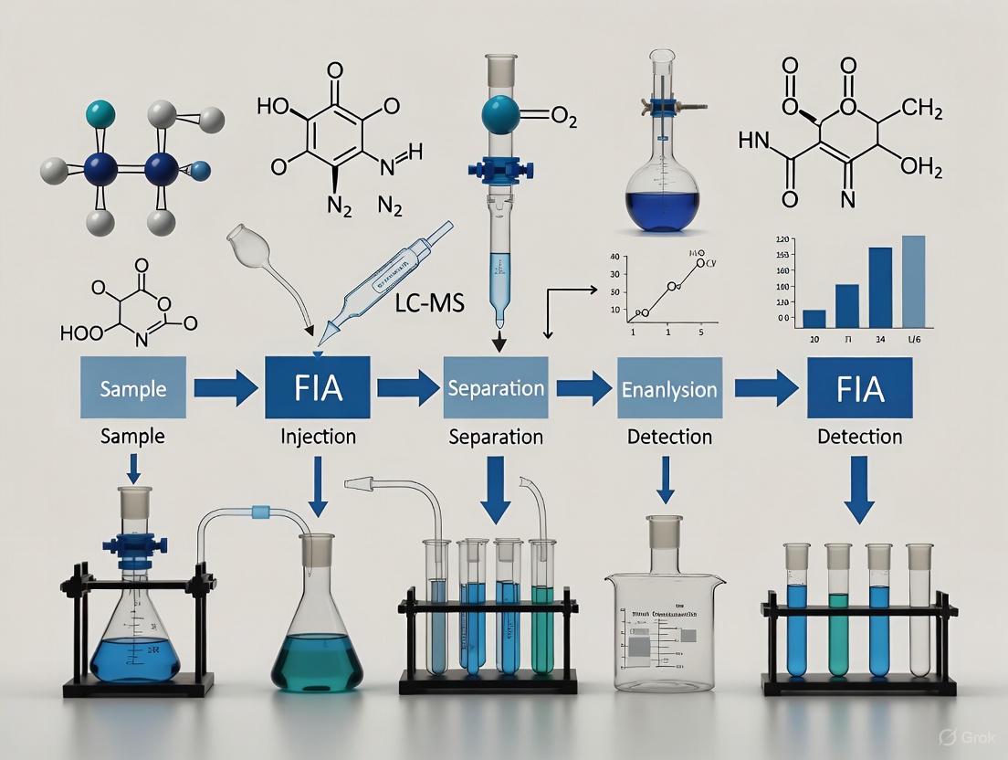
FIA-MS Workflow and System Configuration
Generic FIA-MS Workflow
The following diagram illustrates the logical flow and components of a standard FIA-MS analysis:
Experimental Setup Configurations
Different experimental applications require specific FIA configurations. The diagram below compares setups for general analysis, dynamic titration, and diffusion measurements:
Key Applications and Methodologies
Application Protocol: Dynamic Titration for Binding Constant Determination
Purpose: To determine dissociation constants (Kd) of noncovalent complexes with high throughput and minimal sample consumption [1].
Experimental Setup:
- A known concentration of host molecule is continuously infused via a syringe pump
- A defined volume of guest molecule is injected through a rotary valve into a dispersion loop
- The compositional gradient forms in the dispersion loop and mixes with the host stream
- The mixture is directly analyzed by ESI-MS [1]
Procedure:
- Prepare a stock solution of host molecule at constant concentration
- Prepare a single solution of guest molecule at appropriate concentration
- Set carrier flow rate to achieve medium dispersion (3 ≤ D ≤ 10)
- Infuse host solution at constant flow rate (typically 0.1-0.5 mL/min)
- Inject guest sample (typically 10-100 µL) and acquire MS data in full scan or selected ion monitoring mode
- Monitor intensities of free host, free guest, and complex ions
- Export intensity data and fit to a 1:1 equilibrium binding model using appropriate algorithms [1]
Advantages over Conventional Titration:
- Time efficiency: Complete analysis in minutes versus hours
- Reduced sample consumption: Single guest solution versus multiple preparations
- Automation potential: Easily integrated with autosamplers for high-throughput screening [1]
Application Protocol: Automated Shotgun Lipidomics
Purpose: To perform comprehensive, high-throughput lipid profiling without chromatographic separation [2].
Experimental Setup:
- HPLC system replumbed with PEEKsil tubing to minimize carryover
- Binary pump for mobile phase delivery
- Thermostated autosampler for sample introduction
- High-resolution mass spectrometer (e.g., TripleTOF system) [2]
MS/MSALL Acquisition Parameters:
- Ionization: ESI with positive mode (+4500V) or negative mode (-4200V)
- Source temperature: 400°C
- Gas settings: Curtain gas (25), Gas 1 (18), Gas 2 (30)
- Acquisition: TOF MS scan (0.5-1 sec) followed by sequential MS/MS scans stepped across mass range (200-2250 m/z)
- Isolation windows: Optimized for lipid analyte mass defects [2]
Procedure:
- Prepare lipid extracts using modified Folch method
- Reconstitute in methylene chloride/methanol (50:50) with 5mM ammonium acetate
- Set injection volume to 50 µL
- Use isocratic flow with wash steps between injections
- Acquire data using Infusion MS/MSALL method
- Process data with LipidView software for lipid identification and quantification [2]
Performance Characteristics:
- Reproducibility: %RSD for peak area: 1.83-3.20%
- Carryover: <0.16% after optimized wash protocol
- Throughput: 15 minutes per sample including wash and equilibration [2]
Optimization Strategies and Parameters
Critical FIA-MS Parameters
Table 1: Key Optimization Parameters for FIA-MS Methods
| Parameter Category | Specific Parameters | Optimization Considerations | Impact on Performance |
|---|---|---|---|
| FIA System Parameters | Sample injection volume | Typically 10-100 µL; affects sensitivity and dispersion | Larger volumes increase sensitivity but may broaden peaks |
| Carrier flow rate | 0.1-1.0 mL/min; affects dispersion and analysis time | Higher flow rates reduce analysis time but may decrease ionization efficiency | |
| Tubing length and diameter | Determines extent of dispersion | Longer, narrower tubing increases dispersion | |
| MS Source Parameters | Capillary voltage | 2000-4000 V; critical for ionization efficiency | Higher voltage improves sensitivity but may cause arcing or increased chemical noise [3] |
| Drying gas flow rate and temperature | 8-12 L/min; 300-400°C | Optimizes desolvation; higher temperatures improve desolvation but may degrade thermolabile compounds [3] | |
| Nebulizer gas pressure | 20-50 psi | Affects aerosol formation and ionization stability [4] | |
| MS Analyzer Parameters | Fragmentor voltage | Compound-dependent (e.g., 149 V for forskolin) | Controls in-source fragmentation; higher voltages increase fragmentation [3] |
| Collision energy | Optimized for each transition in MS/MS | Affects fragment ion abundance and signal-to-noise [4] |
Research Reagent Solutions
Table 2: Essential Materials for FIA-MS Experiments
| Reagent/Material | Function/Application | Examples/Specifications |
|---|---|---|
| Mobile Phase/ Carrier Solvents | Transport medium for sample introduction | Methanol, acetonitrile, methylene chloride/methanol (50:50) with 5mM ammonium acetate for lipidomics [2] |
| Additives/ Modifiers | Enhance ionization efficiency and stability | 1 mM sodium acetate, ammonium acetate, formic acid, acetic acid (0.05-0.1%) [3] |
| System Tubing | Sample transport with minimal adsorption | PEEKsil tubing for reduced carryover, especially for lipid analysis [2] |
| Reference Standards | System calibration and method validation | Forskolin for food supplement analysis [3], bovine heart extract for lipidomics [2] |
Troubleshooting and Quality Control
Common FIA-MS Issues and Solutions
Carryover Reduction Strategies:
- Implement aggressive wash protocols with increased flow rates during wash steps
- Use PEEKsil tubing instead of standard PEEK to minimize lipid adsorption
- Include blank injections after high-concentration samples to monitor carryover [2]
Signal Variability Mitigation:
- Ensure stable temperature control for both autosampler and source
- Maintain consistent nebulizer and drying gas flows
- Use internal standards to correct for injection-to-injection variability [5]
Reproducibility Enhancement:
- Implement system suitability testing procedures
- Monitor total ion current (TIC) stability and peak shape consistency
- Use automated quality control metrics within instrument software [5] [2]
Method Validation Parameters
For quantitative FIA-MS methods, key validation parameters include:
- Precision: %RSD for replicate injections (target <5%)
- Carryover: Typically <0.2% for acceptable performance
- Signal stability: Assessed through TIC monitoring over multiple injections
- Linearity: Evaluated through calibration curves in quantitative applications [3] [2]
Flow Injection Analysis coupled with Mass Spectrometry represents a powerful platform for high-throughput analysis in drug development and applied research. By understanding the core principles, optimization strategies, and application-specific protocols detailed in this document, researchers can effectively implement FIA-MS methodologies to accelerate compound screening, binding studies, and lipidomic profiling while maintaining data quality and reproducibility.
In the pursuit of optimization in liquid chromatography-tandem mass spectrometry (LC-MS/MS), the question of whether to include the chromatographic column is central. Flow Injection Analysis (FIA) and liquid chromatography represent two distinct approaches, each with its own performance characteristics. FIA involves the direct injection of samples into the mass spectrometer, bypassing the chromatographic column to maximize analytical throughput. In contrast, LC-MS/MS utilizes on-column separation to resolve analytes from complex matrices prior to detection. This application note details the comparative performance of these techniques and provides structured protocols to guide researchers in selecting and implementing the appropriate method based on their specific analytical requirements in drug development and clinical research.
Comparative Performance Data
The decision between FIA and LC-MS/MS methods hinges on understanding their quantitative performance characteristics. The following tables summarize key metrics derived from published studies across different applications.
Table 1: General Method Performance Comparison between FIA-MS/MS and LC-MS/MS
| Performance Metric | FIA-MS/MS | LC-MS/MS |
|---|---|---|
| Analysis Time | < 60 seconds per sample [6] | ~10 minutes per sample [6] |
| Typical Recovery Range | 79-117% (at higher concentrations) [6] | 100-117% (across tested concentrations) [6] |
| Typical Relative Standard Deviation (RSD) | < 15% [6] | < 9% [6] |
| Instrument LOQ (Matrix-Dependent) | 0.12 - 0.35 ppb [6] | 0.02 - 0.06 ppb [6] |
| Impact of Matrix Effects | High (no chromatographic separation) [6] [7] | Reduced (analytes separated from matrix) [6] [7] |
| Isobaric Interference Risk | High [6] | Low [6] |
Table 2: Application-Specific Performance and Outcomes
| Application Context | Analytes | Key Findings | Recommendation |
|---|---|---|---|
| Newborn Screening [8] | Very-long-chain acylcarnitines (ACs) & lysophosphatidylcholines (LPCs) | FIA-MS/MS optimized as first-tier screen; LC-MS/MS used as second-tier confirmation reduced false positives. | FIA suitable for high-throughput primary screening; LC-MS/MS provides confirmatory precision. |
| Mycotoxin Analysis [6] | Ochratoxin A in food matrices | FIA-MS/MS failed to detect analyte at 1 ppb; LC-MS/MS provided reliable quantification at all levels (1-100 ppb). | LC-MS/MS is essential for low-concentration analytes and complex food matrices. |
| Therapeutic Drug Monitoring [9] | Imatinib in human plasma | Both methods provided comparable results for patient samples; FIA offered higher throughput for routine monitoring. | FIA is viable for high-throughput analysis of specific drugs in clinical matrices. |
| Lipidomics [2] | Complex lipid profiles | Automated FIA MS/MS'ALL workflow demonstrated excellent reproducibility (%RSD 1.83-4.27%) and minimal carryover. | FIA is powerful for untargeted, high-throughput lipidomic profiling. |
Experimental Protocols
Protocol for FIA-MS/MS Analysis of Mycotoxins
This protocol is adapted from a study comparing the determination of ochratoxin A in food commodities [6].
Sample Preparation:
- Fortification: Add 13C uniformly labeled ochratoxin A as an internal standard (13C-IS) to samples.
- Extraction: Prepare samples by solvent extraction, followed by dilution and filtration.
- Preparation for Analysis: Transfer the prepared extract into a suitable vial for the autosampler.
Instrumentation and Conditions:
- Platform: Triple quadrupole tandem mass spectrometer.
- Sample Introduction: Flow injection (bypassing the LC column).
- Mobile Phase: Direct infusion of sample extract.
- Analysis Time: < 60 seconds per sample.
- Detection: Multiple Reaction Monitoring (MRM) mode.
Critical Notes: This method is applicable for concentrations at 5 ppb and above. It is not recommended for detecting ochratoxin A at 1 ppb in complex matrices like corn, oat, or grape juice due to insufficient sensitivity caused by ion suppression [6].
Protocol for LC-MS/MS Analysis of Mycotoxins
This protocol provides a reliable method for quantifying ochratoxin A at lower concentrations, including the 1 ppb level [6].
Sample Preparation:
- Fortification: Use 13C uniformly labeled ochratoxin A as an internal standard (13C-IS).
- Extraction: Prepare samples by solvent extraction, dilution, and filtration.
Instrumentation and Conditions:
- Platform: Triple quadrupole tandem mass spectrometer coupled with a liquid chromatography system.
- Chromatography: Utilize a reverse-phase LC column.
- Mobile Phase: Acid buffer/methanol or acetonitrile gradient.
- Run Time: 10 minutes per sample.
- Ionization: Electrospray Ionization (ESI).
- Detection: Multiple Reaction Monitoring (MRM) mode.
Protocol for FIA-MS/MS in Lipidomics
This protocol outlines an automated, high-throughput shotgun lipidomics approach [2].
Sample Preparation:
- Extraction: Serum samples are extracted using a modified Folch method.
- Reconstitution: Reconstitute the organic phase in methylene chloride/methanol (50/50) with 5mM ammonium acetate in autosampler vials.
Instrumentation and Conditions:
- System: TripleTOF 6600 System or equivalent high-resolution mass spectrometer.
- Infusion: Automated FIA using a standard HPLC system. Increased flow rate during the wash step is critical to minimize carryover. PEEKsil tubing is recommended for all plumbing to reduce analyte adhesion [2].
- Acquisition Method: Infusion MS/MS'ALL, consisting of a TOF MS scan followed by a series of high-sensitivity MS/MS scans stepped across the entire lipidome mass range (e.g., 200-2250 m/z).
- Source Conditions: Optimize for both positive and negative modes (e.g., Spray voltage +4500V/-4200V, Temperature 400°C).
Data Processing: Process acquired data using software (e.g., LipidView) for lipid profile extraction, followed by statistical analysis (e.g., in MarkerView Software).
Workflow and Decision Pathways
The fundamental difference between FIA and LC-MS/MS workflows lies in the presence or absence of chromatographic separation. The following diagram illustrates the core steps and decision points for each method.
The Scientist's Toolkit: Essential Research Reagents and Materials
The following table lists key reagents and materials critical for implementing the FIA and LC-MS/MS protocols discussed.
Table 3: Essential Research Reagent Solutions
| Item | Function / Application | Example from Literature |
|---|---|---|
| 13C Uniformly Labeled Internal Standards | Corrects for matrix effects and losses during sample preparation; essential for accurate quantification in both FIA and LC-MS/MS. | 13C-ochratoxin A used in mycotoxin analysis [6]. |
| Deuterated Internal Standards (e.g., d8-Imatinib) | Serves as an internal standard for therapeutic drug monitoring, compensating for variability in sample preparation and ionization. | d8-Imatinib mesylate used for quantifying imatinib in plasma [9]. |
| LC/MS Grade Solvents (Methanol, Acetonitrile, Formic Acid) | Used in mobile phase and sample preparation to minimize background noise and contamination, ensuring optimal MS performance. | Used in both ochratoxin A and imatinib method development [6] [9]. |
| Characterized Reference Materials (e.g., Bovine Heart Extract) | Complex biological standard used for system suitability testing, workflow development, and assessing reproducibility in lipidomics. | Bovine Heart Extract (BHE) used to evaluate FIA-MS/MS'ALL reproducibility [2]. |
| PEEKsil Tubing | Specialized inert tubing used to replace standard HPLC tubing after the autosampler to minimize carryover, especially critical for sticky molecules like lipids. | Implementation resulted in carryover of <0.16% in lipidomics workflow [2]. |
| Reverse Phase LC Columns (e.g., C18 with sub-2µm particles) | Provides high-resolution separation of analytes from matrix components prior to MS detection, reducing ion suppression and isobaric interference. | C18 column with 1.7µm particles used for fast, high-efficiency separation of imatinib [9]. |
The choice between FIA and LC-MS/MS is not a matter of one technique being superior to the other, but rather of selecting the right tool for the specific analytical question. FIA-MS/MS is the definitive choice for maximizing throughput in targeted, high-throughput applications such as newborn screening [8], routine therapeutic drug monitoring [9], and untargeted lipidomic profiling [2], where analysis speed is critical and analyte concentrations are sufficiently high. Conversely, LC-MS/MS is indispensable when method robustness, sensitivity for trace-level analysis, and specificity in complex matrices are the primary concerns, as demonstrated in mycotoxin testing [6] and when comprehensive analyte identification is required. By applying the structured protocols and decision frameworks outlined in this application note, scientists can strategically bypass the chromatographic column to gain efficiency without compromising the integrity of their data, thereby optimizing resource allocation and accelerating research and development timelines.
Rapid Optimization of MS Parameters and High-Throughput Screening
Flow Injection Analysis (FIA) coupled with mass spectrometry represents a powerful approach for high-throughput screening in modern analytical laboratories. By eliminating chromatographic separation, FIA-MS enables direct sample introduction into the mass spectrometer, significantly reducing analysis times to as little as 60 seconds per sample [6]. This technique is particularly valuable in applications requiring rapid analysis of large sample batches, such as drug discovery, lipidomics, and toxicological screening [2] [10]. However, the absence of chromatographic separation places greater emphasis on optimal mass spectrometer parameter configuration to maintain analytical sensitivity and specificity despite potential matrix effects.
The successful implementation of FIA-MS workflows hinges on two critical components: systematic optimization of instrument parameters to maximize signal response for target compounds, and robust experimental design to ensure reproducibility across large sample sets. This application note details standardized protocols for rapid MS parameter optimization and high-throughput FIA screening, providing researchers with practical methodologies to enhance workflow efficiency while maintaining data quality within the context of LC-MS optimization research.
Key Concepts and Definitions
Flow Injection Analysis (FIA): A technique where samples are directly injected into a flowing carrier stream for introduction into the mass spectrometer without chromatographic separation [11] [6].
Multiple Reaction Monitoring (MRM): A highly sensitive targeted mass spectrometry method that selectively detects and quantifies specific molecules based on precursor-to-fragment ion transitions [12].
Ion Suppression: A phenomenon where co-eluting matrix components reduce ionization efficiency of target analytes, leading to decreased signal intensity [13].
Collision Energy (CE): The voltage applied in the collision cell to fragment precursor ions, optimized based on the mass-to-charge ratio (m/z) of the target compound [12].
Matrix Effects: The influence of co-extracted sample components on the ionization efficiency of target analytes, particularly impactful in FIA due to the absence of chromatographic separation [6].
Protocol 1: Rapid Optimization of MS/MS Parameters
Background and Principles
Sensitivity in MRM-based mass spectrometry depends critically on the tuning of instrument parameters for optimal peptide fragmentation and maximal transmission of desired product ions [12]. While generalized equations exist for parameters like collision energy, these may fail to produce maximum signal response under diverse experimental conditions [12]. Bond fragmentation efficiency depends on peptide residue content and proton mobility, meaning particular residues or residue combinations may not generate maximum response when fragmented under generalized conditions [12]. This protocol describes a streamlined workflow for rapid determination of optimal instrument parameters for each MRM transition.
Experimental Workflow
The following diagram illustrates the comprehensive workflow for rapid optimization of MS parameters:
Materials and Equipment
Table 1: Essential Research Reagent Solutions
| Item | Function/Application | Example Specifications |
|---|---|---|
| Triple quadrupole mass spectrometer | Targeted mass analysis with MRM capability | Waters Quattro Premier or ABI 4000 QTRAP [12] |
| Standard protein/analyte mixture | System performance testing and optimization | 1 nmol of each protein in ammonium bicarbonate [12] |
| Sequencing-grade trypsin | Protein digestion for peptide analysis | Promega, 1:40 (w/w) ratio [12] |
| Solid-phase extraction cartridges | Sample clean-up | Waters Oasis MCX cartridge [12] |
| Mobile phase components | LC-MS analysis | 0.1% formic acid, acetonitrile, HPLC-grade water [12] [11] |
| MRM software | Data analysis and optimization | Mr. M software package [12] |
Step-by-Step Procedure
Prepare MRM Transition List: Generate an initial list of MRM transitions for target compounds. For peptide analysis, use tools like SpectraST to create a consensus spectral library from MS/MS data [12]. Filter to include high-probability transitions.
Reprogram m/z Values: Use a scripting tool (e.g., Perl script) to subtly adjust the second decimal place of precursor and product m/z values to code for different instrument parameters [12]. This creates unique MRM targets for each parameter value while maintaining the same actual transition.
Program Parameter Range: For each MRM transition, program a range of parameter values. For collision energy optimization, use values from 6V less to 6V more than the equation-derived value in 2V steps [12]. The generalized CE equation provided by Waters is: CE = 0.034 × (m/z precursor) + 1.314 [12].
Execute Single Run: Analyze the samples using the modified MRM method with all parameter variations within a single LC-MS run to avoid run-to-run variability [12].
Data Analysis: Process data using MRM software (e.g., Mr. M) to determine optimal instrument parameters for each transition based on maximal product ion signal [12].
Validation: Validate optimized parameters by comparing signal intensity to pre-optimization values.
Protocol 2: High-Throughput Screening Using FIA-MS
Background and Principles
Flow Injection Analysis-MS enables rapid screening by directly introducing samples into the mass spectrometer, bypassing time-consuming chromatographic separation [11] [6]. This approach is particularly valuable in lipidomics [2] and toxicology screening [10], where analysis of hundreds of compounds is required. The MS/MSALL workflow provides an automated, untargeted acquisition strategy that has low carryover and excellent reproducibility [2]. This protocol describes an optimized FIA method for high-throughput screening applications.
Experimental Workflow
The following diagram illustrates the FIA-MS high-throughput screening workflow:
Materials and Equipment
Table 2: FIA-MS Screening Solutions
| Item | Function/Application | Example Specifications |
|---|---|---|
| UHPLC system with autosampler | Automated sample introduction | Shimadzu UHPLC system [2] |
| PEEKsil tubing | Minimize carryover | 50μm I.D. hybrid electrodes [2] |
| TripleTOF mass spectrometer | High-resolution MS and MS/MS analysis | SCIEX TripleTOF 6600 System [2] |
| LipidView software | Automated data processing and lipid profiling | LipidView Software 1.3 [2] |
| MarkerView software | Statistical analysis and visualization | MarkerView Software [2] |
| Mobile phase for lipidomics | Sample delivery and ionization | Methylene chloride/methanol (50/50) with 5mM ammonium acetate [2] |
Step-by-Step Procedure
Sample Preparation: For lipidomics applications, prepare samples using modified Folch method. Extract serum samples and reconstitute the organic phase in methylene chloride/methanol (50/50) with 5mM ammonium acetate [2].
System Configuration: Replace all tubing on the HPLC system after the autosampler with PEEKsil tubing, including the sample loop, to minimize carryover [2]. Use a 50μm I.D. hybrid electrode to maintain low backpressure.
FIA Method Setup: Configure the LC system for flow injection without a column. Use a mobile phase of methylene chloride/methanol (50/50) with 5mM ammonium acetate at a flow rate of 0.2 mL/min [11] [2]. Set injection volume to 50μL [2].
MS/MSALL Acquisition: Program the mass spectrometer to acquire data using Infusion MS/MSALL mode, consisting of a TOF MS scan (5 sec) and series of MS/MS scans (300 msec) stepped across the mass range of interest (e.g., 200-2250 m/z for lipidomics) [2]. Optimize Q1 isolation windows for analyte mass defects.
Ion Source Optimization: Set source parameters for positive and negative mode operation: spray voltage +4500V/-4200V, temperature 400°C, curtain gas 25, Gas 1 18, Gas 2 30 [2].
Data Acquisition: Establish a 5.3-minute data acquisition window to provide stable and reproducible MS signal. Include wash and equilibration steps for a total run time of 15 minutes [2].
Data Processing: Process all data using LipidView Software for lipid profiling, then export to MarkerView Software for principal component analysis and statistical evaluation [2].
Comparative Performance Data
Method Comparison
Table 3: Performance Comparison of FIA-MS/MS vs. LC-MS/MS
| Parameter | FIA-MS/MS | LC-MS/MS | References |
|---|---|---|---|
| Analysis time | <60 seconds/sample | 10 minutes/sample | [6] |
| Recovery (5-100 ppb) | 79-117% | 100-117% | [6] |
| Detection limit (ochratoxin A) | 0.12-0.35 ppb | 0.02-0.06 ppb | [6] |
| Reproducibility (RSD) | 2-15% | 2-8% | [6] |
| Carryover | 0.146-0.156% (reduced to 0.025-0.061% with PEEKsil) | Typically lower | [2] [6] |
| Matrix effects | Significant without separation | Reduced with chromatographic separation | [6] [13] |
Optimization Outcomes
Table 4: Impact of Parameter Optimization on MS Performance
| Optimization Aspect | Before Optimization | After Optimization | Key Factors |
|---|---|---|---|
| MRM sensitivity | Variable signal response | Maximal product ion signal | Collision energy, cone voltage [12] |
| Ion suppression | Up to 20% signal reduction | Minimized through sample cleanup | Solid-phase extraction, mobile phase optimization [13] |
| Run-to-run reproducibility | >15% RSD | 2.1-4.27% RSD for peak height | Automated FIA, system conditioning [2] |
| Lipid profiling variability | Not specified | Minimal variation in class profiles | MS/MSALL workflow, standardized extraction [2] |
| High-throughput screening | Limited by LC separation | 231 compounds in 15 minutes | Optimized MRM transitions, minimal retention [10] |
Troubleshooting and Technical Notes
Common Challenges and Solutions
Ion Suppression in FIA: A primary limitation of FIA-MS is increased susceptibility to ion suppression from co-eluting matrix components [6] [13]. Mitigation strategies include extensive sample dilution, improved sample cleanup techniques such as solid-phase extraction, and implementation of matrix-matched calibration standards [6].
Carryover Issues: Significant carryover (0.146-0.156%) has been observed in FIA systems when analyzing high-concentration samples [2]. This can be reduced to 0.025-0.061% by replacing standard tubing with PEEKsil tubing throughout the system and implementing rigorous wash steps between injections [2].
Parameter Optimization Stability: Optimized instrument parameters may not remain stable over time due to variations in gas pressure or instrument voltage drift [12]. Periodic recalibration is recommended, particularly for quantitative applications requiring long-term reproducibility.
Application-Specific Considerations
Toxicology Screening: For comprehensive drug screening, predefined MRM transitions for 231 compounds including illegal drugs, sedatives, and psychotropic drugs can be implemented with analysis times under 15 minutes [10]. Semi-quantitation is possible using internal standard-based calibration curves.
Lipidomics Profiling: The MS/MSALL workflow enables comprehensive lipid coverage without predefinition of targets [2]. Data can be extracted in silico to mimic precursor ion and neutral loss scans, providing flexibility in data analysis long after acquisition.
Method Selection Criteria: FIA-MS is recommended for high-throughput applications where moderate sensitivity suffices and sample composition is relatively consistent [6]. LC-MS/MS remains preferable for complex matrices or when maximum sensitivity is required, particularly near detection limits [6].
Within the framework of liquid chromatography-mass spectrometry (LC-MS) optimization, Flow Injection Analysis (FIA) serves as a powerful technique for rapid sample introduction and analysis, bypassing the chromatographic column to achieve high throughput. The core principle of FIA involves the injection of a precise sample volume into a continuously flowing, unsegmented carrier stream [14] [15]. The hardware configuration that facilitates this—comprising the pumps, injection valves, tubing, unions, and connectors—is collectively known as the FIA manifold. The reproducibility and quality of FIA-MS data are critically dependent on the precise configuration and selection of these components, as they govern sample dispersion and mixing kinetics prior to MS detection [14] [1]. This application note details the essential hardware setup for a robust and optimized FIA-MS system.
Core Hardware Components of an FIA Manifold
A basic FIA manifold is composed of several key subsystems that work in concert to transport, inject, and prepare the sample for detection.
System Components and Their Functions
Table 1: Core Components of a Flow Injection Analysis Manifold
| Component | Function & Description | Key Configuration Parameters |
|---|---|---|
| Propulsion Unit (Pump) | Generates a constant, pulseless flow of the carrier solvent, transporting the sample plug through the system. | Flow Rate: Typically 0.5-2.0 mL/min; must be stable and reproducible [15]. |
| Sample Injection Valve | Introduces a precise, discrete volume of sample into the flowing carrier stream without flow disruption. A rotary valve with a sample loop is standard. | Injection Volume: Usually 50-150 µL [15]; defined by the loop size. Type: Often a 6-port, 2-position rotary valve [16] [1]. |
| Manifold Tubing / Reactor | The conduit where the sample disperses into the carrier and mixes with reagents. It also serves as the reaction chamber. | Material: Chemically inert (e.g., Teflon, PEEK) [15]. Diameter: Typically 0.5-0.8 mm [15]. Length & Coiling: Determines reaction time; coiled to promote mixing [14] [15]. |
| Connectors & Unions | Low-dead-volume fittings used to connect sections of tubing and other components, ensuring leak-free flow paths. | Type: Low-dead-volume unions, tees, and crosses. Material: Compatible with solvents and pressures used. |
| Detector (MS) | The flow-through sensor that generates the analytical signal. In FIA-MS, this is the mass spectrometer's ion source. | Flow Cell: The ESI or APCI probe serves as the flow cell. Compatibility: Mobile phase must be suitable for MS ionization. |
Research Reagent Solutions and Essential Materials
Table 2: Essential Materials for FIA-MS Manifold Setup
| Item | Typical Specification | Primary Function in FIA-MS |
|---|---|---|
| Carrier Solvent | HPLC-grade methanol, acetonitrile, water, or volatile buffers (e.g., ammonium formate/acetate). | Dissolves the sample and forms the liquid transport stream into the MS ion source. |
| Peristaltic or Syringe Pump | Capable of delivering 0.1-5.0 mL/min with high precision. | Propels the carrier stream and any added reagents through the manifold [14] [15]. |
| Six-Port Rotary Injection Valve | Equipped with a fixed-volume sample loop (e.g., 10-200 µL). | Introduces a sharp, well-defined sample plug into the carrier stream for reproducible injection [16] [1]. |
| Tubing (PEEK or Teflon) | Internal diameter: 0.25-0.75 mm. | Forms the flow path and reactor; smaller diameters limit dispersion [15]. |
| Low-Dead-Volume (LDV) Unions | Zero-dead-volume (ZDV) or nano-volume fittings in 1/16" outer diameter. | Connects tubing segments and injector to detector with minimal band broadening and peak tailing. |
| Mixing Tee | A low-volume "T" or "Y" union. | Merges the main carrier stream with a second reagent stream introduced via a second pump [1]. |
Flow Path Configuration and Dispersion Dynamics
The physical and chemical processes occurring within the flow path after sample injection are fundamental to FIA.
The Dispersion Process
Upon injection, the sample forms a discrete plug within the carrier stream. As it is transported, this plug undergoes dispersion—a controlled mixing process with the carrier stream governed by convection and diffusion [14].
- Convection: Results from the laminar flow profile in narrow-bore tubing, where the fluid at the center moves faster than the fluid at the walls, creating a parabolic profile that stretches the sample plug [14].
- Radial Diffusion: The movement of sample molecules from areas of high concentration (center of the plug) to low concentration (the carrier stream and tubing walls). This is critical for mixing the sample with the carrier and any reagents, and for controlling the shape of the flow profile [14].
The degree of dispersion is quantified by the dispersion coefficient (D), defined as the ratio of the analyte concentration before and after the dispersion process [1]. The configuration can be tailored for different needs:
- Limited Dispersion (D ≤ 3): Used when minimal dilution is desired, achieved with short, straight tubing.
- Medium Dispersion (3 < D < 10): Used when mixing with reagents is required, achieved with coiled reaction tubing.
- Large Dispersion (D ≥ 10): Used for automatic sample dilution [1].
Diagram 1: Sample dispersion process in FIA tubing.
Configuring the Flow Path for MS Detection
For FIA-MS, the flow path must be optimized to deliver a sharp, well-defined analyte band to the mass spectrometer's ion source.
- Minimize Extra-Column Volume: Use the shortest possible length of narrow-bore tubing (e.g., 0.005" - 0.02" inner diameter) between the injector and the MS source. All connections must be made with low-dead-volume unions to reduce peak broadening [15].
- Coiled Reactor: To promote radial diffusion for efficient mixing (e.g., for on-line dilution or dynamic titration) while limiting axial dispersion, the reaction tubing is often coiled. This induces secondary flow patterns that enhance mixing without excessively broadening the sample band [15].
- Multi-Stream Manifolds: More complex experiments may use a multi-channel manifold where a second pump delivers a reagent or a host molecule for binding studies via a mixing tee [1]. This setup is the basis for advanced techniques like dynamic titration for determining dissociation constants (K~d~).
Experimental Protocol: System Setup and a Representative FIA-MS Experiment
Protocol: Assembly and Operational Verification of a Single-Channel FIA-MS Manifold
Objective: To assemble a basic FIA-MS flow path and verify its performance by generating a reproducible transient signal. Materials: See Table 2 for essential materials. Additionally, prepare a carrier stream (e.g., 50/50 methanol/water with 0.1% formic acid) and a standard solution (e.g., 1 µg/mL caffeine in carrier).
Assembly:
- Flush all tubing and the sample loop with the appropriate solvent prior to connection.
- Connect the outlet of the pump to the inlet port of the injection valve using a short piece of PEEK tubing and a LDV union.
- Connect the sample loop to the designated ports on the injection valve.
- Connect the outlet port of the injection valve to one end of the reaction coil (e.g., 50 cm of 0.5 mm ID PEEK tubing, coiled).
- Connect the other end of the reaction coil directly to the MS ion source (e.g., ESI probe) using the shortest possible piece of appropriate tubing.
System Startup:
- Prime the pump and the entire flow path with the carrier solvent at the desired flow rate (e.g., 0.5 mL/min). Ensure there are no leaks.
- Power on the mass spectrometer and establish stable instrument conditions.
Performance Verification:
- Fill the sample loop with the caffeine standard solution using a precision syringe.
- Activate the injection valve to introduce the sample into the carrier stream.
- Simultaneously, start data acquisition on the mass spectrometer, monitoring the ion trace for caffeine (e.g., m/z 195).
- A sharp, Gaussian-like peak should be observed. Record the peak height and peak width at half height.
- Repeat the injection at least five times to calculate the relative standard deviation (RSD) of the peak height and retention time. A well-configured system should yield an RSD of <2% for peak height [15].
Diagram 2: Basic FIA-MS workflow.
Application: Dynamic Titration for Binding Affinity (K~d~) Measurement
Objective: To rapidly determine the dissociation constant (K~d~) of a non-covalent host-guest complex using FIA-MS [1]. Principle: A concentration gradient of the guest molecule is created via FIA dispersion and mixed with a constant concentration of the host. The MS monitors the intensities of the free host, free guest, and the complex in real-time as the gradient passes through.
Materials:
- Two syringe or piston pumps.
- A six-port, two-position rotary valve with a sample loop.
- A dispersion loop (longer coil of tubing, e.g., 200 cm).
- A mixing tee.
- Host and guest molecule solutions in a compatible, volatile buffer.
- Mass spectrometer with an ESI source.
Procedure:
- Manifold Configuration: Configure the system as shown in Diagram 3. The carrier stream (from Pump 1) transports the injected guest plug through the dispersion loop. A second stream containing the host at a fixed concentration is introduced via Pump 2 and merged with the dispersed guest plug just before the MS via a mixing tee.
Execution:
- Set Pump 1 (carrier) and Pump 2 (host) to the same flow rate (e.g., 0.2 mL/min).
- Load the guest solution at a known concentration into the sample loop.
- Activate the injection valve.
- The MS is set to monitor the selected ions for the host, guest, and complex simultaneously.
Data Analysis:
- Export the intensity data for the complex ion over the time of the FIA peak.
- The concentration of the guest at each data point is known from the pre-calibrated dispersion profile of the system.
- Fit the data (guest concentration vs. complex intensity) to a 1:1 binding model (e.g., Langmuir isotherm) to calculate the K~d~ value.
This method offers a high-throughput alternative to conventional titration, completing a measurement in minutes with minimal sample consumption [1].
Concluding Remarks
The performance of an FIA-MS system is intrinsically linked to its hardware configuration. A meticulous approach to selecting and assembling the components—pumps, injection valves, tubing, and unions—is paramount for achieving a flow path with minimal dead volume and controlled dispersion. The protocols outlined herein provide a foundation for establishing a robust FIA-MS setup, enabling applications ranging from simple, high-throughput concentration screening to sophisticated solution-phase binding assays. Proper optimization of this hardware is a critical step in any broader research endeavor aimed at leveraging the full potential of flow injection analysis coupled to mass spectrometry.
Developing a Robust FIA-MS Method: A Step-by-Step Protocol
In liquid chromatography-mass spectrometry (LC-MS), the steps of sample preparation and the choice of solvents are not merely preliminary; they are foundational to the success of the entire analytical workflow. Proper sample preparation aims to remove interfering matrix components, prevent instrument contamination, and concentrate analytes of interest. Concurrently, the selection of solvents and additives directly governs the efficiency of electrospray ionization (ESI), the most prevalent ionization technique in LC-MS, by affecting droplet formation, desolvation, and ultimately, the generation of gas-phase ions [17] [18]. This application note provides detailed protocols and structured data to guide researchers in optimizing these critical first steps for robust and sensitive Flow Injection Analysis (FIA)-MS methods.
Core Principles and Impact on Ionization
The primary goal of sample preparation is to make the sample compatible with the MS system. Complex biological matrices can cause ion suppression, where co-eluting compounds interfere with the ionization of the target analyte, leading to reduced sensitivity and inaccurate quantification [19] [17]. Efficient sample clean-up mitigates this effect.
In ESI, the solvent is integral to the mechanism of ion formation. Key considerations include:
- Elution Strength and Composition: The elution power of the solvent from a prior LC step can be too strong for effective focusing at the head of a subsequent column or for stable ESI formation. Techniques like active solvent modulation (ASM) can be employed to reduce this elution force by adding a weaker solvent [19].
- Additives: Volatile acids (e.g., formic acid) and buffers (e.g., ammonium acetate) are used to enhance analyte ionization. Acids promote positive ion formation by protonating basic analytes, while ammonium acetate is suitable for both positive and negative modes.
- Compatibility: Solvents must be LC-MS grade to minimize background noise and prevent contamination of the ion source.
Optimizing Ion Source and Solvent Parameters
Ion source parameters are highly dependent on the solvent composition and flow rate. A systematic optimization of these parameters is crucial for maximizing signal intensity and stability.
Design of Experiments (DoE) for Systematic Optimization
The one-factor-at-a-time (OFAT) approach is inefficient and can miss interacting effects. Using Design of Experiments (DoE) and Response Surface Methodology (RSM) provides a statistically sound framework for finding optimal conditions [20] [21]. A study optimizing oxylipin analysis used a fractional factorial design to screen relevant factors like interface temperature and CID gas pressure, followed by a central composite design for optimization, which significantly improved signal-to-noise ratios [21].
Key Ion Source Parameters
Optimization of ESI parameters is essential for obtaining high-quality signal across a wide range of analytes. The table below summarizes optimal values for key parameters from an untargeted metabolomics study using an Orbitrap mass spectrometer [17].
Table 1: Optimized ESI Ion Source Parameters for Untargeted Analysis
| Parameter | Optimal Value (Positive Mode) | Optimal Value (Negative Mode) | Impact on Ionization |
|---|---|---|---|
| Spray Voltage | 2.5 – 3.5 kV | 2.5 – 3.0 kV | Applied potential for electrospray formation; critical for stable current. |
| Vaporization Temp. | 250 – 350 °C | 250 – 350 °C | Aids in solvent evaporation from charged droplets. |
| Ion Transfer Tube Temp. | 250 – 350 °C | 250 – 350 °C | Prevents condensation and ensures efficient ion transfer into mass analyzer. |
| Sheath Gas | 30 – 50 (arbitrary units) | 30 – 50 (arbitrary units) | Assists in nebulization and shapes the spray for stability. |
| Auxiliary Gas | ≥10 (arbitrary units) | ≥10 (arbitrary units) | Helps desolvate the droplets by sweeping the spray. |
| Needle Position | Farthest (Z), Closest (Y) to inlet | Farthest (Z), Closest (Y) to inlet | Fine-tunes spray position for maximum ion influx. |
Detailed Experimental Protocols
Protocol 1: Solid Phase Extraction (SPE) for Aqueous Samples
This protocol is adapted from a method developed for the multi-residue analysis of pharmaceuticals, pesticides, and UV filters in water [20].
Application: Pre-concentration and clean-up of micropollutants from surface water or other aqueous matrices. Principle: Analytes are isolated based on affinity to a solid sorbent, followed by washing and elution.
Workflow Overview: SPE for Aqueous Samples
Steps:
- Cartridge Conditioning: Sequentially pass methanol (e.g., 3-5 mL) and then LC-MS grade water (e.g., 3-5 mL) through the hydrophilic-lipophilic balance (HLB) SPE cartridge without letting it run dry.
- Sample Loading: Adjust the sample pH to 3-4 using a volatile acid like formic acid. Load the specified sample volume (e.g., 375 mL for water analysis) onto the cartridge at a steady flow rate (e.g., 5-10 mL/min).
- Washing: Wash the cartridge with LC-MS grade water (e.g., 3-5 mL) to remove polar matrix interferences.
- Drying: Apply a vacuum or air flow (e.g., 10-15 min) to dry the sorbent bed completely. This step is critical for removing water before elution.
- Elution: Elute the target analytes into a clean collection tube using 3.5 mL of ethanol. Let the solvent dwell in the cartridge for ~1 minute before applying gentle vacuum.
- Post-Processing: Evaporate the eluate to dryness under a gentle stream of nitrogen. Reconstitute the dried extract in an initial LC-MS mobile phase (e.g., 100 µL of water/methanol 95:5) and vortex thoroughly before analysis.
Validation: The described method achieved an average absolute recovery of 73% for 32 target micropollutants [20].
Protocol 2: Protein Precipitation for Biofluids
Application: Rapid removal of proteins from biological fluids like plasma or serum. Principle: Organic solvents denature and precipitate proteins, which are then removed by centrifugation.
Workflow Overview: Protein Precipitation
Steps:
- Precipitation: Transfer 100 µL of plasma/serum to a microcentrifuge tube. Add 300 µL of ice-cold acetonitrile or methanol (a 1:3 sample-to-solvent ratio is common).
- Mixing and Incubation: Vortex the mixture vigorously for 30-60 seconds. Incubate at 4°C for 10 minutes to ensure complete protein precipitation.
- Centrifugation: Centrifuge at high speed (e.g., 15,000 x g) for 10 minutes at 4°C to form a compact protein pellet.
- Collection and Reconstitution: Carefully transfer the clear supernatant to a new tube. Evaporate the solvent under nitrogen gas and reconstitute the residue in a compatible FIA solvent (e.g., 50% methanol with 0.1% formic acid).
Protocol 3: Solid-Phase Microextraction (SPME) for Metabolomics
This protocol outlines the preparation of a high-throughput 96-blade SPME platform for metabolite cleaning and enrichment, ideal for small-volume samples and integration with nanoflow LC-MS [22].
Application: Cleaning and enrichment of metabolites from complex biological samples for untargeted metabolomics. Principle: A coated blade extracts analytes directly from the sample, which are then desorbed in a compatible solvent.
Steps:
- Blade Preparation: Sonicate stainless-steel blades in concentrated HCl for 60 minutes. Rinse thoroughly with deionized water and dry in an oven at 150°C for 30 minutes [22].
- Coating Slurry Preparation: Dissolve 10% (w/w) polyacrylonitrile (PAN) particles in N,N-dimethylformamide (DMF) at 90°C. After cooling, mix a 1:1 (w/w) ratio of polystyrene-divinylbenzene with a weak anion exchanger (PS-DVB-WAX) and hydrophilic-lipophilic balance (HLB) sorbent into the PAN glue [22].
- Coating Application: Using a nitrogen-pressurized sprayer (<5 psi), apply a thin layer of the slurry onto the blades. Thermally cure the coating in an oven at 180°C for 2 minutes. Repeat this spray-and-cure process 10 times to achieve a uniform ~120 µm coating thickness [22].
- Metabolite Extraction: Condition the coated blades in methanol and water. Immerse the blades in the processed sample (e.g., biofluid or tissue homogenate) for a defined extraction time with agitation.
- Wash and Desorption: Rinse the blades briefly with water to remove salts. Desorb the metabolites by placing the blades in a desorption solvent (e.g., 80:20 ACN:MeOH with 0.1% formic acid) for a set time. The desorbed solution is then ready for FIA-MS analysis.
The Scientist's Toolkit
Table 2: Essential Reagents and Materials for Sample Preparation and FIA-MS
| Item | Function / Application |
|---|---|
| Hydrophilic-Lipophilic Balance (HLB) Sorbent | A versatile SPE sorbent for extracting a broad range of acidic, basic, and neutral compounds from aqueous matrices [20]. |
| LC-MS Grade Solvents (ACN, MeOH, Water) | High-purity solvents to minimize background noise and contamination in mass spectrometry [17] [22]. |
| Volatile Additives (Formic Acid, Ammonium Acetate) | Enhance ionization efficiency in the ESI source. Formic acid for positive mode, ammonium acetate for both positive and negative mode [17] [21]. |
| Polyacrylonitrile (PAN) Glue | A biocompatible polymer used as a binding agent in the fabrication of SPME coatings [22]. |
| C18 Stationary Phase | The most common reversed-phase material for LC separation and SPE, suitable for non-polar to mid-polar metabolites [17]. |
| Uniformly ¹³C-Labelled Yeast Extract | A complex internal standard mixture for pixel-wise normalization and compensation of matrix effects in quantitative mass spectrometry imaging and metabolomics [23]. |
In Liquid Chromatography-Tandem Mass Spectrometry (LC-MS/MS), optimizing compound-dependent parameters is a critical step for achieving high sensitivity and selectivity. Following the initial dilution of a pure chemical standard, the next pivotal phase involves fine-tuning the mass spectrometer's parameters to uniquely identify and quantify the target analyte [24]. This document details a systematic, experimental protocol for optimizing the fragmentor voltage and collision energy, two parameters that directly control the formation and abundance of precursor and product ions. These optimized settings are foundational for constructing a specific and robust Multiple Reaction Monitoring (MRM) method, which is the cornerstone of quantitative analysis in complex matrices such as clinical and pharmaceutical samples [25] [26].
Key Concepts and Definitions
- Fragmentor Voltage: Also known as the orifice or declustering potential, this voltage is applied in the interface region between the ion source and the first quadrupole. Its primary function is to decluster solvated ions and focus the precursor ion of interest into the mass analyzer. An optimal voltage ensures maximum signal intensity for the precursor ion [24].
- Collision Energy (CE): The energy applied in the collision cell (Q2) to fragment the selected precursor ion through collisions with an inert gas (e.g., nitrogen or argon). The CE dictates the fragmentation pattern, determining the abundance of specific product ions. Optimizing CE is crucial for selecting the most intense and unique transitions for MRM [25] [24].
- Multiple Reaction Monitoring (MRM): A highly specific mass spectrometry mode that monitors one or more predefined precursor ion → product ion transitions. It significantly reduces chemical background noise, thereby enhancing selectivity and sensitivity for quantitative assays [25] [24].
Experimental Protocol: A Step-by-Step Guide
Preparation for Optimization
- Standard Solution: Utilize a pure, high-quality chemical standard of the target compound, dissolved in an appropriate solvent (e.g., a mixture of prospective mobile phases). The concentration should be suitable for instrument detection, typically in the range of 50 ppb to 2 ppm [24].
- Instrumentation: This protocol is applicable to tandem mass spectrometers, including triple quadrupole (QqQ) and Q-Trap instruments. The system should be equipped with an electrospray ionization (ESI) or atmospheric pressure chemical ionization (APCI) source [25] [24].
Optimizing the Fragmentor Voltage
The goal is to determine the voltage that yields the maximum signal for the precursor ion.
- Identify Precursor Ion: Begin by infusing the standard solution directly into the mass spectrometer (bypassing the LC column) in full scan mode. Common precursor ions include
[M+H]⁺or[M-H]⁻. If the signal is low, consider adducts like[M+NH₄]⁺or[M+Na]⁺[24]. - Voltage Ramp: Once the precursor mass is confirmed, switch to selected ion monitoring (SIM) mode for that mass. Ramp the fragmentor voltage over a defined range (e.g., 50V to 250V in 10-20V increments) while monitoring the ion intensity.
- Determine Optimum Value: Plot the signal intensity of the precursor ion against the fragmentor voltage. The voltage that produces the maximum stable signal is selected as the optimal fragmentor voltage. A suboptimal voltage can lead to insufficient declustering (low signal) or in-source fragmentation (premature fragmentation) [24].
Optimizing the Collision Energy
The goal is to determine the energy that generates the most abundant and characteristic product ions.
- Product Ion Scan: Using the optimized fragmentor voltage, introduce the precursor ion into the collision cell and perform a product ion scan. This involves scanning a range of masses in Q3 while ramping the collision energy to generate a spectrum of all possible fragments.
- Select Product Ions: From the product ion spectrum, select at least two abundant and characteristic product ions for each compound. The most intense transition is typically used for quantification, while the second is used for confirmatory identification [24].
- Fine-tune Collision Energy: For each selected precursor→product ion transition (MRM pair), systematically ramp the collision energy to find the value that provides the maximum response for that specific product ion. This can be automated with instrument software. The study on amantadine optimization, for instance, used a collision energy of 25 eV for its specific transitions [25].
Method Verification
- MRM Ratio Confirmation: A compound is positively identified only if both MRM transitions are detected and their intensity ratio matches that of the pure standard within a pre-defined acceptance range [24].
- Calibration Curve: Verify the optimized method by running a series of standard solutions at different concentrations. A linear response (
r > 0.995is typically required) confirms that the optimization is successful and the method is suitable for quantification [25] [24].
The following workflow summarizes the key steps of this optimization process.
Data Presentation and Analysis
The following tables summarize the key parameters and outcomes from the optimization process, drawing on examples from the literature.
Table 1: Optimized MS Parameters for a Model Compound (Amantadine) [25]
| Parameter | Value for Amantadine | Value for Internal Standard (AMT-d15) | Function |
|---|---|---|---|
| Precursor Ion (Q1) | m/z 152.2 | m/z 167.0 | Selects the ion of interest |
| Product Ion 1 (Q2) | m/z 135.3 | m/z 150.3 | First fragmentation product |
| Product Ion 2 (Q3) | m/z 107.4 | m/z 118.1 | Second fragmentation product for MS³ |
| Declustering Potential | 43 V | 43 V | Optimized fragmentor voltage |
| Collision Energy | 25 eV | 25 eV | Energy for generating product ions |
| Dwell Time | 80 ms | 80 ms | Time spent monitoring each transition |
Table 2: Method Validation Metrics for an Optimized LC-MS³ Assay [25]
| Validation Parameter | Result | Acceptance Criteria |
|---|---|---|
| Linear Range | 50 - 1500 ng/mL | - |
| Correlation Coefficient (r) | > 0.995 | > 0.995 |
| Lower Limit of Quantification (LLOQ) | 50 ng/mL | - |
| Intra-day Precision (RSD%) | < 10.7% | < 15% |
| Inter-day Precision (RSD%) | < 8.0% | < 15% |
| Accuracy (Relative Error %) | 90.4 - 102.4% | 85-115% |
The Scientist's Toolkit: Essential Research Reagents and Materials
A successful optimization experiment requires specific, high-quality materials. The following table lists essential items and their functions.
Table 3: Essential Reagents and Materials for Parameter Optimization
| Item | Function | Example/Note |
|---|---|---|
| High-Purity Chemical Standard | Provides the target analyte for parameter optimization without interference. | Ensure purity >95% [24]. |
| HPLC-Grade Solvents | To dissolve the standard and prepare mobile phases; minimizes background noise. | e.g., Methanol, Acetonitrile, Water [24]. |
| Mobile Phase Additives | Enhance ionization efficiency and improve chromatographic peak shape. | e.g., 0.1% Formic Acid, Ammonium Formate [25] [24]. |
| Internal Standard | Corrects for variability in sample preparation and ionization; often a stable isotope-labeled analog. | e.g., Amantadine-d15 [25]. |
| Collision Gas | Inert gas used in the collision cell to induce fragmentation of the precursor ion. | High-purity Nitrogen or Argon [24]. |
| Syringe Pump/Infusion System | For direct introduction of the standard solution during initial MS parameter tuning. | - |
Advanced Applications and Considerations
The principles of parameter optimization are universally applicable across various MS techniques. For instance, Flow Injection Analysis-Mass Spectrometry (FIA-MS) leverages these optimized parameters for rapid, high-throughput screening without chromatographic separation, as demonstrated in the detection of fraud in herbal supplements [3] and newborn screening for X-linked adrenoleukodystrophy [8].
Advanced scanning modes like MS³ build directly upon optimized MRM parameters. In MS³, a product ion from an MRM transition is selected and fragmented further, adding an extra layer of selectivity. This is particularly valuable for eliminating complex matrix interference in biological samples, as shown in the amantadine study where the transition m/z 152.2→135.3→107.4 was used [25]. This highlights that robust optimization of the initial fragmentor voltage and collision energy is a prerequisite for deploying these more advanced, highly specific analytical techniques.
In liquid chromatography-mass spectrometry (LC-MS), the ionization source is a critical interface where analytes in the liquid eluent are converted into gas-phase ions for mass analysis. The performance of this process is highly dependent on the precise tuning of source parameters, which directly influences method sensitivity, robustness, and quantitative accuracy. This document details a systematic approach to optimizing three key source-dependent parameters—desolvation temperature, gas flows, and electrospray ionization (ESI) voltage—within the context of flow injection analysis, providing researchers with detailed protocols for method development.
Parameter Definitions and Optimization Ranges
The table below summarizes the core parameters to be optimized, their fundamental functions, and typical value ranges. These ranges are starting points and should be fine-tuned for specific instrument models and analytical applications.
Table 1: Key Source-Dependent Parameters for Optimization
| Parameter | Primary Function | Typical Optimization Range | Effect of Setting Too Low | Effect of Setting Too High |
|---|---|---|---|---|
| Desolvation Temperature | Evaporates solvent from charged droplets to liberate gas-phase ions [27]. | 200°C to 600°C [27] [28]. | Incomplete desolvation, leading to increased chemical noise and unstable spray. | Thermal degradation of analytes; precipitation of analytes in the capillary, causing clogging [29]. |
| Desolvation Gas Flow | Assists in droplet desolvation and shapes the spray plume [27]. | 800 L/h to 1200 L/h [28]. | Poor desolvation efficiency, reduced signal intensity. | Can cool the spray and deflect ions away from the cone, reducing signal [27]. |
| Nebulizer Gas Flow/Pressure | Pneumatically assists in nebulizing the liquid into fine droplets [27]. | Pressure optimized for specific flow rates (e.g., ~0.2 mL/min for pneumatically assisted ESI) [27]. | Unstable spray formation, large droplet size, reduced sensitivity. | Can cause turbulence, disrupting the stable spray and ion sampling. |
| ESI (Capillary) Voltage | Imparts charge on the liquid eluent, inducing Taylor cone formation and electrospray [27] [30]. | Typically 2-4 kV, depending on solvent composition and flow rate [27]. | Unstable or no electrospray. | Electrical discharge (arc-ing), particularly in negative ion mode; unwanted electrochemical side reactions; rim emission [27]. |
Experimental Protocols for Systematic Optimization
Initial Setup and Sample Infusion
A stable and controlled sample introduction is fundamental for reliable parameter tuning.
- Sample Preparation: Prepare a standard solution of the target analyte(s) at a concentration of approximately 1 µg/mL in a solvent that closely matches the initial mobile phase composition of the intended LC method [31].
- Flow Injection: Use a syringe pump or an LC pump to infuse the standard solution directly into the MS source, bypassing the chromatographic column. A flow rate of 10 µL/min is a common starting point [28].
- Mobile Phase: For initial ionization mode selection, a 50:50 mix of organic solvent (e.g., acetonitrile) and 10 mM ammonium formate buffer (adjusted to both pH 2.8 and 8.2) can be used to evaluate the optimal pH and polarity [31].
- Signal Monitoring: Tune the mass spectrometer to monitor the ion signal for the protonated or deprotonated molecule ([M+H]+ or [M-H]-) of the infused standard.
Detailed Optimization Procedure
The following step-by-step protocol ensures a logical and efficient optimization process.
- Establish a Baseline: Begin with manufacturer-recommended default settings for all source parameters.
- Optimize Gas Flows and Temperature:
- Set the ESI voltage to a moderate value (e.g., 3.0 kV).
- Systematically vary the nebulizer gas and desolvation gas flow rates while monitoring the total ion count (TIC) or the extracted ion chromatogram (XIC) signal for your analyte. The goal is to find values that produce a stable signal with maximum intensity [27].
- Adjust the desolvation temperature. Higher temperatures generally improve desolvation and signal stability but must be balanced against the risk of thermal degradation [29]. The optimal temperature is often linked to the mobile phase flow rate [28].
- Optimize ESI Voltage:
- With gas flows and temperature set to their newly found optimal values, perform a voltage scan (e.g., from 1.5 kV to 4.0 kV).
- Plot the signal intensity against the applied voltage. The goal is not always to find the absolute maximum signal, but to identify a stable plateau where minor fluctuations in voltage do not cause significant signal variation, ensuring method robustness [31].
- In negative ion mode, use lower voltages to avoid electrical discharge [27].
- Seek a Robust Maximum: For all parameters, the objective is to set values on a "maximum plateau" where small, inevitable variations in that variable do not produce a large change in instrument response, thus ensuring a robust analytical method [31].
- Fine-tune Cone Voltage: Although not the primary focus of this protocol, the cone voltage (or declustering potential) should be optimized subsequently. It controls ion declustering and can induce in-source fragmentation. Adjust it to maximize the precursor ion signal while minimizing unwanted fragmentation [27].
Advanced Method: Statistical Design of Experiments (DOE)
For complex methods or when analyzing the interaction of multiple parameters, a DOE approach is highly recommended over the one-factor-at-a-time (OFAT) method.
- Select Critical Parameters: Choose the factors to optimize (e.g., Desolvation Temperature, Nebulizer Gas, ESI Voltage).
- Define Ranges: Set realistic low and high levels for each factor based on preliminary experiments or literature.
- Generate Experimental Design: Use software (e.g., Design-Expert, R) to create a design matrix, such as a Central Composite Design (CCI), which efficiently explores the factor space and their interactions [32].
- Run Experiments and Analyze Response: Execute the experiments in the randomized order prescribed by the design and record the response (e.g., signal intensity, signal-to-noise ratio).
- Model and Predict: Use Response Surface Methodology (RSM) to build a mathematical model and identify the optimal parameter settings that maximize the desired response [33] [32].
Workflow Visualization
The following diagram outlines the logical sequence for tuning source-dependent parameters, integrating both initial optimization and advanced statistical approaches.
Research Reagent Solutions
The table below lists key reagents and materials essential for successfully executing the optimization protocols described in this document.
Table 2: Essential Research Reagents and Materials for LC-MS Optimization
| Reagent/Material | Function/Application | Critical Notes for Optimization |
|---|---|---|
| Ammonium Formate/ Acetate | Volatile buffer for mobile phase; maintains pH for consistent ionization [31] [32]. | Use high-purity LC-MS grade. Test at different pH levels (e.g., 2.8 and 8.2) to determine optimal ionization mode and efficiency [31]. |
| LC-MS Grade Solvents | Mobile phase constituents (e.g., water, acetonitrile, methanol). | Low metal ion content is critical to prevent adduct formation [M+Na]+, [M+K]+ which suppress the protonated molecule signal [27]. |
| Analyte Standards | Model compounds for parameter tuning. | Use pure, well-characterized compounds representative of your target analytes. Start with a concentration of ~1 µg/mL for infusion [31]. |
| Plastic Vials & Inserts | Sample containers for autosampler. | Preferred over glass to avoid leaching of metal ions that cause sodium/potassium adducts [27]. Use glass inserts if plasticizer interference is suspected [29]. |
| Syringe Pump & Infusion Set | For direct infusion of standards during tuning. | Enables stable introduction of analyte solution without chromatographic separation, allowing for direct observation of MS parameter effects [31] [30]. |
In modern drug development and analytical research, the demand for rapid analysis of thousands of samples has made Flow Injection Analysis-Mass Spectrometry (FIA-MS) an indispensable high-throughput technique. Unlike liquid chromatography-mass spectrometry (LC-MS), FIA-MS eliminates chromatographic separation, allowing samples to be directly injected into the mass spectrometer, which drastically reduces analysis time to seconds or minutes per sample [34] [35]. This application note details a strategic, optimized sequence for FIA-MS method development, enabling researchers to efficiently balance speed, sensitivity, and reproducibility in high-throughput screens for reaction optimization, metabolomics, and food supplement authentication [36] [37].
Table 1: Comparison of FIA-MS and LC-MS for High-Throughput Analysis
| Parameter | FIA-MS | Fast LC-MS |
|---|---|---|
| Analysis Time per Sample | ~10 seconds to 2 minutes [34] [37] | Several minutes [37] |
| Throughput | Very High | Medium |
| Chromatographic Separation | None | Required |
| Ion Suppression Effects | Potentially higher due to co-elution [37] | Reduced by separation |
| Ion Competition in Detector | Primary sensitivity limitation [34] | Mitigated by separation |
| Best Application Fit | Qualitative/Semi-quantitative screening of simple mixtures [37] | Quantitative analysis of complex mixtures [37] |
Key Principles and Strategic Advantages of FIA-MS
The core principle of FIA involves the injection of a discrete sample plug into a continuously flowing carrier stream, which transports the sample directly to the MS detector. This setup generates a transient, Gaussian-like signal from which data is extracted [35]. The primary advantage is speed; a typical FIA-MS run can be completed in under 30 seconds, enabling the analysis of a 96-well plate in approximately one hour [37].
A crucial strategic consideration is the ion competition effect. When a complex sample is injected without separation, highly abundant ions can overwhelm the MS detector's capacity (for example, the C-trap in Orbitrap systems), masking lower-abundance ions and reducing sensitivity. This ion competition in the detection system, rather than ion suppression at the ionization source, has been identified as the prime reason for sensitivity loss in FIA-MS [34]. The strategic FIA sequence addresses this directly by using the mass spectrometer's quadrupole to isolate specific m/z ranges, preventing detector overload and significantly enhancing the number of detectable features [34].
Experimental Protocol: A Strategic FIA-MS Sequence
Instrument Setup and Configuration
- Hardware Configuration: An LC-MS system is used, but the chromatographic column is replaced with a zero-dead-volume union or a short piece of PEEKsil tubing to connect the injector directly to the MS source [38] [2]. Using PEEKsil tubing over standard stainless steel is critical for minimizing carryover, especially for sticky molecules like lipids [2].
- Mobile Phase: Choose a solvent compatible with ESI and the samples, typically a methanol/water or acetonitrile/water mixture, often with a volatile additive like 0.1% formic acid or 5mM ammonium acetate [36] [2]. The mobile phase should be delivered at a constant flow rate, commonly between 0.2-0.4 mL/min [36] [2].
Step-by-Step Optimization Sequence
The following sequence ensures method robustness before high-throughput deployment.
Step 1: Infusion Optimization for Compound-Dependent Parameters
- Purpose: To find the optimal fragmentor voltage and collision energies for your specific analytes using a pure standard solution.
- Protocol: Use a syringe pump to continuously infuse a standard solution (e.g., 0.01 mg/mL) into the MS. While infusing, systematically vary parameters like the fragmentor voltage and collision energy to maximize the signal intensity of the precursor and key product ions [38].
Step 2: Initial FIA Scouting for Source Parameters
- Purpose: To rapidly optimize source parameters for maximum ionization efficiency and stability using the actual FIA setup.
- Protocol: With the column bypassed, perform flow injections of the standard. Use an experimental design (e.g., Box-Behnken) to vary and optimize key source parameters [36]:
- Response Variable: Maximize the peak area and stability of the analyte signal.
Step 3: Strategic Mass Range Scoping (Spectral Stitching)
- Purpose: To overcome ion competition and define the optimal m/z scan ranges for sensitive full-scan analysis.
- Protocol: This is a critical step for untargeted analyses. Inject a representative sample and configure the quadrupole to scan a large number of narrow, consecutive m/z windows (e.g., 122 windows of 20 m/z each over 70-2500 m/z) [34]. Analyze the resulting data to map the distribution of detectable m/z features.
- Output: A small set of optimized, non-uniform scan ranges that evenly distribute the ion flux, maximizing the number of reproducibly detected features without unnecessarily extending scan time [34].
Step 4: Carryover Assessment and Wash Cycle Optimization
- Purpose: To ensure that a sample does not contaminate subsequent injections, which is vital for data integrity.
- Protocol: Inject a high-concentration sample followed by a series of blank injections (pure mobile phase). Monitor the signal in the blank injections.
- Acceptance Criterion: Carryover should typically be <0.2%. If exceeded, optimize the wash procedure by increasing the wash solvent strength, volume, and flow rate [2]. An aggressive wash step can be incorporated into the automated sequence.
Step 5: Final Method Validation
- Purpose: To confirm the method's performance is fit-for-purpose.
- Protocol: Perform repeated injections (n=5-9) of a quality control sample to establish precision. The %RSD for peak areas should be <5% [2]. Create a calibration curve to assess linearity if quantitative results are required.
The following workflow diagram summarizes this strategic sequence:
Essential Reagents and Materials
Table 2: Research Reagent Solutions for FIA-MS Workflow
| Item | Function / Role | Example / Specification |
|---|---|---|
| Mobile Phase Solvents | Carrier stream for sample transport and ESI. | HPLC-grade methanol, acetonitrile, water [36] [2]. |
| Ionization Additives | Enhance ionization efficiency and signal stability. | 0.1% Formic Acid, 5mM Ammonium Acetate [36] [2]. |
| Tubing (Post-injector) | Connects autosampler to MS source; critical for low carryover. | PEEKsil tubing [2]. |
| Carrier Phase (for Segmentation) | Creates immiscible plugs for advanced fraction management. | Perfluorotributylamine (FC43) [39]. |
| System Suitability Standard | Verifies instrument performance and method robustness. | A stable compound relevant to the analysis (e.g., SAC for garlic supplements) [36]. |
Data Analysis and Workflow Integration
Modern FIA-MS data analysis leverages the high-resolution and accuracy of the full-scan data. In shotgun lipidomics, for example, the MS/MSALL workflow creates a complete digital record of all precursors and their fragments. This data can later be interrogated in silico to generate precursor ion scans (PIS) or neutral loss (NL) scans for specific lipid classes without re-running the sample [2].
For large-scale screening, data processing software (e.g., LipidView, MarkerView) is used to extract metabolite or lipid profiles, which are then subjected to multivariate statistical analysis like Principal Component Analysis (PCA) to identify differentiating features between sample groups, such as diseased versus control states [2].
Application in Dietary Supplement Authentication
FIA-MS has proven highly effective for the rapid screening of food supplements for quality and authenticity. In a study on aged garlic supplements (AGS), a 4-minute FIA-MS method was successfully validated for the quantitation of the bioactive marker S-allyl-L-cysteine (SAC). This high-throughput approach allowed researchers to quickly identify products with fraudulent compositions, such as those containing undeclared synthetic compounds or discrepancies in bioactive content [36] [3]. This application underscores FIA-MS's role as a powerful tool for rapid quality control and standardization in industries with high sample volumes.
Liquid Chromatography-Mass Spectrometry (LC-MS) represents a cornerstone technology in modern analytical chemistry, providing unparalleled sensitivity and specificity for the quantification of target analytes in complex matrices. Within the broader scope of LC-MS optimization research, Flow Injection Analysis-Mass Spectrometry (FIA-MS) has emerged as a powerful high-throughput alternative that eliminates the chromatographic separation step, instead relying on direct sample injection into the mass spectrometer. This application note details a structured case study investigating the application of FIA-MS for the rapid detection of fraud in Coleus forskohlii food supplements, contrasting this approach with a highly sensitive LC-MS/MS method developed for quantification of the antimalarial drug fosmidomycin in biological fluids [3] [40]. The study is contextualized within a thesis on LC-MS optimization, specifically examining the trade-offs between analysis speed, sensitivity, and selectivity when employing FIA-MS versus conventional LC-MS methodologies.
Theoretical Foundation of Flow Injection Analysis
Flow Injection Analysis (FIA) is a continuous-flow technique where a discrete liquid sample is injected into a moving, non-segmented carrier stream within a manifold [14]. The injected sample forms a transient, well-defined zone that is transported toward a detector. The fundamental processes governing FIA are convection and diffusion, which together produce a characteristic signal profile known as a fiagram [14].
- Dispersion Dynamics: Upon injection, the sample initially exhibits a rectangular flow profile. As it moves through the system, laminar flow creates a parabolic velocity profile, with the sample at the tube center moving twice as fast as the carrier stream. This convective dispersion dominates within the first 100 milliseconds. Subsequently, radial diffusion (diffusion perpendicular to the flow direction) becomes significant, helping to maintain the sample zone's integrity and prevent excessive tailing. The interplay between these forces over the typical 3-20 second FIA timeframe produces the measurable signal [14].
- FIA-Mass Spectrometry Coupling: When coupled with mass spectrometry (MS), the FIA system delivers the sample bolus directly to the ionization source. This allows for extremely rapid analysis cycles (often 30-60 seconds per sample) by omitting the time-consuming chromatographic separation step, making it ideal for high-throughput screening applications where the presence or absence of specific markers is the primary question [3].
Case Study: FIA-MS for Screening of Food Supplement Adulteration
Background and Objective
The market for botanical food supplements is susceptible to fraudulent practices, including adulteration with undeclared synthetic compounds or substitution with inferior material. Coleus forskohlii is a popular supplement, and verifying its authenticity is crucial for consumer safety. The objective of this case study was to develop and validate a high-throughput FIA-MS method capable of rapidly detecting common frauds in Coleus forskohlii products, providing a scalable solution for quality control laboratories [3].
Experimental Protocol: FIA-MS Screening
Sample Preparation
- Extraction: Accurately weigh approximately 100 mg of a homogenized Coleus forskohlii supplement powder.
- Solvent Addition: Add 10 mL of a suitable extraction solvent (e.g., methanol or methanol-water mixture).
- Agitation and Centrifugation: Vortex the mixture for 60 seconds, then subject it to ultrasonic agitation for 15 minutes at room temperature. Follow this by centrifugation at 10,000 × g for 10 minutes.
- Dilution and Filtration: Carefully collect the supernatant and dilute it appropriately with the mobile phase. Pass the diluted extract through a 0.22 µm syringe filter into an LC-MS vial prior to analysis.
FIA-MS Instrumental Configuration
The analysis was performed using a flow injection analysis system coupled to a mass spectrometer.
- Propelling Unit: A high-pressure binary or quaternary pump.
- Injector: An autosampler equipped with a fixed-volume injection loop (e.g., 5-10 µL).
- Tubing: A short length (e.g., 10-50 cm) of narrow-bore PEEK tubing to connect the injector to the MS source.
- Carrier Stream: A composition of 70:30 (v/v) methanol:water with 0.1% formic acid, delivered at a constant flow rate of 0.2 mL/min.
- Mass Spectrometer: A triple quadrupole or high-resolution MS operated in positive and/or negative electrospray ionization (ESI) mode.
- Data Acquisition: Full-scan MS (m/z 100-1000) or Multiple Reaction Monitoring (MRM) of specific ion transitions for target marker compounds and potential adulterants.
Results and Interpretation
The FIA-MS method successfully differentiated authentic Coleus forskohlii extracts from adulterated samples based on the presence or absence of specific mass spectral signatures. The key advantage demonstrated was the analysis speed, with each sample requiring less than one minute of instrument time, enabling the screening of hundreds of samples per day. The fiagrams obtained provided a direct "chemical fingerprint" for rapid pass/fail assessment. This application underscores FIA-MS's role as an optimal tool for high-throughput screening within an analytical workflow, where non-conforming samples can be flagged for more detailed, confirmatory analysis using LC-MS/MS.
Contrasting Case Study: LC-MS/MS for Bioanalytical Quantification
Background and Objective
While FIA-MS excels at screening, many pharmaceutical applications require precise quantification of a drug in a complex biological matrix like plasma. Fosmidomycin is a promising antimalarial drug, and understanding its pharmacokinetic profile is essential for dosing regimen optimization. This requires a method with high sensitivity, selectivity, and robustness to accurately measure drug concentrations amidst biological interferences. The objective here was to develop and validate a selective LC-MS/MS method for the quantification of fosmidomycin in human and rat plasma [40].
Experimental Protocol: LC-MS/MS Bioanalysis
Sample Preparation (Protein Precipitation)
- Aliquot: Transfer 20 µL of plasma (clinical sample, calibration standard, or quality control) into a microcentrifuge tube.
- Add Internal Standard (IS): Add 80 µL of the IS working solution (e.g., 4 µg/mL fosfomycin in ammonium formate buffer).
- Vortex and Precipitate: Vortex the mixture for 5 seconds. Then, add 50 µL of a 10% trichloroacetic acid (TCA) solution in water and vortex for 10 seconds.
- Centrifuge and Inject: Centrifuge the samples at 4°C and 17,968 × g for 20 minutes. Transfer 80 µL of the clear supernatant to an HPLC vial for analysis [40].
LC-MS/MS Instrumental Conditions
- HPLC System: Agilent 1290 Infinity II HPLC.
- Column: Reverse-phase (e.g., Ascentis Express AQ-C18, 150 × 3 mm, 5 µm).
- Mobile Phase: Isocratic elution with 10 mM ammonium formate with 0.1% formic acid.
- Flow Rate: 0.3 mL/min.
- Run Time: 5 minutes.
- Mass Spectrometer: SCIEX QTRAP 5500 with electrospray ionization (ESI) in negative mode.
- MRM Transitions:
- Fosmidomycin: 181.9 → 135.9 (quantifier) and 181.9 → 78.8 (qualifier).
- Fosfomycin (IS): 136.9 → 78.9 [40].
Method Validation and Results
The LC-MS/MS method was rigorously validated according to regulatory guidelines (EMA), demonstrating its fitness for purpose [40].
Table 1: Validation Parameters for the Fosmidomycin LC-MS/MS Assay
| Validation Parameter | Result / Specification | Outcome |
|---|---|---|
| Linearity Range | 0.25 - 15 mg/L | Correlation coefficient (r²) > 0.99 |
| Lower Limit of Quantification (LLOQ) | 0.25 mg/L | Signal ≥ 5x baseline noise; Accuracy & Precision ±20% |
| Accuracy | Within ±15% of nominal value (±20% at LLOQ) | Met criteria at all QC levels |
| Precision (CV%) | ≤15% (≤20% at LLOQ) | Met criteria intra- and inter-day |
| Selectivity | No significant interference from blank plasma | Signal <20% of LLOQ |
| Carry-over | Negligible in blank injection after high calibrator | Signal <20% of LLOQ |
| Matrix Effect | CV of normalized matrix factor ≤ 15% | Minimal matrix interference observed |
The method was successfully applied to clinical samples from a trial in Gabon and a preclinical study in rats, generating viable pharmacokinetic profiles for fosmidomycin [40].
Comparative Analysis: FIA-MS vs. LC-MS/MS
The two case studies highlight the complementary nature of FIA-MS and LC-MS/MS.
Table 2: Comparison of FIA-MS and LC-MS/MS Workflow Attributes
| Attribute | FIA-MS Workflow | LC-MS/MS Workflow |
|---|---|---|
| Primary Application | High-throughput screening, fingerprinting | Precise quantification, complex matrix analysis |
| Analysis Speed | Very High (~1 min/sample) | Moderate to Low (5-20 min/sample) |
| Chromatography | Not applicable | Critical for selectivity and sensitivity |
| Selectivity | Low to Moderate (relies on MS only) | High (separation + MS detection) |
| Sensitivity | Can be compromised by ion suppression | Generally superior due to reduced matrix effects |
| Data Complexity | Lower (simple fiagrams) | Higher (complex chromatograms) |
| Ideal Role in Workflow | First-tier rapid screening | Confirmatory analysis and precise bioquantification |
Workflow Integration and Visualization
In an optimized analytical framework, FIA-MS and LC-MS/MS are not competing techniques but rather sequential, complementary tools. The following workflow diagram illustrates their integration for efficient analysis of large sample sets.
The Scientist's Toolkit: Essential Research Reagents and Materials
The successful implementation of the protocols described above relies on a set of key materials and reagents.
Table 3: Essential Research Reagent Solutions for FIA-MS and LC-MS/MS
| Item | Function / Application | Example from Case Studies |
|---|---|---|
| Reverse-Phase LC Column | Separates analytes based on hydrophobicity; critical for LC-MS/MS selectivity. | Ascentis Express AQ-C18 [40] |
| Mass Spectrometer | Detects and quantifies ions based on mass-to-charge ratio (m/z); the core detector. | Triple Quadrupole (QTRAP) [40] / High-Resolution MS [3] |
| Stable Isotope-Labeled Internal Standard (IS) | Corrects for variability in sample preparation and ionization efficiency; essential for precise bioquantification. | Fosfomycin used as IS for Fosmidomycin [40] |
| LC-MS Grade Solvents & Buffers | High-purity mobile phase components to minimize background noise and contamination. | Methanol, Acetonitrile, Ammonium Formate, Formic Acid [40] |
| Protein Precipitation Reagents | Removes proteins from biological samples (e.g., plasma) to reduce matrix effects and protect the instrument. | Trichloroacetic Acid (TCA) [40] |
| Certified Reference Standards | Provides a known quantity of the target analyte for method development, calibration, and validation. | Fosmidomycin, Forskolin (for Coleus analysis) [3] [40] |
This application note has detailed the practical implementation of two powerful mass spectrometry-based techniques through distinct case studies. FIA-MS serves as an unmatched tool for rapid screening, offering tremendous gains in throughput for applications like quality control and fraud detection, as demonstrated with Coleus forskohlii supplements. In contrast, LC-MS/MS remains the gold standard for precise bioanalytical quantification in complex matrices, a necessity for robust pharmacokinetic studies like that of fosmidomycin. Within a thesis on LC-MS optimization, this comparison underscores that technique selection is not a matter of identifying a single superior method, but rather of strategically deploying complementary tools to create efficient, tiered analytical workflows that balance speed, selectivity, and sensitivity according to the specific analytical question.
Solving Common FIA-MS Challenges: From Peak Anomalies to Sensitivity Issues
Diagnosing and Correcting Peak Tailing and Fronting
In Liquid Chromatography-Mass Spectrometry (LC-MS), particularly in Flow Injection Analysis (FIA) where chromatographic separation is omitted, peak shape is a critical performance indicator. Ideal Gaussian peaks signify robust methods, while tailing or fronting peaks can compromise data integrity, leading to inaccurate quantification, reduced sensitivity, and erroneous conclusions in drug development workflows [41] [42]. FIA, used for rapid ionization assessment and parameter optimization, is highly susceptible to these distortions as there is no column to mask secondary interactions or injection-related issues [11] [2]. This application note provides a detailed framework for diagnosing and correcting peak tailing and fronting within the context of LC-MS FIA optimization.
Theoretical Foundations of Peak Shape
The Ideal Peak and Measurement Parameters
The ideal chromatographic peak is symmetrical and follows a Gaussian distribution. Deviations from this shape are quantified using two primary metrics, which are compared in the table below.
Table 1: Parameters for Quantifying Peak Shape
| Parameter Name | Calculation Formula | Measurement Height | Ideal Value | Common Acceptable Range |
|---|---|---|---|---|
| Tailing Factor (Tf or T) | T = (a + b) / 2a | 5% of peak height | 1.0 | ≤ 1.5 for most assays [42] |
| Asymmetry Factor (As) | As = b / a | 10% of peak height | 1.0 | 0.9 - 1.2 (typical column specification) [42] |
Where 'a' is the width of the front half of the peak, and 'b' is the width of the back half of the peak [41] [42]. A value greater than 1 indicates tailing, while a value less than 1 indicates fronting.
Consequences of Abnormal Peak Shape
Poor peak morphology directly impacts data quality in several ways [41] [42] [43]:
- Compromised Quantitation: Tailing and fronting peaks are harder to integrate accurately due to their gradual return to baseline, leading to poor precision and inaccurate area measurements [41] [43].
- Reduced Sensitivity: Tailing peaks have lower peak heights for the same area, adversely affecting the limit of detection and quantification [42] [43].
- Obscured Peaks: The tail of a major peak can hide or merge with minor peaks (e.g., impurities or metabolites), preventing their detection and accurate reporting [43].
- Increased Run Times: Tailing peaks take longer to elute fully, requiring longer analytical cycles to achieve baseline resolution between peaks, thereby reducing throughput [41].
Figure 1: A systematic diagnostic workflow for troubleshooting peak shape issues in FIA and LC-MS.
Diagnosing Peak Tailing
Peak tailing, where the second half of the peak is broader than the first, is the most common peak shape anomaly [44]. The diagnostic workflow in Figure 1 should be followed.
Chemical Causes and Diagnostics
Secondary Interactions with Silanols: This is a predominant cause for tailing in a single peak or a few peaks, especially for basic analytes. Acidic silanol groups on the silica surface can strongly interact with basic functional groups of the analyte, creating a mixed retention mechanism [41] [44].
- Diagnostic Experiment: Compare the tailing of the basic analyte to that of a neutral compound injected in the same matrix. If the neutral compound is symmetric while the basic compound tails, secondary interactions are likely. Additionally, operating at a lower pH (e.g., pH 2-3) will protonate silanols, reducing interaction; an observed improvement in tailing confirms the diagnosis [44].
Mass Overload: This occurs when the sample amount injected exceeds the column's capacity and can affect all peaks, causing tailing and sometimes a right-triangle shape with reduced retention [42].
- Diagnostic Experiment: Dilute the sample 10-fold and re-inject. If the peak shape improves and retention time increases, mass overload was the cause [41] [44].
Physical Causes and Diagnostics
Packing Bed Deformation and Blocked Frits: A void at the column inlet or a partially blocked inlet frit can cause tailing or splitting for all peaks in the chromatogram [41].
- Diagnostic Experiment: Substitute the column with a new one. If the problem is resolved, the original column was faulty. Reversing the column and flushing with a strong solvent can sometimes clear a blocked frit [41] [42].
Excessive System Dead Volume: Connections between the injector, column, and detector that have unnecessary volume can cause peak broadening and tailing, particularly for early-eluting peaks [41].
- Diagnostic Experiment: Bypass the column (connect the injector directly to the detector with minimal volume tubing) and inject a sample. If tailing persists, the issue is within the instrument's tubing or detector flow cell [41].
Diagnosing Peak Fronting
Peak fronting, where the first half of the peak is broader than the second, is less common than tailing and often related to injection conditions or column integrity.
Injection-Related Causes
Incompatible Injection Solvent: If the sample is dissolved in a solvent stronger than the mobile phase (e.g., higher organic content), the analyte can migrate faster at the center of the solvent band, causing fronting [45].
- Diagnostic Experiment: Re-prepare the sample in a solvent that matches the mobile phase composition or is weaker. If the fronting is eliminated, the injection solvent was the cause [45].
Excessive Injection Volume: Injecting too large a volume can distort peak shape, leading to fronting, especially for early-eluting peaks [45].
- Diagnostic Experiment: Reduce the injection volume by 50% or more. An improvement in peak shape confirms the issue. The maximum allowable volume depends on the system; consult instrument guidelines [45].
Column-Related Causes
Column Collapse: A sudden physical change in the column bed structure can cause severe fronting. This is often a result of operating the column outside its recommended pH or temperature limits [41] [42].
- Diagnostic Experiment: If fronting appears suddenly and affects all peaks similarly, and the column has been subjected to aggressive conditions, collapse is likely. Substituting the column is the only definitive test and solution [42].
Poor Sample Solubility: If the sample has poor solubility in the mobile phase, it cannot be evenly dissolved, which can lead to peak fronting [41] [46].
- Diagnostic Experiment: Reduce the sample concentration or volume. Improving sample dissolution through alternative solvents or additives may also resolve the issue [41].
Corrective Strategies and Experimental Protocols
Protocol 1: Minimizing Silanol Interactions for Basic Analytes
Objective: To achieve symmetric peak shapes for basic compounds by suppressing ionic interactions with residual silanols.
- Switch to a Deactivated Stationary Phase: Use a modern Type B silica column with high purity, low metal content, and extensive end-capping (e.g., Agilent ZORBAX Eclipse Plus) [44] [43].
- Optimize Mobile Phase pH: Prepare a mobile phase at low pH (e.g., pH 2.5-3.0) using formic acid or phosphate buffers. This protonates silanol groups, minimizing their interaction with basic analytes [41] [44].
- Employ Buffers and Additives: For methods requiring higher pH, use 5-10 mM ammonium buffers (e.g., ammonium formate) to mask silanol interactions [41] [11]. Historically, alkylamines like triethylamine were used as tailing suppressors [43].
- Consider Alternative Phases: For persistent issues, use non-silica phases (e.g., polymers, zirconia) or charged surface hybrid (CSH) phases designed to reduce tailing for basic compounds [43].
Objective: To eliminate fronting and tailing caused by the sample itself or its introduction into the LC-MS system.
- Match Injection Solvent to Mobile Phase: Ensure the organic content and pH of the sample solvent are equal to or weaker than the starting mobile phase conditions [45].
- Optimize Injection Volume: Determine the maximum injection volume that does not distort peak shape. As a rule of thumb, the volume should be less than 15% of the volume of the first peak of interest when dissolved in the mobile phase [45].
- Prevent Mass Overload: If dilution improves peak shape, formally determine the linear dynamic range of the assay and ensure all samples fall within this range. Use a column with higher capacity (e.g., larger diameter, higher carbon load) if necessary [41] [44].
- Implement Sample Clean-up: Use solid-phase extraction (SPE) or other techniques to remove matrix components that can interfere with analyte ionization or retention [44].
Protocol 3: System Suitability and Maintenance for Robust FIA
Objective: To ensure the FIA-LC-MS system itself is not a source of peak distortion.
- Minimize System Dead Volume: Use zero-dead-volume fittings and the shortest, narrowest bore tubing appropriate for the system pressure.
- Use In-line Filters and Guard Columns: Protect the system from particulate matter that could block frits [41] [44].
- Regular Flushing and Maintenance: Implement a rigorous flushing protocol to prevent carryover, especially critical in FIA for lipidomics or other sensitive assays [2]. Using PEEKsil tubing can reduce carryover of sticky compounds [2].
Table 2: Troubleshooting Guide for Peak Tailing and Fronting
| Symptom | Likely Cause | Corrective Action |
|---|---|---|
| Tailing of a single peak (Basic Analyte) | Secondary interaction with silanols | 1. Lower mobile phase pH (<3) [44].2. Use a highly deactivated column [41].3. Add buffer to mobile phase (5-10 mM) [41]. |
| Tailing of all peaks | Mass overload | 1. Dilute sample [44].2. Reduce injection volume [45]. |
| Column void or blocked frit | 1. Replace or reverse-flush column [41].2. Use in-line filter/guard column [41]. | |
| Peak Fronting | Injection solvent too strong | Dissolve sample in a solvent matching or weaker than the mobile phase [45]. |
| Injection volume too large | Reduce injection volume [45]. | |
| Column collapse | Replace column and operate within manufacturer's specifications [41] [42]. |
The Scientist's Toolkit: Essential Research Reagent Solutions
Table 3: Key Reagents and Materials for Mitigating Peak Shape Issues
| Item | Function/Description | Application Note |
|---|---|---|
| Type B Silica Columns | High-purity silica with low metal content and reduced acidic silanols. | Foundation for methods analyzing basic compounds; minimizes secondary interactions [43]. |
| Stable Bond (SB) Columns | C18 columns designed for low-pH operation (pH < 3). | Ideal for suppressing silanol activity when using low-pH mobile phases [44]. |
| Extend (Bidentate) Columns | Columns with bridged bidentate ligands for operation at extended pH (pH > 8). | Essential for analyzing basic compounds whose ionization must be suppressed at high pH [44]. |
| Ammonium Formate/Acetate | Volatile buffers for LC-MS. | Used at 5-10 mM concentrations to control mobile phase pH and mask silanol interactions without causing ion source contamination [11] [2]. |
| Formic Acid | Common volatile acidic mobile phase additive. | Used at 0.1% for low-pH mobile phases to protonate silanols and analytes, reducing unwanted interactions [11]. |
| In-line Filters & Guard Columns | Small, replaceable devices containing a frit, installed before the analytical column. | Protects the analytical column from particulate matter, preventing blocked frits and preserving column lifetime [41] [44]. |
| PEEKsil Tubing | Tubing made of PEEK and lined with silica. | Replaces standard stainless steel tubing to minimize carryover, especially for sticky molecules like lipids in FIA workflows [2]. |
Figure 2: A summary of key strategies and reagents for preventing peak tailing and fronting, organized by the critical components of an LC-MS/FIA system.
Effective diagnosis and correction of peak tailing and fronting are non-negotiable for generating reliable quantitative data in LC-MS and FIA. By adopting a systematic diagnostic workflow and implementing targeted corrective protocols—ranging from mobile phase optimization and column selection to meticulous management of sample introduction—researchers can significantly enhance data quality. The strategies and tools outlined herein provide a foundational framework for scientists in drug development and applied research to optimize their methods, ensuring robustness, reproducibility, and regulatory compliance.
In the context of LC-MS optimization for flow injection analysis research, the integrity of analytical data is paramount. Ghost peaks, also referred to as artifact or system peaks, are extraneous signals that appear in chromatograms during the analysis of presumably clean solvents or blank samples [47]. These uninvited guests masquerade as analytes of interest, significantly interfering with accurate quantitation, compromising method sensitivity, and casting doubt on analytical results [47]. For researchers and drug development professionals, these peaks present a particularly challenging problem in gradient elution methods and when detecting low-concentration impurities, potentially jeopardizing experimental outcomes and regulatory submissions.
This application note provides a systematic framework for identifying the sources of ghost peaks, with particular emphasis on carryover and contamination mechanisms within LC-MS systems. By implementing the detailed protocols and diagnostic strategies outlined herein, scientists can effectively troubleshoot their analytical methods, reduce instrument downtime, and ensure the generation of reliable, high-quality data essential for successful drug development pipelines.
Ghost peaks originate from diverse sources within the analytical workflow. Understanding these common origins is the first step in effective troubleshooting. The table below categorizes the primary sources and their characteristics.
Table 1: Common Sources of Ghost Peaks and Their Identifying Features
| Source Category | Specific Source | Typical Manifestation |
|---|---|---|
| Mobile Phase | Contaminated solvents or additives [47] | Ghost peaks that increase in intensity with longer mobile phase equilibration time [48] |
| Microbial growth in aqueous mobile phases | Baseline disturbances, multiple ghost peaks | |
| Leachables from solvent bottles or filters | Consistent ghost peak profiles | |
| LC System | Carryover in autosampler (needle, seat, loop) [49] [50] | Peaks from previous samples appearing in blanks |
| Degraded pump seals or tubing [49] [47] | Increasing baseline noise or new peaks over time | |
| Contamination in injection valve (rotor seal, stator) [50] | Inconsistent carryover despite cleaning protocols | |
| Column | Column bleed (stationary phase degradation) [49] | Rising baseline in gradients, particularly at high temperatures |
| Strongly retained analytes from previous injections [48] | Ghost peaks eluting in subsequent runs | |
| Chemical interactions with active sites [49] | Peak tailing and ghost peaks | |
| Sample Preparation | Contaminated vials, caps, or solvents [47] | Variable ghost peaks between sample batches |
| Impurities from solid-phase extraction cartridges | Consistent ghost peaks specific to preparation lot | |
| Leachables from filters or pipette tips | New peaks not present in unfiltered samples |
Systematic Diagnostic Approach
A structured, step-by-step diagnostic procedure is crucial for efficiently pinpointing the origin of ghost peaks.
Initial System Assessment
Begin by establishing a baseline. Run a gradient blank without injection to identify peaks originating from the mobile phase or system itself [47]. Subsequently, inject a pure solvent in a clean vial to isolate potential contributions from the injector or the vial. Compare these chromatograms to the problematic one to identify the ghost peaks of concern.
Isolating the Source
To isolate the problem, replace the analytical column with a union or a restriction capillary. If the ghost peaks persist, the issue resides within the LC system hardware or the mobile phase [50]. If the ghost peaks disappear, the column is the most likely source, potentially due to carryover or stationary phase degradation.
A key diagnostic test for mobile phase contamination involves running successive null injections (no injection volume) with increasing equilibration times [48]:
- Let the column equilibrate for 5 minutes with starting mobile phase conditions. Inject a null injection.
- Let the column equilibrate for an additional 5 minutes. Inject another null injection.
- Let the column equilibrate for 10 minutes. Inject a third null injection.
- Compare the peak intensity of the contamination peaks across the three injections. If the intensity increases with equilibration time, the mobile phase is contaminated [48].
To test for injector-related carryover, inject a pure solvent sample multiple times, increasing the injection volume with each subsequent injection. If the contamination peak area increases with the injection volume, this indicates contamination in the injector flow path, such as the needle, sample loop, or injection valve [48].
The following workflow diagrams the logical decision process for tracing the root cause of ghost peaks.
Advanced Diagnostic Techniques
For persistent ghost peaks that evade standard diagnostics, advanced techniques may be required:
- UV Wavelength Scanning: Analyze the ghost peaks at different UV wavelengths. The resulting spectrum can provide clues about the chemical nature of the contaminant, helping to identify its origin [47].
- Ghost Traps and Guards: Install specialized cleaning columns (e.g., Ghost Trap) in the mobile phase line. These devices contain cartridges that tightly bind impurities from the solvents, preventing them from reaching the analytical column [47].
- Solvent and Vial Testing: Implement a rigorous testing protocol for all solvents and vials. Run the solvent in a certified clean vial, then rinse the same vial multiple times with the solvent and test again. A change in the ghost peak profile can indicate whether the contamination is from the solvent or the vial itself [47].
Experimental Protocols for Identification and Resolution
Protocol 1: Mobile Phase Contamination Testing
Objective: To confirm or rule out the mobile phase as a source of ghost peaks.
Materials:
- Fresh, high-purity solvents (from a different lot if possible) [48] [47]
- Clean, dedicated glassware for mobile phase preparation
- LC system with a column or a restriction capillary
Method:
- Prepare new mobile phases from fresh solvent lots using scrupulously clean glassware. Ensure the workspace is free from potential airborne contaminants [48].
- Install a restriction capillary in place of the analytical column.
- Set the flow rate and gradient program as used in the analytical method.
- Perform a series of null injections (no injection volume) with increasing equilibration times as described in Section 3.2.
- Record the baseline and note the appearance of any ghost peaks.
- Compare the intensity of the ghost peaks across the injections. An increase in peak intensity with longer equilibration time confirms mobile phase contamination [48].
Resolution: If the mobile phase is contaminated, replace all solvents and additives with new lots. Additionally, replace mobile phase filter frits, inlet lines, and solvent bottles, as contaminants can strongly adsorb to these surfaces [48].
Protocol 2: Autosampler Carryover and Contamination Testing
Objective: To identify carryover originating from the autosampler's injection path.
Materials:
- High-purity solvent for injection
- Clean, contaminant-free vials
Method:
- Ensure the autosampler's needle wash function is active and uses a strong wash solvent. Consider adding additives like Medronic acid or Formic acid to the wash solvent to improve cleaning efficiency [48].
- Inject a pure solvent sample using a typical injection volume.
- Repeat the injection multiple times, progressively increasing the injection volume.
- Monitor the chromatogram for the appearance of ghost peaks. If the area of the ghost peaks increases with the injection volume, this indicates contamination within the injector flow path (e.g., needle, needle seat, sample loop, or rotor seal) [48].
Resolution: If injector contamination is confirmed, systematically replace components in the following order [50]:
- Needle and Needle Seat: These are the most common sources of carryover.
- Sample Loop: Replace or thoroughly flush the loop.
- Rotor Seal and Stator Head: These parts can harbor strongly adsorbed contaminants and often require replacement.
After each replacement step, run a blank to see if the contamination is resolved before proceeding to the next step.
Protocol 3: Column Carryover Evaluation
Objective: To determine if the analytical column is the source of ghost peaks.
Materials:
- A known good, matching column from a different lot or a new column
- High-concentration standard sample
Method:
- Replace the suspected column with a new or different column of the same type.
- Run the method with the highest calibrator or a high-concentration sample, followed by a blank injection.
- Repeat this sequence approximately 10 times.
- Observe the blank injections following the high-concentration samples. If the new column does not show ghost peaks, the original column was the source of the contamination or carryover. If the new column also shows carryover, the method itself may require optimization to include a stronger flush at the end of the gradient to fully elute strongly retained compounds [48].
Resolution:
- For a contaminated column, follow the manufacturer's recommended cleaning and regeneration procedures. This often involves flushing with strong solvents outside the normal method parameters [51].
- To prevent future issues, use a guard column with the same stationary phase as the analytical column. For analytes prone to chelation or specific interactions, consider switching to a column with more inert hardware [48].
The Scientist's Toolkit: Essential Reagents and Materials
Effective troubleshooting and prevention of ghost peaks require the use of specific reagents and materials. The following table details key solutions and their functions.
Table 2: Essential Research Reagent Solutions for Ghost Peak Management
| Item | Function/Application | Key Considerations |
|---|---|---|
| LC-MS Grade Solvents | High-purity mobile phase preparation to minimize baseline noise and ghost peaks. | Lower UV cutoff, reduced inorganic/organic impurities vs. HPLC grade [51]. |
| High-Purity Water | Aqueous mobile phase component and for preparing solutions. | Susceptible to microbial growth; prepare frequently or use preservatives. |
| Needle Wash Solvent | Rinsing the autosampler needle and injection path to prevent carryover. | Should be stronger than the mobile phase; may require additives (e.g., formic acid) [48]. |
| Column Regeneration Solvents | Flushing the column to remove strongly retained contaminants. | Often includes strong solvents like isopropanol or 100% organic; consult column manufacturer's guidelines [51]. |
| Ghost Trap/Guard Column | In-line cartridge placed before the injector to remove impurities from the mobile phase. | Binds contaminants that can accumulate and elute as ghost peaks [47]. |
| Protein Precipitation Agents (e.g., TCA) | Sample preparation for bioanalysis to remove proteins and other matrix components. | Helps reduce matrix-related interferences and potential contamination [52]. |
| Formic Acid / Ammonium Formate | Common mobile phase additives for LC-MS to control pH and improve ionization. | Use high-purity grades to avoid introducing contaminants [52] [53]. |
| In-line Filters / Guard Columns | Placed between the injector and analytical column to capture particulates. | Protects the column and can reduce pressure spikes and contamination [49] [47]. |
Ghost peaks stemming from carryover and contamination represent a significant challenge in LC-MS optimization, particularly for sensitive applications like flow injection analysis in drug development. A systematic and persistent approach is required for identification and resolution. This document provides a comprehensive framework, from initial assessment to advanced protocols, enabling scientists to efficiently diagnose the root cause. By implementing rigorous maintenance schedules, using high-purity materials, and applying structured troubleshooting workflows, researchers can minimize analytical downtime, ensure data integrity, and maintain the robust performance of their LC-MS systems, thereby safeguarding the integrity of their research and development outcomes.
In the context of flow injection analysis (FIA) research, liquid chromatography-mass spectrometry (LC-MS) is prized for its selectivity and broad applicability. However, signal drift and poor sensitivity remain significant challenges, often stemming from ionization inefficiency and system contamination. Signal drift, characterized by a consistent increase or decrease in peak area over time, can severely compromise quantitative accuracy, particularly in methods relying on external calibration without isotopic internal standards [54]. This application note provides detailed protocols for diagnosing these issues, optimizing critical source parameters, and implementing robust maintenance schedules to ensure method reproducibility and enhance signal-to-noise (S/N) ratios.
Systematic Diagnosis of Signal and Sensitivity Issues
A structured approach to troubleshooting is essential for efficiently isolating the root cause of performance issues. The following workflow outlines a step-by-step diagnostic strategy, while Table 1 summarizes common symptoms and their origins.
Table 1: Troubleshooting Guide for Common LC-MS Signal Issues
| Symptom | Potential Causes | Diagnostic Actions |
|---|---|---|
| Signal Drift Over Hours | Unstable electrospray, laboratory temperature fluctuations, vacuum drift, mobile phase degradation [54]. | Monitor laboratory temperature and MS vacuum levels; analyze a purified standard to rule out matrix effects [54]. |
| Poor Peak Shape (Tailing) | Column overloading, secondary interactions with active silanol sites, column contamination, or excessive system volume [49] [55]. | Reduce injection volume or dilute sample; add buffer to mobile phase; use a more inert stationary phase [55]. |
| High Baseline Noise | Contaminated mobile phases or solvents, dirty ion source, or detector lamp issues (for UV) [55] [56]. | Prepare fresh, LC-MS grade mobile phase; clean the ion source and flow path [56]. |
| Low Signal Intensity | Source contamination, suboptimal ionization parameters, incorrect polarity, or matrix suppression [57] [58]. | Optimize source parameters via infusion; check for matrix effects using post-column infusion [57] [58]. |
| Ghost Peaks | Carryover, contaminated solvents or sample vials, or column bleed [49] [56]. | Run blank injections; clean the autosampler; use high-purity solvents and columns designed for LC-MS [49] [56]. |
The Critical Role of System Suitability Testing (SST)
A robust System Suitability Test (SST) is the cornerstone of diagnostic efficiency. The SST involves the injection of a neat standard to decouple instrument performance from sample preparation effects [58]. By comparing SST results—including peak area, shape, retention time, and baseline noise—to archived data from known good performance periods, researchers can quickly determine whether a problem originates from the LC-MS system itself or from the sample preparation workflow. Tracking SST results over time helps establish performance trends and predictive maintenance schedules [58].
Experimental Protocols for Parameter Optimization
Protocol 1: Optimization of ESI Source Parameters
Electrospray Ionization (ESI) efficiency is paramount for sensitivity. This protocol describes a systematic approach to tuning key source parameters to maximize analyte signal [57] [31].
Materials:
- Standard solution of the target analyte (e.g., 1 µg/mL in a suitable solvent)
- LC-MS grade mobile phase (use the intended composition and pH for the method)
- Syringe pump for infusion
Method:
- Polarity and Mobile Phase Screening: Using a tee-piece, infuse the standard solution into the LC eluent at the analytical flow rate. Test both positive and negative ionization modes with mobile phases buffered at two different pH values (e.g., pH 2.8 and 8.2 using ammonium formate) to determine the optimal combination for your analyte [31].
- Parameter Adjustment: Once the optimal polarity and mobile phase are identified, connect the LC system and perform repeated injections of the standard. Systematically adjust the following parameters step-wise with each injection, monitoring the total ion count (TIC) or analyte signal intensity [57]:
- Capillary Voltage: Adjust to achieve a stable and reproducible spray. This voltage is highly dependent on the analyte, eluent, and flow rate [57].
- Nebulizing Gas Flow: Constrains droplet growth. Increase for higher flow rates or highly aqueous mobile phases [57].
- Drying Gas Temperature and Flow: Critical for effective desolvation of the LC eluent. Increase to aid solvent evaporation, but exercise caution with thermally labile compounds [57].
- Source Temperature: Optimize to assist desolvation without degrading the analyte.
- Plateau Optimization: For each parameter, do not simply set the value to an immediate maximum. Instead, identify a "maximum plateau" where small, inevitable variations in the parameter will not produce a large change in instrument response, thereby ensuring method robustness [31].
This optimization process can lead to sensitivity gains of two- to three-fold [57]. Figure 2 demonstrates that optimal settings are analyte-specific; for instance, while one pesticide's signal increased with higher desolvation temperature, another experienced complete signal loss due to thermal lability [57].
Protocol 2: Design of Experiments (DoE) for Multi-Analyte Optimization
For methods involving multiple analytes with diverse physicochemical properties, a one-factor-at-a-time (OFAT) approach is inefficient and may miss parameter interactions. A Design of Experiments (DoE) strategy is superior [59].
Application Example: A recent study optimizing oxylipin analysis used a fractional factorial design to screen factors and a central composite design for optimization. The study found distinct behaviors between polar and apolar oxylipins; prostaglandins and lipoxins benefited from higher collision-induced dissociation (CID) gas pressure and lower interface temperatures, while more lipophilic species like HETEs required different conditions [59].
Method:
- Factor Selection: Identify key factors to optimize (e.g., capillary voltage, fragmentor voltage, collision energy, gas temperature, gas flow).
- Experimental Design: Use software to generate a design matrix (e.g., fractional factorial for screening, central composite for response surface modeling).
- Automated Analysis: Perform the randomized experiments automatically via the LC-MS system.
- Modeling and Optimization: Analyze the data using response surface methodology to build a model that predicts the optimal instrument settings for each analyte or for a global compromise [59].
Outcome: This DoE-guided strategy resulted in a two- to four-fold increase in the signal-to-noise ratio for various oxylipin classes, significantly enhancing trace-level detection [59].
Table 2: Summary of Key MS Parameters and Optimization Guidelines
| Parameter | Function | Optimization Guideline | Impact on Sensitivity |
|---|---|---|---|
| Ionization Polarity | Selects for [M+H]+ or [M-H]- ions. | Match analyte properties: positive for basic, negative for acidic. Screen both [57]. | Fundamental. Incorrect choice results in no signal. |
| Capillary Voltage | Applied potential for electrospray stability. | Dependent on eluent and flow rate. Set for stable spray and maximum signal [57]. | High impact. Affects ionization efficiency and reproducibility. |
| Desolvation Temperature | Evaporates solvent from charged droplets. | Increase for higher aqueous flows. Balance with analyte thermal stability [57]. | High impact. Inadequate temperature reduces ion yield. |
| Nebulizing Gas | Aids in droplet formation and size. | Increase for higher flow rates or aqueous mobile phases [57]. | Moderate impact. Affects spray stability and droplet size. |
| Collision Energy (CE) | Fragments precursor ions in MS/MS. | Optimize for each SRM transition to leave 10-15% of parent ion [31]. | Critical for MS/MS sensitivity and specificity. |
Essential Maintenance and Contamination Control
Consistent signal stability is impossible without rigorous maintenance and contamination control. Contaminants cause ion suppression, elevated baseline noise, and adduct formation [56].
Maintenance Protocol: Ion Source Cleaning and Inspection
A regular cleaning schedule is vital for maintaining a consistent maintenance-free interval [58].
Frequency: Dependent on sample throughput and matrix cleanliness. Monitor SST for early signs of sensitivity loss. Procedure:
- Follow manufacturer instructions to safely access the ion source.
- Clean all components (e.g., capillary, orifice plates, spray shield) by sonicating in a sequence of HPLC-grade water, methanol, and optionally 50:50 water-isopropanol.
- Thoroughly dry all parts with a stream of nitrogen gas before reassembly.
- Pro Tip: Keep a spare set of clean, pre-cleaned source parts on hand. This allows for a quick swap, drastically reducing instrument downtime from hours to minutes [58].
The Scientist's Toolkit: Research Reagent Solutions
The quality of materials used directly impacts sensitivity and background noise. The following table details essential reagents and their functions.
Table 3: Essential Research Reagents for High-Sensitivity LC-MS
| Reagent / Material | Function / Purpose | Quality & Handling Requirements |
|---|---|---|
| LC-MS Grade Solvents | Mobile phase base; dissolves and elutes analytes. | Use "hypergrade" or "LC-MS grade" solvents to minimize background contamination from impurities [56]. |
| Volatile Buffers | Modifies pH to promote analyte protonation/deprotonation. | Use ammonium formate or ammonium acetate. Avoid non-volatile buffers (e.g., phosphates) which cause source contamination [31] [56]. |
| Type I Water | Aqueous component of mobile phase and sample diluent. | Use bottled LC-MS grade water or water from a well-maintained Milli-Q system. Store in sealed amber glass bottles [56]. |
| In-Line Filter / Guard Column | Protects analytical column and ion source from particulates. | Place between injector and column. Use a frit that matches the column's stationary phase. Replace regularly [55] [58]. |
| Passivation Solution | Conditions system surfaces to reduce analyte adsorption. | Use for preliminary injections to coat active sites in the sample loop and flow path, improving response for early injections [55]. |
Addressing signal drift and poor sensitivity in LC-MS-based flow injection analysis requires an integrated strategy of systematic diagnosis, meticulous parameter optimization, and uncompromising maintenance. By implementing the protocols outlined herein—including systematic SSTs, DoE-driven parameter tuning, and stringent contamination control—researchers can achieve robust, sensitive, and reproducible analyses. These practices are fundamental to unlocking the full potential of LC-MS for demanding applications in drug development and biomedical research.
Within the context of LC-MS optimization for flow injection analysis research, maintaining a stable and consistent fluidic path is paramount. Pressure spikes and flow instability are not merely operational nuisances; they are critical diagnostic indicators of underlying system pathologies that can compromise data integrity, detector sensitivity, and column longevity in quantitative analyses [49]. These disruptions are especially detrimental in flow injection analysis LC-MS/MS workflows, where the absence of a column places the integrity of the entire fluidic system—from injector to MS source—under intense scrutiny [38]. This application note provides a systematic framework for diagnosing, resolving, and preventing these issues, ensuring robust and reliable method performance.
Systematic Diagnostic Workflow
A structured approach is essential for efficiently isolating the root cause of pressure anomalies. The following diagram outlines a step-by-step diagnostic procedure. The process begins with the observation of an abnormal pressure event and guides the researcher through a series of isolation tests to pinpoint the faulty component.
Figure 1. Diagnostic workflow for isolating the source of pressure spikes and drops in an LC system. The process involves systematically isolating different sections of the fluidic path to identify the faulty component [49].
Executing the Diagnostic Protocol
- Isolating the Pump and Inlet: Disconnect the tubing at the pump outlet. If pressure remains high during a purge, the issue is proximal to the pump, often a clogged inlet frit or a faulty check valve [49].
- Isolating the Injector: Reconnect the outlet tubing and disconnect the fluidic line before the column inlet. High pressure in this configuration indicates a blockage in the injector loop, rotor seal, or transfer tubing [49].
- Bypassing the Column (Flow Injection Analysis Setup): Replace the column with a zero-dead-volume union [38]. The resumption of normal pressure confirms a blocked column frit or a collapsed column bed. Continued high pressure suggests a blockage persists in the injector or subsequent tubing. In flow injection analysis for MS/MS optimization, this union-based setup is also used to introduce standard solutions directly into the MS source for parameter tuning without column delay [38] [24].
Quantitative Characterization of Pressure Anomalies
Effectively troubleshooting requires understanding the specific signatures of different pressure-related faults. The table below categorizes common anomalies, their primary causes, and immediate investigative actions.
Table 1. Characterization and Initial Response to Common LC Pressure Anomalies
| Anomaly Type | Primary Characteristics | Common Causes | Immediate Diagnostic Actions |
|---|---|---|---|
| Sudden Pressure Spike [49] | Rapid increase to 2-5x normal operating pressure [60]. | Blockage at column inlet frit or guard column [49]. | 1. Measure pressure without column [49]. 2. Reverse-flush column if permitted. |
| Gradual Pressure Increase | Steady climb over multiple runs. | Particulate buildup on frits, column aging [49]. | Check in-line filter and guard column; replace if needed. |
| Sudden Pressure Drop [49] | Pressure falls significantly below baseline. | Fluidic leak (loose fitting, broken pump seal), air in pump, or column void [49]. | 1. Check for visible leaks. 2. Verify pump seal integrity. 3. Confirm solvent levels and inlet line immersion. |
| Pressure Fluctuations | Erratic, oscillating pressure. | Air entrapment in pump, faulty check valve, or poor pump seal [49]. | Purge pump thoroughly; inspect and replace check valves or seals. |
Detailed Experimental Protocols for Resolution
Protocol 1: Clearing a Suspected Column Inlet Frit Blockage
Objective: To restore normal flow and pressure by reversing the flow direction through the column to dislodge debris from the inlet frit. Materials: LC system, appropriate sealing tools, recommended flushing solvents. Procedure:
- Disconnect and Reverse: Carefully disconnect the column from the system. Reinstall it in a reversed direction, connecting the former outlet to the injector.
- Flush at Low Flow: Flush the column at a low flow rate (e.g., 0.2 - 0.5 mL/min) with a strong solvent compatible with the column chemistry (e.g., for reversed-phase, use 100% methanol or acetonitrile). Caution: Do not exceed 50% of the system's maximum pressure limit.
- Monitor Pressure: Flush until pressure stabilizes at a normal level for the reversed configuration. This may take 10-30 minutes.
- Re-equilibrate: Reconnect the column in the correct original orientation and re-equilibrate with the starting mobile phase.
- Validate Performance: Inject a system suitability standard to confirm that efficiency (theoretical plates), peak shape (tailing factor), and retention have been restored [49].
Protocol 2: Identification and Remediation of System Leaks
Objective: To locate and eliminate sources of air introduction or fluid loss causing pressure drops and baseline noise. Materials: Isopropanol wipe, lint-free tissue, torch (optional). Procedure:
- Visual and Tactile Inspection: With the pump running at a moderate flow rate (1.0 mL/min), visually inspect every connection from the solvent bottle to the MS source. Feel for moisture at fittings.
- Wipe Test: Use a clean, lint-free tissue wiped around each connection to check for small, weeping leaks.
- Isopropanol Test (for air leaks): While monitoring the pressure and baseline, gently run a small stream of isopropanol from a wash bottle over suspected fittings. A temporary change in pressure or stabilization of a noisy baseline indicates an air leak at that point as the solvent temporarily seals it.
- Tighten or Replace: For loose fittings, hand-tighten plus an additional one-quarter to one-half turn with a wrench. If a leak persists, replace the ferrule and re-make the connection.
- Inspect Pump Seals: Check the pump seals for signs of wear or leakage. Replace per the manufacturer's schedule or at the first sign of fluid accumulation [49].
Proactive Prevention and Maintenance Strategies
Preventing pressure anomalies is more efficient than resolving them. A robust maintenance regimen is key to system reliability.
Table 2. Essential Preventive Maintenance Schedule for Stable LC-MS Operation
| Component | Preventive Action | Frequency | Purpose |
|---|---|---|---|
| Mobile Phase & Samples | Filter all solvents and samples through a 0.45 µm or 0.22 µm membrane filter. | Before every use. | Prevents particulate introduction, the primary cause of blockages [49]. |
| In-line Filter / Guard Column | Replace or clean the in-line filter and/or guard column. | As needed; monitor pressure trend. | Acts as a sacrificial, inexpensive barrier to protect the analytical column [49]. |
| Pump Seals & Check Valves | Inspect and replace pump seals and check valves. | Per manufacturer's schedule (e.g., every 6-12 months). | Prevents leaks and ensures accurate, pulseless flow [49]. |
| System Monitoring | Record baseline system pressure under standard conditions. | Daily / Start of sequence. | Provides a reference for early detection of drift or blockages [49]. |
The Scientist's Toolkit: Essential Research Reagent Solutions
Table 3. Key materials and reagents for troubleshooting and maintaining LC system fluidic stability.
| Item | Function in Troubleshooting |
|---|---|
| Zero-Dead-Volume Union | Used to bypass the column for diagnostic purposes and for direct flow injection analysis in MS/MS parameter optimization [38]. |
| In-line Filters (0.5 µm or 2 µm) | Placed between the injector and column to capture particulates, protecting the column frit from blockage [49]. |
| Guard Column | A short, disposable cartridge containing the same stationary phase as the analytical column. It retains contaminants that would otherwise bind to the analytical column, preserving its performance and longevity [49]. |
| Seal Wash Kit | Provides solvent to flush the back of the pump seals, preventing buffer crystallization and seal damage, which is a common cause of leaks [49]. |
| High-Purity LC-MS Solvents | Using solvents and additives designed for LC-MS minimizes the introduction of non-volatile deposits that can contaminate the MS source and LC fluid path [24]. |
In flow injection analysis LC-MS/MS research, where the margin for error is minimal, a proactive and systematic approach to fluidic management is non-negotiable. By understanding the characteristic signatures of pressure spikes and flow instability, employing a logical diagnostic workflow, and adhering to a rigorous preventive maintenance schedule, researchers can ensure the generation of high-fidelity, reproducible data critical for successful drug development and analytical research.
The performance of a Liquid Chromatography-Mass Spectrometry (LC-MS) system, particularly when used in Flow Injection Analysis (FIA) mode where chromatographic separation is absent, is profoundly dependent on the efficient transfer of analytes from the liquid phase to the gas phase and their subsequent journey into the mass spectrometer. This process is governed by two critical and interconnected components: the ionization interface and the vaporization channels within the source. Achieving optimal sensitivity, reproducibility, and quantitative accuracy requires a meticulous, systematic approach to configuring these elements. This application note provides detailed protocols for researchers and drug development professionals to leverage advanced optimization strategies for interface and vaporization channel configurations, framed within the broader context of enhancing FIA-based research.
Core Principles of Ionization and Vaporization
In FIA-LC-MS, a discrete sample bolus is injected directly into a flowing carrier stream and transported to the MS ion source without a chromatographic column [61]. This places the entire burden of "separation" from matrix components and sensitivity enhancement on the ionization efficiency and the robustness of the vaporization process. The ionization process at atmospheric pressure and the subsequent transport of ions through sampling cones into the high vacuum of the mass analyzer involve a series of finely balanced physical phenomena.
The initial formation of an analyte-laden spray is followed by solvent evaporation and droplet charging, processes highly sensitive to the chemical and physical environment [62]. The configuration of the vaporization channels—which encompass the nebulizing gas, drying gas, and the physical geometry of the spray chamber—directly controls this desolvation process. Inefficient vaporization can lead to incomplete desolvation, causing chemical noise, ion suppression, and reduced signal intensity. Furthermore, the configuration of the ion sampling interface, including the sprayer position and capillary voltage, dictates how effectively the formed ions are sampled into the mass spectrometer [62]. A holistic optimization strategy that considers the interplay between these components is therefore essential.
Optimizing Ionization Interface Parameters
The ionization interface is the critical junction where liquid sample is transformed into gas-phase ions. The following parameters require careful optimization, often in an interrelated manner.
Table 1: Key Ionization Interface Parameters for Optimization
| Parameter | Impact on Signal | Optimization Goal | Considerations |
|---|---|---|---|
| Ionization Mode | Fundamental to analyte response | Select ESI, APCI, or APPI based on analyte polarity and molecular weight [62] | Screen all available modes; do not assume polarity based on analyte class [62]. |
| Capillary/Sprayer Voltage | Major effect on ionization efficiency [62] | Maximize stable signal for the target analyte. | High voltage can induce non-ideal spray modes; assess quantitative reproducibility [62]. |
| Sprayer Position | Significantly affects ion sampling efficiency [62] | Optimal axial and lateral alignment with the sampling orifice. | Re-optimize when seeking highest sensitivity or after source maintenance [62]. |
| Source Temperatures | Aids in desolvation; can affect analyte stability | Ensure complete solvent vaporization without degrading the analyte. | Highly aqueous eluents require higher desolvation temperatures. |
Experimental Protocol: Ionization Mode Selection and Capillary Voltage Optimization
This protocol is designed for the systematic selection of the ionization technique and the fine-tuning of the capillary voltage.
1. Materials and Reagents:
- Standard solution of the target analyte(s) at a representative concentration.
- Mobile phase (e.g., 1:1 methanol/dichloromethane with 10 mM ammonium acetate for lipidomics [63]).
- LC-MS system with interchangeable ESI, APCI, and (if available) APPI sources.
2. Procedure: a. Ionization Mode Screening: - Prepare the analyte standard solution in the mobile phase. - Using a generic set of source parameters (e.g., Capillary Voltage: 3.5 kV, Drying Gas: 10 L/min, Nebulizer Gas: 30 psi, Source Temperature: 300°C), infuse the standard directly into the mass spectrometer. - Acquire signal in both positive and negative polarities for each available ionization source (ESI, APCI, APPI). - Record the signal-to-noise ratio (S/N) for the primary ion of the analyte (e.g., [M+H]⁺ or [M-H]⁻) in each mode/polarity combination. - Select the ionization mode and polarity that yields the highest and most stable S/N.
b. Capillary Voltage Optimization: - Using the selected ionization mode and polarity, set up a direct infusion or FIA of the standard. - In the instrument method, create a sequence where the capillary voltage is ramped in increments (e.g., from 2.0 kV to 5.0 kV in 0.2 kV steps). - For each voltage step, monitor the intensity and stability of the analyte signal. Also, observe the total ion chromatogram (TIC) background for signs of electrical discharge or increased chemical noise. - Plot the analyte signal intensity against the capillary voltage. The optimal voltage is typically at the plateau region just before the signal becomes unstable or background noise increases significantly.
Optimizing Vaporization and Gas Dynamics
The vaporization channels, defined by the flow and temperature of the nebulizing and drying gases, are responsible for efficiently converting the sprayed droplets into a fine mist and then completely removing the solvent vapor. Their configuration is highly dependent on the eluent composition and flow rate.
Table 2: Key Vaporization and Gas Flow Parameters
| Parameter | Function | Optimization Goal | Considerations |
|---|---|---|---|
| Nebulizing Gas | Breaks the liquid stream into a fine spray of droplets [62] | Produce a stable, conical spray. | Smaller droplets improve ionization efficiency; settings depend on eluent flow rate and organic content [62]. |
| Drying Gas | Evaporates solvent from charged droplets [62] | Achieve complete desolvation without precipitating the analyte. | Critical for highly aqueous eluents; requirements change during gradient elution [62]. |
| Dopant/Additive Use | Can alter droplet surface tension or ionization pathway (e.g., APPI) [62] | Enhance ionization efficiency for problematic analytes. | e.g., Isopropanol can reduce droplet surface tension in ESI; requires re-optimization of gas settings [62]. |
Experimental Protocol: Response Surface Optimization of Gas Flow Parameters
A Design of Experiments (DoE) approach is far more efficient than the "one-variable-at-a-time" method for optimizing interrelated parameters like gas flows and temperatures [64].
1. Materials and Reagents:
- Standard solution of the target analyte(s).
- Mobile phase.
- LC-MS system and software capable of automated sequence runs.
2. Procedure: a. Define Factors and Responses: - Factors: Select Nebulizing Gas Flow (e.g., 20-50 psi) and Drying Gas Flow (e.g., 8-12 L/min) as the two key factors to optimize. - Response: The response variable will be the peak area (or height) of the analyte from an FIA injection.
b. Design the Experiment: - Use a Central Composite Design (CCD) to define the experimental runs. A CCD for two factors typically requires 9-13 individual FIA injections, covering a range of low, medium, and high values for each factor [64].
c. Execute the Experiment: - Create an automated sequence in the LC-MS software where each run corresponds to one of the gas setting combinations defined by the CCD. - For each run, inject a fixed volume of the analyte standard and record the peak area of the analyte.
d. Analyze the Data: - Input the experimental results into statistical software. - Perform a multiple regression analysis to fit a response surface model (e.g., a quadratic model). - The software will generate a contour plot that visually depicts the combination of Nebulizing and Drying Gas flows that maximizes the analyte response. The apex of this response surface indicates the optimal settings.
The Scientist's Toolkit: Research Reagent Solutions
The following table details key materials and reagents essential for implementing the optimized FIA-LC-MS protocols described in this note.
Table 3: Essential Research Reagents and Materials for FIA-LC-MS Optimization
| Item | Function/Application | Example & Notes |
|---|---|---|
| Volatile Buffers | Maintain required pH in mobile phase without causing ion source contamination [62]. | Ammonium acetate, ammonium formate. Ensure buffer pKa is within ±1 pH unit of the eluent system pH [62]. |
| Mass Spectrometry Grade Solvents | Minimize chemical background noise and prevent contamination of the ion source and mass analyzer. | Methanol, Acetonitrile, Water, Dichloromethane (e.g., used in 1:1 with methanol for lipidomics [63]). |
| Stable Isotope-Labeled Internal Standards (IS) | Correct for matrix-induced ion suppression/enhancement and variability in sample preparation and ionization [63]. | e.g., UltimateSPLASH or Lipidyzer IS for lipidomics. Crucial for accurate quantification in complex matrices [63]. |
| Chemical Modifiers / Dopants | Enhance ionization efficiency for specific analyte classes or in specific ionization modes (e.g., APPI) [62]. | Isopropanol (for ESI), Toluene (for APPI). Use with caution as they may require re-optimization of other source parameters [62]. |
| Tuning and Calibration Solutions | Calibrate mass accuracy and optimize instrument parameters for specific ionization modes as per manufacturer's guidelines. | Vendor-provided solutions (e.g., containing sodium formate clusters for high-resolution mass calibration). |
Integrated Workflow and Data Analysis
A systematic workflow is vital for successful method development. The following diagram illustrates the logical sequence for optimizing interface and vaporization channel configurations, incorporating feedback loops for refinement.
Integrated Optimization Workflow
Protocol for Assessing Matrix Effects and Ion Suppression
Even a perfectly optimized source can suffer from ion suppression caused by co-eluting matrix components, a significant challenge in FIA [62].
1. Procedure:
a. Post-extraction Addition:
- Prepare a blank sample matrix (e.g., plasma, urine, cell lysate) and process it through the entire sample preparation protocol.
- Split the resulting matrix extract into two aliquots.
- Spike a known concentration of the analyte standard into one aliquot (the "spiked matrix").
- The other aliquot is the "blank matrix."
- Prepare a neat solution of the analyte in mobile phase at the same concentration as the spiked sample ("neat solution").
b. Analysis and Calculation:
- Analyze all three samples (spiked matrix, blank matrix, neat solution) using the optimized FIA-LC-MS method.
- Calculate the Matrix Effect (ME) using the formula:
ME (%) = (Peak Area of Spiked Matrix / Peak Area of Neat Solution) × 100
- An ME of 100% indicates no matrix effect. Significantly lower values indicate ion suppression; higher values indicate ion enhancement.
The advanced optimization of interface and vaporization channel configurations is a non-negotiable step for developing robust and sensitive FIA-LC-MS methods. By moving beyond default instrument settings and adopting a systematic, rational strategy that includes techniques like Design of Experiments, researchers can significantly enhance ionization efficiency, reduce matrix effects, and achieve superior quantitative performance. This approach is particularly critical in drug development, where the reliability of data generated for compounds in complex biological matrices directly impacts decision-making. The protocols and strategies outlined herein provide a clear roadmap for scientists to leverage these advanced configurations effectively.
Ensuring Method Reliability: Validation, Cross-Platform Comparison, and Performance Metrics
Liquid Chromatography-Mass Spectrometry (LC-MS) and related techniques are foundational tools in modern bioanalysis and drug development. The reliability of data generated by these methods is paramount, particularly in high-throughput environments like Flow Injection Analysis-Mass Spectrometry (FIA-MS) [3]. Method validation provides the rigorous framework that ensures analytical results are trustworthy and fit for their intended purpose. This document details the core validation criteria—Linearity, Limit of Detection (LOD)/Limit of Quantitation (LOQ), Precision, and Accuracy—within the context of LC-MS optimization for FIA research. Adherence to these principles, as guided by organizations like the US-FDA and EMA, is a critical step in the drug development pipeline, from preclinical studies [65] to clinical trials [66].
Core Validation Parameters
Linearity
Linearity refers to the ability of an analytical method to produce results that are directly, or through a well-defined mathematical transformation, proportional to the concentration of the analyte in the sample within a given range [67].
In LC-MS, linearity is not solely about the concentration in neat solutions but, more importantly, about the behavior in samples containing matrix components. Matrix effects, where co-eluting compounds suppress or enhance ionization, are a primary cause of nonlinearity in electrospray ionization (ESI) sources [67]. Furthermore, at high concentrations, the linear response in ESI can be lost as the excess charge on droplet surfaces becomes a limiting factor [67].
Experimental Protocol for Linearity Assessment:
- Preparation: Prepare a series of calibration standards covering the entire anticipated concentration range (e.g., 0.5–500 ng/mL for NC-8 [65] or 75–25,000 ng/mL for TT-478 [66]). Use a blank matrix (e.g., plasma), a zero sample (blank with internal standard), and at least six concentration levels.
- Analysis: Analyze each standard in replicate (e.g., n=5) following the complete analytical procedure [68].
- Calculation & Evaluation: Plot the peak response (often analyte/IS area ratio) against the nominal concentration. Perform a linear regression analysis. The correlation coefficient (r) is often required to be ≥0.99, but the visual inspection of the residual plot is equally critical for identifying deviations from linearity.
Limit of Detection (LOD) and Limit of Quantitation (LOQ)
LOD is the lowest concentration of an analyte that can be reliably detected, but not necessarily quantified, under the stated experimental conditions. LOQ is the lowest concentration that can be quantitatively determined with stated acceptable levels of precision (bias and imprecision) [69].
The LOD is fundamentally a statistical concept that balances the risks of false positives (Type I error, α) and false negatives (Type II error, β) [70]. The International Organization for Standardization (ISO) defines LOD as the true net concentration that will lead, with a high probability (1-β), to the conclusion that the analyte is present [70].
Experimental Protocol for LOD/LOQ Determination: Multiple approaches are acceptable, and the choice may depend on the analytical task [71].
- Signal-to-Noise Ratio (S/N): This common chromatographic approach defines LOD as the concentration yielding a S/N of 3:1, and LOQ as the concentration yielding a S/N of 10:1 [68] [70]. The noise is measured on a blank sample over a distance equivalent to 20 times the peak width at half-height.
- Based on Standard Deviation of the Blank and a Low Concentration Sample: This statistical method, aligned with CLSI guideline EP17, is more rigorous [69].
- Limit of Blank (LoB): Analyze a minimum of 20 replicate blank samples. Calculate the mean and standard deviation (SD~blank~).
LoB = mean~blank~ + 1.645 * SD~blank~(assuming a 5% risk of false positive) [69]. - Limit of Detection (LOD): Analyze a minimum of 20 replicates of a sample with a low concentration of analyte. Calculate the standard deviation (SD~low~).
LOD = LoB + 1.645 * SD~low~(assuming a 5% risk of false negative) [69]. If the standard deviation is constant, this simplifies to LOD ≈ 3.3 * SD [70].
- Limit of Blank (LoB): Analyze a minimum of 20 replicate blank samples. Calculate the mean and standard deviation (SD~blank~).
- Verification: The estimated LOD/LOQ should be verified by analyzing samples at that concentration to confirm they meet the predefined reliability criteria [70].
Table 1: Summary of LOD and LOQ Characteristics
| Parameter | Definition | Typical Criterion | Primary Use |
|---|---|---|---|
| LOD (Limit of Detection) | Lowest concentration that can be detected but not necessarily quantified. | S/N ≥ 3 or defined by statistical error rates (α, β) [68] [70]. | Reporting the presence or absence of an analyte. |
| LOQ (Limit of Quantitation) | Lowest concentration that can be quantified with acceptable precision and accuracy. | S/N ≥ 10 and meets precision/accuracy goals (e.g., CV < 20%) [69] [68]. | The lower limit of the quantitative calibration range. |
Precision
Precision describes the closeness of agreement between a series of measurements obtained from multiple sampling of the same homogeneous sample under the prescribed conditions. It is typically expressed as the coefficient of variation (CV) or relative standard deviation (RSD) [65] [66].
Experimental Protocol for Precision Assessment: Precision should be evaluated at multiple concentrations (e.g., Low, Medium, High QC levels) across different runs [68].
- Within-Run Precision (Repeatability): Analyze a minimum of 5 replicates of each QC level in a single batch [68].
- Between-Run Precision (Intermediate Precision): Analyze each QC level in duplicate or triplicate over at least three separate analytical runs on different days [65].
- Calculation: For each QC level, calculate the mean, standard deviation (SD), and CV (%) (CV = (SD / Mean) * 100). Acceptance criteria are often set at CV < 15% for bioanalytical methods, and < 20% at the LOQ [65] [66].
Accuracy
Accuracy is the closeness of agreement between the measured value and a known reference value or a conventional true value. It is often reported as relative error (RE) or percentage recovery [65].
Experimental Protocol for Accuracy Assessment: Accuracy is assessed concurrently with precision using the same QC samples.
- Preparation: Prepare QC samples at low, medium, and high concentrations by spiking the analyte into the blank matrix at known concentrations.
- Analysis: Analyze the QC samples across multiple runs as described for precision.
- Calculation: For each QC level, calculate the mean measured concentration. Accuracy is calculated as:
Accuracy (%) = (Mean Measured Concentration / Nominal Concentration) * 100Acceptance criteria are typically within 85-115% of the nominal value for bioanalytical methods [65] [66].
Table 2: Summary of Typical Validation Results from Literature
| Analyte | Matrix | Linear Range | LOD / LOQ | Precision (CV) | Accuracy | Source |
|---|---|---|---|---|---|---|
| NC-8 | Rat Plasma | 0.5 - 500 ng/mL | LLOQ: 0.5 ng/mL | < 15% | Within acceptable criteria (<15% RE) | [65] |
| TT-478 | Human Plasma | 75 - 25,000 ng/mL | LLOQ: 75 ng/mL | < 12% | 96 - 107% | [66] |
| trans-ISRIB | Human Plasma | 0.5 - 1000 nM | LLOQ: 0.5 nM | High Precision | High Accuracy | [72] |
Application in FIA-MS Methodology
Flow Injection Analysis-Mass Spectrometry (FIA-MS) bypasses the chromatographic step, offering extreme speed for high-throughput analysis [3] [73]. This gain in speed places a greater burden on the mass spectrometer's selectivity and makes the validation process even more critical.
A key challenge in FIA-MS is matrix effect, where co-injected matrix components can cause significant ion suppression or enhancement [3] [73]. While high dilution factors (e.g., 1000-fold in AEMS) can mitigate this [73], method validation must confirm that linearity, precision, and accuracy are maintained despite the absence of chromatographic separation. The protocol for detecting fraud in Coleus forskohlii supplements demonstrates that FIA-MS can be a valid and fast quantitative tool, producing results comparable to LC-MS when properly validated [3].
The following workflow diagrams a robust method validation process for an FIA-MS application:
The Scientist's Toolkit: Essential Research Reagents & Materials
Table 3: Key Reagents and Materials for LC-MS/FIA-MS Method Validation
| Item | Function / Purpose | Example / Specification |
|---|---|---|
| Analyte Standard | The pure substance used to prepare calibration standards and QC samples; defines the quantitative scale. | High purity (e.g., ≥97% for NC-8 [65]); well-characterized. |
| Internal Standard (IS) | Corrects for variability in sample preparation, injection, and ionization efficiency; improves precision & accuracy. | Stable Isotope-Labeled (SIL) analog of the analyte is ideal; or a structurally similar compound (e.g., Diclofenac sodium for NC-8 [65]). |
| Blank Matrix | The biological or sample material free of the analyte; used to prepare calibrators and QCs. Should be commutable with real samples. | e.g., Drug-free human or rat plasma [65] [66]. |
| HPLC-Grade Solvents | Used for mobile phase, sample reconstitution, and extraction; high purity minimizes background noise and contamination. | Acetonitrile, Methanol, Water (from vendors like Merck [65]). |
| Volatile Additives | Enhance chromatographic separation and ionization efficiency in the MS source. | Formic Acid, Acetic Acid, Ammonium Formate [65] [72] [73]. |
| LC Column (for LC-MS) | Separates the analyte from matrix components before MS detection, critical for reducing ion suppression. | Reverse-phase C18 column (e.g., Phenomenex Luna C18, Waters XSelect HSS T3 [65] [72]). |
| Open Port Interface (OPI) | The core component in AEMS/FIA-MS that captures and transports the acoustically dispensed sample to the MS. | Enables nanoliter volume sampling and high dilution to mitigate matrix effects [73]. |
The establishment of rigorous validation criteria for linearity, LOD/LOQ, precision, and accuracy is a non-negotiable prerequisite for generating reliable data in LC-MS and FIA-MS research. As shown in the protocols and examples herein, this process is rooted in well-defined statistical principles and empirical testing. For FIA-MS, particular attention must be paid to matrix effects and selectivity due to the lack of chromatographic separation. A thoroughly validated method forms the bedrock of trustworthy results, enabling confident decision-making in critical fields like pharmaceutical development and clinical diagnostics.
Liquid chromatography-mass spectrometry (LC-MS) serves as a cornerstone analytical technique in modern laboratories, and the choice of mass analyzer is pivotal to method performance. Within the specific context of LC-MS optimization for flow injection analysis (FIA) research, where chromatographic separation is omitted, the inherent capabilities of the mass spectrometer become paramount. This application note provides a detailed comparison of two dominant technologies: the triple quadrupole (QqQ) and the Orbitrap-based high-resolution mass spectrometer (HRMS). We evaluate their performance for targeted quantification and untargeted screening in FIA workflows, supported by experimental data and optimized protocols.
Performance Comparison: QqQ vs. Orbitrap HRMS
The selection between a QqQ and an Orbitrap instrument involves careful consideration of their respective strengths and limitations, which are summarized in the table below.
Table 1: Key Characteristics of QqQ and Orbitrap Mass Analyzers
| Parameter | Triple Quadrupole (QqQ) | Orbitrap HRMS |
|---|---|---|
| Primary Strength | Highly sensitive and robust targeted quantitative analysis [74] | Superior confirmatory capabilities and untargeted screening [75] |
| Typical Resolution | Unit resolution (Low) | High to Very High (up to 1,000,000 FWHM) |
| Mass Accuracy | Moderate (not typically used for confirmation) | High (< 2-5 ppm) [76] |
| Optimal Workflow | Targeted analysis (e.g., MRM) | Untargeted screening, post-target analysis, structure elucidation |
| Sensitivity | Excellent for targeted compounds, often superior in MRM mode [76] | Excellent; can be compound-dependent, with superior sensitivity reported for some analytes [75] [77] |
| Dynamic Range | Wide (4-6 orders of magnitude) | Wide (4-5 orders of magnitude) |
| Speed | Fast duty cycle for multiple MRMs | Fast scan speeds, but trade-off with resolution |
| Quantification | Gold standard for targeted, multi-analyte quantification | High-quality quantification; requires careful method setup |
Quantitative data from environmental and food safety analyses further highlight these differences. The following table consolidates key performance metrics from comparative studies.
Table 2: Experimental Performance Data from Comparative Studies
| Study Focus & Citation | Instrument Performance | Key Findings |
|---|---|---|
| Veterinary Drugs in Sewage [75] | LOQ/LOD: Similar for glucocorticoids; HRMS slightly better for polyether ionophores.Linear Range & Repeatability: Similar for both methods.Confirmatory Capability: HRMS demonstrated enhanced performance. | |
| Antibiotics in Creek Water [77] | LOD Range: LC-QqQ-MS: 0.11 - 0.23 ng/L; LC-Orbitrap-HRMS: 0.02 - 0.13 ng/L.Linearity (R²): > 0.99 for both instruments.Recoveries: 70-90% for both.Additional Capability: HRMS enabled non-target screening of additional antibiotic classes. | |
| Anabolic Steroids in Meat [76] | Sensitivity: QqQ method was generally more sensitive.Validation: Both methods demonstrated good linearity, precision, and selectivity. |
Experimental Protocols for FIA-MS Method Development
The following section provides detailed protocols for optimizing and executing FIA methods on both QqQ and Orbitrap platforms.
Protocol 1: Basic FIA-MS Setup for Rapid Screening
This protocol outlines a general FIA method suitable for both instrument types, adaptable for high-throughput screening of pesticides, mycotoxins, or lipids [78] [2].
1. Sample Preparation:
- Perform a simple extraction, such as QuEChERS, for complex matrices like grain or feed [78].
- Reconstitute the final extract in a solvent compatible with FIA (e.g., 50:50 or 90:10 methanol/acetonitrile with mobile phase additives).
- Recommended Additives: 5 mM ammonium acetate or 0.1% formic acid, depending on analyte ionization.
2. Instrumental Setup (UHPLC System without Column):
- Injection Volume: 1-10 µL (to minimize matrix effects).
- Mobile Phase: Use an isocratic flow of a solvent matching the reconstitution solvent (e.g., 50:50 acetonitrile/methanol with 5 mM ammonium acetate) [2].
- Flow Rate: 0.1 - 0.4 mL/min.
- Run Time: 1-2 minutes, including a strong wash step (e.g., with a higher organic strength solvent) to minimize carryover [78].
3. Mass Spectrometer Tuning via Infusion:
- Introduce a standard (e.g., 100-500 ng/mL) via a syringe pump or a tee-connection at a typical flow rate of 5-10 µL/min [79].
- Optimize source-dependent parameters for maximum signal intensity of the [M+H]+ or [M-H]- ion:
- Gas 1 (Nebulizer Gas): Shears liquid into droplets.
- Gas 2 (Heating Gas/Temperature): Promotes desolvation.
- IonSpray Voltage: Applied to the needle for ESI.
- Curtain Gas: Protects the orifice from contamination.
- For QqQ: Optimize compound-dependent parameters (DP, CE, CXP) for the most abundant precursor and product ions [79].
Protocol 2: QqQ-Specific Optimization for Targeted FIA-MRM
This protocol details the steps to develop a highly sensitive and specific FIA-MRM method on a QqQ instrument [79].
1. Precursor Ion Selection (Q1 Scan):
- Directly infuse the analyte standard.
- Perform a Q1 scan to identify the intact precursor ion (e.g., [M+H]+).
- Optimize the Declustering Potential (DP), which accelerates ions to knock off solvent clusters, to maximize the precursor ion signal.
2. Product Ion Selection (Product Ion Scan):
- Using the optimized precursor ion, perform a product ion scan.
- Ramp the Collision Energy (CE) to induce fragmentation. Select 2-3 abundant and specific product ions.
- Optimize the Collision Cell Exit Potential (CXP) to efficiently transmit the selected product ions to the detector.
3. Final MRM Method Setup:
- Create a method using the optimized parameters for each analyte.
- Use the most intense transition for quantification and a second transition for confirmation.
- Set a short dwell time (e.g., 5-50 ms) to ensure sufficient data points across the FIA peak.
Protocol 3: Orbitrap-Specific Method for Untargeted FIA Screening
This protocol leverages the high resolution and mass accuracy of the Orbitrap for comprehensive untargeted analysis, such as lipidomics or contaminant screening [2] [76].
1. Full-Scan Data Acquisition:
- Acquire data in full-scan mode with a resolving power of at least 60,000 (at m/z 200) to ensure selectivity in the absence of chromatography [76].
- Set the mass range appropriately (e.g., m/z 150-1500 for small molecules; m/z 200-2250 for lipids) [2].
- Use an automatic gain control (AGC) target to maintain optimal ion population and mass accuracy.
2. Data-Dependent MS/MS (DDA) for Identification:
- To obtain structural information, incorporate a data-dependent acquisition (DDA) step.
- Set a survey full scan, then automatically isolate and fragment the most intense ions using higher-energy collisional dissociation (HCD).
- Fragment at multiple normalized collision energies (e.g., 20, 35, 50 eV) to obtain comprehensive fragmentation spectra.
3. Data-Independent Acquisition (DIA) as an Alternative:
- For a more comprehensive and quantitative record of all fragments, use a DIA approach such as MSALL [2].
- This sequentially isolates and fragments all ions across the entire mass range in a single injection, creating a complete digital record of the sample that can be interrogated in silico later.
Workflow Visualization
The logical process for selecting and implementing an FIA-MS strategy based on the analytical question is summarized in the workflow below.
The Scientist's Toolkit: Essential Reagents and Materials
The following table lists key materials required for the FIA-MS experiments described in this note.
Table 3: Essential Research Reagent Solutions for FIA-MS
| Item | Function / Application | Brief Explanation |
|---|---|---|
| Ammonium Acetate | Mobile phase additive for LC-MS | Volatile salt that promotes ionization in both positive and negative ESI modes; ideal for lipidomics [2]. |
| Formic Acid | Mobile phase additive for LC-MS | Provides a low pH to promote [M+H]+ ionization in positive ESI mode. |
| Optima LC/MS Grade Solvents | Mobile phase and sample reconstitution | High-purity solvents (Acetonitrile, Methanol, Water) minimize chemical noise and background interference. |
| QuEChERS Extraction Kits | Sample preparation for complex matrices | Provides rapid, efficient extraction and clean-up for food, feed, and biological samples [78]. |
| PEEKsil Tubing | Plumbing for automated FIA | Reduces analyte carryover, especially critical for sticky molecules like lipids [2]. |
| Chemical Standards | System tuning, method development, and quantification | Pure analyte standards are essential for parameter optimization (DP, CE), calibration, and identification. |
The choice between triple quadrupole and Orbitrap mass spectrometers for flow injection analysis is not a matter of one being superior to the other, but rather of selecting the right tool for the specific analytical objective. QqQ systems remain the gold standard for sensitive, robust, and high-throughput targeted quantification of known compounds using MRM. In contrast, Orbitrap HRMS platforms offer unparalleled capabilities for untargeted screening, discovery, and retrospective data analysis due to their high resolution and mass accuracy. By leveraging the optimized protocols and performance data outlined in this application note, researchers can effectively implement and maximize the potential of FIA-MS in their analytical workflows.
Assessing Matrix Effects and Developing Effective Mitigation Strategies
Matrix effects (MEs) represent a significant challenge in liquid chromatography-mass spectrometry (LC-MS), particularly in electrospray ionization (ESI), where co-eluting compounds interfere with the ionization of target analytes [80] [81]. These effects manifest as ion suppression or, less commonly, ion enhancement, leading to diminished accuracy, sensitivity, and reproducibility in quantitative bioanalysis [81] [13]. In the context of flow injection analysis, where chromatographic separation is absent or minimal, the risk of matrix effects is substantially heightened, as all dissolved components enter the ionization source simultaneously [3].
The mechanisms behind matrix effects are multifaceted. Co-eluting interferents may compete for available charge during ionization, neutralize gas-phase analyte ions, or alter droplet formation and evaporation efficiency in the ESI process [80] [81]. Complex matrices—such as biological fluids, environmental samples, and food extracts—contain numerous compounds like salts, lipids, pigments, and organic matter that can induce these effects [80] [82] [83]. Consequently, developing robust strategies to assess and mitigate matrix effects is paramount for generating reliable analytical data, especially in regulated environments like drug development [13].
Quantitative Assessment of Matrix Effects
Accurate quantification of matrix effects is the foundational step toward their mitigation. Several established methodologies enable analysts to evaluate the presence and magnitude of ionization suppression or enhancement.
Established Assessment Methodologies
Table 1: Methods for Assessing Matrix Effects in LC-MS
| Method | Description | Key Advantages | Key Limitations |
|---|---|---|---|
| Post-Extraction Spiking [81] | Compares analyte signal in neat solvent versus a post-extraction blank matrix spike. | Quantifies the precise extent of suppression/enhancement. | Requires a true, analyte-free blank matrix, which is unavailable for endogenous compounds. |
| Post-Column Infusion [81] | A constant analyte infusion is combined with HPLC eluent; blank extract is injected, and signal stability is monitored. | Qualitatively identifies chromatographic regions of ionization interference. | Time-consuming; requires specialized hardware; not ideal for multi-analyte methods. |
| Relative Enrichment Factor (REF) Analysis [84] | Evaluates signal suppression across different sample enrichment or dilution factors. | Helps determine the optimal sample loading to balance sensitivity and matrix effects. | Requires analysis of the same sample at multiple concentrations, increasing analytical runs. |
The post-extraction spiking method is widely used for its quantitative nature. The matrix effect (ME) is typically calculated as: ME (%) = (B / A) × 100 where A is the peak area of the analyte in neat solvent and B is the peak area of the analyte spiked into the post-extracted blank matrix. A value of 100% indicates no matrix effect, <100% indicates suppression, and >100% indicates enhancement [81].
Recent research on urban runoff analysis demonstrates the utility of the REF approach, revealing high variability in signal suppression (0–67% median suppression at REF 50) between samples from different catchment areas [84]. This highlights that matrix effects are not constant and must be evaluated across the expected sample range.
The following workflow outlines the strategic process for assessing and diagnosing matrix effects:
Experimental Protocols for Mitigation
This section provides detailed, executable protocols for the primary techniques used to overcome matrix effects.
Protocol 1: Solid-Phase Extraction (SPE) for Matrix Cleanup
This protocol is adapted from a method developed for analyzing ethanolamines in high-salinity oil and gas wastewater, a notoriously complex matrix [80].
1. Objective: To remove interfering salts and organic matter from produced water samples prior to LC-MS/MS analysis, thereby mitigating ion suppression. 2. Materials and Reagents:
- Mixed-mode SPE cartridges (e.g., Oasis HLB or similar)
- Methanol (LC-MS grade)
- Acetonitrile (LC-MS grade)
- Deionized water (18 MΩ·cm)
- Formic acid (≥95%)
- Ammonium formate or ammonium acetate
- Stable isotope-labeled internal standards (SIL-IS) for each target analyte
3. Procedure:
- Acidify Sample: Adjust the pH of the water sample to approximately 3.0 using formic acid.
- Add Internal Standards: Spike the sample with the appropriate SIL-IS mixture.
- Condition SPE Cartridge: Sequentially pass 5 mL of methanol and 5 mL of deionized water (acidified to pH 3) through the cartridge. Do not allow the sorbent to dry.
- Load Sample: Load the acidified sample onto the conditioned cartridge at a flow rate of 1-2 mL/min.
- Wash Interferents: Wash the cartridge with 5 mL of a 5:95 (v/v) methanol:water solution to remove polar salts and impurities.
- Elute Analytes: Elute the target analytes into a clean collection tube using 5-10 mL of methanol containing 2% formic acid.
- Concentrate and Reconstitute: Evaporate the eluent to dryness under a gentle stream of nitrogen at 40°C. Reconstitute the dry extract in 1 mL of initial mobile phase for LC-MS/MS analysis. 4. Notes: The use of one SIL-IS per target analyte is critical to correct for any residual matrix effects and SPE recovery losses [80].
Protocol 2: Dispersive Solid-Phase Extraction (d-SPE) Cleanup for Complex Food Matrices
This protocol is effective for challenging matrices like chili powder, which is rich in pigments, capsaicinoids, and oils [83].
1. Objective: To remove specific matrix interferents (lipids, pigments, organic acids) from a chili powder extract prior to pesticide residue analysis. 2. Materials and Reagents:
- d-SPE sorbents: Primary Secondary Amine (PSA), C18, and Graphitized Carbon Black (GCB)
- Centrifuge tubes (50 mL)
- Acetonitrile (LC-MS grade)
- Magnesium sulfate (anhydrous)
- Sodium chloride
3. Procedure:
- Extract Sample: Homogenize 5 g of chili powder with 10 mL acetonitrile and shake vigorously for 1 minute.
- Add Salts: Add a mixture of 1 g NaCl and 4 g MgSO4 to the tube. Shake immediately and vigorously for 1 minute.
- Centrifuge: Centrifuge at >3000 RCF for 5 minutes.
- Clean Extract: Transfer 1 mL of the upper acetonitrile layer to a 2 mL d-SPE tube containing:
- 50 mg PSA (removes organic acids and sugars)
- 50 mg C18 (removes non-polar interferents like lipids)
- 10 mg GCB (removes pigments; use cautiously as it can adsorb planar pesticides)
- Shake and Centrifuge: Shake the mixture for 30 seconds and centrifuge at >3000 RCF for 2 minutes.
- Filter and Analyze: Filter the supernatant through a 0.2 μm syringe filter into an LC vial for analysis. 4. Notes: The sorbent combination and amounts must be optimized to prevent "over-cleaning" and subsequent loss of target analytes [83].
Protocol 3: Standard Addition for Endogenous Analytes
This protocol is valuable when a blank matrix is unavailable, such as when quantifying endogenous metabolites like creatinine in urine [81].
1. Objective: To accurately quantify an endogenous analyte in a biological fluid by compensating for matrix effects without a blank matrix. 2. Materials and Reagents:
- High-purity analyte standard
- The sample matrix (e.g., urine, plasma)
- Volatile LC-MS compatible solvents (e.g., methanol, acetonitrile, water)
3. Procedure:
- Prepare Aliquots: Pipette equal volumes (e.g., 100 μL) of the sample into four separate vials.
- Spike Standards: Spike the vials with increasing volumes of a known standard solution:
- Vial 1: No spike (original sample)
- Vial 2: Low-level spike
- Vial 3: Medium-level spike
- Vial 4: High-level spike
- Dilute to Volume: Add an appropriate solvent to ensure all vials have the same final volume.
- Analyze and Plot: Analyze all four vials by LC-MS/MS. Plot the measured analyte concentration (y-axis) against the spiked standard concentration (x-axis.
- Calculate Original Concentration: Extrapolate the line of best fit to the x-axis. The absolute value of the x-intercept represents the original concentration of the analyte in the unspiked sample. 4. Notes: This method is robust but requires multiple analyses per sample, reducing throughput.
Key Reagents and Materials
The successful implementation of mitigation strategies relies on specific research reagents and materials.
Table 2: Essential Research Reagent Solutions for Mitigating Matrix Effects
| Reagent / Material | Function / Purpose | Application Context |
|---|---|---|
| Stable Isotope-Labeled Internal Standards (SIL-IS) [80] [81] | Corrects for ionization suppression/enhancement, SPE losses, and instrument variability by behaving identically to the analyte. | The gold standard for quantitative targeted LC-MS/MS across all application fields. |
| Mixed-Mode SPE Sorbents [80] | Provides multiple interaction mechanisms (e.g., reverse-phase, ion-exchange) for superior cleanup of complex samples. | Effective for high-salinity wastewaters and biological fluids. |
| d-SPE Sorbents (PSA, C18, GCB) [83] | Selectively removes specific matrix components (acids, lipids, pigments) during a quick, dispersive cleanup. | Ideal for complex food matrices (e.g., chili powder, avocados). |
| Immunocapture Antibodies [85] | Uses molecular recognition to selectively isolate and concentrate the target analyte from a complex sample. | Used for high-sensitivity bioanalysis of specific molecules like proteins or peptides. |
| Matrix-Matched Calibration Standards [83] | Calibration standards prepared in a processed blank matrix to mimic the sample's residual matrix effects. | Used when SIL-IS are unavailable for all analytes, common in multi-residue analysis. |
Integrated Mitigation Workflow
A modern, robust approach to handling matrix effects combines several techniques rather than relying on a single method. The following diagram illustrates a comprehensive, integrated strategy:
This integrated workflow emphasizes that mitigation is a multi-stage process. It begins with effective sample preparation (e.g., SPE, d-SPE) to physically remove interferents [80] [83]. This is followed by optimized chromatography; using UHPLC with sub-2μm particles or multi-dimensional chromatography (column switching) provides superior separation, reducing the number of co-eluting compounds [85]. Finally, intelligent data correction is applied. While SIL-IS is the gold standard [80] [81], a novel approach like Individual Sample-Matched Internal Standard (IS-MIS) normalization can be more effective for highly variable samples. The IS-MIS strategy analyzes each sample at multiple dilutions to select the best-matched internal standard for each feature, significantly improving accuracy in heterogeneous sample sets like urban runoff [84].
Matrix effects are an inherent challenge in LC-MS analysis, but they can be effectively managed through a systematic and layered strategy. The protocols and workflows detailed in this application note provide a clear roadmap for researchers. The cornerstone of success lies in a thorough initial assessment of matrix effects, followed by the judicious application of sample cleanup, chromatographic optimization, and, crucially, the use of appropriate internal standards or calibration methods. As demonstrated by recent advances like the IS-MIS approach, the field continues to evolve, offering ever more sophisticated tools to ensure data accuracy and reliability, which is fundamental to progress in drug development, environmental monitoring, and food safety.
Flow Injection Analysis coupled with tandem mass spectrometry (FIA-MS/MS) presents a compelling alternative to conventional Liquid Chromatography-MS/MS (LC-MS/MS) for high-throughput bioanalysis in drug development. The direct infusion of samples, bypassing chromatographic separation, reduces analysis times to less than 60 seconds per sample compared to 5-10 minutes for typical LC methods [6] [52]. This dramatic increase in throughput must be carefully balanced against potential compromises in sensitivity, specificity, and robustness. Cross-validation serves as the critical scientific bridge, ensuring that data generated by rapid FIA-MS/MS methods maintain the analytical rigor required for pharmaceutical development and therapeutic drug monitoring [9] [6]. This application note details standardized protocols for cross-validation, leveraging case studies to demonstrate how FIA-MS/MS can be effectively deployed while maintaining data integrity comparable to established LC-MS/MS methods.
Experimental Protocols for Cross-Validation
Sample Preparation and General Workflow
A standardized sample preparation protocol forms the foundation for a valid cross-validation study. For small molecule analysis (e.g., imatinib, ochratoxin A, fosmidomycin), protein precipitation with organic solvents like acetonitrile or methanol is effective [9] [6] [52]. Samples should be centrifuged and the supernatant diluted as needed to mitigate matrix effects. For monoclonal antibodies, commercial kits such as the mAbXmise kit can streamline multiplexed sample preparation, incorporating stable-isotope-labeled internal standards to control for variability [86].
The core principle of cross-validation is to analyze an identical set of study samples using both the candidate FIA-MS/MS method and the reference LC-MS/MS method. This set should include calibration standards, quality controls (QCs) at multiple concentrations, and real study samples spanning the expected concentration range.
FIA-MS/MS Method Configuration
- Instrument Setup: Utilize a standard UHPLC system for automated flow injection. Replace all post-autosampler tubing with PEEKsil tubing to minimize carryover [2] [63].
- Mobile Phase and Flow Rate: A typical isocratic mobile phase consists of a 50:50 mixture of methanol and dichloromethane with 5-10 mM ammonium acetate, delivered at a low flow rate (e.g., 8 µL/min) [63]. The sample is injected as a discrete bolus.
- Mass Spectrometry: Employ a triple quadrupole mass spectrometer operating in Multiple Reaction Monitoring (MRM) mode. Electrospray ionization (ESI) parameters should be optimized for the target analytes via flow injection analysis of standard solutions [52].
- Analysis Cycle: The total cycle time, including injection, data acquisition, and system wash, can typically be optimized to under 3 minutes [2].
Reference LC-MS/MS Method Configuration
- Chromatography: Perform separation using a reversed-phase C18 column (e.g., 150 mm x 3 mm, 5 µm) maintained at 30-40°C. A binary gradient of water and methanol, both modified with 0.1% formic acid, is standard [87] [52].
- Gradient Elution: A typical run time is 5-10 minutes, with the target analyte eluting around 2-4 minutes [6] [52].
- Mass Spectrometry: Use the same mass spectrometer as for FIA-MS/MS to ensure comparability. The MRM transitions and source parameters should be identical between the two methods.
The following workflow diagram illustrates the parallel paths of method development and cross-validation:
Key Performance Metrics and Data Comparison
A rigorous cross-validation requires a head-to-head comparison of fundamental analytical figures of merit. The data extracted from the cited literature reveals a consistent performance profile.
Table 1: Quantitative Comparison of FIA-MS/MS and LC-MS/MS Performance Across Applications
| Analyte / Matrix | Performance Metric | FIA-MS/MS | LC-MS/MS | Citation |
|---|---|---|---|---|
| Ochratoxin A (Corn, Oat, Juice) | Analysis Time | < 60 sec/sample | 10 min/sample | [6] |
| Instrument LOQ | 0.24 - 0.35 ppb | 0.02 - 0.06 ppb | [6] | |
| Recovery (at 5 ppb) | 79 - 117% | 103 - 109% | [6] | |
| RSD | < 15% | < 9% | [6] | |
| S-Allyl-L-Cysteine (Garlic Supplements) | Analysis Time | ~4 min/sample | >~4 min/sample | [87] |
| Lipidomics (Cell Extracts) | Analysis Time | ~25 min/sample | Typically >60 min | [2] [63] |
| Reproducibility (%CV) | 2.1 - 4.3% | N/R | [2] | |
| Imatinib (Human Plasma) | Result Agreement | Yes (Bland-Altman) | Reference Method | [9] |
Table 2: Summary of Advantages and Limitations of FIA-MS/MS vs. LC-MS/MS
| Aspect | FIA-MS/MS | LC-MS/MS |
|---|---|---|
| Throughput | Very High | Moderate |
| Sensitivity | Lower (due to ion suppression) | Higher |
| Specificity | Lower (risk of isobaric interference) | High (separation + MRM) |
| Matrix Effects | Pronounced, requires mitigation | Mitigated by chromatographic separation |
| Solvent Consumption | Lower (isocratic flow) | Higher (gradient flow) |
| Data Complexity | Lower (peak for each analyte) | Higher (chromatograms) |
| Ideal Use Case | High-throughput screening of clean samples or well-characterized targets | Regulated bioanalysis, complex matrices, low-abundance analytes |
The data shows that while FIA-MS/MS offers a significant speed advantage, it often comes with a trade-off in sensitivity. For instance, in the analysis of ochratoxin A, the FIA method failed to detect the analyte at 1 ppb in all tested matrices, whereas the LC-MS/MS method achieved confident quantification at this level [6]. This is directly linked to the higher instrument limit of quantification (LOQ) for FIA-MS/MS. Furthermore, the lack of separation can lead to issues with specificity, as evidenced by the FIA-MS/MS method's failure to determine ochratoxin A in two incurred wheat flour samples due to co-eluted interferences [6].
The Scientist's Toolkit: Essential Research Reagent Solutions
Successful implementation of a cross-validated FIA-MS/MS method relies on a set of key reagents and materials.
Table 3: Essential Research Reagents and Materials for FIA-MS/MS Cross-Validation
| Reagent / Material | Function / Description | Application Example |
|---|---|---|
| Stable Isotope-Labeled Internal Standards (SIL-IS) | Corrects for matrix effects and variability in sample preparation and ionization; essential for accurate quantification in FIA [9] [6] [86]. | d8-Imatinib for imatinib quantification [9]; 13C-ochratoxin A for mycotoxin analysis [6]. |
| PEEKsil Tubing | Low-adsorption tubing used in the flow path to minimize carryover of analytes, especially critical for phospholipids and sticky compounds [2] [63]. | Lipidomics workflow to prevent false positives and maintain sensitivity [2]. |
| Mobile Phase Additives | Volatile salts (e.g., ammonium acetate) and acids (e.g., formic acid) promote ionization and adduct formation. Acetate is key for forming [M+Ac]- adducts for PCs in negative mode [63]. | Lipid class analysis using FIA with differential mobility separation [63]. |
| Differential Mobility Spectrometry (DMS) Modifiers | Chemical modifiers (e.g., 1-propanol) introduced into the DMS cell to induce a dipole moment, providing class-based separation and reducing isobaric overlap [63]. | Separation of phospholipid classes (PC, PE, PI, etc.) without chromatography [63]. |
| Commercial Sample Prep Kits | Standardized kits for specific analyte classes that simplify and harmonize sample preparation across labs. | mAbXmise kit for multiplexed quantification of monoclonal antibodies in plasma [86]. |
Concluding Remarks
Cross-validation is the essential process that establishes the credibility of FIA-MS/MS data by tethering it to the gold-standard LC-MS/MS reference method. The presented protocols and data demonstrate that FIA-MS/MS is a viable, high-throughput tool for applications where speed is critical and potential compromises in sensitivity and specificity are acceptable or can be managed. Its ideal niche includes high-throughput screening, therapeutic drug monitoring of high-concentration drugs [9] [86], and lipidomics profiling [2] [63]. For regulated bioanalysis supporting drug registration, or for analyzing complex matrices and low-abundance analytes, LC-MS/MS remains the unequivocal standard. By implementing a rigorous cross-validation framework, scientists can confidently deploy FIA-MS/MS to accelerate research and development while ensuring the reliability of their analytical results.
Liquid chromatography-mass spectrometry (LC–MS) has become a cornerstone technique in analytical science, prized for its selectivity, sensitivity, and broad applicability [57]. However, the complexity of LC–MS systems often leaves analysts struggling to meet stringent method detection limits, a challenge acutely felt in high-throughput environments like clinical diagnostics [88]. The transition of a method from a research tool to a clinical application necessitates a focus on absolute quantification and high sample throughput, where chromatography often becomes the rate-limiting step [88].
This case study examines strategies for enhancing detectability, with a specific focus on innovations that address the throughput-sensitivity trade-off. We evaluate traditional optimization methods alongside a novel approach, Sequential Quantification Using Isotope Dilution (SQUID), which employs serial sample injections into a continuous isocratic mobile phase to maximize analytical speed without sacrificing quantitative rigor [88]. The performance of different setups is critically compared to provide a framework for selecting and optimizing instrumental configurations for demanding applications.
Comparative Analysis of Instrumental Setups and Performance
The choice of instrumental setup and optimization strategy has a profound impact on the detectability and throughput of an LC-MS assay. The following table summarizes the key characteristics, advantages, and limitations of different approaches.
Table 1: Comparison of LC-MS Configurations and Optimization Strategies for Enhanced Detectability
| Configuration / Strategy | Key Principle | Reported Performance Metrics | Primary Advantages | Key Limitations |
|---|---|---|---|---|
| Conventional Gradient LC-MS | Balanced separation of analytes using a timed mobile-phase gradient [88]. | Gradient lengths of 10-20 min/sample; can be reduced to ~3 min/sample with potential quantitative issues [88]. | Effective metabolite resolution from complex samples [88]. | Throughput limited by chromatographic gradient length [88]. |
| Optimized ESI Source | Enhancement of gas-phase ion production and transmission via parameter adjustment [57]. | Potential for 2- to 3-fold sensitivity gains; 20% increase for specific compounds (e.g., methamidophos) [57]. | Directly addresses ionization efficiency, a key sensitivity factor [57]. | Optimization can be time-consuming; parameters are compound- and mobile-phase-dependent [57]. |
| SQUID (Serial Injection) | Serial sample injection into a continuous isocratic flow with isotope dilution [88]. | ~57 s/sample; LLOQ = 106 nM for agmatine; NRMSE < 0.019 [88]. | Maximizes throughput; reduces need for multi-step sample cleanup [88]. | Requires narrow range of analyte chemical properties; relies on effective isotope dilution [88]. |
The data illustrates a clear trade-off. While conventional gradients provide robust separation, and source optimization directly boosts signal, the SQUID approach fundamentally rethinks the workflow to achieve a order-of-magnitude improvement in throughput while maintaining quantitative reliability, as evidenced by the low normalized root mean square error (NRMSE) [88].
Detailed Experimental Protocols
Protocol 1: SQUID for High-Throughput Metabolite Quantification in Urine
This protocol details the application of SQUID for quantifying microbial polyamines in human urine, as described by [88].
1. Reagent Preparation:
- Internal Standard Solution: Prepare [U-13C]-agmatine in 50% methanol to a final concentration of 250 nM relative to the original urine content.
- Solid Phase Extraction (SPE) Solvents: Prepare HPLC-grade solvents for SPE: water, methanol, and methanol with 0.1% formic acid.
- Elution & Neutralization Solutions: Prepare water with 2% formic acid for elution and a concentrated ammonium bicarbonate solution (pH 8.0) for neutralization.
2. Sample Preparation:
- Collection and Fixation: Collect mid-stream urine samples and immediately fix them 1:1 (v/v) in methanol. Store samples at -80°C until analysis.
- Internal Standard Addition: Combine 350 µL of thawed sample with 150 µL of the internal standard solution in a 96-well plate.
- Solid Phase Extraction:
- Use a 96-well silica SPE plate.
- Equilibrate the plate sequentially with 400 µL water and 400 µL methanol.
- Load the 500 µL sample solution onto the plate.
- Wash the plate with 1 mL methanol, followed by 1 mL water, and then 250 µL of methanol with 0.1% formic acid.
- Elute the target analytes using 125 µL of water with 2% formic acid into a new 96-well plate.
- Partially neutralize the eluent by adding 25 µL of concentrated ammonium bicarbonate to achieve a pH above 3.0.
3. LC-MS Analysis with SQUID:
- Chromatography:
- Technique: Serial injection into an isocratic mobile phase.
- Column: Use a HILIC stationary phase.
- Mobile Phase: A carefully calibrated isocratic solvent that selectively elutes target metabolites while retaining biological salts.
- Injection: Serially inject samples with a cycle time as low as 57 seconds.
- Mass Spectrometry:
- Ionization: Electrospray Ionization (ESI), positive mode.
- Quantification: Use the ratio of unlabelled to [U-13C]-labelled agmatine for absolute quantification via isotope dilution.
4. Data Analysis:
- Calculate the concentration of the target analyte based on the response ratio of the native analyte to its isotope-labelled internal standard.
- Determine method performance using a standard curve to establish the Lower Limit of Quantification (LLOQ) and calculate the Normalized Root Mean Square Error (NRMSE) [88].
Protocol 2: General ESI Source Optimization for Sensitivity Enhancement
This protocol provides a general method for optimizing the electrospray ionization (ESI) source parameters to improve signal-to-noise ratio, as derived from [57].
1. Standard and Mobile Phase Preparation:
- Prepare a standard solution of the target analyte(s) at a concentration expected to be near the limit of quantification.
- Use the exact LC mobile phase and flow rate intended for the final method.
2. Systematic Parameter Optimization:
- Capillary Voltage: Make stepwise adjustments to the applied potential difference to achieve a stable and reproducible spray. This parameter is highly dependent on the analyte, eluent, and flow rate.
- Nebulizing Gas Flow and Temperature: Increase these parameters to constrain droplet growth and facilitate desolvation, particularly for higher flow rates or highly aqueous mobile phases.
- Drying Gas Flow and Temperature: Optimize to ensure effective desolvation of the LC eluent and production of gas-phase ions. Exercise caution with thermally labile analytes to prevent degradation.
- Source Geometry: Adjust the position of the capillary tip relative to the sampling orifice. For slower flow rates, a closer position can increase ion plume density and improve sensitivity.
3. Data Collection and Evaluation:
- Continuously infuse the standard solution or make repeated injections while monitoring the total ion current (TIC) or extracted ion chromatogram for the target analyte.
- For each parameter, record the signal intensity (or area) and the signal-to-noise ratio.
- The optimal setting for each parameter is the one that yields the highest signal-to-noise ratio without compromising stability.
Workflow and Signaling Pathway Diagrams
Diagram 1: SQUID assay workflow for urine metabolite analysis.
Diagram 2: ESI source parameter optimization logic for sensitivity.
Research Reagent Solutions
The following table lists key reagents and materials critical for implementing the described protocols, particularly the SQUID assay.
Table 2: Essential Research Reagents and Materials for High-Throughput LC-MS Metabolite Assay
| Reagent / Material | Function / Application | Specific Example / Note |
|---|---|---|
| Isotope-Labelled Internal Standards | Enables absolute quantification via isotope dilution; corrects for instrument variability and sample preparation losses [88]. | [U-13C]-agmatine; [U-13C]-putrescine. Critical for SQUID normalization. |
| HyperSep Silica SPE Plate | Sample clean-up and pre-concentration of target analytes from complex biological matrices like urine [88]. | 96-well format for high-throughput processing. |
| HILIC Chromatography Column | Stationary phase for retaining and separating hydrophilic metabolites in the SQUID isocratic method [88]. | Allows target elution while retaining salts to reduce ion suppression. |
| High-Purity Solvents & Buffers | Mobile phase generation and sample preparation. Purity is critical to minimize background noise [89]. | HPLC-grade water, methanol, formic acid, ammonium bicarbonate. |
| Chemical Modifiers | Can be added to samples in GF-AAS or to mobile phases in LC-MS to reduce interferences and improve volatility or ionization [90]. | Palladium nitrate, magnesium nitrate [90]. |
Conclusion
Flow Injection Analysis stands as a powerful, efficient technique within the LC-MS workflow, particularly for rapid method development and optimization. By mastering its foundational principles, implementing robust methodological protocols, proactively troubleshooting common issues, and rigorously validating performance against established criteria, researchers can significantly accelerate analytical timelines. The future of FIA in biomedical and clinical research is bright, with implications for streamlining high-throughput drug screening, validating biomarker assays, and implementing rapid quality control checks. As mass spectrometry technology continues to advance, the integration of FIA with automated workflows and intelligent data analysis promises to further enhance its utility in driving drug discovery and development forward.
References
- 1. Hyphenation of Flow-Injection Analysis with Mass ... [https://www.chromatographyonline.com/view/hyphenation-flow-injection-analysis-mass-spectrometry-versatile-and-high-throughput-technique]
- 2. MS/MSALL workflow with optimized automated flow ... [https://sciex.com/tech-notes/life-science-research/lipidomics/ms-msall-workflow-with-optimized-automated-flow-injection-platfo]
- 3. Flow Injection Analysis–Mass Spectrometry for the fast ... [https://www.sciencedirect.com/science/article/pii/S002196732400921X]
- 4. Optimizing LC–MS and LC–MS-MS Methods [https://www.chromatographyonline.com/view/optimizing-lc-ms-and-lc-ms-ms-methods]
- 5. Flow Injection Analysis Masterclass: Benefits ... [https://www.selectscience.net/article/flow-injection-analysis-masterclass-benefits-troubleshooting-and-assay-development]
- 6. Comparison of Flow Injection-MS/MS and LC ... [https://pmc.ncbi.nlm.nih.gov/articles/PMC8402343/]
- 7. Sensitivity of LC-MS/MS: understanding the difference ... [https://www.linkedin.com/pulse/sensitivity-lc-msms-understanding-difference-between-stephane-vdt9e]
- 8. Optimization of the Performance of Newborn Screening for ... [https://pubmed.ncbi.nlm.nih.gov/40981302/]
- 9. Flow injection analysis vs. ultra high performance liquid ... [https://www.sciencedirect.com/science/article/abs/pii/S0009898110005115]
- 10. LC/MS/MS Rapid Toxicology Screening System Ver.3 [https://www.shimadzu.com/an/products/liquid-chromatograph-mass-spectrometry/lc-ms-system/lcmsms-rapid-toxicology-screening-system-ver3/index.html]
- 11. Flow Injection Analysis [https://www.shimadzu.com/an/service-support/technical-support/analysis-basics/lib/lctalk/61/61lab.html]
- 12. Rapid Optimization of MRM-MS Instrument Parameters by ... [https://pmc.ncbi.nlm.nih.gov/articles/PMC2811718/]
- 13. Optimize LC‑MS/MS Sensitivity: Ion Suppression in ... [https://amsbiopharma.com/lc-ms-ms-sensitivity/]
- 14. 13.4: Flow Injection Analysis [https://chem.libretexts.org/Courses/Northeastern_University/CHEM_1000%3A_General_Chemistry/13%3A_Kinetic_Methods/13.4%3A_Flow_Injection_Analysis]
- 15. Flow Injection Analysis - an overview [https://www.sciencedirect.com/topics/chemistry/flow-injection-analysis]
- 16. Flow injection analysis – Knowledge and References [https://taylorandfrancis.com/knowledge/Engineering_and_technology/Chemical_engineering/Flow_injection_analysis/]
- 17. Evaluation of ion source parameters and liquid ... [https://www.sciencedirect.com/science/article/abs/pii/S1570023225001163]
- 18. LC-MS Sample Preparation: Techniques & Challenges [https://opentrons.com/applications/lc-ms-sample-preparation?srsltid=AfmBOopBkYIILvTc_rCO3XTeLNSfxcgANFDNv1FtFsDvDRvYO-ReBkrE]
- 19. HPLC 2025 Preview: Boosting the Separation Power of LC ... [https://www.chromatographyonline.com/view/boosting-the-separation-power-of-lc-lc]
- 20. SPE-LC-MS/MS optimisation using response surface ... [https://www.sciencedirect.com/science/article/pii/S0021967325004005]
- 21. Species-specific optimization of oxylipin ionization in LC–MS [https://pmc.ncbi.nlm.nih.gov/articles/PMC11913994/]
- 22. Protocol for dual metabolomics and proteomics using ... [https://pmc.ncbi.nlm.nih.gov/articles/PMC12001105/]
- 23. Spatial quantitative metabolomics enables identification of ... [https://www.nature.com/articles/s42255-025-01340-8]
- 24. 4 Steps to Successful Compound Optimization on LC-MS/MS [https://www.technologynetworks.com/analysis/how-to-guides/4-steps-to-successful-compound-optimization-on-lc-msms-311382]
- 25. Comparison of LC-MS3 and LC-MRM Methods for ... [https://pmc.ncbi.nlm.nih.gov/articles/PMC9655109/]
- 26. HPLC 2025 Update Day 2: Pharmaceutical Separations ... [https://www.chromatographyonline.com/view/hplc-2025-update-day-2-pharmaceutical-separations-session]
- 27. 10 Tips for Electrospray Ionisation LC-MS [https://www.elementlabsolutions.com/uk/chromatography-blog/post/10-tips-for-electrospray-ionisation-lc-ms]
- 28. Re-optimizing the ESI probe position - Waters Knowledge Base [https://support.waters.com/Select/cIMS/User_Manuals_and_Video_Training/Troubleshoot_and_maintain_my_system/Re-optimizing_the_ESI_probe_position]
- 29. Electrospray helpful hints [http://web.mit.edu/toxms/www/esi_hints.htm]
- 30. Critical Role of Voltage Application Points in “Analog” and “ ... [https://pmc.ncbi.nlm.nih.gov/articles/PMC12063163/]
- 31. Tips for Optimizing Key Parameters in LC–MS [https://www.chromatographyonline.com/view/tips-optimizing-key-parameters-lc-ms]
- 32. Optimization of Electrospray Ionization by Statistical Design of ... [https://pmc.ncbi.nlm.nih.gov/articles/PMC4972871/]
- 33. HPLC-Electrospray Ionization-Mass Spectrometry (LC-ESI-MS ... [https://pmc.ncbi.nlm.nih.gov/articles/PMC7874509/]
- 34. Fast and sensitive flow-injection mass spectrometry ... [https://www.nature.com/articles/s41467-020-17026-6]
- 35. Hyphenation of Flow-Injection Analysis with Mass ... [https://www.spectroscopyonline.com/view/hyphenation-flow-injection-analysis-mass-spectrometry-versatile-and-high-throughput-technique-0]
- 36. A Comparative Study of LC-MS and FIA-(ESI) ... [https://pmc.ncbi.nlm.nih.gov/articles/PMC11394311/]
- 37. High-Throughput Experimentation: Where Does Mass ... [https://www.chromatographyonline.com/view/high-throughput-experimentation-where-does-mass-spectrometry-fit]
- 38. Flow Injection Analysis LCMSMS [http://www.chromforum.org/viewtopic.php?t=111614]
- 39. Segmented Flow Strategies for Integrating Liquid ... [https://pmc.ncbi.nlm.nih.gov/articles/PMC10870252/]
- 40. Development and application of an LC–MS/MS method for ... [https://malariajournal.biomedcentral.com/articles/10.1186/s12936-025-05489-1]
- 41. An Introduction to Peak Tailing, Fronting and Splitting in ... [https://www.acdlabs.com/blog/an-introduction-to-peak-tailing-fronting-and-splitting-in-chromatography/]
- 42. Troubleshooting Basics, Part IV: Peak Shape Problems [https://www.chromatographyonline.com/view/troubleshooting-basics-part-iv-peak-shape-problems]
- 43. How to Reduce Peak Tailing in HPLC? [https://www.phenomenex.com/knowledge-center/hplc-knowledge-center/how-to-reduce-peak-tailing-in-hplc?srsltid=AfmBOoruYZz1yzlmM98aao8vF14JMPP660_ex73ZQjYH6JNJpfPUKYRI]
- 44. Peak Tailing in HPLC [https://www.elementlabsolutions.com/uk/chromatography-blog/post/peak-tailing-in-hplc]
- 45. Peak Fronting . . . Some of the Time [https://www.chromatographyonline.com/view/peak-fronting-some-time-0]
- 46. Understanding Chromatogram Peaks – Fronting, Tailing ... [https://chemtech-us.com/understanding-chromatogram-peaks-fronting-tailing-ghosting-rounded-explained/]
- 47. Conquer Ghost Peaks in HPLC: Identification and Elimination [https://www.sepscience.com/conquer-ghost-peaks-in-hplc-identification-and-elimination-10277]
- 48. Tips for Troubleshooting Analyte Contamination in the LC [https://www.restek.com/chromablography/tips-for-troubleshooting-analyte-contamination-in-the-lc?srsltid=AfmBOopEt0jqY4RJKGsk3wEu0z-zk55iSrMjKTw-lce-wFOnSLjjOEvX]
- 49. LC Troubleshooting Essentials: A Guide to Common ... [https://www.chromatographyonline.com/view/lc-troubleshooting-essentials-a-guide-to-common-problems-and-solutions-for-peak-tailing-ghost-peaks-and-pressure-spikes]
- 50. Troubleshooting Source of Contamination on Samplers [https://community.agilent.com/knowledge/lc-portal/kmp/lc-articles/kp1097.troubleshooting-source-of-contamination-on-samplers]
- 51. Effective LC Troubleshooting: Symptom-Based Strategies ... [https://www.restek.com/articles/effective-lc-troubleshooting-symptom-based-strategies-and-solutions?srsltid=AfmBOoqb6LuRVduQ20cQMtl7A7i63Hav-wHUO1wfyHN3T8cobz4CfuqN]
- 52. Development and application of an LC–MS/MS method for ... [https://pmc.ncbi.nlm.nih.gov/articles/PMC12297723/]
- 53. Development and validation of an LC–MS/MS method for ... [https://fjps.springeropen.com/articles/10.1186/s43094-025-00886-4]
- 54. Causes for drifting signal intensity in LC/MS over time? - Chromatography Forum [https://www.chromforum.org/viewtopic.php?t=82364]
- 55. Effective LC Troubleshooting: Symptom-Based Strategies ... [https://www.restek.com/articles/effective-lc-troubleshooting-symptom-based-strategies-and-solutions?srsltid=AfmBOoqq7qy7q3soUst0gv4fuhEaAWWKDpX8_KFnYMHj60YNQXLWFp68]
- 56. How to Maximize Sensitivity in LC-MS [https://www.sigmaaldrich.com/US/en/technical-documents/technical-article/analytical-chemistry/small-molecule-hplc/tips-and-tricks-sensitivity-gains-in-lcms]
- 57. LC–MS Sensitivity: Practical Strategies to Boost Your Signal and Lower Your Noise | LCGC International [https://www.chromatographyonline.com/view/lc-ms-sensitivity-practical-strategies-boost-your-signal-and-lower-your-noise]
- 58. Troubleshooting Liquid Chromatography-Tandem Mass ... [https://myadlm.org/cln/articles/2015/august/troubleshooting-liquid-chromatography-tandem-mass-spectrometry-in-the-clinical-laboratory]
- 59. Species-specific optimization of oxylipin ionization in LC–MS: a design of experiments approach to improve sensitivity | Analytical and Bioanalytical Chemistry [https://link.springer.com/article/10.1007/s00216-025-05759-6]
- 60. Why Pressure Spikes Kill Hydraulic Components [https://gesrepair.com/why-pressure-spikes-kill-hydraulic-components/]
- 61. 13.4: Flow Injection Analysis [https://chem.libretexts.org/Bookshelves/Analytical_Chemistry/Analytical_Chemistry_2.1_(Harvey)/13%3A_Kinetic_Methods/13.04%3A_Flow_Injection_Analysis]
- 62. Optimizing LC–MS and LC–MS-MS Methods [https://www.chromatographyonline.com/view/optimizing-lc-ms-and-lc-ms-ms-methods-1]
- 63. Quantification of lipids by flow injection analysis and ... [https://sciex.com/tech-notes/life-science-research/lipidomics/quantification-of-lipids-by-flow-injection-analysis-and-differen]
- 64. A rational strategy based on experimental designs to ... [https://www.sciencedirect.com/science/article/abs/pii/S0039914019306885]
- 65. Development and validation of an LC–MS/MS method for ... [https://pmc.ncbi.nlm.nih.gov/articles/PMC9332635/]
- 66. Development and validation of an LC-MS/MS method for ... [https://pubmed.ncbi.nlm.nih.gov/41104540/]
- 67. 3.1. Linearity – Validation of liquid chromatography mass ... [https://sisu.ut.ee/lcms_method_validation/31-linearity/]
- 68. LC-MS/MS Method Validation [https://bio-protocol.org/exchange/minidetail?id=2125400&type=30]
- 69. Limit of Blank, Limit of Detection and Limit of Quantitation [https://pmc.ncbi.nlm.nih.gov/articles/PMC2556583/]
- 70. The Limit of Detection | LCGC International [https://www.chromatographyonline.com/view/limit-detection]
- 71. Tutorial on estimating the limit of detection using LC-MS ... [https://www.sciencedirect.com/science/article/abs/pii/S0003267016310364]
- 72. Development and validation of a UHPLC-MS/MS method ... [https://www.sciencedirect.com/science/article/pii/S1570023225000212]
- 73. Notes on AEMS Methods Development for High Throughput ... [https://pmc.ncbi.nlm.nih.gov/articles/PMC12551195/]
- 74. Current Role and Potential of Triple Quadrupole Mass ... [https://pmc.ncbi.nlm.nih.gov/articles/PMC11643727/]
- 75. Comparison of triple quadrupole mass spectrometry and ... [https://pubmed.ncbi.nlm.nih.gov/25044843/]
- 76. High resolution orbitrap mass spectrometry in comparison ... [https://www.sciencedirect.com/science/article/abs/pii/S0003267013001189]
- 77. Comparative performance evaluation of triple quadrupole ... [https://pubs.rsc.org/en/content/articlelanding/2025/an/d5an00482a]
- 78. Evaluation of flow injection mass spectrometry approach ... [https://www.sciencedirect.com/science/article/abs/pii/S0956713520302395]
- 79. Learn with Lin about LC–MS bioanalysis: part III tune ... [https://www.bioanalysis-zone.com/learn-with-lin-about-lc-ms-bioanalysis-part-iii-tune-the-triple-quadrupole-system_covance/]
- 80. Mitigating matrix effects in oil and gas wastewater analysis [https://pubs.rsc.org/en/content/articlehtml/2025/em/d4em00716f]
- 81. Strategies for the Detection and Elimination of Matrix ... [https://www.chromatographyonline.com/view/strategies-detection-and-elimination-matrix-effects-quantitative-lc-ms-analysis]
- 82. Overcoming Matrix Interference in LC-MS/MS [https://www.sepscience.com/overcoming-matrix-interference-in-lc-ms-ms-12015]
- 83. Overcoming Matrix Challenges in Pesticide Residue ... [https://www.chromatographyonline.com/view/tackling-the-heat-overcoming-matrix-challenges-in-pesticide-residue-analysis-of-chili-powder-using-lc-ms-ms]
- 84. Overcoming Matrix Effects in Non-Target Screening [https://pmc.ncbi.nlm.nih.gov/articles/PMC12224152/]
- 85. Strategies for Dealing with Matrix Suppression in LC/MS ... [https://labscievents.pittcon.org/event/pittcon-2025/planning/UGxhbm5pbmdfMjQ1MzY4OQ==]
- 86. Cross-Validation of a Multiplex LC-MS/MS Method for ... - PMC [https://pmc.ncbi.nlm.nih.gov/articles/PMC8401780/]
- 87. A Comparative Study of LC-MS and FIA-(ESI) ... [https://www.mdpi.com/2304-8158/13/17/2645]
- 88. Rapid LC – MS assay for targeted metabolite quantification by serial... [https://link.springer.com/article/10.1007/s00216-022-04384-x]
- 89. LC-MS Sample Preparation [https://www.thermofisher.com/us/en/home/industrial/mass-spectrometry/mass-spectrometry-learning-center/liquid-chromatography-mass-spectrometry-lc-ms-information/lc-ms-sample-preparation.html]
- 90. The assessment of the usability of selected instrumental ... [https://www.nature.com/articles/s41598-021-82179-3]