HPLC Method Development for Drug Substance and Product Assay: A Comprehensive Guide from Theory to Regulatory Compliance
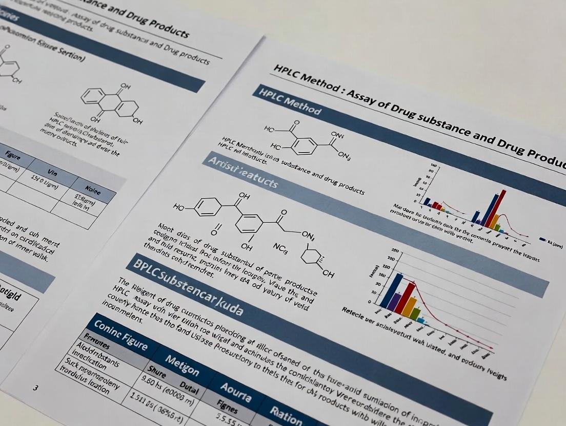
This comprehensive guide details the essential principles and practical applications of High-Performance Liquid Chromatography (HPLC) for the quantitative analysis of both drug substances (active pharmaceutical ingredients, APIs) and finished drug...
HPLC Method Development for Drug Substance and Product Assay: A Comprehensive Guide from Theory to Regulatory Compliance
Abstract
This comprehensive guide details the essential principles and practical applications of High-Performance Liquid Chromatography (HPLC) for the quantitative analysis of both drug substances (active pharmaceutical ingredients, APIs) and finished drug products. Targeting researchers, analytical scientists, and drug development professionals, the article systematically covers foundational HPLC theory, method development and application strategies, troubleshooting and optimization techniques, and the critical requirements for method validation and comparative analysis according to current ICH guidelines. By integrating foundational knowledge with advanced practical insights, this resource aims to equip professionals with the tools to develop robust, reliable, and regulatory-compliant HPLC assays for quality control and stability studies throughout the drug development lifecycle.
Understanding HPLC Fundamentals: The Science Behind Drug Substance and Product Analysis
Within the framework of developing a robust HPLC method for the assay of drug substance and drug products, understanding the foundational principles is paramount. The core separation mechanism and the optimization of key parameters directly dictate the method's selectivity, sensitivity, precision, and overall suitability for regulatory submission. This document details these principles as applied to pharmaceutical analysis.
Separation Mechanisms in Pharmaceutical HPLC
The interaction between analyte, stationary phase, and mobile phase dictates separation.
Reversed-Phase (RP-HPLC)
The dominant mode for pharmaceutical analysis due to its compatibility with most organic and ionic drugs.
- Mechanism: Partitioning based on hydrophobicity. A nonpolar stationary phase (e.g., C18) and a polar mobile phase (e.g., water/acetonitrile) are used. Analytes are retained based on their affinity for the nonpolar phase; increasing mobile phase polarity (by reducing organic % elutes compounds more quickly.
- Application in Thesis: Primary mode for assay of drug substances and formulated products, separating active pharmaceutical ingredients (APIs) from excipients and degradation products.
Normal-Phase (NP-HPLC)
- Mechanism: Separation based on analyte polarity. A polar stationary phase (e.g., silica) and a nonpolar mobile phase (e.g., hexane/chloroform) are used. Polar analytes are retained more strongly.
- Application in Thesis: Useful for very hydrophobic compounds, isomers, or chiral separations when RP-HPLC fails.
Ion-Exchange (IEC)
- Mechanism: Electrostatic interaction between charged analytes and oppositely charged stationary phase. Retention is controlled by mobile phase pH and ionic strength.
- Application in Thesis: Analysis of ionic drugs, peptides, proteins, or nucleotides.
Size-Exclusion (SEC)
- Mechanism: Physical sieving based on hydrodynamic volume. Larger molecules elute first, smaller molecules last.
- Application in Thesis: Determining aggregation state of biologic drugs or molecular weight distribution of polymer excipients.
Key Parameters and Their Optimization
Critical method parameters (CMPs) are systematically varied during development to achieve critical quality attributes (CQAs) of the method.
Table 1: Key HPLC Parameters and Their Impact on Separation
| Parameter | Definition & Control | Primary Impact on Separation | Typical Optimization Goal |
|---|---|---|---|
| Mobile Phase Composition | Type and ratio of solvents (e.g., Water:ACN), buffer type/concentration, pH. | Selectivity (α), Retention (k). | Maximize resolution of API from all potential impurities. |
| Stationary Phase | Chemistry (C18, C8, phenyl, etc.), particle size (e.g., 5µm, 3µm), column dimensions (L x ID). | Selectivity, Efficiency (N), Backpressure. | Achieve required selectivity and efficiency within pressure limits. |
| Flow Rate | Rate of mobile phase delivery (mL/min). | Efficiency, Analysis Time, Backpressure. | Balance efficiency and run time (Van Deemter curve). |
| Column Temperature | Temperature of the column oven (°C). | Retention, Efficiency, Selectivity. | Improve efficiency, reproducibility, and sometimes selectivity. |
| Gradient Profile | Programmed change in mobile phase composition over time. | Elution of analytes with a wide range of hydrophobicity. | Resolve all components in a single run with acceptable peak shape. |
| Detection Wavelength | UV-Vis wavelength selected for analyte detection (nm). | Sensitivity, Specificity. | Maximize signal-to-noise for the API and key impurities. |
Experimental Protocols for Method Scouting
Protocol 4.1: Initial Scouting of Selectivity (Stationary Phase/Mobile Phase)
Objective: To identify the best column/mobile phase combination for separating the API from its known impurities. Materials: See Scientist's Toolkit. Procedure:
- Prepare stock solutions of API and known impurities (e.g., synthesis intermediates, degradation products) at ~1 mg/mL in a suitable solvent (e.g., diluent).
- Prepare a test mixture containing all analytes at appropriate levels.
- Equilibrate three different HPLC columns (e.g., C18, C8, Phenyl-hexyl) with a generic gradient (e.g., 5-95% ACN in water over 20 min, 0.1% Formic acid).
- Inject the test mixture onto each column using the same gradient. Monitor at a universal UV wavelength (e.g., 220 nm) or using a diode array detector (DAD).
- Compare chromatograms for peak resolution (Rs > 2.0 for baseline separation), peak symmetry, and overall analysis time.
- Select the column providing the best overall selectivity for further optimization.
Protocol 4.2: Optimization of Critical Mobile Phase pH (For Ionizable Compounds)
Objective: To determine the optimal mobile phase pH for controlling retention and selectivity of ionizable APIs. Procedure:
- Prepare three separate, filtered and degassed mobile phase A buffers: pH 2.5 (e.g., phosphate or formate), pH 4.5 (e.g., acetate), and pH 7.0 (e.g., phosphate). Mobile phase B is acetonitrile.
- Use the selected column from Protocol 4.1. Set an isocratic method (e.g., 30% B) or a shallow gradient.
- Sequentially equilibrate the system with each pH buffer and inject the test mixture.
- Record the retention time (tR) and peak shape for each analyte. Plot tR vs. pH.
- Select the pH that provides the most robust separation (greatest resolution between critical pairs) and acceptable peak shape. A pH where the API is in its non-ionized form often provides better retention in RP-HPLC.
Diagrams
Title: HPLC Method Development Workflow for Drug Analysis
Title: Reversed-Phase HPLC Separation Mechanism
The Scientist's Toolkit: Key Research Reagent Solutions
Table 2: Essential Materials for HPLC Method Development
| Item | Function & Rationale |
|---|---|
| HPLC-Grade Water | Ultra-pure water to eliminate background UV absorbance and prevent column contamination. |
| HPLC-Grade Organic Solvents (Acetonitrile, Methanol) | Low UV-cutoff, high purity mobile phase components. ACN offers low viscosity and high elution strength. |
| Buffer Salts & pH Modifiers (e.g., Potassium phosphate, Sodium acetate, Formic acid, Trifluoroacetic acid) | Control mobile phase pH to suppress analyte ionization, ensuring reproducible retention and peak shape. |
| Stationary Phase Columns (C18, C8, Phenyl, HILIC, etc.) | Different selectivities to resolve complex mixtures. Particle size (e.g., 3-5µm) affects efficiency and backpressure. |
| Reference Standards (Drug Substance, Impurities) | For peak identification, calibration, and determining method specificity and accuracy. |
| Volumetric Glassware & Precision Balances | Essential for accurate and precise preparation of mobile phases, standards, and samples. |
| Syringe Filters (0.45 µm or 0.22 µm, Nylon/PVDF) | Removal of particulate matter from samples and mobile phases to protect the HPLC column and system. |
| Vials & Caps (LC-MS approved, low adsorption) | To hold samples without leaching contaminants or adsorbing analytes. |
Within the broader thesis on HPLC method development for the assay of drug substances and drug products, a fundamental principle is the clear delineation of scope between the Active Pharmaceutical Ingredient (API) and the Finished Product (Drug Product). The assay scope defines the analytical target, methodology, and validation parameters, which differ significantly between the pure drug substance and its formulated, often multi-component, final dosage form. This document provides application notes and protocols for establishing these distinct analytical scopes.
Key Differences in Assay Scope
Table 1: Comparative Assay Scope for API vs. Finished Product
| Parameter | Drug Substance (API) Assay | Finished Product (Drug Product) Assay |
|---|---|---|
| Primary Objective | To determine the purity of the active moiety, excluding process impurities and degradation products. | To quantify the amount of API in a dosage unit, assessing uniformity and content. |
| Analytical Target | The chemical entity itself, often as a free acid/base or salt. | The API within a complex matrix (excipients, coatings, preservatives). |
| Sample Preparation | Typically direct dissolution in a suitable solvent. | Often requires extraction, solubilization, or matrix destruction to liberate the API. |
| Chromatographic Focus | High-resolution separation from closely related impurities (synthesis by-products, intermediates). | Separation from excipient peaks and potential degradation products formed during formulation/storage. |
| Method Validation | Emphasis on specificity against impurities, accuracy, and precision. | Emphasis on specificity against excipients, accuracy (via recovery studies), robustness for routine use. |
| Acceptance Criteria | Often tighter (e.g., 98.0-102.0% on dried basis). | Broader to account for manufacturing variability (e.g., 90.0-110.0% of label claim at release). |
| Reference Standard | Highly purified API, characterized for identity and purity. | Usually the same API standard, but calculations are based on label claim per unit. |
Application Notes
Note 1: Interference from Excipients. The finished product assay must demonstrate specificity against common formulation components like lactose, microcrystalline cellulose, magnesium stearate, and colorants. Forced degradation studies should be performed on the formulated product, not just the API, as excipients can catalyze unique degradation pathways.
Note 2: Sample Heterogeneity. Unlike the homogeneous API, solid dosage forms require a representative sampling and homogenization protocol. For suspensions or creams, ensuring homogeneity of the analytical sample is critical.
Note 3: Potency Calculation. The API assay result is typically expressed as a percentage of the theoretical pure substance. The drug product assay result is expressed as a percentage of label claim (e.g., mg/tablet), linking directly to dosage.
Experimental Protocols
Protocol 1: HPLC Assay for Drug Substance (API)
Objective: To determine the percentage purity of an API batch by HPLC. Materials: See "The Scientist's Toolkit" below. Method:
- Mobile Phase: Prepare a filtered and degassed mixture of phosphate buffer (pH 3.0) and acetonitrile (65:35, v/v).
- Chromatographic Conditions:
- Column: C18, 250 mm x 4.6 mm, 5 μm.
- Flow Rate: 1.0 mL/min.
- Detection: UV at 254 nm.
- Injection Volume: 10 μL.
- Column Temperature: 30°C.
- Standard Solution: Accurately weigh ~10 mg of API reference standard into a 100 mL volumetric flask. Dissolve and dilute to volume with diluent (mobile phase). This is the primary standard solution (100 μg/mL).
- Sample Solution: Prepare identically using the unknown API batch.
- System Suitability: Inject six replicates of the standard solution. The %RSD for peak area should be ≤1.0%. The theoretical plate count (N) should be >2000.
- Procedure: Separately inject the standard and sample solutions. Record the chromatograms and integrate the main peak areas.
- Calculation:
% Purity = (A_Sample / A_Standard) x (W_Standard / W_Sample) x P x 100Where A = Peak area, W = Weight, P = Potency of Reference Standard (%).
Protocol 2: HPLC Assay for Finished Product (Tablet)
Objective: To determine the content of API per tablet relative to the label claim. Materials: See "The Scientist's Toolkit" below. Method:
- Mobile Phase & Chromatographic Conditions: As per Protocol 1.
- Standard Solution: Prepare as per Protocol 1 (100 μg/mL).
- Sample Preparation: a. Accurately weigh and finely powder not less than 20 tablets. b. Transfer an accurately weighed portion of powder, equivalent to ~10 mg of API, to a 100 mL volumetric flask. c. Add ~70 mL of diluent, sonicate for 30 minutes with intermittent shaking to ensure complete extraction. d. Cool to room temperature, dilute to volume with diluent, and mix. e. Filter a portion through a 0.45 μm nylon membrane, discarding the first 5 mL of filtrate.
- Placebo Solution: Prepare a solution containing all excipients at their nominal concentration in the formulation, omitting the API.
- Specificity Check: Inject placebo solution to confirm no interference at the retention time of the API.
- Procedure & System Suitability: As per Protocol 1.
- Calculation:
mg API per Tablet = (A_Sample / A_Standard) x (W_Standard / P_Std) x (100 / W_Sample) x Avg. Tablet Weight% Label Claim = (mg API per Tablet / Label Claim mg per Tablet) x 100Where P_Std = Potency of Reference Standard (as a decimal, e.g., 0.995).
Diagrams
Title: HPLC Assay Scope Decision Flow
Title: Assay Methodology Decision Tree
The Scientist's Toolkit: Key Reagent Solutions & Materials
Table 2: Essential Research Materials for HPLC Assay Development
| Item | Function | Example for API/Product Assay |
|---|---|---|
| HPLC Reference Standard | Highly characterized material used as the benchmark for quantification. | USP-grade API with certified purity. |
| Chromatographic Column | Stationary phase for separation. | Reverse-phase C18 column (e.g., 250 x 4.6 mm, 5 μm). |
| HPLC-Grade Solvents | Low UV absorbance and purity for mobile phase preparation. | Acetonitrile, Methanol, Water. |
| Buffer Salts | To control mobile phase pH, affecting selectivity and peak shape. | Potassium/sodium phosphate, ammonium acetate, trifluoroacetic acid. |
| Volumetric Glassware | For accurate preparation of standard and sample solutions. | Class A volumetric flasks, pipettes. |
| Membrane Filters | To remove particulate matter from samples and mobile phases. | 0.45 μm (or 0.22 μm) Nylon or PVDF syringe filters. |
| Ultrasonic Bath | To aid dissolution and degassing of solutions. | For extracting API from tablets and degassing mobile phase. |
| Analytical Balance | For precise weighing of standards and samples. | Microbalance (0.01 mg sensitivity). |
| Placebo/Excipient Blend | A mixture of all inactive components. | Critical for drug product method specificity testing. |
Within the framework of a thesis on HPLC method development for the assay of drug substances and drug products, the robustness, accuracy, and precision of the analytical method are paramount. The reliability of the generated data, which underpins critical decisions in pharmaceutical development and quality control, is directly contingent upon the performance and understanding of the HPLC system's core components. This document details the function, selection criteria, and practical protocols for operating the pumps, columns, detectors, and data systems, with a focus on application in pharmaceutical analysis.
Critical Components: Function and Selection
Pumps
The HPLC pump delivers the mobile phase at a constant, precise, and pulse-free flow rate. Isocratic (constant composition) or gradient (changing composition) elution is possible. For drug assay methods, reproducibility of retention times is critical, demanding high pump precision (<0.5% RSD).
Key Selection Parameters:
- Type: Quaternary pumps for method development; binary pumps for robust gradient methods.
- Pressure Limit: 6000 psi (400 bar) standard; up to 18,000 psi (1200 bar) for UHPLC.
- Flow Accuracy and Precision: <±1% accuracy, <0.1% RSD precision.
- Gradient Performance: Low delay volume and high compositional accuracy for reproducible gradients.
Columns
The column is the heart of the separation, where partitioning of analytes between the stationary and mobile phases occurs. Selection is the most critical factor in method development.
Key Selection Parameters:
- Chemistry: C18 (most common), C8, phenyl, HILIC, etc.
- Particle Size: 5 µm (conventional), 3.5 µm or sub-2 µm (for UHPLC, offering higher efficiency).
- Dimensions: 150 mm x 4.6 mm is common; 50-100 mm x 2.1 mm for UHPLC.
- Pore Size: 80-120 Å for small molecules (drug substances).
Detectors
The detector converts the physical or chemical property of eluting analytes into an electrical signal. The choice depends on the analyte's properties and the required sensitivity.
Key Selection Parameters:
- UV/Vis PDA: Most common for drugs with chromophores. Provides spectral data for peak purity.
- Mass Spectrometer (MS): For identification, structural elucidation, and ultra-sensitive quantification.
- Fluorescence (FLD): High selectivity and sensitivity for native fluorescent compounds or derivatized analytes.
- Refractive Index (RID): Universal detector for compounds lacking UV absorption (e.g., sugars, polymers).
Data Systems
Modern chromatography data systems (CDS) control the instrument, acquire data, process peaks, and generate reports. Compliance with 21 CFR Part 11 regulations (electronic records, electronic signatures) is essential for regulated laboratories.
Key Selection Parameters:
- Functionality: Instrument control, data acquisition, integration, calibration, reporting.
- Compliance: Audit trail, user access levels, electronic signature capability.
- Data Integrity: Secure storage, backup, and retrieval mechanisms.
Table 1: Quantitative Comparison of Common HPLC Detectors for Drug Assay
| Detector Type | Typical Sensitivity | Dynamic Range | Selectivity | Suitability for Drug Assay |
|---|---|---|---|---|
| UV/Vis | 10 pg - 1 ng | 10³ - 10⁴ | Low to Moderate (λ-dependent) | Excellent for most APIs with chromophores. |
| PDA | 100 pg - 1 ng | 10³ - 10⁴ | Moderate (Spectral ID) | Excellent for method development and peak purity. |
| Fluorescence | 1 fg - 1 pg | 10³ - 10⁵ | Very High | Ideal for specific, fluorescent analytes (e.g., vitamins). |
| MS (Single Quad) | 1 fg - 100 pg | 10⁴ - 10⁵ | Extremely High | Gold standard for bioanalysis, trace impurity testing. |
Application Notes and Protocols
Protocol: Pump Performance Qualification (Flow Rate Accuracy & Precision)
Objective: To verify the delivered flow rate matches the set point and is precise over time, ensuring method reproducibility. Materials: HPLC pump, calibrated digital thermometer, 50 mL volumetric flask, stopwatch, HPLC-grade water. Procedure:
- Place a beaker of HPLC-grade water on the balance and record its temperature (T°C).
- Prime the pump with water and set to 1.000 mL/min. Allow to stabilize for 15 min.
- Disconnect the column. Route the pump outlet tubing to the 50 mL volumetric flask placed on the balance.
- Simultaneously start the stopwatch and record the initial weight (W1).
- Collect effluent for exactly 30 minutes. Record the final weight (W2).
- Calculate the measured flow rate: F_meas (mL/min) = (W2 - W1) / (ρ_water at T°C * 30 min).
- Repeat steps 3-6 four more times (n=5).
- Calculate Accuracy: %Accuracy = [(Set Flow - F_meas_avg) / Set Flow] * 100.
- Calculate Precision: %RSD of the five F_meas values. Acceptance Criteria: Accuracy within ±2%; Precision (RSD) <0.5%.
Protocol: Column Screening for Drug Substance Assay Method Development
Objective: To rapidly evaluate different stationary phases to identify the best starting point for separation. Materials: HPLC system with PDA detector, 3-5 candidate columns (e.g., C18, C8, Phenyl, Polar Embedded), drug substance and related impurities stock solutions, mobile phase buffers (e.g., phosphate or formate) at pH 3.0 and 7.0, acetonitrile, water. Procedure:
- Prepare a test mixture containing the drug substance and all available known impurities (~0.1 mg/mL each).
- For each column, create two generic gradient methods:
- Method A: Acidic pH. Mobile Phase A: 0.1% Formic acid in water; B: Acetonitrile. Gradient: 5-95% B in 20 min.
- Method B: Neutral pH. Mobile Phase A: 10 mM Ammonium formate pH 7.0; B: Acetonitrile. Gradient: 5-95% B in 20 min.
- Inject the test mixture onto each column using Method A. Record chromatograms, noting retention, peak shape (asymmetry factor, As), and resolution (Rs) between critical pairs.
- Repeat with Method B.
- Compare results. Select the column/pH condition providing the best overall peak shape, resolution (>2.0 between all peaks), and analysis time.
Protocol: Detector Linear Range and Limit of Quantification (LOQ) Determination
Objective: To establish the concentration range over which the detector response is linear and determine the lowest quantifiable level for the drug assay. Materials: HPLC system with relevant detector, drug substance reference standard, mobile phase. Procedure:
- Prepare a stock solution of the drug reference standard at a concentration near the expected assay concentration (e.g., 1 mg/mL).
- Serially dilute to prepare at least 6 standard solutions covering a wide range (e.g., from 0.001% to 150% of target concentration).
- Inject each solution in triplicate using the developed chromatographic method.
- Plot the peak area (average) versus concentration.
- Perform linear regression analysis. The correlation coefficient (r) should be >0.999.
- Calculate the LOQ as the concentration yielding a signal-to-noise ratio (S/N) of 10:1 from a low-level standard injection.
Visualizations
The Scientist's Toolkit: Research Reagent Solutions
Table 2: Essential Materials for HPLC Method Development in Drug Analysis
| Item | Function & Importance in Drug Assay Research |
|---|---|
| HPLC-Grade Water | Low UV absorbance and particulate-free; essential for mobile phase preparation to ensure low background noise and prevent system blockages. |
| HPLC-Grade Acetonitrile & Methanol | Primary organic modifiers for reversed-phase mobile phases. High purity minimizes baseline drift and ghost peaks. |
| Ammonium Formate/Acetate, Trifluoroacetic Acid (TFA), Formic Acid | Buffering agents and ion-pairing modifiers. Critical for controlling mobile phase pH, which governs analyte ionization, retention, and peak shape. |
| Certified Reference Standards | High-purity, well-characterized drug substance. Essential for accurate method calibration, quantification, and validation. |
| Column Regeneration & Storage Solutions | (e.g., 80% Water/20% ACN). Proper column cleaning and storage in appropriate solvent extend column lifetime and maintain performance. |
| Vial Inserts & Low-Volume Vials | Minimize sample evaporation and dead volume, crucial for accurate and precise injections, especially in automated systems. |
| In-Line Filters & Guard Columns | Protect the analytical column from particulate matter and strongly retained contaminants, preserving column efficiency and lifetime. |
| pH Meter with ATC Probe | Accurate pH adjustment of aqueous mobile phases is critical for reproducible retention times and robust method performance. |
Within the broader context of developing robust and selective HPLC methods for the assay of drug substance and drug products, the selection of the appropriate chromatographic mode is the foundational decision. This choice dictates selectivity, efficiency, and overall method success. This application note provides a comparative analysis of Reversed-Phase (RP), Normal-Phase (NP), Ion-Exchange (IEX), and Hydrophilic Interaction Liquid Chromatography (HILIC), supported by protocols and data to guide researchers in pharmaceutical development.
The following table summarizes the key characteristics, applications, and quantitative performance metrics of the four primary HPLC modes.
Table 1: Comparison of HPLC Modes for Pharmaceutical Analysis
| Mode | Typical Stationary Phase | Typical Mobile Phase | Primary Separation Mechanism | Ideal Analyte Properties | Typical Efficiency (Plates/m) | Ruggedness |
|---|---|---|---|---|---|---|
| Reversed-Phase (RP) | C18, C8, Phenyl | Water + Organic Modifier (ACN, MeOH) | Hydrophobic partitioning | Medium to non-polar, neutral | 80,000 - 120,000 | Excellent |
| Normal-Phase (NP) | Silica, Diol, Amino | Organic (Hexane) + Polar Modifier (IPA, EA) | Adsorption (polar interactions) | Polar, non-ionic | 40,000 - 70,000 | Poor (hydration sensitive) |
| Ion-Exchange (IEX) | Cationic or Anionic Resin | Aqueous Buffer (pH-controlled) | Ionic attraction/repulsion | Charged (acids, bases, peptides) | 20,000 - 50,000 | Good (buffer dependent) |
| HILIC | Bare Silica, Amide, Diol | ACN (>60%) + Aqueous Buffer | Partitioning + Secondary Interactions | Polar, hydrophilic, ionizable | 70,000 - 100,000 | Good (equilibrium critical) |
Table 2: Application Suitability for Drug Analysis
| Mode | Common Drug Substance/Product Applications | Key Advantages | Key Limitations |
|---|---|---|---|
| RP | >80% of small molecule drugs, stability-indicating methods, impurities. | Robust, reproducible, compatible with MS. | Poor retention of very polar compounds. |
| NP | Isomer separation, lipophilic compounds, chiral separations. | Unique selectivity for structural isomers. | Less reproducible, not MS-friendly, long equilibration. |
| IEX | Biologics (mAbs, proteins), nucleotides, charged APIs, counter-ion analysis. | High selectivity for ionic species. | Limited to ionic analytes, slow, requires buffer cleanup for MS. |
| HILIC | Polar APIs (e.g., metformin), glycosylated compounds, small polar impurities. | Excellent retention of polar compounds, MS compatible. | Method development complex, long equilibration times. |
Experimental Protocols
Protocol 1: Initial Mode Selection & Scouting Gradient
This protocol provides a systematic approach for selecting the appropriate chromatographic mode for a new drug substance.
Objective: To rapidly assess the retention and peak shape of an unknown Active Pharmaceutical Ingredient (API) across different HPLC modes to guide final mode selection.
Materials: (See "The Scientist's Toolkit" section) Procedure:
- Prepare a 1 mg/mL solution of the API in a solvent compatible with all modes (e.g., 50:50 ACN:Water or a solvent matching the initial mobile phase).
- Equilibrate four HPLC systems (or columns in parallel) with the following initial conditions:
- RP: 95% Water / 5% Acetonitrile (0.1% Formic Acid)
- HILIC: 90% Acetonitrile / 10% Water (10 mM Ammonium Formate, pH 3.0)
- IEX (Anionic): 20 mM Sodium Phosphate, pH 7.0
- IEX (Cationic): 20 mM Sodium Phosphate, pH 3.0
- Inject 5 µL of the API solution onto each column.
- Run a generic scouting gradient for each mode:
- RP & HILIC: Linear gradient from initial conditions to 95% organic (RP) or 50% aqueous (HILIC) over 20 minutes.
- IEX: Linear salt gradient (e.g., 0 to 500 mM NaCl in buffer) over 20 minutes.
- Monitor retention time (tR) and peak shape (asymmetry factor, As).
- Decision Logic: If tR > 2 min in RP, proceed with RP optimization. If unretained in RP but retained in HILIC, proceed with HILIC. If peak tails/shows multiple peaks in IEX, consider IEX for ionic impurities or related substances.
Protocol 2: HILIC Method Development for a Polar Drug Substance
Objective: To develop a validated HILIC-UV method for the assay of a highly polar, water-soluble API (e.g., metformin hydrochloride).
Procedure:
- Column Selection: Install an amide-bonded HILIC column (e.g., 150 x 4.6 mm, 3.5 µm).
- Initial Conditions: Equilibrate with 85% Acetonitrile / 15% 50 mM Ammonium Acetate buffer (pH 5.0). Flow: 1.0 mL/min. Temperature: 30°C.
- Scouting Injection: Inject the API standard. If retention is too strong (>15 min), increase % aqueous. If too weak (<2 min), increase % ACN.
- Buffer Optimization: Prepare mobile phase buffers at constant organic ratio (85% ACN) but varying buffer pH (3.0, 5.0, 7.0) and concentration (5 mM, 20 mM). Assess impact on retention, peak shape, and selectivity from related polar impurities.
- Temperature Gradient: Evaluate column temperatures (25°C, 35°C, 45°C). In HILIC, increased temperature typically decreases retention.
- Final Method: Based on data, select conditions that yield tR of ~5-10 minutes for the API, baseline resolution from all known impurities (Rs > 2.0), and peak asymmetry between 0.9-1.2.
- Equilibration: Ensure a minimum of 10-15 column volumes of the initial mobile phase for full equilibration before each sequence.
Logical Workflow & Visualization
Diagram Title: HPLC Mode Selection Decision Tree
Diagram Title: HILIC Method Development Workflow
The Scientist's Toolkit
Table 3: Essential Research Reagent Solutions for HPLC Mode Scouting
| Item | Function & Rationale |
|---|---|
| C18 Column (e.g., 150 x 4.6 mm, 3 µm) | Standard workhorse for Reversed-Phase (RP) screening. Provides hydrophobic interaction surface. |
| HILIC Column (e.g., Amide, 150 x 4.6 mm, 3 µm) | Essential for evaluating retention of polar compounds unretained in RP. Operates via hydrophilic partitioning. |
| Weak Anion Exchange (WAX) Column | Used for separating acidic compounds and anions via ionic interaction with positively charged groups. |
| Weak Cation Exchange (WCX) Column | Used for separating basic compounds and cations via ionic interaction with negatively charged groups. |
| HPLC-Grade Acetonitrile & Methanol | Primary organic modifiers. ACN is preferred for RP and HILIC due to low viscosity and UV cutoff. |
| Ammonium Formate & Ammonium Acetate | Volatile buffers (pH 2-8) compatible with mass spectrometry for RP, HILIC, and IEX screening. |
| Trifluoroacetic Acid (TFA) / Formic Acid | Ion-pairing agents and pH modifiers for RP. Improve peak shape for ionizable analytes. |
| Phosphate Buffer Salts | Non-volatile buffers for UV-detection methods requiring precise pH control in IEX or RP. |
| Column Heater/Chiller | Critical for maintaining retention time reproducibility, especially in HILIC and IEX modes. |
1. Introduction: Role in HPLC Method Validation
Within the research framework of developing a High-Performance Liquid Chromatography (HPLC) method for the assay of drug substance and drug product, adherence to regulatory guidelines is paramount. The International Council for Harmonisation (ICH) guideline Q2(R1), "Validation of Analytical Procedures: Text and Methodology," and the United States Pharmacopeia (USP) general chapter <621>, "Chromatography," provide the foundational principles. ICH Q2(R1) defines the validation parameters and methodology required to demonstrate that an analytical procedure is suitable for its intended purpose. USP <621> provides the system suitability tests and allowable adjustments to chromatographic conditions, ensuring the method's performance at the time of execution. Together, they form the core regulatory and scientific standards for method validation and routine control in pharmaceutical analysis.
2. Comparative Analysis of ICH Q2(R1) and USP <621>
The table below summarizes the key focus areas and requirements of both guidelines, highlighting their complementary roles.
Table 1: Core Focus and Scope of ICH Q2(R1) vs. USP <621>
| Aspect | ICH Q2(R1) | USP <621> |
|---|---|---|
| Primary Purpose | Validation of analytical procedures to prove fitness for purpose. | Establishment of system suitability criteria and rules for adjusting chromatographic parameters. |
| Key Parameters | Accuracy, Precision (Repeatability, Intermediate Precision), Specificity, Detection Limit (DL), Quantitation Limit (QL), Linearity, Range, Robustness. | Parameters defining system suitability: Plate count (N), Tailing factor (T), Resolution (Rs), Relative Standard Deviation (RSD) for replicate injections. |
| Stage of Use | Applied during method development and pre-validation/validation. | Applied during method validation (as part of robustness) and every time the method is used for sample analysis. |
| Adjustability | Does not address operational adjustments. | Explicitly defines permissible adjustments to chromatographic conditions (e.g., flow rate, column length, particle size, pH, mobile phase ratio) within defined limits to maintain validity. |
3. Application Notes: Integrating Guidelines into HPLC Method Development
3.1. Validation Protocol Based on ICH Q2(R1) for an Assay Method For a stability-indicating HPLC assay method, the following validation parameters, as per ICH Q2(R1), must be addressed experimentally.
Table 2: Validation Parameters and Typical Acceptance Criteria for an HPLC Assay
| Validation Parameter | Protocol Summary | Typical Acceptance Criteria |
|---|---|---|
| Specificity | Inject blank, placebo, drug substance, degraded samples (acid/base/oxidative/thermal stress). | Peak purity passes (e.g., by PDA). Resolution from nearest known impurity > 2.0. No interference from blank/placebo. |
| Linearity & Range | Prepare standard solutions at minimum 5 concentrations (e.g., 50-150% of target assay concentration). Plot response vs. concentration. | Correlation coefficient (r) ≥ 0.999. Residuals randomly scattered. |
| Accuracy (Recovery) | Spike placebo with drug at 3 levels (e.g., 80%, 100%, 120%) in triplicate. Compare measured vs. added amount. | Mean recovery 98.0–102.0% per level. Overall mean 98.0–102.0%. |
| Precision | 1. Repeatability: 6 replicate injections of 100% standard. 2. Intermediate Precision: Perform on different day, different analyst, different instrument. | RSD of assay results ≤ 2.0% for drug substance; ≤ 2.0% for product (small molecule). |
| Robustness | Deliberately vary parameters (flow rate ±10%, column temp ±2°C, mobile phase pH ±0.2, organic ratio ±2% absolute). | System suitability passes in all varied conditions. Assay results consistent. |
3.2. System Suitability Protocol as per USP <621> System suitability is an integral part of both method validation and routine use. USP <621> provides the governing rules.
Protocol: System Suitability Test Execution
- Preparation: Prepare the standard solution as per the analytical method.
- Injection: Inject the standard solution a minimum of 5 times.
- Calculation & Evaluation: Calculate the following from the standard chromatogram:
- Theoretical Plates (N): > 2000 is typical for a well-packed column.
- Tailing Factor (T): ≤ 2.0 for the analyte peak.
- Resolution (Rs): ≥ 2.0 between the analyte peak and the closest eluting known impurity or degradation product.
- Relative Standard Deviation (RSD): For peak area (or height) of replicate injections, typically ≤ 2.0% for assay methods.
- Pass/Fail: The analysis batch can proceed only if all system suitability criteria are met.
Table 3: USP <621> Allowable Adjustments for HPLC Methods
| Parameter | General Allowable Adjustment | Key Constraints |
|---|---|---|
| Flow Rate | ±50% | Must meet system suitability. Pressure limits must not be exceeded. |
| Column Dimensions | Length: ±70% | The ratio of column length to particle size (L/dp) must not vary by more than ±25%. |
| Internal Diameter: ±25% | Adjusted flow rate to maintain linear velocity. | |
| Particle Size | May decrease by up to 50% | Must meet system suitability and pressure limits. Increases not permitted. |
| pH of Aqueous Buffer | ±0.2 pH units | Absolute change, not relative. |
| Buffer Concentration | ±10% | Relative change. |
| Organic Modifier Ratio | Relative adjustment up to ±30% of the absolute value stated (e.g., 30% ± 10% absolute → 20-40% allowed). | Final composition cannot be less than zero. Must meet system suitability. |
4. The Scientist's Toolkit: Essential Reagents and Materials
Table 4: Key Research Reagent Solutions for HPLC Method Validation
| Item | Function & Importance |
|---|---|
| Drug Substance Reference Standard | Certified, high-purity material used as the primary standard for preparing calibration solutions. Essential for accuracy, linearity, and identification. |
| Placebo Formulation | Contains all excipients of the drug product without the Active Pharmaceutical Ingredient (API). Critical for specificity testing to confirm no interference. |
| Chromatography Columns | Multiple lots from the same manufacturer/specification. Required for robustness testing and verifying method reproducibility. |
| HPLC-Grade Solvents & Reagents | High-purity mobile phase components (water, acetonitrile, methanol) and buffer salts (e.g., potassium phosphate). Minimizes baseline noise and ghost peaks. |
| Forced Degradation Materials | Reagents for stress studies: Acid (e.g., 0.1M HCl), Base (e.g., 0.1M NaOH), Oxidant (e.g., 3% H₂O₂), Thermal/Photolytic chambers. Used to demonstrate specificity and stability-indicating capability. |
5. Workflow and Relationship Diagrams
Title: HPLC Method Validation Workflow Integrating ICH & USP
Title: Relationship Between ICH Q2(R1) and USP <621> Objectives
Within the broader thesis on HPLC method development for the assay of drug substances and products, System Suitability Tests (SST) serve as the critical quality control checkpoint. Their execution validates that the analytical system—comprising the instrument, reagents, column, analyst, and the method itself—is performing adequately at the time of analysis. This ensures the integrity, reliability, and reproducibility of the generated data, which is fundamental for making definitive conclusions about drug potency, purity, and stability.
Key SST Parameters: Definitions and Acceptance Criteria
SST parameters are derived from the analysis of a standard solution, typically a drug reference standard at or near the target concentration. The table below summarizes the core parameters, their definitions, and typical acceptance criteria as per ICH Q2(R1) and USP <621> guidelines.
Table 1: Core System Suitability Parameters for HPLC Assay Methods
| Parameter | Definition | Typical Acceptance Criteria (for assay) | Rationale |
|---|---|---|---|
| Theoretical Plates (N) | A measure of column efficiency. | > 2000 | Ensures sufficient peak sharpness and resolving power. |
| Tailing Factor (Tf) | A measure of peak symmetry. | ≤ 2.0 | Indicates proper column condition and absence of unwanted interactions. |
| Resolution (Rs) | Degree of separation between two adjacent peaks. | > 1.5 between drug and closest eluting impurity | Confirms the method's specificity and ability to separate analytes. |
| Repeatability (RSD) | Precision of the system, measured by relative standard deviation of replicate injections. | RSD ≤ 1.0% for peak area (n=5 or 6) | Demonstrates the instrument's precision and injection reproducibility. |
| Relative Retention Time | Consistency of a peak's retention time relative to a reference. | RSD ≤ 1.0% | Confirms system stability over the sequence. |
Detailed Experimental Protocol for SST Execution
This protocol is designed for the SST injection sequence within an HPLC assay method for a drug product.
A. Materials and Reagent Preparation
- Mobile Phase: Prepare as per the validated method. Filter through a 0.45 µm membrane filter and degas.
- Diluent: Appropriate solvent (e.g., water, buffer, organic mix).
- System Suitability Standard Solution: Precisely weigh and dissolve the drug reference standard in diluent to obtain a solution at the target assay concentration (e.g., 100 µg/mL). Prepare in duplicate from independent weighings.
- Test Samples: Drug substance or product samples prepared as per the method.
B. Instrumental Setup and Conditioning
- Install the specified HPLC column (C18, 150 x 4.6 mm, 5 µm).
- Set the chromatographic conditions: flow rate (1.0 mL/min), column temperature (25°C), detection wavelength, and injection volume (10 µL).
- Prime the system with mobile phase and initiate the method. Allow the system to equilibrate until a stable baseline is achieved (typically 30-60 minutes).
C. SST Injection Sequence and Evaluation
- Initial Blank: Inject the diluent to confirm no interfering peaks at the retention time of the analyte.
- System Conditioning Injections: Inject the SST standard solution until consistent retention times and peak responses are observed (typically 3-5 injections). Data from these injections are not used for SST calculation.
- SST Evaluation Injections: Make six consecutive injections from the same vial of the SST standard solution.
- Data Analysis: Using the chromatography data system (CDS) software, calculate the parameters from the five (or six) replicate injections:
- Theoretical Plates (N): Calculated as N = 16 (tR/w)2, where tR is retention time and w is peak width at baseline.
- Tailing Factor (T): Calculated as T = W0.05 / 2f, where W0.05 is the peak width at 5% height and f is the distance from the peak front to the peak maximum at 5% height.
- Resolution (Rs): For methods requiring separation, calculate between the drug peak and a specified impurity or degradation product peak injected in a resolution solution. Rs = 2(tR2 - tR1) / (w1 + w2).
- Repeatability: Calculate the %RSD of the peak areas (and retention times) of the replicate injections.
- Acceptance: Compare calculated values against pre-defined criteria (Table 1). The system is deemed suitable only if all criteria are met. If any parameter fails, troubleshoot the system (e.g., check for air bubbles, column degradation, improper mobile phase preparation) and repeat the SST sequence.
- Proceed with Analysis: Once SST passes, inject samples, controls, and standards in the planned sequence. SST check standards are often interspersed throughout the run to monitor ongoing performance.
Visualizing the SST Decision Workflow
SST Pass/Fail Decision Logic
The Scientist's Toolkit: Essential Research Reagent Solutions
Table 2: Key Materials for HPLC SST Execution
| Item | Function in SST Context |
|---|---|
| Drug Reference Standard | Certified material with high purity (>99%) used to prepare the SST solution. Serves as the benchmark for system performance. |
| HPLC-Grade Solvents | Acetonitrile, methanol, water. High purity minimizes baseline noise and UV absorbance, ensuring accurate detection. |
| Buffer Salts (e.g., Potassium Phosphate) | For mobile phase pH control. Critical for achieving consistent retention and peak shape of ionizable compounds. |
| Volumetric Glassware (Class A) | Precise flasks and pipettes for accurate preparation of mobile phase and standard solutions. |
| Syringe Filters (0.45 µm or 0.22 µm) | For removing particulate matter from samples and standards, protecting the HPLC column from blockage. |
| Certified HPLC Column | The stationary phase specified in the method. Its condition is the primary determinant of parameters like N, Tf, and Rs. |
| Resolution Test Mixture | A solution containing the analyte and a closely eluting impurity or analog. Used to specifically demonstrate resolution (Rs) capability. |
Step-by-Step HPLC Method Development and Real-World Applications
Within the broader thesis on HPLC method development for the assay of drug substance and drug products, the initial scouting phase is critical for establishing a robust, selective, and efficient analytical method. This Application Note details a systematic protocol for concurrent mobile phase composition screening, stationary phase selection, and aqueous phase pH optimization. This orthogonal approach rapidly identifies the starting conditions for further method optimization and validation, significantly reducing development time for new chemical entities.
High-Performance Liquid Chromatography (HPLC) is the cornerstone for the quantitative analysis of active pharmaceutical ingredients (APIs) and formulated products. The initial scouting experiments aim to explore the multidimensional chromatographic parameter space to find a combination that provides adequate retention, peak shape, and selectivity for the API and its potential impurities. A poorly designed scouting phase can lead to prolonged development, method failure during validation, or lack of specificity. This protocol outlines a structured, efficient approach grounded in Quality by Design (QbD) principles.
Research Reagent Solutions & Essential Materials
The following table lists the key reagents and materials required to execute the scouting protocols.
Table 1: Essential Research Reagent Solutions and Materials
| Item | Function in Scouting Experiments |
|---|---|
| HPLC System (e.g., Agilent 1260, Waters Alliance) | Instrument capable of handling solvent gradients, multiple columns, and providing stable pH and temperature control. |
| Diode Array Detector (DAD) | For multi-wavelength detection and peak purity assessment across different conditions. |
| Automated Column Switcher (e.g., 6-position valve) | Enables rapid, unattended screening of multiple stationary phases using a single instrument. |
| ChemStation/EMPOWER or equivalent CDS | Software for instrument control, data acquisition, and analysis. |
| Reference Standards (API and known impurities) | Critical for assessing selectivity, resolution, and peak identity under various conditions. |
| pH Meter (with certified buffers) | Accurate calibration for precise pH adjustment of aqueous mobile phases. |
| Buffer Salts (Ammonium formate, ammonium acetate, phosphate salts) | Provides buffering capacity at targeted pH ranges for reproducible retention. |
| Mobile Phase Modifiers (Trifluoroacetic acid, formic acid, ammonium hydroxide) | Used to adjust pH in acidic and basic ranges and influence ionization and peak shape. |
| Scouting Columns (C18, C8, Phenyl, Polar Embedded, HILIC) | Diverse stationary phases to probe different selectivity mechanisms (hydrophobicity, π-π, H-bonding). |
| HPLC Grade Solvents (Acetonitrile, Methanol, Water) | Primary solvents for mobile phase preparation; low UV cutoff and high purity are essential. |
| Vials and Caps (LC-MS certified) | For sample and standard preparation, minimizing contaminant introduction. |
Experimental Protocols
Protocol: Concurrent pH and Organic Modifier Scouting
Objective: To determine the optimal pH and organic modifier (acetonitrile vs. methanol) for analyte ionization control, retention, and peak shape.
Procedure:
- Buffer Preparation: Prepare 10 mM ammonium formate buffers at pH 3.0, 4.5, and 6.0. Prepare 10 mM ammonium bicarbonate at pH 8.0 and 10.0. Filter all through a 0.22 µm membrane.
- Mobile Phase Setup: For each pH, create two solvent systems:
- System A (Acetonitrile): Buffer pH X / Acetonitrile (95:5, v/v)
- System B (Methanol): Buffer pH X / Methanol (95:5, v/v) Use the organic percentage (5%) to ensure all analytes are retained at the start of the gradient.
- Gradient Program: Employ a linear gradient from 5% to 95% organic modifier over 20 minutes. Hold at 95% for 3 min, then re-equilibrate.
- Column: Use a generic, high-quality C18 column (e.g., 150 x 4.6 mm, 3.5 µm) for this initial screening.
- Detection: Monitor at 210 nm, 254 nm, and the λ-max of the API.
- Data Analysis: Plot retention factor (k) vs. pH for the API and available impurities. Note peak asymmetry (As) for each condition.
Protocol: Orthogonal Stationary Phase Screening
Objective: To evaluate selectivity differences across diverse column chemistries at a narrowed pH range identified from Protocol 3.1.
Procedure:
- Column Selection: Install the following column types (all 50 x 4.6 mm, 3 µm for rapid screening) into an automated column switcher:
- C18 (L1)
- Polar-Embedded C18 (e.g., amide)
- Phenyl-Hexyl or Phenyl
- Cyano (CN)
- Pentafluorophenyl (PFP)
- Mobile Phase: Use the optimal pH buffer (from 3.1) mixed with the optimal organic modifier (ACN/MeOH) in a 95:5 ratio as the starting mobile phase.
- Gradient Program: Use a fast, linear gradient from 5% to 95% organic over 10 minutes.
- System Setup: The CDS method includes valve switching commands to sequentially route the flow through each column.
- Data Analysis: Calculate the resolution (Rs) between the closest eluting critical pair (e.g., API and its primary impurity) on each column. Rank columns based on Rs and peak symmetry.
Protocol: Isocratic Scouting for Initial Method Conditions
Objective: To approximate the optimal isocratic organic percentage for further fine-tuning after selecting pH and column.
Procedure:
- Condition: Use the selected buffer pH, organic modifier, and column from previous protocols.
- Run a Series of Isocratic Methods: Perform injections at 10%, 20%, 30%, 40%, and 50% organic modifier (balance is aqueous buffer).
- Analysis: For the API, calculate the retention factor (k) at each organic percentage.
- Modeling: Plot log(k) vs. % organic. Use linear regression to determine the % organic required to achieve a target k value (typically 2-10 for assay methods).
Data Presentation and Analysis
Table 2: Summary of Scouting Results for API X-123 and Primary Impurity (Imp-A)
| Condition (Column / pH / Modifier) | k (API) | k (Imp-A) | Selectivity (α) | Resolution (Rs) | Peak Asymmetry (API) |
|---|---|---|---|---|---|
| C18 / pH 3.0 / ACN | 4.2 | 4.5 | 1.07 | 1.2 | 1.05 |
| C18 / pH 6.0 / ACN | 5.8 | 6.9 | 1.19 | 2.8 | 1.10 |
| C18 / pH 8.0 / ACN | 2.1 | 2.0 | 0.95 | Co-elution | 1.35 |
| C18 / pH 3.0 / MeOH | 6.5 | 7.1 | 1.09 | 1.5 | 1.15 |
| Phenyl / pH 6.0 / ACN | 7.2 | 8.9 | 1.24 | 3.5 | 1.08 |
| Polar-Embedded / pH 6.0 / ACN | 5.1 | 5.3 | 1.04 | 0.8 | 1.02 |
| PFP / pH 6.0 / ACN | 8.5 | 10.1 | 1.19 | 3.1 | 1.20 |
Optimal condition selected based on highest Rs and acceptable asymmetry is highlighted.
Table 3: Isocratic Scouting Data on Selected Conditions (Phenyl, pH 6.0, ACN)
| % ACN (Isocratic) | k (API) | log(k) | Plate Count (N) |
|---|---|---|---|
| 30% | 15.2 | 1.18 | 12500 |
| 35% | 9.1 | 0.96 | 11200 |
| 40% | 5.5 | 0.74 | 10500 |
| 45% | 3.3 | 0.52 | 9800 |
| 50% | 2.0 | 0.30 | 8900 |
Linear regression of log(k) vs. %ACN indicates ~38% ACN is required to achieve k=5.
Visualized Workflows and Relationships
Title: Initial HPLC Scouting Sequential Workflow
Title: Key Parameter Interactions in HPLC Scouting
Within the broader thesis on HPLC method development for drug substance and product assay, this document details the application notes and protocols for identifying, evaluating, and controlling Critical Method Parameters (CMPs). Robustness is a key validation parameter per ICH Q2(R2), ensuring method reliability during routine use and technology transfer.
Critical Method Parameters: Identification & Screening
CMPs are variables in the analytical procedure that, when varied within a reasonable range, may significantly influence measurement results, system suitability criteria, or the validity of the analytical procedure. For a typical reversed-phase HPLC assay, these include factors related to the mobile phase, column, temperature, and flow rate.
Table 1: Typical HPLC Assay Parameters for Screening
| Parameter Category | Specific Parameter | Normal Operating Condition (NOC) | Tested Range (±) |
|---|---|---|---|
| Mobile Phase | pH | 2.7 (e.g., Phosphate buffer) | ±0.2 units |
| Organic % (B) | 45% Acetonitrile | ±3% absolute | |
| Buffer Conc. | 25 mM | ±5 mM | |
| Chromatographic | Column Temp. | 30°C | ±5°C |
| Flow Rate | 1.0 mL/min | ±0.1 mL/min | |
| Wavelength | 220 nm | ±5 nm | |
| Column | Different Lot/Brand | Specified L1 | 2 additional lots/vendors |
Experimental Protocols for Robustness Testing
Protocol 3.1: Univariate (One-Factor-at-a-Time, OFAT) Preliminary Assessment
Objective: To rapidly gauge the individual effect of a single parameter variation on key chromatographic outputs. Materials: See "The Scientist's Toolkit" below. Procedure:
- Prepare the drug substance standard solution at the target concentration (e.g., 100 µg/mL) per the candidate method.
- Set the HPLC system to the NOC.
- Perform a system suitability test (SST) injection: Ensure tailing factor <2.0, theoretical plates >2000, and %RSD of replicate injections <2.0%.
- Select one parameter (e.g., mobile phase pH). Vary it to the extremes of the proposed range (e.g., pH 2.5 and 2.9) while holding all other parameters at NOC.
- Inject the standard solution in triplicate at each condition.
- Record the retention time (tR), peak area, tailing factor (T), and resolution (Rs) from any critical pair.
- Return the system to NOC and re-inject to confirm system performance.
- Repeat steps 4-7 for each parameter to be screened. Analysis: Calculate the % change relative to NOC for each response. Parameters causing a change greater than a pre-defined threshold (e.g., >10% in tR or >2% in area) are flagged for further multivariate study.
Protocol 3.2: Multivariate (Design of Experiments, DoE) Robustness Study
Objective: To systematically evaluate the main effects and interactions of multiple CMPs simultaneously. Design: A 2-level fractional factorial design for 5-6 parameters is often sufficient. Example Design for 4 Factors: pH (±0.1), %B (±2%), Temp. (±3°C), Flow (±0.05 mL/min). Procedure:
- Design Setup: Use statistical software (e.g., JMP, Minitab, Design-Expert) to generate a randomized run order for the experimental design (e.g., 8 runs + 3 center point replicates).
- Sample Preparation: Prepare a single, homogenous batch of standard and sample solutions sufficient for all design runs to minimize preparation variability.
- Sequential Analysis: Follow the randomized run order, equilibrating the system at each new set of conditions for at least 5 column volumes before injection.
- Data Collection: For each run, inject the standard solution and record tR, area, T, and Rs.
- Center Points: The center point (NOC) replicates are interspersed to estimate experimental error and check for curvature. Analysis:
- Perform ANOVA to identify statistically significant factors (p-value < 0.05).
- Generate main effects and interaction plots.
- Set acceptance criteria for robustness: e.g., assay result must remain within 98.0-102.0% of label claim across all design runs, and Rs > 2.0.
- Define a Method Operable Design Region (MODR): the multidimensional combination of parameter ranges where the method meets all acceptance criteria.
Diagram Title: Robustness Testing & MODR Establishment Workflow
Protocol 3.3: Forced Degradation Sample Analysis
Objective: To verify the stability-indicating capability of the method and ensure specificity near CMP boundaries. Procedure:
- Stress the drug product separately under acid, base, oxidative, thermal, and photolytic conditions per ICH Q1B.
- Prepare samples of stressed material, blank, and unstressed control.
- Analyze these samples using the HPLC method at the NOC and at the extreme edges of the CMP ranges identified in Protocol 3.2.
- Assess chromatograms for:
- Peak Purity: Using a diode array detector (PDA) to ensure main peak homogeneity.
- Resolution: Resolution between the main peak and the nearest degradation product must be >2.0 under all tested method conditions. Analysis: This confirms the method's ability to accurately quantify the active ingredient in the presence of degradants, even with minor, expected method fluctuations.
Data Presentation: Robustness Study Results
Table 2: Representative DoE Results for Critical Responses (NOC: Area = 10000, tR = 10.0 min)
| Run | pH | %B | Temp (°C) | Flow (mL/min) | Peak Area | tR (min) | Resolution (Rs) |
|---|---|---|---|---|---|---|---|
| 1 | 2.6 | 43 | 27 | 0.95 | 10052 | 10.8 | 3.5 |
| 2 | 2.8 | 43 | 27 | 1.05 | 9985 | 8.9 | 3.2 |
| 3 | 2.6 | 47 | 27 | 1.05 | 10023 | 8.5 | 3.8 |
| 4 | 2.8 | 47 | 27 | 0.95 | 10048 | 11.5 | 3.1 |
| 5 | 2.6 | 43 | 33 | 1.05 | 10012 | 8.7 | 3.7 |
| 6 | 2.8 | 43 | 33 | 0.95 | 10061 | 11.2 | 3.0 |
| 7 | 2.6 | 47 | 33 | 0.95 | 10044 | 10.5 | 4.0 |
| 8 | 2.8 | 47 | 33 | 1.05 | 9988 | 8.2 | 3.6 |
| CP1 | 2.7 | 45 | 30 | 1.00 | 10005 | 10.0 | 3.5 |
| CP2 | 2.7 | 45 | 30 | 1.00 | 9998 | 10.1 | 3.5 |
| CP3 | 2.7 | 45 | 30 | 1.00 | 10011 | 10.0 | 3.4 |
Table 3: Statistical Summary of Main Effects
| Factor | Effect on Peak Area | p-value | Effect on Retention Time | p-value | Conclusion |
|---|---|---|---|---|---|
| pH | +15.2 | 0.12 | +0.05 | 0.85 | Not Significant |
| %Organic (B) | -8.5 | 0.30 | -1.10 | <0.01* | Critical for tR |
| Temperature | +10.8 | 0.18 | -0.80 | <0.01* | Critical for tR |
| Flow Rate | -12.4 | 0.14 | -1.05 | <0.01* | Critical for tR |
| Model | Robust across ranges |
Diagram Title: CMP Influence Map on HPLC Assay Performance
The Scientist's Toolkit: Key Research Reagent Solutions
Table 4: Essential Materials for HPLC Robustness Studies
| Item | Function & Specification | Example/Catalog |
|---|---|---|
| HPLC-Grade Solvents | Low UV absorbance, minimal impurities for reproducible baselines and retention. | Acetonitrile (CH3CN), Methanol (MeOH). |
| Buffer Salts & pH Modifiers | Provide consistent mobile phase ionic strength and pH. Use high-purity (>99%). | Potassium Phosphate, Sodium Phosphate, Trifluoroacetic Acid (TFA). |
| Volumetric Glassware (Class A) | Precise preparation of mobile phases and standard solutions. Critical for accuracy. | 1 L, 500 mL, 100 mL volumetric flasks. |
| Reference Standard | Highly characterized material of known purity for system suitability and calibration. | USP/EP Reference Standard or internally qualified working standard. |
| HPLC Column (Multiple Lots) | The primary CMP. Test at least 3 different lots from the same supplier and/or columns from 2 different vendors. | e.g., Waters XBridge C18, 4.6 x 150 mm, 3.5 µm. |
| PDA or DAD Detector | Essential for peak purity assessment during forced degradation studies. | Diode Array Detector capable of 200-400 nm scanning. |
| Statistical Software | For designing DoE studies and performing ANOVA, effects analysis, and MODR mapping. | JMP, Minitab, Design-Expert. |
| Column Heater/Oven | Provides precise and stable temperature control (±0.5°C). A key CMP. | Thermostatted column compartment. |
Sample Preparation Protocols for APIs and Complex Dosage Forms (Tablets, Capsules, Injections)
Within the broader thesis on HPLC method development for the assay of drug substance and drug products, robust and reproducible sample preparation is the critical first step. This document provides detailed application notes and protocols for preparing Active Pharmaceutical Ingredients (APIs) and complex dosage forms for subsequent chromatographic analysis. The goal is to ensure complete extraction of the analyte, removal of matrix interferences, and compatibility with the HPLC mobile phase.
Table 1: Common Dosage Form Excipients and Sample Preparation Challenges
| Dosage Form | Typical Excipients (Interferents) | Primary Sample Prep Challenge | Typical Solution |
|---|---|---|---|
| API (Drug Substance) | Residual solvents, synthesis impurities | Homogeneity, solubility | Direct dissolution in suitable solvent |
| Immediate-Release Tablet | Binders (e.g., MCC), disintegrants, lubricants (Mg stearate), fillers | Binding of API, insoluble particulates | Sonication with solvent, filtration, centrifugation |
| Modified-Release Tablet | Polymer matrices (e.g., HPMC, EC), coatings | Incomplete extraction due to slow release | Extended stirring, use of surfactant, or organic solvent |
| Hard Gelatin Capsule | Gelatin shell, lubricants, fillers | Gelatin cross-linking, insolubility | Capsule shell removal or dissolution in warmed solvent |
| Injectable Solution | Buffers, preservatives, antioxidants, solubilizers | Low API concentration, protein binding (biologics) | Dilution, protein precipitation, solid-phase extraction |
| Injectable Suspension | Suspending agents, stabilizers | Homogeneity of sample aliquot | Intensive mixing/sonication prior to sampling, filtration |
Table 2: Typical Solvent Volumes and Extraction Parameters
| Protocol Step | Parameter | Typical Range | Notes |
|---|---|---|---|
| Solvent Addition | Volume for Tablet/Capsule | 50-1000 mL | Based on target concentration & solubility |
| Sonication | Time & Temperature | 10-30 min at 25-40°C | Avoid for thermolabile compounds |
| Mechanical Shaking | Time & Speed | 15-60 min at 150-200 rpm | For modified-release forms |
| Centrifugation | Speed & Time | 3000-5000 rpm for 5-15 min | Clarify supernatant |
| Filtration | Membrane Pore Size | 0.45 µm or 0.22 µm | Nylon for aqueous, PTFE for organic |
Detailed Experimental Protocols
Protocol 3.1: API (Drug Substance) Preparation
Objective: To prepare a stock solution of the pure API for use in calibration standard preparation.
- Accurately weigh an appropriate amount of API (e.g., 25 mg) using an analytical balance.
- Quantitatively transfer to a 25 mL volumetric flask using the chosen diluent (e.g., HPLC grade methanol or mixture with buffer).
- Add diluent to ~80% of flask volume and sonicate for 5-10 minutes to ensure complete dissolution.
- Allow to cool to room temperature, then dilute to volume with diluent and mix thoroughly.
- This yields a primary stock solution (e.g., 1 mg/mL). Further dilute serially with diluent to prepare working standards.
Protocol 3.2: Immediate-Release Tablet Preparation (Sonication-Extraction)
Objective: To completely extract the API from a tablet matrix for assay.
- Accurately weigh and finely powder not less than 10 tablets using a mortar and pestle or a mechanical mill.
- Accurately weigh a portion of the powder equivalent to the label claim of one tablet (e.g., 100 mg API) into a 100 mL volumetric flask.
- Add ~70 mL of extraction solvent (e.g., 70:30 methanol:pH 4.5 buffer). Sonicate in a water bath for 25 minutes, swirling intermittently.
- Allow to cool to room temperature. Dilute to volume with the same solvent and mix well.
- Filter a portion through a 0.45 µm nylon syringe filter, discarding the first 2-3 mL of filtrate.
- Further dilute the filtrate as needed to fall within the HPLC calibration range.
Protocol 3.3: Hard Gelatin Capsule Preparation
Objective: To extract API from capsule contents, avoiding interference from the gelatin shell.
- Carefully open not less than 10 capsules and collect the combined contents.
- Mix the powder thoroughly. Accurately weigh an amount equivalent to one capsule's label claim.
- Transfer the powder to a suitable flask. Add a known volume (e.g., 50 mL) of warmed (40-45°C) extraction solvent.
- Shake mechanically for 30 minutes. Cool, then quantitatively transfer to a volumetric flask, rinsing the original flask.
- Dilute to volume, mix, and filter (0.45 µm) before HPLC analysis.
Protocol 3.4: Injectable Solution Preparation
Objective: To prepare injectable solutions, often requiring simple dilution or matrix removal. For Small Molecule Injections:
- For concentrated solutions, make an appropriate direct dilution with mobile phase or a compatible solvent.
- For solutions in oily vehicles, perform a liquid-liquid extraction (e.g., with hexane and acetonitrile). For Protein-Based Biologics (Requiring Protein Precipitation):
- Accurately pipette a volume of the injection (e.g., 100 µL) into a microcentrifuge tube.
- Add 300 µL of cold acetonitrile (or a mixture with methanol) to precipitate proteins.
- Vortex vigorously for 1 minute, then centrifuge at 10,000 rpm for 10 minutes.
- Carefully collect the clear supernatant, filter (0.22 µm), and inject into the HPLC.
Visualization of Workflows
Figure 1. General Workflow for Solid Oral Dosage Form Preparation
Figure 2. Core Principle of Sample Preparation for HPLC Assay
The Scientist's Toolkit
Table 3: Essential Research Reagent Solutions for Sample Prep
| Item / Reagent | Function & Rationale | Key Considerations |
|---|---|---|
| HPLC Grade Solvents (Methanol, Acetonitrile, Water) | Primary extraction/dilution solvents. High purity minimizes UV interference and baseline noise. | Use appropriate grade to avoid ghost peaks. Acetonitrile is a stronger eluent. |
| Buffer Salts (e.g., Potassium Phosphate, Ammonium Acetate) | Control pH of extraction to maintain analyte stability and solubility, especially for ionizable compounds. | Buffer must be compatible with HPLC column and mobile phase. Filter before use. |
| Dilute Acid/Base (e.g., 0.1N HCl, NaOH) | Aid in dissolving APIs by forming salts; can break down certain matrix components. | Neutralize after extraction if needed for HPLC compatibility. |
| Surfactants (e.g., SDS, Polysorbate 80) | Improve wetting and extraction efficiency from polymeric or lipid-based matrices. | Must be removable (e.g., via SPE) or not interfere with chromatography. |
| Protein Precipitants (Cold ACN, MeOH, TCA) | Denature and precipitate proteins in biological injectables, freeing the analyte into solution. | Precipitation efficiency varies; supernatant must be clear. |
| Solid-Phase Extraction (SPE) Cartridges (C18, HLB, Ion Exchange) | Selective clean-up of complex matrices (e.g., plasma in PK studies) to remove interferences. | Condition, load, wash, and elute steps must be optimized. |
| Syringe Filters (Nylon, PTFE, PVDF, 0.45/0.22 µm) | Remove insoluble particulates to protect HPLC column and system. | Choose membrane compatible with solvent (PTFE for organics). Discard first filtrate. |
Within the broader thesis on HPLC method development for the assay of drug substances and products, the chapter on forced degradation studies is pivotal. It establishes the specificity and stability-indicating nature of the proposed analytical method. This section provides the experimental proof that the method can accurately and reliably measure the active pharmaceutical ingredient (API) while resolving it from its degradation products, which is a fundamental requirement for ICH Q1A(R2) and Q2(R1) guidelines.
Theoretical Background and Regulatory Imperatives
Forced degradation, also known as stress testing, is a deliberate, aggressive degradation of a drug substance and product under conditions more severe than accelerated stability. Its primary goal is to elucidate intrinsic stability characteristics and identify likely degradation products. The developed HPLC method must then demonstrate its ability to “indicate stability” by separating the API from all generated degradation peaks. Key regulatory guidelines include ICH Q1A(R2) Stability Testing of New Drug Substances and Products, ICH Q1B Photostability Testing, and ICH Q2(R1) Validation of Analytical Procedures.
Diagram: Role of Forced Degradation in Method Development
Diagram Title: Flow of SIA Development
Key Experimental Protocols for Forced Degradation
Protocol 1: Acid and Base Hydrolytic Stress
Objective: To evaluate the susceptibility of the API to hydrolysis and generate relevant degradants.
- Preparation: Prepare separate solutions of the drug substance (~1 mg/mL) in 0.1 M HCl (for acid stress) and 0.1 M NaOH (for base stress). For drug products, use a suspension or solution in the same media.
- Stress Condition: Heat the solutions at 60°C (±2°C) for a period ranging from 1 to 24 hours. Shorter intervals (e.g., 1, 4, 8 hr) are recommended for time-point sampling.
- Neutralization: After the desired time, cool and neutralize the samples immediately. Acid-stressed samples are neutralized with 0.1 M NaOH, and base-stressed samples with 0.1 M HCl, to a final pH of ~7.
- Analysis: Dilute the neutralized samples with mobile phase or a suitable solvent to the target concentration. Inject onto the HPLC system.
- Control: Run a parallel control sample (API in neutral solvent, e.g., water/methanol) under the same temperature conditions.
Protocol 2: Oxidative Stress
Objective: To induce and study oxidative degradation pathways.
- Preparation: Prepare a solution of the drug substance (~1 mg/mL) in 3% w/v hydrogen peroxide (H₂O₂). For sensitive compounds, start with 0.3% H₂O₂.
- Stress Condition: Keep the solution at room temperature (25°C ± 2°C) or 40°C for up to 24 hours. Sample at intervals (e.g., 1, 6, 24 hr).
- Quenching (if necessary): Oxidation may be quenched by dilution with mobile phase or by adding a reducing agent like methionine, if it does not interfere.
- Analysis: Dilute and inject directly. Include a control sample in solvent without H₂O₂.
Protocol 3: Thermal (Solid-State) and Photolytic Stress
Objective: To assess degradation under dry heat and UV/Vis light exposure.
- Thermal (Solid):
- Spread the solid drug substance or product (in its final packaging and opened) in a thin layer in a petri dish.
- Place in a stability chamber at 70°C (±2°C) for 1-4 weeks.
- Periodically withdraw samples, prepare solutions, and analyze.
- Photolytic:
- Expose solid samples and/or solutions to ICH-specified light conditions (Option 2: 1.2 million lux hours of visible light and 200 watt-hours/square meter of UV light).
- Use a qualified photostability chamber.
- Protect control samples with opaque wrapping (e.g., aluminum foil).
- After exposure, prepare samples and analyze alongside controls.
Protocol 4: Thermal (Solution) and Humidity Stress
Objective: To assess solution-state thermal degradation and moisture sensitivity.
- Thermal (Solution): Heat drug solutions in neutral pH buffer (e.g., phosphate buffer pH 7.0) at 70°C for 24-72 hours. Analyze versus a room-temperature control.
- Humidity: Expose solid samples to 75% ± 5% relative humidity at 25°C for 1-4 weeks. Use a saturated salt solution (e.g., NaCl) in a closed desiccator to maintain humidity.
Data Presentation: Typical Degradation Acceptance Criteria and Results
Table 1: Common Forced Degradation Conditions and Targets
| Stress Condition | Typical Parameters | Target Degradation | Key Considerations |
|---|---|---|---|
| Acid Hydrolysis | 0.1-1 M HCl, 40-70°C, 1-72 h | 5-20% degradation | Neutralize before analysis to stop reaction. |
| Base Hydrolysis | 0.1-1 M NaOH, 40-70°C, 1-72 h | 5-20% degradation | Neutralize before analysis to stop reaction. |
| Oxidation | 0.3%-3% H₂O₂, RT-40°C, 1-48 h | 5-20% degradation | May proceed rapidly; monitor closely. |
| Thermal (Solid) | 70°C, dry, 1-4 weeks | 5-20% degradation | For drug product, test in and out of package. |
| Thermal (Solution) | Neutral pH, 70°C, 24-72 h | 5-20% degradation | Use buffer to control pH. |
| Photolysis | ICH Q1B Option 2 | Evidence of change | Compare with dark control. |
| Humidity | 75% RH, 25°C, 1-4 weeks | Evidence of change | Often combined with heat. |
Table 2: Example Forced Degradation Results for a Hypothetical API
| Stress Condition | Time Point | % API Remaining | % Major Degradant | Purity Angle / Purity Threshold* | Peak Purity Pass? |
|---|---|---|---|---|---|
| Control (Initial) | 0 h | 100.0 | 0.00 | 0.501 / 1.032 | Yes |
| Acid (0.1M HCl, 60°C) | 8 h | 88.5 | 8.2 (Deg A) | 0.890 / 1.245 | Yes |
| Base (0.1M NaOH, 60°C) | 8 h | 85.2 | 12.1 (Deg B) | 1.102 / 1.567 | Yes |
| Oxidation (3% H₂O₂, RT) | 24 h | 90.1 | 7.5 (Deg C) | 0.732 / 1.110 | Yes |
| Heat (Solid, 70°C) | 7 days | 95.8 | 2.5 (Deg D) | 0.610 / 1.085 | Yes |
| Light (ICH) | Final | 98.5 | <0.5 | 0.520 / 1.041 | Yes |
*Data from Photodiode Array (PDA) detector; Purity Angle < Purity Threshold indicates a spectrally pure peak.
The Scientist's Toolkit: Essential Research Reagent Solutions
Table 3: Key Reagents and Materials for Forced Degradation Studies
| Item | Function & Rationale |
|---|---|
| High-Purity APIs & Drug Products | The starting material for stress studies; must be well-characterized to attribute changes to degradation. |
| Concentrated Acids & Bases (HCl, NaOH) | To prepare hydrolytic stress media. Analytical grade ensures no interfering impurities. |
| Hydrogen Peroxide (30% w/v) | Standard oxidant for oxidative stress testing. Fresh stock is critical as it decomposes. |
| pH Buffers (e.g., Phosphate) | To maintain specific pH during solution thermal stress, mimicking physiological conditions. |
| HPLC-Grade Solvents (Methanol, Acetonitrile, Water) | For sample preparation, dilution, and as mobile phase components. Purity is essential for accurate chromatography. |
| Qualified Stability/Photostability Chambers | To provide precise, controlled, and documented temperature, humidity, and light conditions. |
| PDA or Mass Spectrometry Detector | PDA is mandatory for peak purity assessment. MS is used for identification of unknown degradants. |
| Validated HPLC/UHPLC System | The core analytical instrument. Must be qualified and calibrated to generate reliable data. |
Diagram: Forced Degradation Experimental Workflow
Diagram Title: Forced Degradation Study Workflow
Within the broader thesis on HPLC method development for drug substance and product analysis, a pivotal challenge is the efficiency of analytical control strategies. Traditionally, separate methods are validated for assay (potency) and related substances (impurities), demanding significant resources, time, and sample. This application note details a practical, modern approach for consolidating these determinations into a single, stability-indicating reversed-phase HPLC method. The unified method must satisfy the distinct validation requirements for both quantitative purposes: precision and accuracy for assay, and sensitivity and selectivity for impurities.
Key Methodological Considerations
The primary challenge lies in optimizing chromatographic conditions to elute and resolve the main active pharmaceutical ingredient (API) from all potential impurities (process-related and degradants) while maintaining detection parameters suitable for widely differing concentrations (e.g., 100% for assay vs. 0.1% for impurities). Key parameters include column selection, gradient profile, detection wavelength, injection volume, and sample concentration.
Table 1: Comparison of Single vs. Dual Method Approaches
| Parameter | Traditional Dual-Method Approach | Consolidated Single Method | Advantage of Single Method |
|---|---|---|---|
| Development Time | ~8-12 weeks | ~6-10 weeks | Reduced time to method readiness |
| Validation Runs | 2 full sets (assay & impurities) | 1 integrated set | ~40% reduction in validation workload |
| Sample Consumption | Higher (two separate preparations) | Lower (one preparation) | Preserves scarce drug substance |
| System Suitability | Two distinct sets of criteria | One comprehensive set | Simplified QC testing routine |
| Risk | Method alignment issues | Higher initial development complexity | Streamlined lifecycle management |
Experimental Protocol: Unified HPLC Method Development
Objective: To develop and validate a single RP-HPLC method for the simultaneous determination of Assay (% purity) and Related Substances for Drug Substance X.
I. Materials & Equipment (The Scientist's Toolkit) Table 2: Key Research Reagent Solutions & Materials
| Item | Function & Specification |
|---|---|
| Reference Standard | Highly characterized material of known purity for accurate quantification. |
| Forced Degradation Samples | Stressed samples (acid, base, oxidative, thermal, photolytic) to establish method specificity. |
| Hybrid Silica C18 Column | Stationary phase (e.g., 150 x 4.6 mm, 2.7 µm particle size) offering high efficiency and pH stability (pH 1.5-10). |
| HPLC-MS Grade Mobile Phase Solvents | Acetonitrile and Methanol for consistent baseline and MS compatibility if needed. |
| High-Purity Buffer Salts | e.g., Potassium dihydrogen phosphate or Ammonium formate, for precise pH control. |
| pH Meter with Temperature Probe | Accurate (±0.01 units) adjustment of aqueous buffer. |
| Variable Wavelength or Diode Array Detector (DAD) | For multi-wavelength monitoring and peak purity assessment. |
| Automated Injector & Column Heater | Ensures precise injection volumes (±1%) and stable retention times. |
II. Detailed Protocol
Step 1: Preliminary Scouting & Sample Preparation
- Prepare stock solutions of Drug Substance X and all available impurity standards (~1 mg/mL) in a suitable diluent (e.g., water:acetonitrile 70:30).
- Prepare a sample solution at the target assay concentration (typically ~0.1-1 mg/mL).
- Perform gradient scouting runs (e.g., 5-95% organic over 30 mins) on 2-3 different column chemistries (C18, phenyl, polar-embedded) at pH 3.0, 4.5, and 7.0. Use a flow rate of 1.0 mL/min and DAD detection from 200-400 nm.
Step 2: Forced Degradation for Specificity
- Subject Drug Substance X to stress conditions: 0.1N HCl & NaOH (4h, 60°C), 3% H₂O₂ (24h, RT), heat (70°C, 48h), and light (1.2 million lux-hours).
- Neutralize or dilute stressed samples as required and analyze using the preliminary gradient.
- Assess chromatograms for peak purity (using DAD) and resolution between the main peak and all degradants. The method must be "stability-indicating."
Step 3: Method Optimization
- Based on scouting data, select the column and pH providing the best overall peak shape and separation of critical pairs.
- Fine-tune the gradient profile to elute all impurities within a reasonable runtime (e.g., 45-60 minutes), ensuring the API elutes near the mid-point.
- Optimize the flow rate (e.g., 0.8-1.2 mL/min) for efficiency.
- Set detection wavelength at the λ-max of the API, ensuring sufficient sensitivity for impurities (confirm all are detectable at 0.1% level).
Step 4: Final Method Parameters
- Column: Zodiac C18 (150 x 4.6 mm, 2.7 µm)
- Mobile Phase A: 10 mM Potassium Phosphate Buffer, pH 5.0
- Mobile Phase B: Acetonitrile
- Gradient: 0 min (15% B), 0-25 min (15→50% B), 25-30 min (50→95% B), 30-35 min (95% B), 35-36 min (95→15% B), 36-45 min (15% B)
- Flow Rate: 1.0 mL/min
- Column Temp: 30°C
- Injection Volume: 10 µL
- Detection: 254 nm (DAD for peak purity 210-400 nm)
- Sample Temp: 5°C
- Sample Concentration: 0.5 mg/mL for assay and impurities.
Step 5: Validation Experiments Perform an integrated validation assessing parameters for both assay and impurity determinations per ICH Q2(R1).
- Specificity: Verify resolution from all known and forced degradation impurities.
- Linearity: For Assay: 50-150% of target concentration (e.g., 0.25-0.75 mg/mL). For Impurities: LOQ to 1.5% of assay concentration.
- Accuracy: For Assay: Spiked recovery at 80%, 100%, 120% levels. For Impurities: Spiked recovery at LOQ, 0.5%, 1.0% levels.
- Precision: Repeatability (n=6) of assay and impurity quantification at specification level.
- Sensitivity: Determine LOD and LOQ for main impurity (S/N ~3:1 and 10:1).
- Robustness: Deliberate variations in pH (±0.2), temperature (±2°C), gradient profile (±2%).
Table 3: Example Validation Results for Drug Substance X
| Validation Parameter | Assay (API at 100%) | Impurity A (at 0.5% level) | Acceptance Criteria Met? |
|---|---|---|---|
| Linearity (R²) | 0.9999 | 0.9995 | Yes (R² > 0.999 / >0.995) |
| Accuracy (% Recovery) | 99.8% ± 0.5 | 100.2% ± 2.1 | Yes (98-102% / 90-110%) |
| Repeatability (%RSD) | 0.15% | 1.8% | Yes (<1.0% / <10.0%) |
| LOQ (as % of API) | Not Required | 0.03% | Yes (≤ Reporting Threshold) |
| Robustness (Worst-case %RSD) | 0.35% | 2.5% | Yes (System suitability passed) |
Visualization of Workflow and Logical Structure
Title: Unified HPLC Method Development Workflow
Title: Logical Relationship of Single Method Goals
This practical approach demonstrates that determining assay and related substances via a single, well-designed HPLC method is not only feasible but advantageous. It enhances analytical efficiency, reduces resource consumption, and simplifies the control strategy—a significant advancement within drug development research. The success hinges on rigorous upfront scouting and forced degradation studies to ensure the method is fundamentally stability-indicating, followed by a comprehensive, integrated validation protocol.
Application Notes
Small Molecule Drug Substance Assay
The quantification of a small molecule API, such as the anticoagulant Apixaban, is a cornerstone of drug substance research. The primary challenge is achieving separation from closely related process impurities and degradation products (e.g., hydrolyzed or oxidized species). A robust Reverse-Phase (RP) method using a C18 column (150 x 4.6 mm, 2.7 µm) with a mobile phase of 0.1% trifluoroacetic acid (TFA) in water (A) and acetonitrile (B) is standard. Detection at 280 nm provides the requisite sensitivity.
Table 1: Validated HPLC Method Parameters for Apixaban Assay
| Parameter | Specification |
|---|---|
| Column | C18, 150 x 4.6 mm, 2.7 µm |
| Mobile Phase | A: 0.1% TFA in H₂O; B: Acetonitrile |
| Gradient | 30% B to 70% B over 15 min |
| Flow Rate | 1.0 mL/min |
| Column Temperature | 30°C |
| Detection | UV at 280 nm |
| Injection Volume | 10 µL |
| Retention Time | ~8.2 min |
| Linearity (R²) | >0.999 (2-150 µg/mL) |
| Assay Precision (%RSD) | <1.0% |
Peptide Drug Product Analysis
The analysis of Glucagon-like peptide-1 (GLP-1) analogs (e.g., Liraglutide) in a formulation matrix presents challenges of separating the target peptide from deamidated, truncated, and dimerized variants. A UHPLC method with a superficially porous particle (SPP) C18 column (100 x 2.1 mm, 1.7 µm) offers high resolution. The use of 0.1% Formic Acid in water and acetonitrile as modifiers is critical for maintaining peptide solubility and enhancing MS-compatibility for identity confirmation.
Table 2: UHPLC-MS Method for Liraglutide and Related Substances
| Parameter | Specification |
|---|---|
| Column | SPP C18, 100 x 2.1 mm, 1.7 µm |
| Mobile Phase | A: 0.1% FA in H₂O; B: 0.1% FA in Acetonitrile |
| Gradient | 25% B to 40% B over 20 min |
| Flow Rate | 0.3 mL/min |
| Temperature | 50°C |
| Detection | UV at 214 nm, MS (ESI+) |
| Key Impurities | Deamidated (ΔRt ~ -0.5 min), Dimer (ΔRt ~ +2.1 min) |
| Mass Accuracy (MS) | < 2 ppm |
Complex Formulation: Ointment Assay
Assaying a corticosteroid (e.g., Betamethasone Dipropionate) in a complex ointment base containing petroleum, waxes, and lanolin requires extensive sample preparation to eliminate interfering lipids. The method employs a solid-phase extraction (SPE) clean-up step prior to RP-HPLC analysis with a C8 column, which provides better selectivity for the moderately hydrophobic analyte against residual matrix components.
Table 3: HPLC Method for Betamethasone Dipropionate in Ointment
| Parameter | Specification |
|---|---|
| Sample Prep | Dissolve in hexane, SPE (C18) clean-up, elute with MeOH |
| Column | C8, 250 x 4.6 mm, 5 µm |
| Mobile Phase | Methanol:Water (75:25 v/v) |
| Flow Rate | 1.2 mL/min |
| Detection | UV at 240 nm |
| Recovery | 98.5% ± 1.2% |
| Specificity | Resolved from matrix peaks and degradation product |
Experimental Protocols
Protocol 1: Assay of Apixaban Drug Substance
Objective: To determine the purity and assay of Apixaban API by HPLC-UV.
Materials:
- HPLC system with UV detector, C18 column (150 x 4.6 mm, 2.7 µm)
- Apixaban reference standard and test sample
- TFA, HPLC-grade water, and acetonitrile
- Volumetric flasks, pipettes, syringe filters (0.22 µm, Nylon)
Procedure:
- Mobile Phase: Prepare 1.0 mL of TFA in 1 L of HPLC-grade water (Solvent A). Use HPLC-grade acetonitrile as Solvent B.
- Standard Solution: Accurately weigh ~25 mg of Apixaban RS into a 50 mL volumetric flask. Dissolve and dilute to volume with a 50:50 mixture of Solvent A and B. Further dilute 5 mL to 50 mL with the same diluent (~50 µg/mL).
- Test Solution: Prepare the test sample identically to the standard solution.
- System Suitability: Inject the standard solution five times. The %RSD of peak area must be ≤1.0%. The theoretical plates for the Apixaban peak should be >5000.
- Analysis: Separately inject the standard and test solutions (10 µL). Use the gradient program: 0-2 min (30% B), 2-15 min (30-70% B), 15-17 min (70% B), 17-18 min (70-30% B), 18-23 min (30% B).
- Calculation: Calculate the % assay using the formula:
(Area_Test / Area_Standard) x (Wt_Standard / Wt_Test) x Purity_of_Standard x 100.
Protocol 2: Related Substances Method for Liraglutide Injection
Objective: To quantify Liraglutide and its related impurities in a drug product using UHPLC-UV.
Materials:
- UHPLC system with PDA or UV detector, SPP C18 column (100 x 2.1 mm, 1.7 µm)
- Liraglutide RS and impurity standards (if available)
- Formic acid, water, acetonitrile (LC-MS grade)
- Refrigerated centrifuge
Procedure:
- Mobile Phase: Prepare 0.1% v/v formic acid in water (A) and in acetonitrile (B). Filter and degas.
- Resolution Solution: Sponge the main peptide standard with stressed sample (e.g., heat-treated) to generate impurities.
- Test Solution: For a multi-dose pen, pool contents from 5 pens. Dilute an aliquot equivalent to ~1 mg of Liraglutide to 10 mL with 20% acetonitrile in water.
- Centrifuge: Centrifuge the test solution at 10,000 rpm for 5 min at 4°C to remove any particulates.
- Chromatography: Inject 2 µL using the gradient: 0-20 min (25-40% B), 20-21 min (40-95% B), 21-24 min (95% B), 24-25 min (95-25% B), 25-30 min (25% B). Maintain column at 50°C, flow at 0.3 mL/min. Detect at 214 nm.
- Evaluation: Identify impurities by relative retention time and/or spiking. Report any impurity >0.1%.
Protocol 3: Assay of Betamethasone in Ointment with SPE Clean-up
Objective: To extract and quantify Betamethasone Dipropionate from a complex hydrophobic ointment matrix.
Materials:
- HPLC system, C8 column (250 x 4.6 mm, 5 µm)
- Betamethasone Dipropionate RS
- SPE cartridges (C18, 500 mg), vacuum manifold
- Hexane, methanol, water (HPLC grade)
Procedure:
- SPE Conditioning: Condition the C18 SPE cartridge sequentially with 5 mL methanol followed by 5 mL water. Do not let the bed dry.
- Sample Extraction: Accurately weigh ointment (~500 mg) into a tube. Add 10 mL hexane and vortex to dissolve the base. Load this hexane solution onto the conditioned SPE cartridge.
- Wash & Elution: Wash with 5 mL of 40% methanol in water to remove polar interferences. Elute the drug with 5 mL of pure methanol into a 10 mL volumetric flask.
- Dilution: Bring to volume with methanol and filter (0.45 µm PTFE).
- HPLC Analysis: Use an isocratic mobile phase of methanol:water (75:25). Inject 20 µL. Flow rate: 1.2 mL/min. UV detection at 240 nm.
- Quantification: Compare peak areas of the sample against a reference standard solution of known concentration prepared in methanol.
Visualization
HPLC Workflow for Small Molecule API Assay
Peptide Analysis with UHPLC-MS Workflow
Complex Formulation Analysis with SPE Workflow
The Scientist's Toolkit: Research Reagent Solutions
Table 4: Essential Materials for HPLC Method Development in Drug Analysis
| Item | Function & Rationale |
|---|---|
| High-Purity Solvents & Buffers (ACN, MeOH, Water, TFA, FA) | Ensure reproducible chromatography, low UV background, and MS compatibility. |
| Pharmaceutical Grade Reference Standards | Provide the primary benchmark for identity, potency, and impurity quantification. |
| Stationary Phases (C18, C8, SPP, Polar-Embedded) | Select based on analyte hydrophobicity/ polarity; crucial for resolution and peak shape. |
| Syringe Filters (0.22 µm, Nylon, PTFE) | Protect HPLC column from particulate matter in samples. |
| Solid-Phase Extraction (SPE) Cartridges (C18, HLB) | Essential for extracting analyte from complex matrices (ointments, creams, plasma). |
| Vial Inserts (Low Volume, 250 µL) | Maximize injection precision and minimize sample waste for precious peptides/ compounds. |
| Column Heater/Oven | Maintains consistent retention times and improves separation efficiency. |
| pH Meter & Standards | Critical for reproducible buffer preparation in ionizable compound methods. |
Solving Common HPLC Problems: Troubleshooting and Advanced Optimization Strategies
Within the broader thesis on HPLC method development for the assay of drug substance and drug products, robustness and reliability are paramount. Common chromatographic issues like tailing peaks, peak splitting, and baseline noise directly impact method validation parameters such as precision, accuracy, and sensitivity. This document provides detailed application notes and protocols for diagnosing and resolving these critical issues to ensure method suitability for regulatory submission.
Tailing Peaks: Diagnosis and Resolution
Primary Cause: Secondary interactions of the analyte with active sites on the stationary phase (e.g., silanol groups on silica-based columns). Other Causes: Column overload, mismatch between sample solvent and mobile phase, void at column inlet, or incorrect mobile phase pH.
Table 1: Common Causes and Corrective Actions for Tailing Peaks
| Cause | Diagnostic Test | Typical Tailing Factor (T) | Corrective Action | Expected Outcome |
|---|---|---|---|---|
| Silanol Activity | Inject a basic probe (e.g., amitriptyline) | T > 2.0 | Use a lower pH mobile phase (pH < 3), add amine modifiers (e.g., TEA), or use a specialty endcapped column | T reduced to 1.0 - 1.5 |
| Column Void | Visual inspection of peak shape for all analytes | Progressive increase in T over time | Replace column inlet frit or repack column inlet | Restoration of original T value |
| Mobile Phase pH Mismatch | Compare tailing at different pH values | T varies significantly with pH | Adjust pH to be ≥2 units away from analyte pKa | Optimized, consistent T |
| Sample Solvent Stronger than MP | Inject sample in different solvents | High T only for strong solvent | Dilute sample in mobile phase or a weaker solvent | T reduced to match standard |
Experimental Protocol: Diagnosing Silanol Activity
Objective: To determine if peak tailing is due to ionic interaction with residual silanols. Materials: As per "Scientist's Toolkit" below. Procedure:
- Prepare a standard solution of a basic test compound (e.g., 0.1 mg/mL amitriptyline) in the mobile phase.
- Perform chromatography using the current method. Record the tailing factor (T) at 5% peak height.
- Modify the mobile phase by adding 0.1% (v/v) triethylamine (TEA).
- Re-inject the standard. Record the new T value.
- Interpretation: A reduction in T by >20% confirms silanol activity. Permanent resolution may require switching to a column designed for basic compounds.
Peak Splitting: Diagnosis and Resolution
Primary Cause: Column hardware issues (e.g., mismatched frits, improperly cut tubing) or a void at the column inlet. Other Causes: Contamination at the column inlet, sample solvent mismatch, or presence of multiple conformational isomers.
Table 2: Troubleshooting Guide for Splitting Peaks
| Symptom | Likely Location of Issue | Diagnostic Step | Resolution Protocol |
|---|---|---|---|
| Doublet or shoulder on all peaks | Column inlet/connections | Replace column with a known good column | Re-tighten or replace connections (zero-dead-volume fittings); replace inlet frit |
| Splitting on one specific analyte | Sample/chemical | Check for isomerization or pH-sensitive forms | Adjust mobile phase pH; use a stabilizing buffer; change column chemistry |
| Sudden onset of splitting after many runs | Column inlet contamination/void | Check system pressure (often reduced) | Reverse and flush column; replace inlet frit; refill column void with packing material |
| Random splitting | Detector flow cell | Install a flow cell bypass tube | Clean or replace detector flow cell |
Experimental Protocol: Resolving a Column Inlet Void
Objective: To repair a column with degraded performance due to a void at the inlet. Materials: HPLC column repair kit, fresh packing material (same as column), sonicator, slurry solvent. Procedure:
- Remove the column and note the flow direction.
- Carefully open the column inlet end fitting and remove the worn frit.
- Gently remove approximately 1-2 mm of the discolored or settled packing material.
- Prepare a slurry of fresh packing material in an appropriate solvent (e.g., isopropanol).
- Using the kit tools, add the new slurry to the column inlet to fill the void.
- Insert a new frit, reassemble the column end fitting, and tighten to manufacturer specifications.
- Reconnect the column in the correct flow direction, condition with mobile phase, and evaluate performance with a test mix.
Baseline Noise: Diagnosis and Resolution
Primary Causes: Contaminated mobile phase or column, air bubbles in the system, leak, or detector lamp failure.
Table 3: Classification and Resolution of Baseline Noise
| Noise Type | Characteristic | Common Source | Corrective Action |
|---|---|---|---|
| High-Frequency (Short-Term) | Rapid, jagged signal fluctuations | Detector lamp (UV), electronic noise, bubbles in flow cell | Replace lamp; degas mobile phase; check detector grounding |
| Low-Frequency (Long-Term) | Slow, rolling drifts | Temperature fluctuations, mobile phase mixing issues, column equilibration | Use a column heater; ensure proper mobile phase mixing; allow for equilibration |
| Regular Spikes | Sharp, periodic spikes | Pump seal chatter, injection valve malfunction, electrical interference | Replace pump seals; clean or rebuild injector; use dedicated power line |
| Irregular Drift & Noise | Combined slow drift and high noise | Contaminated mobile phase or column, microbial growth | Prepare fresh mobile phase with HPLC-grade water; flush/clean column |
Experimental Protocol: Systematic Isolation of Noise Source
Objective: To identify the component responsible for excessive baseline noise. Procedure:
- Isolate the detector: Disconnect the column and connect a piece of tubing from the injector outlet directly to the waste. Place a restrictor capillary after the detector if needed. Observe baseline. High noise implicates the detector or mobile phase.
- Change mobile phase: Replace with fresh, freshly prepared, and degassed mobile phase from different solvent bottles. Observe change.
- Re-introduce column: Reconnect the column. A significant increase in noise indicates a contaminated column.
- Check injector: Perform multiple blank injections (mobile phase). Spikes correlated with injection cycle point to a contaminated or worn injector rotor seal.
- Check pump: Monitor pressure trace for regularity. Erratic pressure correlates with noise indicates failing pump seals or check valves.
Visualization of Troubleshooting Workflows
Title: Tailing Peaks Diagnostic Decision Tree
Title: Systematic Baseline Noise Isolation Protocol
The Scientist's Toolkit: Research Reagent Solutions
Table 4: Essential Materials for HPLC Troubleshooting
| Item Name | Function/Application | Example/Brand |
|---|---|---|
| Silanol Test Mix | Diagnoses secondary interactions with acidic silanols on the column. Contains basic, neutral, and acidic probes. | USP L7, or custom mix (e.g., amitriptyline, acetophenone, benzoic acid) |
| Triethylamine (TEA) | Mobile phase additive to mask residual silanol activity for basic compounds. | HPLC-grade, ≥99.9% purity |
| Column Inlet Frit Repair Kit | For replacing clogged or damaged frits to restore column efficiency and prevent splitting. | Vendor-specific kits (e.g., Phenomenex, Agilent, Waters) |
| In-line Degasser | Removes dissolved air from mobile phase to reduce baseline noise and drift. | Integral part of modern HPLC systems or standalone modules |
| Pump Seal Kit | For replacing worn pump seals that cause pressure fluctuations and baseline spikes. | Must match specific HPLC pump model |
| Rheodyne Injector Seal Kit | Rebuilds the injection valve to eliminate leaks and sample carryover contributing to noise/peaks. | For specific valve models (e.g., 7725i, 7125) |
| Restrictor Capillary | Provides backpressure when column is bypassed during system diagnostics to protect the flow cell. | Polyether ether ketone (PEEK) tubing, 0.005" ID x several meters |
| HPLC-Grade Water System | Produces ultrapure, low-UV-absorbance water to prevent contamination and microbial growth in mobile phases. | Milli-Q or equivalent system with 0.22 µm filtration |
Within the development and validation of HPLC methods for drug substance and drug product assay, system pressure is a critical performance parameter. Unexpected pressure fluctuations often precede catastrophic column failure, leading to method irreproducibility, loss of critical samples, and costly downtime. This document details the causes, preventative protocols, and solutions for pressure-related issues, framed within robust HPLC method development.
Causes of Pressure Fluctuations and Column Failure
Primary Causes
The etiology of abnormal pressure can be categorized by its manifestation: sudden increases, gradual increases, or erratic fluctuations.
Table 1: Root Causes of Pressure Anomalies in HPLC Assay Methods
| Pressure Symptom | Primary Causes | Underlying Mechanism | Risk to Column Integrity |
|---|---|---|---|
| Sudden Pressure Increase | Column frit blockage, particle precipitation (sample/eluent), pump seal failure, tubing obstruction. | Physical occlusion of flow path, often at system or column inlets. | High. Immediate risk of crushing stationary phase bed. |
| Gradual Pressure Increase | Accumulation of strongly retained sample components/impurities, mobile phase microbial growth, stationary phase degradation (e.g., silica dissolution at low pH). | Progressive reduction of interstitial volume and flow paths within the column. | Moderate-High. Leads to reduced efficiency and eventual blockage. |
| Erratic Fluctuations | Inadequate mobile phase degassing (cavitation), leak (especially before the column), failing pump check valve, improper pump blending. | Introduction of compressible gas or inconsistent solvent delivery. | Moderate. Causes retention time shifts and baseline noise, stressing hardware. |
| Sudden Pressure Drop | Major leak, column fracture (channeling), or catastrophic frit failure. | Loss of system integrity or column bed structure. | Catastrophic. Column is typically non-functional. |
Application Notes & Experimental Protocols
Protocol 1: Systematic Diagnosis of Pressure Fluctuations
Objective: To isolate the source of anomalous pressure in an HPLC assay system. Materials: HPLC system, diagnostic toolkit (zero-dead-volume unions, 0.5µm in-line filter, pressure sensor), spare column inlet frit, appropriate wrenches. Procedure:
- Baseline Pressure: Record the normal operating pressure for the method with a new column.
- Disconnect Column: Replace the column with a zero-dead-volume union. Flush the system at the method's flow rate.
- Observation: If pressure remains high, the issue is in the HPLC system (pump, autosampler, tubing, in-line filter).
- Observation: If pressure is normal (~10-20 bar), the issue is in the column or its connections.
- System Diagnosis: For high system pressure, sequentially remove and clean/replace components (in-line filter, guard column, sample loop) upstream, checking pressure after each step.
- Column Diagnosis: For suspected column issues, visually inspect for voids or cracks. Replace the inlet frit if possible. Test the column with a known standard to assess changes in efficiency (plate count) and peak asymmetry.
- Leak Test: For erratic pressure/loss, inspect all fittings with system running. Use a dry tissue to check for minute leaks, especially at the pump and autosampler injection valve.
Protocol 2: Prevention through Method Optimization and Maintenance
Objective: To develop an HPLC assay method with inherent robustness against pressure-related failure. Materials: Drug substance, drug product placebo, HPLC columns (≥2 from different lots), 0.22µm or 0.45µm membrane filters, in-line 0.5µm frit, sonicator, vacuum pump. Procedure:
- Sample Preparation: Ensure complete solubility of the analyte. Centrifuge or filter all samples (0.22µm) prior to injection to remove particulate matter. Validate that filtration does not adsorb the analyte.
- Mobile Phase Preparation: Use high-purity solvents and water (HPLC or LC-MS grade). Degas mobile phases thoroughly by sparging with helium or using an in-line degasser. For buffer solutions, prepare fresh regularly (<48 hours) and store sealed to prevent microbial growth and evaporation.
- Guard Column Usage: Always use a guard column containing the same stationary phase as the analytical column. Establish a scheduled replacement based on injection count (e.g., every 100-200 injections) or when pressure increases by 10%.
- Column Equilibration and Storage: Document a standardized equilibration protocol (e.g., 10-20 column volumes). For storage, flush the column with a compatible solvent (e.g., high organic for reversed-phase) and seal ends tightly.
- System Suitability with Pressure Monitoring: Incorporate pressure trend monitoring into system suitability tests. Define an action limit (e.g., 15% increase from initial method pressure) that triggers column maintenance or investigation.
Protocol 3: Column Cleaning and Salvage
Objective: To attempt restoration of a column exhibiting increased backpressure or loss of efficiency. Materials: HPLC system, a series of cleaning solvents (e.g., water, acetonitrile, isopropanol, 0.1% formic acid, 0.1% trifluoroacetic acid), strong wash solvent compatible with column chemistry. Warning: Confirm solvent compatibility with column stationary phase. Procedure:
- Reverse Flush: Disconnect the column and re-install it in the reverse flow direction. Do not reverse-flush a column with a guard column in line.
- Gradient Clean: At a reduced flow rate (e.g., 0.2 mL/min for a 4.6mm ID column), run a step gradient: 1) 20 column volumes of water, 2) 20 column volumes of isopropanol (or other strong solvent), 3) 20 column volumes of acetonitrile, 4) re-equilibrate with starting mobile phase.
- Assessment: Re-install the column in the correct direction. Test with a standard mixture. Compare plate count, asymmetry, and pressure against the column's performance log. Document the outcome.
Visual Summaries
Diagnostic Flowchart for HPLC Pressure Issues
Key Pillars of Preventive HPLC Column Care
The Scientist's Toolkit
Table 2: Essential Research Reagents & Materials for Pressure Management
| Item | Function & Rationale |
|---|---|
| In-line Filter (0.5 µm) | Placed between pump and autosampler to protect the column from pump seal debris and mobile phase particulates. |
| Guard Column (matched phase) | Pre-column that traps strongly retained compounds and particulates from the sample, sacrificing itself to protect the costly analytical column. |
| Zero-Dead-Volume (ZDV) Unions | Used for system diagnostics (replacing the column) and for making leak-free connections with minimal peak broadening. |
| Column Frit Replacement Kit | Allows replacement of clogged inlet frits on some column models, potentially salvaging the column. |
| HPLC-Grade Solvents & Water | Minimizes baseline noise and prevents contamination from UV-absorbing impurities or particulates that can clog frits. |
| Syringe Filters (0.22 µm, PTFE/Nylon) | For filtering samples and mobile phases. PTFE is preferred for organics, nylon for aqueous. Validate for non-adsorption of analyte. |
| Seal Wash Kit | For systems with seal wash capability, using a compatible wash solvent (e.g., 10% isopropanol) prolongs pump seal life. |
| Check Valve & Seal Kit | Maintenance parts for the pump. Worn check valves cause pressure fluctuations; failing seals cause leaks and buffer crystallization. |
Managing Method Transfer Challenges Between Labs and Instrument Platforms
Within the broader thesis on "Development and Validation of a Robust HPLC Method for Assay of Drug Substance and Drug Products," the successful transfer of the analytical method is a critical milestone. It confirms the method's robustness and ensures consistent results across different quality control and development laboratories, which may utilize varying instrument platforms (e.g., Waters, Agilent, Thermo). This document details application notes and protocols to mitigate common transfer challenges, including system suitability failures, retention time shifts, and sensitivity variations.
Common quantitative discrepancies observed during HPLC method transfers are summarized below.
Table 1: Common Quantitative Discrepancies in HPLC Method Transfer
| Challenge Parameter | Typical Acceptance Criteria | Observed Variance in Transfer | Primary Root Cause |
|---|---|---|---|
| Retention Time (tR) | RSD ≤ 1.0% for standards | Shifts of 2-15% | Differences in dwell volume, gradient delay volume |
| Peak Area/Assay Result | ≤ 2.0% difference from sending lab | Differences of 3-10% | Detector response variation, injection volume precision |
| Theoretical Plates (N) | ≥ 2000 | Decrease of 20-30% | Differences in column temperature, flow cell design |
| Tailing Factor (T) | ≤ 2.0 | Increase of 0.2-0.8 | Differences in injection solvent mixing, detector cell volume |
| Signal-to-Noise (S/N) | ≥ 10 for LOQ | Reduction of 15-40% | Detector lamp age, bandwidth settings, data acquisition rate |
Experimental Protocols for Method Transfer
Protocol 3.1: Pre-Transfer Equivalency Testing
Objective: To establish baseline performance of the receiving lab's instrument before formal transfer. Procedure:
- Standard Preparation: Prepare six replicate injections of the drug substance reference standard per the original method.
- System Suitability: Execute the HPLC method. Calculate and report tR, plate count (N), tailing factor (T), %RSD of peak area, and S/N.
- Comparison: Compare results against the sending lab's historical system suitability data. A deviation > 20% for key parameters (N, T, tR shift) triggers an investigation (see Protocol 3.2).
- Documentation: All data must be recorded in a pre-approved transfer protocol.
Protocol 3.2: Diagnostic Gradient Delay Volume Determination
Objective: To accurately measure and compensate for instrumental dwell volume differences. Procedure:
- Mobile Phase: Use 0.1% acetone in water (A) and 0.1% acetone in acetonitrile (B). Monitor at 265 nm.
- Gradient Program: 5% B to 95% B over 10 minutes. Flow rate: 1.0 mL/min.
- Execution: Run the gradient with the column disconnected, connecting the injector directly to the detector with a zero-dead-volume union.
- Calculation: Inject at time zero. Measure the time from injection start to the midpoint of the absorbance step gradient. This is the system dwell volume (mL) = time (min) × flow rate (mL/min).
- Adjustment: The difference in dwell volume between sending and receiving instruments must be compensated by adjusting the gradient start time or program in the receiving lab's method.
Visualization of Workflows
Diagram Title: HPLC Method Transfer Investigation Workflow
Diagram Title: Core Elements of HPLC Method Transfer
The Scientist's Toolkit: Research Reagent Solutions
Table 2: Essential Materials for HPLC Method Transfer
| Item | Function & Rationale |
|---|---|
| Certified Reference Standard | Provides the benchmark for identity, retention time, and response factor. Essential for system suitability and assay calculation. |
| HPLC Grade Solvents & Buffers | Ensures reproducibility and prevents ghost peaks or baseline drift caused by impurities. |
| Characterized Column Lot | A specific, well-documented column from the same manufacturing lot reduces variability in stationary phase chemistry. |
| Dwell Volume Test Mix (e.g., Acetone/NaNO2) | Used to accurately measure the instrument's gradient delay volume for method adjustment. |
| Performance Check Standard (e.g., USP EP System Suitability Mix) | Independent test mixture to verify detector wavelength accuracy, chromatographic efficiency, and gradient performance. |
| Low-Volume Vials & Caps | Minimizes evaporation and ensures consistent injection volume, especially for methods with weak or strong injection solvents. |
| In-Line Mobile Phase Degasser | Critical for stable baselines and retention times by removing dissolved air that can form bubbles in the detector. |
| Calibrated Digital Thermometer | To verify column oven temperature accuracy, a key factor affecting retention time and plate count. |
Within the broader context of developing a robust, stability-indicating HPLC method for the assay of drug substance and drug products, advanced optimization of separation parameters is critical. This application note details the systematic approach to optimizing gradient elution profiles and column temperature to maximize resolution (Rs) between critical pairs, specifically the active pharmaceutical ingredient (API) and its close-eluting impurities/degradants. The goal is to achieve baseline resolution (Rs ≥ 1.5) while maintaining a practical run time, as mandated by ICH Q2(R1) guidelines for analytical method validation.
Core Principles & Optimization Targets
Resolution Equation and Controllable Parameters
The resolution between two adjacent peaks is governed by: Rs = (1/4)√N * [(α-1)/α] * [k₂'/(1+k₂')] Where:
- N = Plate number (column efficiency)
- α = Selectivity (impacted by gradient slope and temperature)
- k' = Retention factor (impacted by gradient steepness)
For gradient elution, the effective k' is controlled by the gradient time (tG), flow rate (F), column volume (Vm), and the change in solvent strength (ΔΦ). The primary levers for optimization are:
- Gradient Steepness (b):
b = (Vm * ΔΦ * S) / (tG * F), where S is a compound-specific constant. - Column Temperature (T): Impacts retention, selectivity (α), and mobile phase viscosity.
Quantitative Optimization Targets
| Parameter | Target Value | Rationale |
|---|---|---|
| Resolution (Rs) | ≥ 1.5 (Baseline) | ICH requirement for precise quantitation of impurities. |
| Peak Symmetry (As) | 0.9 - 1.2 | Indicates optimal column loading and interaction kinetics. |
| Run Time | Minimized, ideally < 15 min | Throughput consideration for routine analysis. |
| Column Pressure | < 200 bar (for 4.6 mm ID) | To extend column life and ensure system compatibility. |
Experimental Protocols
Protocol 1: Scouting Initial Gradient and Temperature
Objective: Identify starting conditions for separating the API (Compound A) and its primary degradant (Impurity B). Materials: See Scientist's Toolkit. Procedure:
- Prepare a test solution containing API and stressed sample (e.g., heat, acid, base, peroxide) at ~0.5 mg/mL total.
- Install a C18 column (e.g., 150 x 4.6 mm, 2.7 µm) in the HPLC and thermostat at 30°C.
- Use a binary mobile phase: A = 0.1% Formic Acid in Water; B = 0.1% Formic Acid in Acetonitrile.
- Inject 10 µL and run a linear gradient: 5% B to 95% B over 20 minutes at 1.0 mL/min. Detect at λmax of the API.
- Identify the retention times (tR) of the critical pair (API and closest impurity).
- Repeat the run at 40°C and 50°C, keeping all other conditions constant.
- Record tR, peak width (w), and calculate Rs, α, and N for the critical pair at each temperature.
Data Analysis: Initial scouting data typically yields a table like the one below.
| Condition (Temp) | tR API (min) | tR Imp B (min) | α | Peak Width API (min) | Rs |
|---|---|---|---|---|---|
| 30°C | 10.2 | 10.8 | 1.06 | 0.25 | 1.0 |
| 40°C | 9.8 | 10.2 | 1.04 | 0.22 | 0.8 |
| 50°C | 9.2 | 9.5 | 1.03 | 0.20 | 0.6 |
Protocol 2: Iterative Gradient Slope Optimization
Objective: Fine-tune selectivity by adjusting gradient time around the elution window of the critical pair. Procedure:
- From Protocol 1, note the %B at which the critical pair elutes (e.g., ~45-55%B at 30°C).
- Design a segmented gradient focusing on this window. Example initial condition:
- 0-5 min: Hold at 40% B.
- 5-15 min: Ramp from 40% B to 60% B (ΔΦ=0.2).
- 15-17 min: Ramp to 95% B.
- 17-20 min: Hold at 95% B.
- Run this method at 30°C. Calculate Rs.
- Iterate by varying the ramp rate in the critical segment. Adjust the segment time (e.g., try 10 min and 20 min for the 40-60%B ramp) while keeping initial and final conditions constant.
- For each iteration, calculate the effective gradient steepness parameter (b) and plot Rs vs. b to find the optimum.
Protocol 3: Combined Temperature-Gradient Design of Experiments (DoE)
Objective: Model the interaction effect of temperature and gradient time on Resolution. Procedure:
- Define factors and levels:
- Factor X1: Column Temperature (Levels: 25°C, 35°C, 45°C)
- Factor X2: Gradient Time for critical segment (Levels: 8 min, 12 min, 16 min)
- Perform all 9 experiments in randomized order to avoid bias.
- Measure the Response (Y): Resolution between API and Impurity B.
- Analyze data using statistical software to generate a response surface model and identify the optimum factor combination.
Representative DoE Results Table:
| Run | Temp (°C) | Grad Time (min) | Resolution (Rs) |
|---|---|---|---|
| 1 | 25 | 8 | 1.1 |
| 2 | 25 | 12 | 1.5 |
| 3 | 25 | 16 | 1.7 |
| 4 | 35 | 8 | 0.9 |
| 5 | 35 | 12 | 1.3 |
| 6 | 35 | 16 | 1.6 |
| 7 | 45 | 8 | 0.7 |
| 8 | 45 | 12 | 1.1 |
| 9 | 45 | 16 | 1.4 |
Visualization of the Optimization Workflow
Title: HPLC Resolution Optimization Workflow
The Scientist's Toolkit: Key Reagents & Materials
| Item | Function & Specification | Rationale |
|---|---|---|
| High-Purity Acetonitrile (HPLC Grade) | Organic mobile phase component (≥99.9% purity, low UV cutoff). | Ensures low baseline noise, critical for gradient elution and sensitive detection. |
| Ultrapure Water (Type I, 18.2 MΩ·cm) | Aqueous mobile phase component. | Prevents column contamination and system blockages from particulates/ions. |
| Ammonium Formate or Formic Acid | Buffer/ion-pairing agent. Formic Acid (0.1%) for low pH; Ammonium Formate (e.g., 10 mM, pH 3.5) for buffering capacity. | Controls ionization of analytes (esp. for APIs with acidic/basic groups), improving peak shape and reproducibility. |
| pH Meter with Micro Electrode | For accurate mobile phase pH adjustment (±0.02 unit accuracy). | Essential for consistent method performance, as retention is highly pH-sensitive for ionizable compounds. |
| C18 Column (2.7 µm Core-Shell) | Stationary phase (e.g., 150 x 4.6 mm). | Provides high efficiency (N > 150,000 plates/m) for resolving complex mixtures with shorter run times. |
| Thermostatted Column Oven | Precise temperature control (±0.5°C). | Mandatory for investigating temperature effects and ensuring retention time reproducibility. |
| Stressed API Sample | Sample containing degradants (e.g., from heat, light, acid/base hydrolysis). | Provides the critical pair (API/degradant) necessary for meaningful resolution optimization studies. |
This application note details advanced protocols for enhancing the sensitivity and specificity of High-Performance Liquid Chromatography (HPLC) methods used in the assay of drug substances and products. Within the context of a comprehensive thesis on HPLC method development, this document focuses on practical, evidence-based modifications to detector parameters and chromatographic conditions to meet stringent regulatory requirements for selectivity, limit of detection (LOD), and limit of quantification (LOQ).
The following table summarizes quantitative improvements achievable through specific modifications, based on current literature and instrument specifications.
Table 1: Impact of Optimization Strategies on Method Performance Parameters
| Optimization Strategy | Typical Improvement in Sensitivity (LOD/LOQ) | Typical Improvement in Specificity (Resolution) | Key Consideration |
|---|---|---|---|
| Detector: Transition to UHPLC | LOD reduction by ~30-70% | Improved peak capacity; up to 50% increase in resolution. | Requires compatible (sub-2µm) columns & high-pressure systems. |
| Detector: Post-column Derivatization | Fluorescence/UV signal enhancement by 10-100 fold. | Specific to functional groups (e.g., primary amines). | Adds complexity; may increase peak broadening. |
| Detector: Advanced Data Acquisition (HRMS) | Exact mass improves specificity; LOD in low ng/mL range. | Unmatched specificity via exact mass & isotopic patterns. | High capital and operational cost. |
| Method: Column Temperature Increase | ~1-2% decrease in retention time per °C; can sharpen peaks. | Can improve resolution of critical pairs. | Must consider analyte stability. |
| Method: Gradient Time Extension | Mitigates peak overlap, improving effective sensitivity. | Resolution increase proportional to √(gradient time). | Increases analysis time. |
| Method: pH Adjustment (±0.5 units) | Can significantly alter ionizable compound retention. | Major impact on selectivity for ionizable analytes. | Requires robust buffer capacity. |
| Method: Additive Modification (Ion-pairing, Complexation) | Can enhance UV response or MS ionization. | Dramatic selectivity shifts for ionic/planar molecules. | MS compatibility; difficult to remove from system. |
Experimental Protocols
Protocol 3.1: Systematic Optimization of Fluorescence Detector Parameters
Objective: To maximize signal-to-noise ratio (S/N) for a low-abundance analyte using fluorescence detection. Materials: HPLC/UHPLC system with programmable fluorescence detector, standard solutions of analyte (e.g., 1 µg/mL and 100 ng/mL in mobile phase), mobile phase. Procedure:
- Initial Setup: Install a suitable C18 column. Set a generic mobile phase (e.g., 50:50 Acetonitrile:Water). Set flow rate to 1.0 mL/min (or 0.4 mL/min for UHPLC). Set detector to a reported excitation/emission (Ex/Em) wavelength.
- Excitation Wavelength Scan:
- Fix the emission wavelength at the reported value.
- Inject the 1 µg/mL standard.
- Program the detector to scan excitation from 200 nm to 400 nm during the analyte's elution window.
- Plot signal intensity vs. excitation wavelength. Identify λ_ex(max).
- Emission Wavelength Scan:
- Fix excitation at the new λex(max).
- Repeat injection and program an emission scan from λex(max) + 10 nm to 600 nm.
- Identify λ_em(max).
- PMT Voltage/Gain Optimization:
- Set Ex/Em to optimized pair.
- Inject the 100 ng/mL standard (low concentration).
- Run sequential injections, increasing PMT voltage/gain in steps (e.g., from Medium to High).
- For each setting, record the peak height of the analyte and the baseline noise in a region without peaks.
- Calculate S/N for each setting:
S/N = (2 * Peak Height) / (Peak-to-Peak Noise). - Select the setting yielding the highest S/N without saturating the detector for the higher standard.
- Validation: Using the optimized parameters, perform a linearity and LOD/LOQ study per ICH Q2(R1) guidelines.
Protocol 3.2: Modifying Selectivity via Strategic pH Adjustment in Reversed-Phase HPLC
Objective: To resolve a co-eluting impurity from the main drug peak by modulating the mobile phase pH. Materials: HPLC system with UV detector, buffer salts (e.g., potassium phosphate, ammonium formate), pH meter, standards of drug substance and known impurity. Procedure:
- pKa Determination: Obtain or estimate the pKa values of the drug and impurity (e.g., via software prediction or literature).
- Buffer Preparation: Prepare three separate 20 mM aqueous buffer stocks:
- Buffer A: pH = pKa(analyte) - 2 (fully ionized for acids/unionized for bases).
- Buffer B: pH = pKa(analyte) (50% ionized).
- Buffer C: pH = pKa(analyte) + 2 (unionized for acids/ionized for bases).
- Filter all buffers (0.45 µm).
- Mobile Phase Preparation: For each pH condition, mix the appropriate buffer stock with organic modifier (e.g., acetonitrile) to the desired starting isocratic or gradient condition. Ensure final pH is verified after mixing.
- Chromatographic Evaluation:
- Using a reversed-phase C18 column stable across the pH range, equilibrate with each mobile phase separately.
- Inject a mixture of the drug and impurity.
- Record retention times, peak asymmetry, and resolution (Rs).
- Data Analysis:
- Plot retention factor (k) vs. mobile phase pH for each analyte. Expect a "V" or inverted "V" shape for ionizable compounds.
- Identify the pH that provides maximum resolution (Rs > 2.0). This often occurs where the ionization states of the two compounds differ most.
- Fine-tune pH in 0.1-0.2 unit increments around the optimal value.
- Robustness Test: Perform a deliberate variation of pH (±0.2 units) around the selected optimum to ensure the method's robustness.
Visual Workflows
Diagram 1: Optimization Decision & Workflow Path
Diagram 2: Simplified HPLC Instrument Flow Path
The Scientist's Toolkit: Essential Research Reagents & Materials
Table 2: Key Reagents and Materials for HPLC Sensitivity/Specificity Optimization
| Item | Function & Rationale | Example/Note |
|---|---|---|
| MS-Grade Solvents | Minimize baseline noise and ion suppression in MS detection. Essential for low LOD/LOQ work. | Acetonitrile, Methanol, Water (low UV cutoff, < 10 ppb residue). |
| LC-MS Grade Buffers & Additives | Provide volatile buffers compatible with MS detection (e.g., formate, acetate). Avoid non-volatile salts (phosphate, sulfate). | Ammonium formate, Ammonium acetate, Trifluoroacetic acid (TFA, use cautiously). |
| Ultrapure Water System | Produces Type I water (18.2 MΩ·cm) to prevent contamination, microbial growth, and baseline drift. | Critical for all aqueous mobile phases and sample prep. |
| Certified Reference Standards | High-purity drug substance and impurity standards for accurate calibration, identification, and specificity studies. | USP, EP, or certified reference material (CRM). |
| Tuned LC/MS Calibration Solution | For mass spectrometers, ensures accurate mass measurement and optimal sensitivity. | Vendor-specific mixtures (e.g., ESI Positive/Negative Ion Mix). |
| Post-column Derivatization Reagents | Chemically react with eluting analytes to introduce a chromophore or fluorophore, dramatically enhancing sensitivity. | o-Phthaldialdehyde (OPA) for primary amines, Dansyl chloride for phenols/amines. |
| High-Efficiency (UHPLC) Columns | Columns packed with sub-2µm particles provide higher peak efficiency (N) and resolution, improving effective sensitivity. | C18, 50-100mm length, 2.1mm ID, 1.7-1.8µm particle size. |
| In-line Degasser | Removes dissolved gases from solvents to prevent baseline noise, pump cavitation, and retention time variability. | Standard module on modern HPLC systems. |
| Pre-column Filter & Guard Column | Protects the expensive analytical column from particulate matter and strongly retained components, preserving performance. | 0.5µm frit; guard cartridge with same stationary phase as analytical column. |
Preventive Maintenance Schedules to Ensure Consistent HPLC Performance
Within the rigorous framework of drug substance and drug product assay development, the reliability of High-Performance Liquid Chromatography (HPLC) data is non-negotiable. Consistent system performance is a cornerstone of method validation, transfer, and routine quality control. This application note details proactive, scheduled preventive maintenance (PM) protocols designed to minimize unplanned downtime, reduce data variability, and extend instrument lifespan, thereby safeguarding the integrity of pharmaceutical research and development.
Key Preventive Maintenance Components & Schedules
The following tables summarize critical maintenance tasks, recommended frequencies, and quantitative performance benchmarks. Frequencies are guidelines and should be adjusted based on usage, method requirements, and laboratory SOPs.
Table 1: High-Frequency (Daily/Weekly) Maintenance Tasks
| Component | Task | Frequency | Acceptance Criteria / Target Value |
|---|---|---|---|
| Mobile Phase | Prepare fresh, degassed eluents; Check for microbial growth/buffer expiration. | Daily | Clear, particle-free; pH within ±0.05 of target. |
| Pump | Check for leaks; Monitor pressure noise and baseline. | Daily | Pressure fluctuation < ±2% of set point. |
| Autosampler | Check for leaks; Ensure sufficient vial/loop wash solvent. | Daily | No visible leaks; Precise injection volume (RSD <0.5%). |
| System Suitability | Perform test injection using reference standard. | Daily/Per Run | Meet all method criteria (e.g., Plate count, Tailing Factor, RSD). |
Table 2: Scheduled Periodic Maintenance Tasks & Indicators
| Component | Task | Frequency | Key Performance Indicators & Thresholds for Action |
|---|---|---|---|
| Pump | Replace inlet & outlet check valves, piston seals, and seal wash solvent. | Every 3-6 months or 2000 hours | Pressure spikes >10%, retention time drift >1%. |
| Injector | Replace rotor seal, needle seal, and wash needle if clogged. | Every 6-12 months or 5000 injections | Carryover >0.1%, poor injection reproducibility. |
| Detector (UV/VIS) | Clean flow cell with 20% nitric acid or isopropanol; Replace lamp if intensity drops. | Lamp: 1000-2000 hrs; Cell: As needed | Baseline noise >±1.5x10⁻⁵ AU; Lamp energy below threshold. |
| Column Oven | Verify temperature calibration. | Annually | Actual temp vs. set point deviation >±2°C. |
| Tubing & Fittings | Inspect and replace capillary tubing, ferrules, and unions showing wear. | As needed/Annually | Peak broadening, increased backpressure, or leaks. |
Detailed Experimental Protocols for Key PM Activities
Protocol 3.1: Pump Seal and Check Valve Replacement
Objective: To restore consistent flow rate and pressure stability. Materials: Seal replacement kit, appropriate wrenches, isopropanol, lint-free wipes.
- Power Down: Shut off the HPLC pump and disconnect from power.
- Access: Remove the front cover of the pump module to expose the piston and seal assemblies.
- Remove Old Seal: Using the provided tools, carefully remove the compression fitting and old piston seal. Clean the piston surface gently with isopropanol and a lint-free wipe.
- Install New Seal: Lubricate the new seal with the provided lubricant or methanol and install it into the seal housing. Reassemble the compression fitting hand-tight, then give a quarter-turn with a wrench.
- Replace Check Valves: Locate the inlet and outlet check valve cartridges. Unscrew and replace them with new ones. Do not overtighten.
- Prime & Purge: Reconnect lines, prime the pump with methanol/water, and perform a high-flow purge to remove air bubbles and seat the seals.
- Pressure Test: Run the pump at 1 mL/min with methanol/water against a restricted flow (e.g., column in place). Monitor pressure for stability and absence of leaks.
Protocol 3.2: UV/VIS Detector Flow Cell Cleaning
Objective: To reduce baseline noise and drift caused by cell contamination. Materials: 20% v/v nitric acid, HPLC-grade water, isopropanol, syringes (without needles), tubing.
- Bypass Column: Disconnect the column and connect detector inlet directly to a union or the injector outlet.
- Prepare Solutions: Prepare fresh 20% nitric acid (HNO₃ in water) and have water and isopropanol ready.
- Flush with Acid: Using a syringe, slowly draw ~10 mL of 20% HNO₃ through the flow cell inlet line. Let it sit in the cell for 30-60 minutes. CAUTION: Use appropriate PPE.
- Rinse Thoroughly: Flush the cell extensively with at least 50 mL of HPLC-grade water to remove all acid.
- Final Rinse: Flush with 20 mL of isopropanol, then return to your starting mobile phase.
- Reconnect System: Reconnect the column and equilibrate the system.
- Performance Check: Run a blank injection. Baseline noise and drift should return to specifications (<±1x10⁻⁵ AU).
Protocol 3.3: Autosampler Needle and Seal Maintenance
Objective: To eliminate carryover and ensure injection volume accuracy. Materials: Replacement needle, needle seat (rotor seal), wash solvent (e.g., 10% isopropanol), sonicator.
- Access: Open the autosampler cover to access the injection assembly.
- Inspect Needle: Visually inspect for bending or tip damage. If damaged, replace.
- Clean Needle: If only soiled, sonicate the needle in a suitable solvent (e.g., 10% isopropanol) for 15 minutes. Rinse with clean solvent and dry.
- Replace Needle Seat: Locate the needle seat (seal) in the injector valve. Use the appropriate tool to remove the old seat. Install the new seat, ensuring it is seated evenly.
- Test for Leaks: Reassemble, prime the wash station, and perform several wash/inject cycles. Monitor for leaks at the valve.
- Carryover Test: Inject a high-concentration standard followed by multiple blank injections. Measure peak area in the first blank. Carryover should be <0.1%.
Diagrams
Preventive Maintenance Decision Workflow
HPLC PM Impact on Data Integrity & Drug Development
The Scientist's Toolkit: Key Reagents & Materials for HPLC PM
Table 3: Essential PM Reagents and Materials
| Item | Function & Purpose |
|---|---|
| HPLC-Grade Water & Organic Solvents (MeOH, ACN) | For preparing fresh mobile phases and flushing lines to prevent salt precipitation and microbial growth. |
| Nitric Acid (20% v/v) | Primary solution for cleaning UV detector flow cells to remove adsorbed contaminants. |
| Isopropanol (HPLC Grade) | Versatile solvent for cleaning seals, needles, and for final rinsing of aqueous systems. |
| Seal Wash Solution (10% IPA) | Continuously lubricates and cleans pump piston seals, preventing buffer crystallization and wear. |
| Needle Wash Solvent (e.g., 90:10 Water:Organic) | Minimizes carryover in autosampler by washing the injection needle between samples. |
| Replacement Piston Seals & Check Valves | Critical pump consumables to maintain accurate flow rates and pressure. |
| Injector Rotor Seals & Needle Seats | Ensure leak-free injections and precise sample volumes in the autosampler/injector valve. |
| Degassing Kit or In-line Degasser | Removes dissolved air from mobile phase to prevent baseline drift and pump cavitation. |
| Standard Reference Mixture (e.g., USP) | Used for daily system suitability tests to verify overall chromatographic performance. |
Ensuring Reliability: A Complete Guide to HPLC Method Validation and Comparative Analysis
Within the broader thesis on "Development and Validation of a Novel Stability-Indicating HPLC Method for the Assay of Drug Substance and Drug Products," method validation stands as the critical pillar ensuring the reliability, reproducibility, and regulatory acceptance of the analytical procedure. This document provides detailed application notes and experimental protocols for the validation of a High-Performance Liquid Chromatography (HPLC) method, structured around the core validation characteristics as defined by the International Council for Harmonisation (ICH) guideline Q2(R1): Specificity, Accuracy, Precision, Linearity, and Range. The protocols are designed for the assay of an active pharmaceutical ingredient (API) in both its pure form (drug substance) and in a formulated tablet (drug product).
The Scientist's Toolkit: Essential Research Reagent Solutions
| Item | Function in HPLC Method Validation |
|---|---|
| Certified Reference Standard | Highly characterized API with known purity; serves as the primary benchmark for quantification and calibration. |
| Placebo/Excipient Blend | A mixture of all formulation components except the API; crucial for specificity testing to prove no interference. |
| Forced Degradation Samples | API and product samples subjected to stress conditions (acid, base, oxidation, heat, light); used to demonstrate specificity and stability-indicating capability. |
| HPLC-Grade Solvents | Acetonitrile, methanol, and water with low UV absorbance and particulate matter to ensure baseline stability and column longevity. |
| Buffer Salts | e.g., Potassium dihydrogen phosphate or ammonium formate/acetate; used to prepare mobile phase for pH control, influencing selectivity. |
| Volumetric Glassware (Class A) | Precise pipettes, flasks, and cylinders for accurate preparation of standard and sample solutions, directly impacting accuracy. |
Validation Characteristics: Protocols & Data
Specificity
Objective: To unequivocally assess the analyte in the presence of components that may be expected to be present (impurities, degradants, excipients). Experimental Protocol:
- Sample Preparation:
- Standard Solution: Prepare API solution at target concentration (e.g., 100 µg/mL).
- Placebo Solution: Prepare a solution of the drug product placebo at nominal concentration.
- Forced Degradation Samples: Treat API and drug product separately with 0.1M HCl, 0.1M NaOH, 3% H₂O₂, heat (e.g., 80°C), and UV light (~1.2 million lux hours). Neutralize/stop reactions and dilute to target concentration.
- Spiked Solution: Prepare a mixture of placebo and API standard.
- Chromatographic Analysis: Inject all solutions into the HPLC system. Use a photodiode array (PDA) detector to record peak purity.
- Data Analysis: Compare chromatograms. The method is specific if:
- The analyte peak is baseline resolved from all other peaks (resolution, Rs > 2.0).
- No peak interference at the retention time of the analyte from placebo or degradants.
- Peak purity index from PDA confirms a homogeneous peak.
Table 1: Specificity Data Summary
| Sample | Retention Time (min) | Peak Purity Index (Threshold > 990) | Resolution from Closest Peak |
|---|---|---|---|
| API Standard | 8.5 | 999.2 | N/A |
| Placebo | No peak at 8.5 min | N/A | N/A |
| Acid Degraded API | 8.5 (API) | 998.7 | 4.5 (from degradant at 6.2 min) |
| Spiked Product | 8.5 | 998.9 | 3.8 (from excipient at 7.1 min) |
Accuracy
Objective: To establish the closeness of agreement between the value found and the value accepted as a true or reference value. Experimental Protocol (Recovery Study):
- Preparation: Prepare a placebo blend equivalent to one tablet weight. Spike it with API at three levels: 80%, 100%, and 120% of the target concentration (n=3 per level).
- Analysis: Analyze each sample as per the test method.
- Calculation: Calculate %Recovery = (Found Amount / Spiked Amount) × 100.
Table 2: Accuracy (Recovery) Data Summary
| Spiking Level (%) | Mean % Recovery | % RSD (n=3) |
|---|---|---|
| 80 | 99.3 | 0.8 |
| 100 | 100.1 | 0.5 |
| 120 | 99.8 | 0.6 |
| Overall Mean | 99.7 | - |
Precision
Objective: To express the closeness of agreement between a series of measurements. Experimental Protocols:
- Repeatability (Intra-day): Analyze six independent sample preparations of drug product at 100% test concentration on the same day by the same analyst.
- Intermediate Precision (Ruggedness): Repeat the repeatability study on a different day, with a different analyst, and on a different HPLC system.
Table 3: Precision Data Summary
| Precision Type | Assay Result (% of Label Claim) | Mean (%) | % RSD |
|---|---|---|---|
| Repeatability | 99.5, 100.2, 99.8, 100.5, 99.3, 100.1 | 99.9 | 0.45 |
| Intermediate Precision | 98.9, 100.8, 99.6, 101.0, 99.1, 100.4 | 99.9 | 0.78 |
| Pooled Data (n=12) | - | 99.9 | 0.63 |
Linearity & Range
Objective: To demonstrate that the analytical procedure produces results directly proportional to analyte concentration within a specified range. Experimental Protocol:
- Preparation: Prepare standard solutions at a minimum of five concentrations, e.g., 50%, 80%, 100%, 120%, 150% of target concentration.
- Analysis: Inject each solution in triplicate.
- Data Analysis: Plot mean peak area vs. concentration. Perform linear regression analysis.
Table 4: Linearity Data Summary
| Level (%) | Concentration (µg/mL) | Mean Peak Area | Residual |
|---|---|---|---|
| 50 | 50 | 1024505 | +1250 |
| 80 | 80 | 1638920 | -1840 |
| 100 | 100 | 2048760 | +980 |
| 120 | 120 | 2456500 | -1550 |
| 150 | 150 | 3071200 | +1160 |
| Linearity Results | Equation: y = 20475x + 10500 | Correlation Coefficient (r): 0.9999 | Range: 50-150% |
Method Validation Workflow & Relationships
Diagram Title: ICH Q2(R1) HPLC Method Validation Sequential Workflow
Diagram Title: ICH Q2 Attributes Relationship for Assay Methods
Robustness testing is a critical element of analytical method validation, establishing a method's reliability during normal usage by evaluating its capacity to remain unaffected by small, deliberate variations in method parameters. For an HPLC method for the assay of drug substance (DS) and drug product (DP), this demonstrates that the method will provide consistent, accurate, and precise results under typical operational and environmental conditions encountered in quality control laboratories. This application note details a systematic protocol for robustness testing within a broader HPLC method development and validation thesis.
Key Parameters for HPLC Robustness Testing
Based on current regulatory guidelines (ICH Q2(R1), USP <1225>) and industry practice, the following parameters are typically evaluated for an HPLC assay method.
| Parameter Category | Typical Variable Studied | Normal Operating Condition (Example) | Variation Range (Example) |
|---|---|---|---|
| Chromatographic | Mobile Phase pH (± 0.2 units) | pH 3.0 | 2.8, 3.2 |
| Organic Modifier Ratio (± 2-5% absolute) | 65% Acetonitrile | 63%, 67% | |
| Buffer Concentration (± 10%) | 25 mM Potassium Phosphate | 22.5 mM, 27.5 mM | |
| Flow Rate (± 10%) | 1.0 mL/min | 0.9 mL/min, 1.1 mL/min | |
| Column Temperature (± 2-5°C) | 30°C | 28°C, 32°C | |
| Sample Related | Extraction Time (for DP) (± 20%) | 15 minutes sonication | 12 min, 18 min |
| Diluent Composition | Specific Solvent Mixture | Alternative, similar-strength solvent | |
| System & Operational | Detection Wavelength (± 3-5 nm) | 254 nm | 251 nm, 257 nm |
| Injection Volume* (± 25-50% for low vol.) | 10 µL | 8 µL, 12 µL | |
| Column Lot/Supplier | Primary Column | 2 Different lots/brands |
*Variation often limited by autosampler precision.
Experimental Protocol: A One-Factor-At-A-Time (OFAT) Approach
This detailed protocol outlines a structured OFAT study for an HPLC assay method of "Compound X" in tablet formulation.
3.1. Materials and Equipment
- HPLC System: With quaternary pump, autosampler, column thermostat, and PDA/UV detector.
- Columns: Primary C18 column (e.g., 150 x 4.6 mm, 3.5 µm). Two additional columns from different lots or manufacturers of similar chemistry.
- Chemicals: Drug Substance (Compound X) reference standard, placebo excipient blend, commercial tablet (Drug Product). HPLC-grade water, acetonitrile, methanol, and buffer salts (e.g., potassium dihydrogen phosphate).
- Software: Chromatography Data System (CDS) for data acquisition and analysis.
3.2. Preparation of Solutions
- Standard Solution: Accurately weigh and dissolve Compound X DS in diluent to obtain a solution at the target assay concentration (e.g., 0.1 mg/mL).
- Sample Solution: Accurately weigh and powder tablets. Transfer an equivalent weight of powder to contain ~0.1 mg/mL of Compound X into a volumetric flask. Extract using the prescribed diluent and method (e.g., sonicate for 15 minutes, dilute to volume, and filter).
- Placebo Solution: Prepare a solution of the placebo excipients at the same nominal concentration as in the sample solution.
- Mobile Phase: Prepare the nominal mobile phase (e.g., 65:35 v/v Acetonitrile: 25 mM Potassium Phosphate, pH 3.0). Prepare variants according to the robustness table.
3.3. Experimental Design & Procedure
- System Suitability: Under nominal conditions, inject six replicates of the standard solution. Confirm system suitability criteria (e.g., %RSD of peak area ≤ 2.0%, tailing factor ≤ 2.0, theoretical plates > 2000) are met before proceeding.
- Baseline Run: Inject the standard, sample, and placebo solutions in triplicate under nominal conditions. Record retention time (tR), peak area, and assess resolution (Rs) from any placebo peaks.
- Parameter Variation: For each parameter in Section 2, alter only that parameter while keeping all others at nominal conditions.
- Example for Flow Rate: Set flow rate to 0.9 mL/min. Equilibrate the column. Inject the standard and sample solutions in triplicate.
- Return flow to 1.0 mL/min, equilibrate, and confirm system performance.
- Set flow rate to 1.1 mL/min. Repeat injections.
- Column Variation: After testing all liquid/operational parameters on the primary column, replace with the alternative column(s). Condition with nominal mobile phase. Perform the baseline run (Step 2) on each new column.
3.4. Data Analysis & Acceptance Criteria For each varied parameter, calculate the following versus the nominal condition baseline:
- Assay result (% of label claim) for the drug product.
- Change in retention time (ΔtR).
- Critical resolution (Rs) between the analyte and the closest eluting peak.
- Tailing factor and theoretical plates.
Typical acceptance criteria for assay robustness:
- All assay results remain within 98.0–102.0% of the nominal condition result.
- Resolution remains > 2.0 in all cases.
- System suitability criteria are maintained under all conditions.
Summarize all quantitative outcomes in a comprehensive table.
| Varied Parameter | Condition | Assay Result (% LC) Mean ± SD | Δ tR (min) | Resolution (Rs) | Tailing Factor | Outcome (Pass/Fail) |
|---|---|---|---|---|---|---|
| Nominal | As Validated | 100.2 ± 0.3 | - | > 5.0 | 1.1 | Baseline |
| Mobile Phase pH | 2.8 | 99.8 ± 0.5 | +0.15 | 4.8 | 1.2 | Pass |
| 3.2 | 100.5 ± 0.4 | -0.10 | > 5.0 | 1.1 | Pass | |
| Organic Ratio | 63% ACN | 101.0 ± 0.6 | +0.80 | 4.5 | 1.1 | Pass |
| 67% ACN | 99.5 ± 0.5 | -0.75 | > 5.0 | 1.2 | Pass | |
| Flow Rate | 0.9 mL/min | 100.1 ± 0.3 | +0.90 | > 5.0 | 1.1 | Pass |
| 1.1 mL/min | 100.3 ± 0.4 | -0.70 | > 5.0 | 1.1 | Pass | |
| Column Temperature | 28°C | 100.4 ± 0.5 | +0.25 | > 5.0 | 1.2 | Pass |
| 32°C | 99.9 ± 0.3 | -0.20 | > 5.0 | 1.1 | Pass | |
| Column Lot | Supplier B Lot Y | 99.7 ± 0.6 | -0.05 | 4.2 | 1.3 | Pass* |
| *Resolution remains > 2.0, meeting criteria. |
Visualizing the Robustness Testing Workflow
Title: HPLC Robustness Testing Experimental Workflow
The Scientist's Toolkit: Key Reagents & Materials
| Item | Function & Importance in Robustness Testing |
|---|---|
| Drug Substance (DS) | Serves as the primary reference standard for quantifying the active ingredient. Must be of certified high purity and well-characterized. |
| Drug Product (DP) & Placebo | The formulated product and its matching inert excipient blend are essential for evaluating specificity and interference under varied conditions. |
| HPLC-Grade Organic Solvents (ACN, MeOH) | Mobile phase modifiers. Variability in ratio is tested. Low UV absorbance and high purity are critical for reproducible chromatography. |
| Buffer Salts (e.g., KH₂PO₄) | Provides pH control and ionic strength in the mobile phase. Concentration and pH are key robustness variables. |
| pH Standard Buffers (pH 2.0, 4.0, 7.0, 10.0) | Used for precise calibration of the pH meter, which is crucial for accurate mobile phase pH adjustment (±0.05 units). |
| Characterized HPLC Columns | Multiple columns (C18, etc.) of identical dimensions but different lot numbers or from different suppliers are used to test method ruggedness. |
| Certified Volumetric Glassware | Ensures accurate preparation of standard and sample solutions, a foundational requirement for any quantitative assay. |
| Inline Solvent Filters & Degasser | Maintains consistent mobile phase delivery and prevents pump and detector issues caused by particles or air bubbles during long sequences. |
Setting Science-Based Acceptance Criteria for System Suitability and Assay Results
Within the broader research thesis on "Development and Validation of a Robust HPLC Method for Assay of Drug Substance and Drug Products," establishing science-based acceptance criteria is paramount. System suitability tests (SSTs) and assay acceptance limits are not arbitrary but must be derived from method capability, analytical target profile (ATP), and robust statistical analysis of validation and routine data. This ensures the method consistently delivers results with the required accuracy and precision to support drug development and quality control.
Foundational Principles for Setting Criteria
Acceptance criteria must align with regulatory guidance (ICH Q2(R1), USP <621>) and be grounded in the method's performance during validation. Key principles include:
- Analytical Target Profile (ATP): The ATP defines the required quality of the reportable value (e.g., assay result ± 2.0% of true value). Acceptance criteria are operational limits that ensure the ATP is met.
- Method Capability: The inherent precision and accuracy of the method, determined during validation, define the achievable limits.
- Risk Assessment: Criteria should control variables critical to method performance (e.g., resolution, tailing).
- Statistical Rationale: Use data from method validation, robustness studies, and statistical process control (SPC) to set statistically justified limits.
Application Notes: Deriving Criteria from Validation Data
Application Note 1: Setting System Suitability Criteria from Robustness Studies A robustness study, employing a fractional factorial design (e.g., variations in column temperature, flow rate, pH, and gradient slope), provides data to set SST limits that ensure method resilience.
Table 1: Summary of Robustness Study Data for a Hypothetical API Assay
| Parameter (Variation) | Impact on Resolution (Rs) | Impact on Tailing Factor (Tf) | Impact on %Assay |
|---|---|---|---|
| pH (±0.2 units) | Rs: 5.2 ± 0.3 | Tf: 1.05 ± 0.04 | 99.8% ± 0.6% |
| Temp. (±3°C) | Rs: 5.2 ± 0.1 | Tf: 1.05 ± 0.02 | 99.8% ± 0.3% |
| Flow Rate (±5%) | Rs: 5.1 ± 0.2 | Tf: 1.08 ± 0.03 | 99.7% ± 0.5% |
| Combined Worst-Case | Min Rs = 4.6 | Max Tf = 1.15 | Max SD = 0.7% |
Derived SST Criteria: Based on the worst-case observed resolution (4.6), a justified, rounded SST limit for Resolution (Rs) is set at NLT 5.0. Similarly, Tailing Factor is set at NMT 1.2.
Application Note 2: Setting Assay Acceptance Criteria from Precision Data The repeatability (intra-assay precision) and intermediate precision data from validation define the expected normal variation in the assay result.
Table 2: Precision Data from Method Validation
| Precision Level | % Assay Results (n=6) | Mean (%) | Standard Deviation (SD) | Relative Standard Deviation (RSD%) |
|---|---|---|---|---|
| Repeatability | 99.2, 100.1, 99.5, 100.3, 99.8, 100.0 | 99.82 | 0.40 | 0.40% |
| Intermediate Precision | Data from 2 analysts, 2 days, 2 columns | 99.75 | 0.55 | 0.55% |
Derived Assay Criteria: Using the total error approach and intermediate precision SD (0.55%), and assuming a true value of 100.0%, the reportable value for a single determination should fall within ±3SD (~±1.65%) with high confidence. Aligning with common pharmacopeial standards and method capability, justified limits of 98.0% - 102.0% are set for the drug product assay.
Detailed Experimental Protocols
Protocol 1: Conducting a Robustness Study for SST Limits
- Objective: To determine the effect of small, deliberate variations in HPLC parameters on SST criteria.
- Materials: See "Scientist's Toolkit" below.
- Method:
- Define Variables & Ranges: Select critical parameters (e.g., column temp: ±3°C, mobile phase pH: ±0.2, flow rate: ±5%). Use a Plackett-Burman or fractional factorial design to minimize runs.
- Prepare Solutions: Prepare system suitability solution and assay standard per the validated method.
- Execute Experimental Runs: Perform HPLC injections according to the experimental design matrix. For each run, record all SST parameters (resolution, tailing, plate count, %RSD of replicate injections).
- Data Analysis: Use statistical software to evaluate the main effects of each parameter on SST responses. Determine the worst-case combined effect on each SST parameter.
- Set Criteria: Establish SST limits tighter than the worst-case observed value to ensure system performance within the validated space. For example, if the lowest resolution observed was 4.7, set the SST limit at NLT 5.0.
Protocol 2: Using Control Charts for Ongoing Verification of Assay Limits
- Objective: To implement statistical process control (SPC) for monitoring the assay method's performance over time.
- Method:
- Initial Data Collection: Plot the results of the standard (reference) preparation from at least 20 consecutive routine assay runs on an Individual Moving Range (I-MR) control chart.
- Calculate Control Limits: Establish the central line (CL) as the mean of the standards. Calculate the upper and lower control limits (UCL, LCL) as mean ± 3*SD of the individual values.
- Ongoing Monitoring: For each new run, plot the standard assay result on the I-MR chart.
- Interpretation & Action: Investigate any out-of-trend (OOT) result or violation of control rules (e.g., point outside 3-sigma limits, 7 points in a row trending up). This data can be used to scientifically justify or refine long-term assay acceptance criteria.
Visualized Workflows & Relationships
Title: Science-Based Criteria Setting Workflow
Title: Logical Basis for Key Acceptance Criteria
The Scientist's Toolkit: Essential Research Reagent Solutions
| Item/Category | Function & Rationale |
|---|---|
| Reference Standard (Drug Substance) | Certified material with high purity and known identity. Serves as the primary benchmark for calculating assay content and system suitability parameters. |
| System Suitability Mixture | A solution containing the analyte and critical potential impurities/degradants. Used to directly measure resolution, tailing, and theoretical plates before sample analysis. |
| QC Check Standard/Control Sample | A homogeneous, stable sample with a known concentration (often in-house reference). Analyzed with each batch to verify the method's ongoing accuracy and precision (acts as a process control). |
| Robustness Test Kit | Pre-measured buffers for pH variation, calibrated columns from different lots, and precision pipettes to execute the designed robustness study accurately. |
| Statistical Analysis Software | Software (e.g., JMP, Minitab, or R) capable of Design of Experiments (DoE) analysis, analysis of variance (ANOVA), and control chart generation to derive statistically sound criteria. |
This document, framed within broader thesis research on HPLC methods for the assay of drug substance (DS) and drug product (DP), provides a comparative analysis of High-Performance Liquid Chromatography (HPLC) and Ultra-High-Performance Liquid Chromatography (UHPLC). The shift towards higher throughput and efficiency in pharmaceutical analysis drives the adoption of UHPLC, which utilizes sub-2-µm particles and higher operating pressures. This analysis details the practical implications for method development, validation, and transfer in drug research and quality control.
Quantitative Comparison: HPLC vs. UHPLC
The core differences between the two techniques are summarized in the table below, with data compiled from current instrument specifications and peer-reviewed literature.
Table 1: Core Technical Specifications and Performance Metrics
| Parameter | Conventional HPLC | UHPLC | Impact on Throughput & Efficiency |
|---|---|---|---|
| Typical Particle Size | 3 µm, 5 µm, or larger | 1.7 µm - 1.8 µm | Smaller particles increase efficiency (theoretical plates, N). |
| Operating Pressure Range | Up to 400 bar (6,000 psi) | 600 - 1300+ bar (9,000 - 19,000+ psi) | Higher pressure enables use of smaller particles and longer columns at optimal linear velocity. |
| System Dispersion (Extra-Column Volume) | Higher (≥ 10 µL) | Very Low (≤ 2 µL) | Minimized dispersion preserves efficiency from small-particle columns. |
| Typical Column Dimensions (L x id) | 150 mm x 4.6 mm | 50-100 mm x 2.1 mm | Shorter, narrower columns reduce run time and solvent consumption. |
| Optimal Flow Rate | 1.0 - 1.5 mL/min (4.6 mm id) | 0.4 - 0.6 mL/min (2.1 mm id) | Lower flow rates reduce solvent consumption per run. |
| Peak Width | 10 - 30 seconds | 2 - 5 seconds | Sharper peaks improve resolution and detection sensitivity. |
| Analysis Time (Example) | 10 - 30 minutes | 3 - 10 minutes | Throughput increased by 3-5x. |
| Solvent Consumption per Run | 10 - 45 mL | 2 - 6 mL | Efficiency improved by 5-10x (reduced waste). |
Table 2: Method Comparison for a Hypothetical API Assay
| Metric | HPLC Method | UHPLC Method (Direct Transfer) | UHPLC Method (Optimized) |
|---|---|---|---|
| Column | 150 mm x 4.6 mm, 5 µm | 150 mm x 2.1 mm, 1.7 µm | 50 mm x 2.1 mm, 1.7 µm |
| Flow Rate | 1.2 mL/min | 0.42 mL/min (scaled) | 0.6 mL/min |
| Injection Volume | 10 µL | 2 µL (scaled) | 1.5 µL |
| Gradient Time | 20 min | 20 min | 6.5 min |
| Cycle Time | 25 min | 25 min | 8 min |
| Theoretical Plates (N) | ~12,000 | ~20,000 | ~10,000 |
| Peak Capacity | ~100 | ~150 | ~100 |
| Pressure | 180 bar | 580 bar | 400 bar |
| Mobile Phase Used | 30 mL | 10.5 mL | 4.5 mL |
Application Notes and Protocols
Protocol: Direct Method Transfer from HPLC to UHPLC
Aim: To transfer an existing isocratic HPLC assay for Drug Substance purity to a UHPLC platform with maintained resolution. Materials: See "The Scientist's Toolkit" (Section 6). Procedure:
- Calculate Scaling Factors: Using column geometry (particle size, length, internal diameter), calculate the linear velocity-scaling factor to maintain the same volumetric flow rate.
- Adjust Flow Rate: New Flow Rate = (Original Flow Rate) * [(rnew² * Lold) / (rold² * Lnew)].
- Adjust Injection Volume: Scale injection volume proportional to column volume to maintain mass load and avoid overloading. Note: Consider lower system dispersion of UHPLC.
- Adjust Gradient Program (if applicable): Maintain the same number of column volumes. New Gradient Time = (Original Gradient Time) * (New Flow Rate / Original Flow Rate) * (Column VolumeNew / Column VolumeOld).
- System Setup: Equilibrate UHPLC system with scaled method parameters. Use a low-dispersion vial and needle settings.
- Execution & Validation: Inject system suitability standard and samples. Assess critical parameters: resolution, tailing factor, plate count, and retention time reproducibility against original HPLC method specifications.
Protocol: Developing a New UHPLC Method for Higher Throughput Assay
Aim: To develop a fast UHPLC method for the assay of a Drug Product (tablet) with multiple active ingredients. Procedure:
- Scouting Gradient: Perform an initial fast gradient (e.g., 5-95% organic modifier in 5 min) on a 50 mm UHPLC column with a wide pH range (e.g., C18 column, pH 2.5 and pH 7 buffers) to identify the best starting conditions for separation.
- Initial Optimization: Adjust gradient slope and temperature to achieve baseline separation of all critical peak pairs (APIs, degradants, excipient interferences).
- Design of Experiments (DoE): Use a 2-factor (e.g., gradient time, temperature) or 3-factor (adding pH) DoE to model the design space and identify robust method conditions that meet all system suitability criteria.
- Method Finalization: Set the final method parameters based on DoE results. Perform a robustness test by varying key parameters (± 0.1% organic modifier, ± 1°C, ± 0.1 pH unit).
- Forced Degradation Study: Apply the finalized UHPLC method to stressed samples (acid, base, oxidative, thermal, photolytic) of the Drug Product to demonstrate stability-indicating capability (peak purity, resolution from main peak).
Visualized Workflows and Relationships
Title: HPLC vs UHPLC Method Selection Workflow
Title: HPLC to UHPLC Method Transfer Logic
The Scientist's Toolkit
Table 3: Essential Research Reagent Solutions and Materials
| Item | Function in HPLC/UHPLC Analysis | Key Consideration for UHPLC |
|---|---|---|
| UHPLC-Grade Acetonitrile & Methanol | Primary organic modifiers in reversed-phase chromatography. | Low UV absorbance, minimal particle impurities to prevent system clogging and baseline noise. |
| MS-Grade or LC-MS Grade Water | Aqueous component of mobile phase. | Very low ionic and organic impurities, 0.22 µm filtered. |
| Volatile Buffers (Ammonium Formate/Acetate) | For pH control in methods compatible with Mass Spectrometry detection. | Use at low concentrations (e.g., 5-10 mM) to prevent salt precipitation at high pressure. |
| Phosphate or Phosphate-Free Buffers | For robust, non-MS methods requiring precise pH control. | Must be 0.22 µm filtered. Risk of salt precipitation increases with pressure. |
| TFA / FA (Trifluoroacetic / Formic Acid) | Ion-pairing agents and pH modifiers for peptide/protein separations or acidic conditions. | Can increase backpressure. Use high-purity grades to reduce baseline drift. |
| Vial Inserts (Low Volume, Polymer) | Hold small sample volumes in autosampler vials. | Critical: Must be low-dispersion, conical bottom, for accurate and precise sub-µL injections. |
| UHPLC Column (e.g., C18, 1.7-1.8 µm) | Stationary phase for separation. | Match column chemistry (ligand, pore size) to original HPLC method if transferring. Ensure hardware is rated for system pressure. |
| In-Line Filters (0.2 µm) & Guard Columns | Protect analytical column from particulate matter and strongly retained compounds. | Use low-volume (e.g., 0.1 µL) designs to minimize extra-column band broadening. |
| System Suitability Standard Mix | Contains analytes and impurities to confirm column performance and system readiness. | Should be stable and produce sharp, well-resolved peaks under the method conditions. |
In the development and lifecycle management of an HPLC method for the assay of drug substance and product, changes are inevitable due to improvements in technology, reagent availability, or scalability needs. Such changes, performed in a regulated environment (e.g., under ICH, FDA, EMA guidelines), require a formal assessment of method equivalency to ensure the analytical procedure remains fit for purpose. This application note details a systematic protocol for demonstrating equivalency between an established (original) HPLC method and a modified one, justifying the change within a quality-by-design (QbD) framework.
Key Regulatory Principles and Pre-Change Assessment
A justified change is not merely a technical demonstration but a documented, risk-based process. The core principles are:
- Defining the Analytical Target Profile (ATP): The method's purpose—to quantify the active pharmaceutical ingredient (API) with specific accuracy, precision, and selectivity—remains the unchanged benchmark.
- Change Classification: Determine if the change is minor, major, or critical per regulatory guidance. Examples include column supplier change (minor), detector type change (major), or a complete separation mechanism alteration (critical).
- Risk Assessment: Utilize a prior knowledge and risk assessment tool (e.g., Fishbone diagram, FMEA) to identify potential impact on method performance attributes.
Protocol for Method Equivalency Testing
The following protocol outlines a comprehensive equivalency study design, comparing the original (Method A) and modified (Method B).
3.1. Experimental Design A pre-plighted, statistically sound study comparing both methods using the same set of samples.
- Samples: A minimum of six independent preparations each of Drug Substance (DS) and Drug Product (DP) batches spanning the expected potency range (e.g., 50%, 80%, 100%, 120% of target concentration). Include placebo for specificity.
- System Suitability: Verify both methods meet pre-defined system suitability criteria (e.g., tailing factor, plate count, %RSD of standard replicates) before proceeding.
- Execution: Analyze all samples in a randomized sequence by two analysts on different days to incorporate intermediate precision variability.
3.2. Data Analysis and Acceptance Criteria Equivalency is typically concluded if no statistically significant difference is found and the differences are within pre-specified, clinically/analytically justified limits.
Table 1: Summary of Statistical Acceptance Criteria for Method Equivalency
| Performance Attribute | Experimental Approach | Acceptance Criterion for Equivalency |
|---|---|---|
| Accuracy/Recovery | Spike recovery at multiple levels in placebo. Compare mean % recovery. | Difference in mean recovery between methods ≤ 2.0%. |
| Precision (Repeatability) | %RSD of six sample preparations (100% level) per method. | %RSD of each method ≤ 2.0%. No statistically significant difference (F-test, p > 0.05). |
| Intermediate Precision | %RSD from full study (2 analysts, 2 days). Compare pooled variance. | No statistically significant difference (F-test, p > 0.05). |
| Systematic Bias (Primary Equivalency Metric) | Compare individual results for all samples (n≥24) by Equivalence Test (Two One-Sided T-tests - TOST) or by constructing a 90% Confidence Interval (CI) for the mean difference. | The 90% CI for the mean difference must fall entirely within the equivalence interval (e.g., ±1.5% absolute). |
| Specificity | Chromatographic comparison: resolution from nearest eluting peak. | Resolution remains ≥ 2.0 in modified method. No co-elution. |
3.3. Detailed Experimental Methodology
Protocol 1: Primary Equivalency Testing via TOST
- Preparation: Prepare a single, homogeneous stock solution of DS. From this, prepare 30 independent weighings and dilutions to target concentration (100%).
- Randomization: Assign 15 preparations to Method A and 15 to Method B in a randomized order.
- Analysis: Execute analyses per respective validated procedures, interspersed with appropriate standards.
- Calculation: Calculate the assay value (%) for each preparation. Perform TOST using statistical software (e.g., JMP, Minitab).
- Null Hypothesis (H0): The mean difference (Method B – Method A) is outside the interval [-Δ, +Δ] (where Δ=1.5%).
- Alternative Hypothesis (H1): The mean difference is inside the interval [-Δ, +Δ].
- Decision Rule: If both one-sided tests reject H0 (p < 0.05 for each), conclude equivalence.
Protocol 2: Robustness Assessment of the Modified Method
- Define Factors: Identify critical method parameters (e.g., column temperature (±2°C), flow rate (±5%), mobile phase pH (±0.1 units), wavelength (±2nm)).
- Design of Experiments (DoE): Employ a fractional factorial design (e.g., Plackett-Burman) to efficiently evaluate effects.
- Response Monitoring: Key responses are assay result, tailing factor, and resolution from critical peak.
- Analysis: Use statistical software to determine which factors have a significant effect (p < 0.05) on the responses. Ensure the method is robust (no significant effect on assay value) within the normal operational variations.
Visualization of the Method Change Justification Workflow
Diagram Title: Method Change & Equivalency Justification Process
The Scientist's Toolkit: Key Research Reagent Solutions
Table 2: Essential Materials for HPLC Method Equivalency Studies
| Item / Reagent Solution | Function & Rationale |
|---|---|
| Pharmaceutical Grade API Reference Standard | Provides the definitive benchmark for identity, purity, and potency. Essential for preparing calibration standards for both methods. |
| Qualified/Validated Impurity Standards | Used to demonstrate specificity and resolution are maintained in the modified method, ensuring selectivity for the API. |
| HPLC Columns (Original & New Supplier/Lot) | The primary variable in many changes. Columns must be of identical dimensions and chemistry (L1, C18) per USP classification. |
| MS-Grade Mobile Phase Solvents & Buffers | High-purity solvents minimize baseline noise and ghost peaks, ensuring consistent detector response and accurate integration. |
| In-House or Commercial Placebo Formulation | Critical for specificity testing in drug product assays. Must match the commercial product composition without the API. |
| Certified Volumetric Glassware & Calibrated Balances | Ensures accuracy in sample and standard preparation, a fundamental prerequisite for any quantitative comparison. |
| Stability-Indicating Sample (Forced Degraded) | A sample containing known degradation products. Used to prove the modified method can adequately separate and quantify the API amid impurities. |
| Statistical Analysis Software (e.g., JMP, Minitab) | Required for performing advanced equivalence tests (TOST), ANOVA, and calculating confidence intervals with proper statistical rigor. |
Within the thesis context of developing and maintaining robust HPLC methods for the assay of drug substances and drug products, formalized lifecycle management is critical. This document outlines Application Notes and Protocols for method revalidation and continuous improvement, ensuring methods remain fit-for-purpose in a regulated environment.
Triggers for Method Revalidation
A risk-based approach dictates when a method should be re-evaluated. Revalidation is not a periodic event but is initiated by specific, documented triggers.
Table 1: Common Triggers for HPLC Method Revalidation
| Trigger Category | Specific Example | Recommended Action |
|---|---|---|
| Change in Drug Substance Synthesis | New starting material, change in synthetic route, new impurity profile. | Partial or full revalidation focusing on specificity, impurity separation, and accuracy. |
| Change in Drug Product Composition | Change in excipient grade or supplier, modification of formulation ratio. | Revalidation for specificity (excipient interference) and accuracy/recovery. |
| Change in Analytical Procedure | Transition to a new HPLC instrument model, column supplier change, minor method optimization (e.g., gradient slope). | Revalidation for system suitability, precision, and robustness. |
| Transfer to a New Laboratory | Method moved to a QC site or CRO. | Comparative testing and partial revalidation (precision, accuracy) at the receiving site. |
| Out-of-Trend (OOT) or Out-of-Specification (OOS) Results | Unexplained shift in system suitability or assay results. | Investigation followed by targeted revalidation to confirm method performance. |
| Regulatory Requirement | Major pharmacopoeial update (e.g., USP general chapter <621> revision). | Assessment and revalidation against new standards. |
Protocol for Tiered Revalidation
A full revalidation (as per ICH Q2(R2)) is not always required. The extent of revalidation is based on the nature of the change.
Experimental Protocol 1: Risk Assessment and Revalidation Scoping
- Document the Change: Clearly describe the change initiating the revalidation (e.g., "Change of column from Supplier A to Supplier B for Column X").
- Impact Assessment: Form a cross-functional team (Analytical, Manufacturing, Quality) to assess the potential impact on the method's validated state. Use a risk assessment matrix (severity x probability).
- Define Revalidation Scope: Based on the assessment, define which validation characteristics require re-evaluation. Reference Table 2.
- Protocol Approval: Draft and approve a revalidation protocol detailing the scope, acceptance criteria, and procedures.
Table 2: Tiered Revalidation Scopes Based on Trigger
| Trigger | Likely Scope | Key Parameters to Re-assess |
|---|---|---|
| Column Supplier Change | Partial | Specificity, Resolution, System Suitability, Robustness (deliberate variation in flow rate, temperature). |
| New Unknown Impurity | Partial | Specificity, Forced Degradation, LOQ for the new impurity, Linearity (impurity range). |
| Instrument Platform Change | Partial | System Suitability, Precision (repeatability), Carryover. |
| Change in Formulation Buffer pH | Full | Specificity, Accuracy/Recovery, Precision, Linearity, Range, Robustness. |
Experimental Protocol 2: Executing a Partial Revalidation for a Column Change Objective: To demonstrate equivalent chromatographic performance with a new column from a different supplier, maintaining separation of all critical pairs. Materials: HPLC system, Column A (original, 4.6 x 150 mm, 3.5 μm C18), Column B (new supplier, 4.6 x 150 mm, 3.5 μm C18), Drug substance, known impurities mixture, mobile phase components. Procedure:
- System Suitability Test (SST): Perform the existing SST injection (n=6) on Column A to establish baseline performance. Record retention time (RT) of active, key impurities, resolution (Rs) between critical pair, tailing factor (T), and plate number (N).
- Equivalency Testing: Install Column B. Condition as per method. Inject the SST solution (n=6).
- Data Comparison: Compare the SST results from both columns. Key acceptance criteria:
- Resolution between the critical pair must be ≥ 2.0 on both columns.
- %RSD of RT for the main peak must be ≤ 1.0% on Column B.
- The elution order must remain identical.
- Tailing factor for main peak must be ≤ 2.0.
- Robustness Check: On Column B, deliberately vary method conditions per a predefined plan (e.g., flow rate ±0.1 mL/min, temperature ±2°C). The critical resolution must be maintained above 2.0 in all variations.
- Reporting: Document all data, compare against acceptance criteria, and write a revalidation report. Update the method document to include the new column as an acceptable alternative.
Framework for Continuous Improvement
Continuous improvement is a proactive, data-driven process to enhance method reliability and efficiency.
Data Sources for Continuous Improvement:
- Annual Method Review Trends: Compile system suitability data, control chart results, and investigation reports.
- New Technology Assessments: Evaluate new column chemistries, detector technologies, or software enhancements.
- Feedback from Routine Users: Gather input from QC analysts on method pain points (e.g., long run times, difficult preparation).
Protocol for Implementing a Method Improvement
- Proposal: Submit a formal change proposal detailing the proposed improvement, justification (e.g., reduce analysis time by 40%, reduce solvent consumption), and risk assessment.
- Experimental Design: Develop a study to demonstrate improved method performance without compromising validation. This often involves a Design of Experiments (DoE) approach.
- Comparative Testing: Run the old and new methods in parallel using a representative set of samples (stability, batches, placebo).
- Statistical Analysis: Use appropriate tests (e.g., t-test, F-test) to demonstrate equivalence or superiority.
- Change Management: Submit data through the formal change control system. Update validation documentation, SOPs, and train personnel.
HPLC Method Lifecycle Management Flow
The Scientist's Toolkit: Key Research Reagent Solutions
Table 3: Essential Materials for HPLC Method Revalidation & Improvement
| Item / Reagent Solution | Function / Purpose | Critical Quality Attribute |
|---|---|---|
| Pharmaceutical Grade Reference Standards | Provides the benchmark for identity, potency, and impurity quantification. | Certified purity, stability, traceability to USP/EP or primary standard. |
| System Suitability Test (SST) Solution | A ready-to-inject mixture containing drug and critical impurities to verify chromatographic performance daily. | Stability, accurate composition mimicking worst-case separation. |
| Forced Degradation Sample Library | Samples of drug substance/product stressed under acid, base, oxidative, thermal, and photolytic conditions. | Used during revalidation to demonstrate specificity and stability-indicating capability. |
| Column Equivalency Test Kit | A set of columns from different suppliers or lots that meet the method's stationary phase description. | Used to qualify alternative columns, ensuring method robustness. |
| High-Purity Mobile Phase Solvents & Buffers | Consistent mobile phase composition is vital for retention time reproducibility and peak shape. | HPLC-grade or better, low UV absorbance, specified pH and buffer capacity. |
| Mass Spectrometry-Compatible Buffers (e.g., Ammonium Formate) | For methods requiring mass spec detection for impurity identification during investigations. | Volatility, compatibility with MS ionization. |
| Column Performance Check Standards | A separate test mixture (e.g., pharmacopoeial) to independently assess column health and efficiency. | Contains well-characterized probes for efficiency, tailing, and hydrophobic selectivity. |
Revalidation Decision Logic Based on Change Impact
Conclusion
Developing a robust, validated HPLC method for drug substance and product assay is a cornerstone of modern pharmaceutical analysis, integrating deep scientific understanding with meticulous practical application. This guide has traversed the journey from foundational principles and systematic method development to proactive troubleshooting and rigorous validation, underscoring that a successful method is both scientifically sound and demonstrably fit for its intended regulatory purpose. As drug modalities evolve—from traditional small molecules to complex biologics and advanced therapies—the principles of HPLC remain vital, but the techniques must adapt. Future directions point toward increased automation, integration with mass spectrometry for heightened specificity, and the application of quality-by-design (QbD) and analytical lifecycle management (ALM) principles to build greater robustness and efficiency from the start. Ultimately, a well-crafted HPLC method is more than a laboratory procedure; it is a critical tool for ensuring drug safety, efficacy, and quality, directly impacting patient health and the success of biomedical research.