Mastering UV-Vis Spectrometer Performance: A Comprehensive Guide to Alignment, Calibration, and Troubleshooting for Scientists
This article provides a complete guide for researchers and drug development professionals on ensuring optimal UV-Vis spectrometer performance.
Mastering UV-Vis Spectrometer Performance: A Comprehensive Guide to Alignment, Calibration, and Troubleshooting for Scientists
Abstract
This article provides a complete guide for researchers and drug development professionals on ensuring optimal UV-Vis spectrometer performance. Covering foundational principles to advanced applications, it details systematic alignment and calibration procedures, offers comprehensive troubleshooting for common issues like drift and inconsistent readings, and establishes robust method validation protocols following ICH guidelines. The content integrates the latest spectroscopic research, including insights on machine learning interpretability and calibration transfer, to deliver practical solutions for pharmaceutical quality control and complex sample analysis.
Understanding UV-Vis Spectrometer Fundamentals: Core Principles and Common Challenges
FAQs: Core Principles and Relationships
Q1: What is the relationship between transmittance and absorbance? Absorbance (A) and transmittance (T) have a logarithmic relationship. Transmittance is the ratio of the intensity of light passing through a sample (I) to the initial intensity of light (I₀) [1]. Absorbance is defined as the negative logarithm of transmittance: ( A = \log{10} \left( \frac{I0}{I} \right) ) [2]. This means that as absorbance increases, transmittance decreases exponentially [1].
Q2: What is the Beer-Lambert Law and what is its primary application? The Beer-Lambert Law (or Beer's Law) states a linear relationship between the absorbance of a solution and its concentration [1]. It is expressed as ( A = \epsilon l c ), where:
- ( A ) is the absorbance [2]
- ( \epsilon ) is the molar absorptivity (a measure of how strongly a substance absorbs light at a specific wavelength) [2]
- ( l ) is the optical path length through the solution (e.g., the cuvette width) [1]
- ( c ) is the concentration of the analyte [1]
The primary application of this law is to determine the concentration of a solution by measuring its absorbance, often through the use of a calibration curve [1].
Q3: Why are my absorbance readings fluctuating or inconsistent? Inconsistent readings are most commonly caused by an aging or degraded light source [3]. UV-Vis spectrophotometers typically use deuterium (1,000–3,000 hours) or xenon lamps (~500 hours), which lose intensity and stability as they approach end-of-life [3]. Other frequent causes include:
- Dust or contamination on the cuvette, detector optics, or sample compartment [3] [4].
- Inadequate instrument warm-up time before measurement [4].
- Stray light in the sample compartment or temperature fluctuations [3].
Troubleshooting Guide: Common UV-Vis Instrument Issues
The following table summarizes common problems and their solutions.
| Problem Symptom | Possible Cause | Recommended Action |
|---|---|---|
| Fluctuating readings or drift [3] [4] | Aging lamp; Insufficient warm-up; Dust/contamination. | Replace lamp if near end-of-life [3]; Allow 30-60 min for instrument warm-up [5]; Clean optics and cuvettes [4]. |
| Low light intensity or signal error [4] | Misaligned or dirty cuvette; Debris in light path. | Ensure proper cuvette alignment and clean it; Inspect and clean the sample compartment [4]. |
| Blank measurement errors [4] | Incorrect reference solution; Dirty reference cuvette. | Re-blank with the correct solvent; Clean and properly fill the reference cuvette [4]. |
| Unexpected baseline shifts [4] | Residual sample contamination; Need for recalibration. | Perform a baseline correction; Ensure cuvette or flow cell is thoroughly clean [4]. |
| Calibration failures [5] | Expired or contaminated standards; Wavelength accuracy drift. | Use fresh, certified reference standards; Check wavelength accuracy with holmium oxide filter [5]. |
Troubleshooting Logic and Workflow
The diagram below outlines a systematic approach to diagnosing and resolving common UV-Vis instrument problems.
Experimental Protocols
Protocol 1: Spectrophotometer Performance Validation and Calibration
This protocol ensures your instrument provides accurate and reliable data, which is critical for research and drug development [5].
1. Instrument Warm-up and Baseline Setting:
- Turn on the instrument and allow the lamp to stabilize for 30-60 minutes [5].
- Fill a clean cuvette with the appropriate blank solvent (e.g., pure water or buffer).
- Place the blank in the compartment and perform a baseline correction to set the 0% T / 0 A reference [5].
2. Wavelength Accuracy Check:
- Use a certified reference material with sharp, known absorption peaks, such as a holmium oxide filter [5].
- Scan the standard and record the wavelength at which its peak absorbance occurs.
- Compare the measured peak wavelength to the certified value. The deviation should be within the manufacturer's specification (e.g., ±1 nm) [5].
3. Photometric Accuracy Check:
- Use NIST-traceable absorbance filters or solution standards (e.g., potassium dichromate) [5].
- Measure the absorbance of the standard at the specified wavelength(s).
- Compare the measured absorbance value to the certified value. The deviation should be within the instrument's specified tolerance [5].
4. Stray Light Check:
- Use a specialized stray light filter or a solution that absorbs strongly at a specific wavelength (e.g., a high-concentration salt solution) [5].
- Measure the absorbance at the wavelength where the sample blocks all light. Any signal detected is stray light, which should be below an acceptable threshold [5].
Protocol 2: Creating a Beer-Lambert Law Calibration Curve
This method is fundamental for determining unknown concentrations of a target analyte [1].
1. Preparation of Standard Solutions:
- Prepare a stock solution of the analyte with known concentration.
- Using precise serial dilution, create a series of standard solutions of known concentrations, covering the expected range of the unknown samples [1].
2. Absorbance Measurement:
- Ensure the spectrophotometer is warmed up and blanked with the solvent.
- Measure the absorbance of each standard solution at the analyte's wavelength of maximum absorption (λ_max) [1].
- Use a consistent, clean cuvette for all measurements.
3. Plotting the Calibration Curve:
- Plot the measured absorbance (y-axis) against the known concentration (x-axis) for each standard [1].
- Perform linear regression to obtain the equation of the best-fit line, which should be in the form ( y = mx + b ), where ( m ) is the slope (related to ( \epsilon l )) [1].
4. Determining Unknown Concentration:
- Measure the absorbance of the unknown sample under the same conditions.
- Use the equation of the calibration curve to calculate the concentration: ( x = (y - b) / m ) [1].
Calibration Curve Workflow
The following diagram illustrates the logical workflow for creating and using a calibration curve based on the Beer-Lambert Law.
The Scientist's Toolkit: Essential Research Reagents and Materials
The table below lists key materials required for the experiments and calibration procedures described in this guide.
| Item | Function/Brief Explanation |
|---|---|
| NIST-Traceable Absorbance Filters [5] | Certified reference materials used to verify the photometric accuracy of the spectrophotometer. |
| Holmium Oxide Filter [5] | A wavelength standard with sharp absorption peaks used to validate the accuracy of the instrument's wavelength scale. |
| Stray Light Filter [5] | A solid filter or solution that blocks nearly all light at a specific wavelength, used to check for stray light effects in the instrument. |
| Spectrophotometric Cuvettes | High-quality, matched cuvettes (typically with 1 cm path length) to hold liquid samples without contributing to measurement error. |
| Powder-Free Gloves [5] | Essential for handling cuvettes and optical standards to prevent fingerprints, oils, and dust from affecting measurements. |
| Lint-Free Wipes [5] | Used for gently cleaning cuvettes and optical surfaces to avoid scratches and contamination. |
| Certified Reference Materials (CRMs) | Solutions like potassium dichromate with certified absorbance values, used for comprehensive performance verification [5]. |
Troubleshooting Guide: Common Issues and Solutions
This guide addresses frequent problems related to the core components of UV-Vis spectrophotometers, providing a systematic approach to diagnosis and resolution for researchers.
Fluctuating or Noisy Absorbance Readings
- Symptoms: Erratic baseline, non-reproducible absorbance values, high signal-to-noise ratio.
- Underlying Cause: Most commonly related to instability in the light source or detection system [3] [6].
- Troubleshooting Steps:
- Check Lamp Hours and Status: Confirm the lamp usage hours. Deuterium lamps typically last 1,000–3,000 hours, and xenon flash lamps about 500 hours. Replace lamps approaching or exceeding their rated lifespan [3].
- Inspect for Lamp Degradation: A failing deuterium lamp often shows low energy in the UV region, leading to fluctuations and error messages like "NG9" (insufficient deuterium lamp energy) or "ENERGY ERROR" [7].
- Verify Warm-up Time: Ensure the instrument has warmed up adequately. Tungsten halogen or arc lamps can require ~20 minutes to stabilize; LED lights may need a few minutes [8].
- Inspect Optics and Detector: Check for dust or contamination on the detector optics, which can cause instability [3]. Schedule a service call to evaluate electronics or detector performance if simpler steps fail [3].
Wavelength Inaccuracy or Drift
- Symptoms: Absorption peaks appear at incorrect wavelengths, quantitative results are inaccurate.
- Underlying Cause: Failure of the monochromator (which contains gratings, filters, and slits) to maintain proper calibration or alignment [5] [9].
- Troubleshooting Steps:
- Perform Wavelength Calibration: Use certified reference materials with well-defined spectral peaks, such as Holmium oxide filter or a Mercury/Vapor lamp [5] [9].
- Check for Moisture Damage: In humid environments, optical filters within the monochromator can be damaged by deliquescence (moisture absorption), leading to wavelength check failures [7].
- Inspect for Stray Light: Stray light can affect both photometric and wavelength accuracy. Use specialized filters, like sodium nitrite (for 340 nm) or potassium chloride (for 200 nm), to evaluate stray light levels [6] [9].
Low Light Intensity or Signal Error
- Symptoms: Instrument reports "Low Signal" or "Energy Error," or absorbance readings are suddenly much higher than expected.
- Underlying Cause: An obstruction or failure in the optical path, affecting components from the lamp to the detector [10] [7].
- Troubleshooting Steps:
- Verify Lamp Operation: For a "Tungsten lamp energy high" fault, suspect a malfunction in the light source switching motor or its control circuit [7]. For a deuterium lamp that won't ignite, check both the lamp and its power supply, including high-voltage components like ignition electrodes [7].
- Clear the Optical Path: Ensure there is no foreign object blocking the internal light path [7].
- Check Cuvette and Alignment: Inspect the sample cuvette for scratches, residue, or improper alignment in the holder [10]. Ensure the light beam passes through a uniform area of your sample [8].
- Inspect Optical Fibers (if applicable): If using a modular system, ensure optical fibers are not damaged, bent, or twisted, and that all connectors are secure [8].
Unstable or Drifting Baseline
- Symptoms: The baseline (blank measurement) shows a continuous upward or downward trend over time [6].
- Underlying Cause: Instrumental instability or environmental interference [6] [9].
- Troubleshooting Steps:
- Record a Fresh Blank: Compare the drifting sample spectrum against a new blank measurement. If the blank is also unstable, the issue is instrumental [6].
- Control Environmental Factors: Baseline drift can be caused by temperature fluctuations in the lab or mechanical vibrations from adjacent equipment [6] [9]. Ensure stable ambient conditions.
- Clean Optics: Dust or contamination on optical components can cause shifts. Clean optics according to the manufacturer's instructions [5].
- Utilize Double-Beam Design: If possible, use a double-beam instrument, which splits the light to simultaneously measure sample and reference, automatically correcting for baseline shifts caused by light source fluctuations [10] [9].
Abnormal Peaks or Unexpected Spectral Features
- Symptoms: Peaks appear where none are expected, or the overall shape of the spectrum is distorted.
- Underlying Cause: Often related to sample preparation or contamination, but can also indicate issues with optics or the sample compartment [8] [6].
- Troubleshooting Steps:
- Verify Sample and Cuvette Purity: Ensure the sample is not contaminated and that cuvettes are meticulously clean. Handle cuvettes only with gloved hands to avoid fingerprints [8].
- Check Solvent Compatibility: Verify that the solvent does not have significant absorption in the measured wavelength region, which could introduce unexpected features [9].
- Inspect for Stray Light: As with wavelength inaccuracy, stray light can cause spectral distortions and unexpected features, particularly at high absorbances [5] [9].
Component Lifespan and Calibration Data
Table 1: Typical Lifespan and Calibration Schedule for Critical UV-Vis Components
| Component | Typical Lifespan | Common Failure Signs | Recommended Check/Calibration Frequency |
|---|---|---|---|
| Deuterium Lamp | 1,000 – 3,000 hours [3] | Fluctuating readings, low UV energy, "ENERGY ERROR" messages [3] [7] | Verify performance weekly/monthly; NIST-traceable wavelength check annually [5] |
| Tungsten/Halogen Lamp | Varies by manufacturer & use | Low visible light signal, discoloration of lamp envelope | Verify performance weekly/monthly; NIST-traceable photometric check annually [5] |
| Xenon Flash Lamp | ~500 hours [3] | Signal intensity drops, increased noise | Verify performance weekly/monthly [5] |
| Monochromator Grating | Long-lived, but sensitive to misuse | Wavelength inaccuracy, failed calibration checks | Perform wavelength accuracy check weekly/monthly using Holmium oxide [5] [9] |
| Optical Windows & Lenses | Indefinite, but prone to contamination | Drift, need for frequent recalibration, poor analysis readings [11] | Clean regularly as part of routine maintenance; inspect for dust or damage [11] |
Table 2: Essential Research Reagent Solutions for Calibration and Validation
| Reagent/Standard | Function | Application Example |
|---|---|---|
| Holmium Oxide (Ho₂O₃) Filter | Wavelength accuracy standard [5] [9] | Verify the instrument reports correct wavelengths using its sharp, well-defined spectral peaks. |
| NIST-Traceable Neutral Density Filters | Photometric accuracy standard [5] | Certify that absorbance or transmittance readings are correct against certified reference values. |
| Stray Light Solutions (e.g., Sodium Nitrite, KCl) [6] | Stray light evaluation | Check for unwanted light deviations at specific wavelengths (e.g., 340 nm with NaNO₂). |
| Nicotinic Acid Solutions | Linearity verification [9] | Ensure the instrument's response is proportional across the measurement range (Beer-Lambert law compliance). |
| Certified Reference Materials (CRMs) | System suitability and validation [5] | Confirm the entire instrument and method are performing reliably before critical QC or research experiments. |
Frequently Asked Questions (FAQs)
Q1: My spectrophotometer shows "L0" or "over" in absorbance mode. The lamp seems to be on. What should I do? This indicates a low light energy error [7]. First, determine if the issue is in the UV or Vis region. If in UV, your deuterium lamp is likely failing or has failed and needs replacement [7]. If in Vis, check the tungsten lamp. Also, ensure nothing is blocking the light path and that the cuvette is clean and correctly aligned [10].
Q2: How often should I perform a complete calibration of my UV-Vis instrument? The frequency depends on usage, application criticality, and regulatory requirements. A typical schedule includes: a quick blank verification daily or at the start of a shift; a full photometric and wavelength check weekly or monthly; and an annual factory or accredited (e.g., ISO/IEC 17025) certification for formal traceability and documentation [5].
Q3: I've replaced the lamp, but my readings are still fluctuating. What is the next step? After confirming the new lamp is properly installed and has been allowed to warm up, the issue likely lies elsewhere [3]. Proceed to check the detector optics for dust or contamination [3]. Inspect the sample compartment for signs of stray light. Fluctuations can also be caused by temperature variations or electronic issues, at which point a professional service call is recommended [3] [9].
Q4: Why is the baseline of my instrument unstable even after warming up for an hour? An unstable baseline is often environmental [6] [9]. Check for drafts, temperature cycling from air conditioning, or mechanical vibrations from nearby equipment [6]. Ensure the sample compartment door is fully closed and sealed. If using a double-beam instrument, its design should compensate for minor light source fluctuations; if not, the baseline stability may need professional servicing [10] [9].
Experimental Protocol: Comprehensive Performance Validation
This protocol provides a detailed methodology for verifying the key performance parameters of a UV-Vis spectrophotometer, as required for high-quality research and regulatory compliance.
1. Instrument Warm-up and Baseline Stabilization
- Turn on the instrument and allow it to warm up for 30–60 minutes to ensure the lamp, optics, and electronics have reached a stable operating temperature [5].
- Follow the manufacturer's procedure to set a baseline (zero absorbance) using the appropriate pure solvent in a clean, matched quartz cuvette [5].
2. Wavelength Accuracy Verification
- Principle: Confirm that the monochromator accurately selects and delivers the specified wavelengths.
- Procedure: a. Place a certified Holmium oxide (Ho₂O₃) filter or solution in the light path. b. Scan the region from 240 nm to 650 nm or use the peak-searching function. c. Record the measured wavelengths of the characteristic peaks (e.g., 241.0 nm, 287.5 nm, 360.8 nm, 536.4 nm) [5] [9]. d. Compare the measured values to the certified values. The deviation should be within the manufacturer's specification (typically ±0.5 nm or better for research-grade instruments) [5].
3. Photometric Accuracy Verification
- Principle: Verify that the detector and electronics report the correct absorbance values.
- Procedure: a. Obtain a set of NIST-traceable neutral density glass filters with certified absorbance values at specific wavelengths [5]. b. Measure the absorbance of each filter at the specified wavelength (e.g., 440 nm, 465 nm, 590 nm, 740 nm). c. Record the measured absorbance values and compare them to the certified values. The deviation should be within the instrument's specified tolerance (e.g., ±0.001 A or ±0.5% T) [5].
4. Stray Light Check
- Principle: Detect the presence of unwanted light outside the intended bandwidth, which can cause deviations from the Beer-Lambert law, especially at high absorbances [9].
- Procedure: a. Use a solution that completely absorbs light at a specific wavelength. For example, a 1.2% w/v Potassium Chloride (KCl) solution for checking stray light at 200 nm [6]. b. Fill a quartz cuvette with the solution and measure the transmittance at the target wavelength. c. The measured %T should be very low (e.g., <0.1% T). A higher value indicates significant stray light, which may require optical servicing [5].
5. Resolution Check
- Principle: Assess the ability of the monochromator to distinguish between closely spaced spectral features.
- Procedure: a. Use a sample with very sharp peaks, such as a holmium oxide solution or a vapor lamp (e.g., deuterium or mercury). b. Examine the recorded spectrum. The instrument should clearly resolve the doublet at 656.1 nm and 656.3 nm in a deuterium lamp spectrum, for example. The ability to resolve these peaks indicates good monochromator slit width and optical alignment.
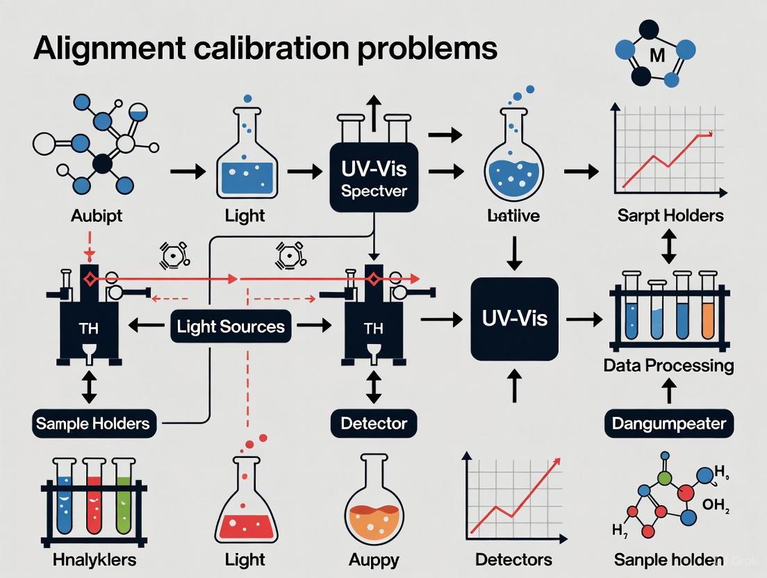
Systematic Troubleshooting Workflow
The following diagram outlines a logical decision-making process for diagnosing problems with UV-Vis instruments, helping to efficiently identify the root cause.
This guide provides a structured troubleshooting resource for researchers, scientists, and drug development professionals, framed within a thesis on UV-Vis spectrometer alignment and calibration. The content is organized by problem source—instrument, sample, and methodology—to help you efficiently diagnose and resolve experimental issues.
Instrument-Related Issues and Solutions
Instrumental errors often stem from the spectrophotometer's components and require systematic checking and maintenance.
Frequently Asked Questions (FAQs)
Q: The spectrophotometer fails its self-test, showing errors related to "stray light" (NG9) or "wavelength repeatability." What should I do?
- A: An "NG9" error often indicates insufficient energy from the deuterium lamp, suggesting it is aging and needs replacement. If you are only working in the visible range, you may continue temporarily, but the lamp should be replaced for UV work. Wavelength repeatability failures can be caused by moisture-damaged optical filters or general misalignment [7].
Q: The instrument display shows "ENERGY ERROR" or "L0," and it fails to zero. What is the cause?
- A: This indicates a low light energy error. The most common causes are a failed or failing light source (deuterium lamp for UV, tungsten lamp for visible), a blocked light path, or, in rare cases, a faulty power supply component like a resistor [7].
Q: The absorbance or transmittance readings are unstable and drift over time.
Q: My readings are consistently inaccurate, but the instrument shows no explicit errors.
- A: The likely culprits are incorrect wavelength calibration or stray light. Calibrate the wavelength using certified reference materials like holmium oxide filters. Stray light, which is light outside the intended bandwidth reaching the detector, can be checked using potassium chloride (KCl) solutions for the UV range [12] [13].
Experimental Protocol: Wavelength Accuracy Calibration
Principle: Verifies that the wavelength scale of the spectrophotometer is correct, which is critical for obtaining accurate absorption spectra [12].
Materials:
- Holmium oxide filter or solution (primary standard)
- Didymium filter (secondary check for visible range)
- Lint-free gloves and lens cleaning tissues
Methodology:
- Allow the instrument to warm up for the manufacturer-specified time (typically 20-30 minutes).
- Place the holmium oxide filter or filled cuvette in the sample compartment.
- Perform a spectral scan over the range of 240-650 nm.
- Identify the characteristic absorption peaks of holmium oxide (e.g., 241.5 nm, 279.4 nm, 287.5 nm, 360.9 nm, 418.4 nm, 453.2 nm, 536.2 nm, and 637.5 nm).
- Compare the measured peak wavelengths to the certified values. The deviation should typically be within ±0.5 nm. If outside the tolerance, follow the manufacturer's procedure for wavelength adjustment [14] [13].
Sample-Related Issues and Solutions
Errors arising from the sample itself or its container are among the most common in daily use.
Frequently Asked Questions (FAQs)
Q: I see unexpected peaks or a noisy baseline in my spectrum.
Q: The absorbance reading is unstable or non-linear, especially at high values.
Q: The results are inconsistent between replicate measurements of the same sample.
- A: Inconsistent results can be caused by air bubbles in the cuvette, inhomogeneous samples, or variations in sample positioning. Tap the cuvette gently to dislodge bubbles, vortex samples to ensure homogeneity, and always place the cuvette in the holder facing the same direction and orientation [13].
Q: I cannot zero the instrument with my blank solution.
- A: First, ensure you are using the correct blank—a solution containing all components except the analyte. Check that the cuvette is clean and that the blank is properly prepared. If the problem persists, the issue may be instrumental, such as a failing lamp [7].
Experimental Protocol: Cuvette Selection and Handling
Principle: Using the correct, clean cuvette is fundamental for accurate light transmission measurements [15].
Materials:
- Quartz cuvettes (for UV range below 300 nm)
- Glass or optical-quality plastic cuvettes (for visible range only)
- HPLC-grade or spectrophotometric-grade solvents (e.g., methanol, water)
- Lint-free wipes
- Disposable gloves
Methodology:
- Selection: Choose a quartz cuvette for measurements in the UV range. For visible-only measurements, glass or specific solvent-compatible plastic cuvettes are acceptable [8].
- Cleaning: Rinse the cuvette several times with the solvent used in your sample. Avoid using harsh acids or bases that might etch the optical surfaces.
- Handling: Always wear gloves. Hold the cuvette by its upper, opaque ridges to avoid fingerprints on the optical windows.
- Inspection: Visually inspect the cuvette against light for scratches, chips, or residue. Scratched cuvettes should be discarded.
- Filling: Fill the cuvette with enough volume to ensure the light beam passes entirely through the solution, not the meniscus or air [8].
Methodology-Related Issues and Solutions
These issues arise from incorrect experimental setup, calibration procedures, or data analysis methods.
Frequently Asked Questions (FAQs)
Q: My calibration curve has a poor correlation coefficient (R²).
- A: This can be caused by incorrect preparation of standard solutions (e.g., volumetric errors), using expired or degraded standards, or selecting an inappropriate wavelength for analysis. Ensure accurate dilution techniques, use fresh standards, and verify the analyte's absorption maximum via a full wavelength scan [15].
Q: Why is it crucial to use a blank, and what should it contain?
- A: The blank corrects for absorbance from the solvent, cuvette, and other non-analyte components. Failing to use a proper blank will result in overestimation of the analyte's absorbance. The blank must contain all the components of the sample solution except the analyte [15].
Q: The measured absorbance for a known standard has changed over time.
Q: What is the impact of bandwidth and slit width on my measurement?
- A: A wider slit width increases light throughput but reduces spectral resolution, potentially obscuring fine spectral features. A narrower slit improves resolution but may reduce signal-to-noise ratio. Use the smallest slit width that provides an acceptable signal level for your application [14] [13].
Experimental Protocol: Establishing a Photometric (Absorbance) Scale
Principle: This procedure verifies the linearity and accuracy of the spectrophotometer's photometric scale (Absorbance or %Transmittance) [14].
Materials:
- Solid neutral-density glass filters with certified transmittance values OR
- Potassium dichromate (K₂Cr₂O₇) in acidic solution (a common standard for UV-Vis)
- Volumetric flasks, pipettes
- 0.01 M Perchloric acid (HCIO₄) or 0.001 M HCIO₄ as a solvent for potassium dichromate
Methodology (using Potassium Dichromate):
- Prepare a stock solution of potassium dichromate with known, high purity in 0.01 M HCIO₄.
- Precisely dilute the stock solution to create a series of standards covering a range of absorbance values (e.g., from 0.2 to 1.0 AU) at its peak wavelength (e.g., 235 nm, 257 nm, 313 nm).
- Measure the absorbance of each standard.
- Plot the measured absorbance against the calculated absorbance or concentration. The plot should be linear. Deviations from linearity indicate issues with photometric accuracy or stray light.
Table 1: Quantitative Data from Inter-Laboratory Comparison Study
This table, adapted from a classic study, highlights the real-world variability in spectrophotometric measurements across different laboratories, underlining the importance of standardized procedures [14].
| Solution & Type | Concentration (mg/L) | Wavelength (nm) | Absorbance (A) | Transmittance (%T) | Coefficient of Variation in Absorbance (ΔA/A %) |
|---|---|---|---|---|---|
| Acidic Potassium Dichromate | 100 | 240 | 1.262 | 5.47 | 2.8 |
| Acidic Potassium Dichromate | 100 | 366 | 0.855 | 14.0 | 5.8 |
| Alkaline Potassium Chromate | 40 | 340 | 0.318 | 48.3 | 9.2 |
| Acidic Potassium Dichromate | 20 | 380 | 0.109 | 77.8 | 11.1 |
Table 2: The Scientist's Toolkit - Key Research Reagent Solutions
This table lists essential materials and standards used for the calibration and validation of UV-Vis spectrophotometers.
| Item | Function | Key Application / Note |
|---|---|---|
| Holmium Oxide Filter | To verify the wavelength accuracy of the spectrophotometer. | Provides sharp, known absorption peaks across UV-Vis range. Primary standard for wavelength calibration [14] [13]. |
| Neutral-Density Glass Filters | To check the photometric linearity and accuracy of the Absorbance/Transmittance scale. | Certified for specific transmittance values at given wavelengths [14]. |
| Potassium Dichromate Solutions | A chemical standard for verifying photometric performance and stray light. | Used in acidic solution (e.g., 0.001 M HCIO₄) for calibration in the UV region [14] [15]. |
| Potassium Chloride (KCl) Solution | To test for stray light in the UV region. | A 1.2% w/v KCl solution is used to check for stray light at 200 nm [13]. |
| Quartz Cuvettes | To hold liquid samples for measurement. | Required for UV measurements due to high transparency below 300 nm [8] [15]. |
Troubleshooting Workflow and Experimental Setup
The following diagrams outline systematic approaches to diagnosing problems and ensuring proper instrument setup.
Troubleshooting Workflow
Proper Cuvette Alignment Setup
FAQs: Fundamental Challenges and Instrument Limitations
Q1: What are the most significant unsolved problems in quantitative UV-Vis analysis for drug development? Current research highlights several persistent challenges. Key among them are the difficulties in achieving robust calibration transfer between different instruments and accounting for sample heterogeneity, which can introduce significant bias in quantitative results. Furthermore, accurate uncertainty estimation for multivariate calibration models remains a non-trivial task, complicating the reliability of concentration predictions in critical pharmaceutical applications [17].
Q2: Why do my absorbance readings become unstable or non-linear, especially at higher values? This is a common instrumental limitation. For optimal results, absorbance values should ideally be maintained between 0.1 and 1.0 absorbance units. Highly concentrated samples can cause readings to become noisy, unstable, or max out (e.g., at an absorbance of 3.0), indicating insufficient light is reaching the detector. The solution is to dilute the sample or use a cuvette with a shorter path length [18]. This issue is related to the broader unsolved challenge of detecting and correcting for nonlinearities in spectral calibration models [17].
Q3: What causes unexpected peaks or a noisy baseline in my UV-Vis spectrum? Unexpected spectral features often originate from sample and setup issues rather than the instrument itself. Primary causes include:
- Contaminated Samples or Cuvettes: Impurities introduce unexpected absorbance [8].
- Unclean ATR Crystals or Optics: In Fourier transform infrared (FT-IR) spectroscopy, a dirty crystal can cause negative peaks and distorted baselines. This translates to UV-Vis as the need for pristine quartz cuvettes [19].
- Instrument Vibration: External vibrations can introduce false spectral features, a problem also documented in FT-IR troubleshooting [19].
- Solvent Interference: The solvent itself may absorb strongly in the UV region, especially if using plastic cuvettes which block UV light. Always use quartz cuvettes for UV-Vis work and blank with your correct solvent [18].
Q4: How can I ensure my spectrophotometer's calibration is accurate and traceable? Regular calibration is a cornerstone of reliable data. The process involves [20]:
- Warm-up: Power on the instrument for at least 30 minutes to stabilize the light source.
- Blanking: Use a pure solvent or buffer in a clean, matching cuvette to set the baseline.
- Wavelength Accuracy: Use a holmium oxide filter with known peak wavelengths to verify the instrument's wavelength scale.
- Photometric Accuracy: Use a standard, such as potassium dichromate solution, to verify the accuracy of absorbance measurements.
- Linearity: Check the detector's response across a range of concentrations using standard solutions.
Troubleshooting Guides
Guide: Resolving High Noise and Signal Instability
| Symptom | Possible Cause | Solution |
|---|---|---|
| Noisy, fluctuating absorbance values | Weak or aging light source | Switch to uncalibrated mode to check lamp output spectrum; replace lamp if necessary [18]. |
| Insufficient warm-up time | Allow the lamp (tungsten halogen or arc) to warm up for at least 20 minutes before measurement [8]. | |
| Contaminated or scratched cuvette | Thoroughly clean or replace the cuvette. Handle only with gloved hands [8]. | |
| Light path obstruction | Ensure the cuvette is correctly aligned and filled, and the beam path is clear [18]. | |
| Low light transmission | For high-concentration samples, dilute the sample or use a cuvette with a shorter path length [8] [18]. |
Guide: Addressing Calibration and Quantitative Errors
| Symptom | Possible Cause | Solution |
|---|---|---|
| Inconsistent results between instruments | Lack of calibration transfer | This is a major research frontier. Apply techniques like Direct Standardization (DS) or Piecewise Direct Standardization (PDS) to transfer models between devices [17]. |
| Poor reproducibility on the same sample | Sample heterogeneity | Ensure consistent sample preparation (grinding, mixing). Chemometric models are being developed to better handle this inherent variability [17]. |
| Drifting calibration | Dirty optics/windows | Clean the external windows of the instrument's sample compartment regularly. Contamination causes analysis drift [11]. |
| Inaccurate quantitative results | Improvent blanking or incorrect calibration standards | Always use a blank that matches the sample matrix. Use certified reference materials for calibration and follow a documented calibration procedure [20]. |
Current Research Frontiers: Unsolved Problems in Spectroscopic Analysis
The field continues to grapple with fundamental challenges that limit the accuracy, robustness, and interoperability of spectroscopic methods. The table below summarizes key unsolved problems as identified in current literature.
Table: Key Unsolved Problems in Modern Spectroscopy
| Research Frontier | Core Challenge | Impact on Pharmaceutical Analysis |
|---|---|---|
| Calibration Transfer [17] | Spectral models trained on one instrument fail on another due to hardware variability. | Hinders method validation and deployment across multiple sites or over time as instruments age. |
| Uncertainty Estimation [17] [21] | Difficulty in providing reliable confidence intervals for predictions from multivariate models. | Undermines risk assessment in drug quality control and compliance. |
| Sample Heterogeneity [17] | Physical and chemical inhomogeneity in samples leads to unrepresentative spectra. | Causes inaccurate potency and content uniformity measurements in solid dosage forms. |
| Net Analyte Signal [17] | Ensuring analyte specificity in complex mixtures with severe spectral overlap. | Critical for accurately quantifying individual components in fixed-dose combination drugs. |
| Machine Learning Interpretability [17] | Deep learning models are "black boxes," making it hard to trust or validate their outputs. | A barrier to regulatory acceptance of advanced AI-driven analytical methods. |
| Baseline/Scatter Correction [17] | Accurately separating the analyte's signal from complex background effects. | Directly affects the accuracy of quantitative results, especially in turbid or scattering samples. |
Advanced Methodologies & Experimental Protocols
Protocol: A Sustainable UV-Spectrophotometric Method for Combination Drugs
Recent research demonstrates a green analytical method for simultaneous determination of Meloxicam and Rizatriptan in a newly approved fixed-dose tablet (Symbrao) [22].
1. Principle: Employ chemometric models (PCR, PLS, MCR-ALS) to resolve the severely overlapping UV spectra of the two drugs, using an environmentally friendly solvent system.
2. Materials and Equipment:
- Instrument: Double-beam UV-Vis spectrophotometer (e.g., Shimadzu UV-1800) with 1.0 cm quartz cuvettes.
- Solvent: Binary green solvent Water:Ethanol (1:1, v/v).
- Standards: Pure reference standards of Meloxicam and Rizatriptan.
- Software: Chemometric software for implementing PCR, PLS, and MCR-ALS algorithms.
3. Procedure:
- Stock Solution Preparation: Accurately weigh and dissolve Meloxicam and Rizatriptan in the water:ethanol solvent to prepare primary stock solutions.
- Calibration Set Design: Use an optimization algorithm (e.g., Fedorov algorithm) to select the most informative mixture ratios for the calibration set, minimizing experimental runs [22].
- Spectra Acquisition: Scan the absorption spectra of all calibration and validation samples over the appropriate wavelength range (e.g., 200-400 nm).
- Chemometric Modeling:
- Build Partial Least Squares (PLS) or Multivariate Curve Resolution-Alternating Least Squares (MCR-ALS) models using the calibration set spectra and known concentrations.
- Optimize model parameters using variable selection algorithms like the Genetic Algorithm (GA) or Firefly Algorithm (FA) [22].
- Method Validation: Validate the model using an external validation set, assessing accuracy, precision, and robustness.
4. Sustainability Assessment: The method's greenness is quantitatively evaluated using tools like the Multi-color Assessment (MA) tool and the Need-Quality-Sustainability (NQS) index, aligning with UN Sustainable Development Goals [22].
Workflow for sustainable UV-spectrophotometric analysis of combination drugs.
Visualization: The Interrelationship of Unsolved Problems
The core challenges in spectroscopic analysis are not isolated; they are interconnected, as shown in the following diagram.
Logical relationships between unsolved problems in spectroscopy.
The Scientist's Toolkit: Essential Research Reagents & Materials
Table: Key Reagents for Advanced Spectroscopic Analysis
| Item | Function & Application |
|---|---|
| Holmium Oxide Filter | A certified reference material for validating the wavelength accuracy of a UV-Vis spectrophotometer [20]. |
| Potassium Dichromate Solution | A standard solution used to verify the photometric accuracy and linearity of a UV-Vis instrument's absorbance scale [20]. |
| Quartz Cuvettes | Essential for UV-range measurements, as they are transparent to ultraviolet light, unlike plastic or glass cuvettes [8] [18]. |
| Green Solvents (e.g., Water, Ethanol) | Environmentally benign solvents used in sustainable method development to reduce toxic waste, as demonstrated in the Meloxicam/Rizatriptan assay [22]. |
| Chemometric Software | Software packages capable of implementing multivariate algorithms (PLS, MCR-ALS, GA) are crucial for resolving complex, overlapping spectra [22] [17]. |
Procedures and Protocols: Step-by-Step Alignment, Calibration, and Sample Preparation
Pre-Measurement Environmental and Instrument Setup
Q: What are the critical environmental factors to check before starting measurements?
A stable operating environment is crucial for obtaining reliable and reproducible data. Key factors to verify include:
- Temperature and Humidity: Operate your spectrophotometer in an environment with a temperature between 15°C and 35°C and relative humidity below 80%. High humidity can cause condensation on optical components, interfering with light transmission [23].
- Cleanliness: The instrument should be located in a dust-free area to prevent particles from settling on optical components, which can reduce light transmittance and impact accuracy [23].
- Vibration and Magnetic Interference: Place the device on a stable, sturdy workbench away from equipment that generates strong vibrations or magnetic fields, such as large motors or transformers, as these can disrupt the delicate optical system and electronics [23].
Q: What is the correct procedure for instrument warm-up and why is it necessary?
Allowing the spectrophotometer to warm up is essential for the optical system and electronic components to reach a stable operating state, which minimizes drift and ensures measurement accuracy.
- Warm-up Time: Before use, turn on the spectrophotometer and allow it to warm up for 15 to 30 minutes. Note that specific warm-up times may vary by model, so always consult your user manual [23].
- Light Source Stability: For instruments using tungsten halogen or arc lamps, you should wait approximately 20 minutes after turning the lamp on before measuring. LED light sources may stabilize in just a few minutes [8].
Cuvette Selection and Handling
Q: How do I select the correct cuvette material for my experiment?
The choice of cuvette material is determined by the wavelength range of your analysis and the chemical compatibility with your samples. The following table summarizes the key properties:
| Feature | Quartz (Fused Silica) | Optical Glass | Plastic (PS/PMMA) |
|---|---|---|---|
| UV Transmission | Excellent (190–2500 nm) [24] | Limited (>320 nm) [24] | Not supported [24] |
| Visible Transmission | Excellent [24] | Excellent [24] | Good [24] |
| Autofluorescence | Low [24] | Moderate [24] | High [24] |
| Chemical Resistance | High (av. HF & hot strong bases) [24] | Moderate [24] | Low [24] |
| Max Temperature | 150–1200 °C [24] | ≤90 °C [24] | ≤60 °C [24] |
| Best Use | UV-Vis, fluorescence, solvents [24] | Visible-only assays [24] | Teaching, colorimetric assays [24] |
- Application-Specific Selection:
- UV Spectroscopy (<300 nm): Quartz cuvettes are essential for applications like DNA (260 nm) and protein (280 nm) analysis [24].
- Fluorescence Spectroscopy: Use 4-window quartz cuvettes for their low autofluorescence and polished sides for 90° detection [24].
- Visible Spectroscopy: For colorimetric assays in the visible range (400-800 nm), glass or plastic cuvettes may be sufficient and more cost-effective [24].
Q: What are the common mistakes to avoid when handling cuvettes?
Proper cuvette handling is a simple yet critical step for accuracy.
- Incorrect Orientation: Always align the clear, polished optical windows with the spectrometer's light path. Placing the frosted sides in the beam will cause errors [25].
- Improper Filling: Overfilling can cause spills that contaminate the instrument, while underfilling creates air gaps in the light path. Fill the cuvette to about ¾ of its height to ensure the beam passes entirely through the sample [25].
- Contamination and Damage: Always handle cuvettes with gloved hands to avoid fingerprints. Rinse thoroughly with distilled water or appropriate solvents after use and clean with soft tissues. Do not reuse disposable plastic cuvettes, as this leads to contamination [8] [25]. Inspect cuvettes for scratches or residue before use [26].
System Stability and Baseline Verification
Q: How do I verify that my spectrophotometer system is stable and ready for measurement?
After the initial warm-up period, perform these checks to confirm system stability.
- Baseline/Blank Correction: Perform a baseline correction or full recalibration using your certified reference solution (e.g., the pure solvent) in a clean, compatible cuvette. Ensure no residual sample is left in the compartment from previous runs [26] [8].
- Signal Stability Check: Observe the baseline or 100% Transmittance (T%) reading for a few minutes. The reading should be stable. If the T% reading fluctuates significantly (e.g., by about 8%), it may indicate an unstable light source, such as a failing deuterium lamp, or environmental issues [7].
- Light Path Inspection: Visually check for any debris in the sample compartment that might be obstructing the light path [26].
Troubleshooting Common Pre-Measurement Issues
Q: The instrument fails to calibrate or the blank measurement shows high/unstable absorbance. What should I do?
This is a common issue often caused by insufficient light reaching the detector [27]. Follow this troubleshooting workflow:
Q: I see an error code related to lamp energy or wavelength during startup. What does this mean?
Error messages like "NG9" (insufficient deuterium lamp energy), "D2-failure," or "energy-low" typically point to a problem with the light source [7].
- Lamp Life: Deuterium lamps have a finite lifespan (several hundred to one thousand hours) and tungsten lamps last for several hundred to a few thousand hours. An aged lamp is a common cause of low energy errors, particularly in the UV region [7] [23].
- Electrical Issues: If a new lamp does not resolve the issue, the problem could lie with the lamp's power supply or control circuitry, which requires professional technical service [7].
- Optical Component Failure: In instruments that have been unused for long periods, optical components like filters can be damaged by moisture, leading to wavelength check failures [7].
Essential Research Reagent and Material Solutions
The following table details key materials required for reliable UV-Vis spectroscopy.
| Item | Function & Application | Critical Notes |
|---|---|---|
| Quartz Cuvettes (Fused Silica) | Holds liquid samples for analysis; essential for UV light transmission (<300 nm) and fluorescence assays [24]. | 2-window: Standard for absorbance [24]. 4-window: Required for fluorescence measurements [24]. |
| Certified Reference Standards | Used for wavelength calibration and verifying instrument accuracy [26] [23]. | Use traceable standards as specified in the instrument's manual. |
| Deuterium & Tungsten Halogen Lamps | Light sources for UV and Visible/NIR regions, respectively [26] [7]. | Monitor usage hours; replace when output degrades or errors appear [7] [23]. |
| High-Purity Solvents | Used for preparing blank/reference solutions and sample dilution [8] [27]. | Ensure solvent is transparent at your measurement wavelength. Can be a source of high blank absorbance [27]. |
| Appropriated Cleaning Solvents | For cleaning quartz cuvettes after use to prevent contamination [25]. | Must be chemically compatible with quartz (e.g., avoid HF and hot concentrated strong bases) [24] [28]. |
| Cuvette Cleaning Kit | Includes soft lint-free tissues, pipettes, and mild detergent for proper cuvette maintenance [25]. | Avoid abrasive materials that can scratch optical surfaces [25]. |
Wavelength and Photometric Accuracy Calibration Using Certified Standards
FAQs on Calibration Principles and Procedures
What is the difference between wavelength accuracy and photometric accuracy, and why are both critical?
Wavelength Accuracy ensures the spectrophotometer correctly selects and reports the specific wavelength of light. It verifies the x-axis of the instrument's output is correct. Inaccuracy, for instance the instrument reporting 280 nm while actually outputting 282 nm, causes flawed measurements, especially on the steep slope of an absorption peak, leading to errors in quantification and potential misidentification of compounds [29].
Photometric Accuracy ensures the instrument's detector and electronics correctly measure the amount of light absorbed by the sample, providing a true absorbance (or transmittance) value. It validates the y-axis of the output. Error here directly translates into an incorrect calculated concentration of the analyte [29]. Both parameters are fundamental for data integrity and adherence to regulatory pharmacopeias like USP <857> and Ph. Eur. 2.2.25 [30] [29].
How often should I perform a full instrument qualification with certified standards?
The frequency depends on your instrument's usage, criticality of measurements, and regulatory requirements. A best practice includes performance verification after any major maintenance, lamp replacement, or instrument relocation [31] [3]. For routine monitoring, schedules can be based on elapsed time or usage hours. Proactive replacement of deuterium lamps (typically 1,000–3,000 hours) or xenon lamps (~500 hours) is recommended, as a degrading lamp is a primary cause of performance drift [3].
What are the key regulatory compliance considerations for spectrophotometer calibration?
Major international pharmacopeias provide explicit guidance. The United States Pharmacopeia (USP) General Chapter <857> and the European Pharmacopoeia (Ph. Eur.) Chapter 2.2.25 define parameters and acceptance criteria for calibration [30] [29]. Compliance requires using appropriate Certified Reference Materials (CRMs) whose values are traceable to national standards bodies like NIST [29]. The latest versions of these standards, such as the updated USP <857> effective December 2022, may introduce more stringent requirements, like multiple replicate measurements for statistical validation [29].
Troubleshooting Guides
Guide 1: Addressing Wavelength Accuracy Failures
A wavelength accuracy failure indicates the instrument's monochromator is misaligned or has drifted.
- Observed Symptom: Measured absorption peaks of a reference material are shifted from their certified positions.
- Potential Root Causes: Mechanical shock or vibration, thermal expansion/contraction of components, aging light source, or gradual misalignment of the diffraction grating [29].
- Corrective Actions:
- Verify Warm-Up: Ensure the instrument has stabilized (typically 30-60 minutes after lamp ignition) [31].
- Re-measure Standard: Confirm the holmium oxide filter or solution is clean, properly positioned, and scanned at a slow speed with a small bandwidth [32] [33].
- Check for Obstructions: Inspect the light path for any debris [31].
- Service Intervention: If the shift persists and exceeds specifications, the instrument likely requires professional service for optical re-alignment [34].
Guide 2: Addressing Photometric Accuracy & Linearity Failures
A photometric accuracy failure means the instrument reports incorrect absorbance values, directly impacting quantitative results.
- Observed Symptom: Measured absorbance values for a set of neutral density filters or potassium dichromate solutions fall outside the certified uncertainty range [32] [35].
- Potential Root Causes: Stray light is the most common cause, especially for high absorbance values. Other causes include a failing or aged lamp, dirty optics (cuvette, lenses), or detector issues [14] [3] [29].
- Corrective Actions:
- Check the Light Source: Examine lamp usage hours. A lamp near or beyond its rated life should be replaced [3] [36].
- Inspect and Clean: Check the sample cuvette for scratches, residue, or improper alignment. Clean the cuvette and inspect the sample compartment for dust on optics [31] [34].
- Test for Stray Light: Perform a stray light test using recommended cut-off filters (e.g., KCl solution for 200 nm or NaI for 220 nm) [32] [29]. High stray light often explains linearity failure at high absorbances.
- Verify Sample Concentration: Ensure samples and standards are within the ideal absorbance range (0.1-1.0 A) and are not overly concentrated, which can cause non-linearity [36].
Guide 3: Resolving Instability and Fluctuating Readings
Erratic or drifting readings, even after blanking, indicate instrument instability.
- Observed Symptom: Absorbance readings for a stable standard drift over time or show high noise.
- Potential Root Causes: Aging lamp, insufficient warm-up time, temperature fluctuations in the sample compartment, dirty optics, or electronic instability [31] [3] [34].
- Corrective Actions:
- Allow Sufficient Warm-Up: Let the instrument stabilize for the manufacturer-recommended time before use [31].
- Check Lamp Hours and Output: Replace the lamp if it is near end-of-life or if a check in uncalibrated mode shows a flat or low-intensity spectrum in certain regions [3] [36].
- Ensure Environmental Stability: Operate the instrument away from drafts, direct sunlight, and vibrating equipment. Use grounded outlets to prevent electrical noise [34].
- Perform a Power Reset: For instruments connected to interfaces like LabQuest, a full power reset can resolve communication or software-related instability [36].
The Scientist's Toolkit: Research Reagent Solutions
The following table details key Certified Reference Materials (CRMs) used for spectrophotometer qualification.
| Material Name | Primary Function | Key Application Wavelength Range | Brief Description & Function |
|---|---|---|---|
| Holmium Oxide Solution/Glass [32] [33] | Wavelength Accuracy | 240-650 nm | The most widely used reference, cited by pharmacopeias, with multiple sharp absorption peaks for verifying wavelength scale [32]. |
| Potassium Dichromate Solutions [32] | Photometric Accuracy & Linearity | 235-430 nm | A classic solution reference for verifying absorbance accuracy and instrument linearity across a range of UV wavelengths [32]. |
| Neutral Density Glass Filters [32] [35] | Photometric Accuracy & Linearity | 250-635 nm | Durable solid filters with certified absorbance values at specific wavelengths, used for checking photometric scale without preparation [32] [35]. |
| Stray Light Cut-off Filters [32] | Stray Light Testing | 175-385 nm | Solutions or filters that block all light below a specific wavelength. Used to quantify stray light levels at critical UV wavelengths [32]. |
| Combined Holmium/Neutral Density Glass [32] | Combined Wavelength & Photometry | 360-640 nm | A dual-purpose filter that allows simultaneous checking of wavelength accuracy (via Holmium peaks) and photometric accuracy (at ~1 A) [32]. |
| NIST-Traceable CRM [29] | Regulatory Compliance | Varies | CRMs from accredited suppliers with certificates providing an unbroken chain of calibration to primary national standards, essential for defensible data [29]. |
Experimental Protocol: Full Instrument Qualification
This protocol outlines the methodology for a comprehensive performance check of a UV-Vis spectrophotometer against pharmacopeial standards [32] [29].
1. Scope: This procedure applies to the qualification of UV-Vis spectrophotometers used for quantitative and qualitative analysis in research and quality control.
2. Pre-Qualification Prerequisites:
- Instrument: Ensure the instrument is clean, level, and has been powered on with a stable lamp for at least 30 minutes.
- Standards: Use certified, traceable reference materials. Allow liquid standards to reach room temperature.
- Cuvettes: Use matched quartz cuvettes. Clean them thoroughly before use.
3. Step-by-Step Procedure:
Step 1: Wavelength Accuracy Check
- Method: Scan the holmium oxide (solution or glass filter) across its entire range (e.g., 240-650 nm) using a slow scan speed and narrow spectral bandwidth.
- Measurement: Record the wavelength at which each characteristic absorption peak occurs.
- Acceptance Criterion: The measured peak positions must be within ±1.0 nm of the certified values [32] [33].
Step 2: Photometric Accuracy Check
- Method: Measure the absorbance of a set of neutral density glass filters or potassium dichromate solutions at their specified wavelengths.
- Measurement: Record the average of multiple readings for each standard.
- Acceptance Criterion: The measured absorbance values must be within the certified tolerance (e.g., ±0.01 A or as per CRM certificate) of the stated values [35].
Step 3: Stray Light Check
- Method: Use a stray light cut-off filter (e.g., KCl solution for 200 nm). Fill a cuvette with the solution and measure the transmittance at the specified wavelength.
- Measurement: The recorded value corresponds to the instrument's stray light level at that wavelength.
- Acceptance Criterion: Stray light must be less than 0.1% (Absorbance > 3.0) at the tested wavelength [32] [29].
Step 4: Resolution (Bandwidth) Check (if required)
- Method: Scan a toluene in hexane solution and measure the depth of the fine structure valley at 269 nm versus the peak at 267 nm.
- Measurement: The ratio of these values is calculated.
- Acceptance Criterion: The ratio must meet or exceed the minimum value specified by the pharmacopeia [32].
Workflow Visualization
The following diagram illustrates the logical workflow for diagnosing and resolving common calibration failures, integrating the troubleshooting concepts from the guides above.
Diagnostic Workflow for Calibration Failures
Advanced Sample Preparation Techniques for Pharmaceutical and Biological Matrices
In the context of research on UV-Vis spectrometer alignment and calibration, consistent and accurate results are fundamentally dependent on proper sample preparation. Biological and pharmaceutical matrices are highly complex, containing various interfering components, broad molecular weight distributions, and target analytes that can exist at very low content levels [37]. The most critical step in analyzing these samples is, therefore, the preparation process. Without proper pretreatment, which serves to extract, separate, purify, and enrich target analytes, even a perfectly calibrated UV-Vis instrument will yield unreliable data [37]. This guide addresses common preparation-related issues that manifest as spectroscopic problems, providing targeted troubleshooting FAQs and detailed protocols to ensure data integrity.
Essential Knowledge: Core Principles and Reagents
Key Research Reagent Solutions for Sample Preparation
The selection of appropriate media and reagents is paramount for effective sample preparation. The table below details key materials that significantly enhance performance when integrated with extraction technologies like Solid-Phase Extraction (SPE) and its variants [37].
| Reagent/Media | Primary Function | Key Applications |
|---|---|---|
| Porous Organic Frameworks | High-performance sorbent for extraction; offers high surface area and tunable porosity [37]. | Extraction of drugs and metabolites; improved selectivity and sensitivity [37]. |
| Molecularly Imprinted Polymers (MIPs) | Synthetic polymers with tailor-made recognition sites for specific target molecules [37]. | Selective separation of trace chiral enantiomers; analysis of complex body fluids [37]. |
| Bioactive Media | Utilizes biological interactions (e.g., antibody-antigen) for highly specific capture [37]. | Targeting specific disease markers or proteins in biological samples [37]. |
| Spectrophotometric-Grade Solvents | High-purity solvents that minimize background absorbance and impurity interference [13]. | Used as the mobile phase or for dissolving samples for UV-Vis analysis to avoid distorted readings [13]. |
| Certified Reference Materials (CRMs) | Standards with precisely known absorbance values for method validation [5]. | Verifying the accuracy and recovery of your sample preparation method and instrument calibration [5]. |
Troubleshooting FAQs: Sample Preparation and UV-Vis Performance
This section addresses specific, common problems users encounter, linking issues in sample preparation directly to their symptoms in spectroscopic analysis.
FAQ 1: My UV-Vis baseline is noisy and drifts unpredictably after analyzing multiple biological samples. What is the cause?
- Potential Cause: Contamination of the spectrophotometer's optical path or cuvette from insufficiently cleaned samples or sample carryover. Residues from complex biological matrices can deposit on the cuvette walls or the instrument's optics.
- Solution:
- Cuvette Handling: Always use high-quality quartz cuvettes for UV work and clean them thoroughly immediately after use. Handle them with gloves or lint-free tissues to avoid smudges and scratches that scatter light [13].
- Optical Maintenance: Regularly clean the instrument's optical components, such as lenses and the sample holder, according to the manufacturer's instructions using lint-free cloths and appropriate solvents like ethanol [38].
- Validation: After cleaning, validate the process by running a blank solvent. A stable, flat baseline confirms the system is clean.
FAQ 2: I observe inconsistent absorbance readings and poor reproducibility between sample replicates. How can I fix this?
- Potential Cause: Inhomogeneous sample solutions or improper handling leading to air bubbles. Biological samples require careful preparation to ensure homogeneity [13].
- Solution:
- Ensure Proper Mixing: Use vortex mixers or sonication to achieve a homogenous sample solution before pipetting it into the cuvette [13].
- Avoid Air Bubbles: Bubbles in the cuvette can act as lenses and scatter light, causing significant errors. After filling, tap the cuvette gently to dislodge any trapped air, or degas the solution beforehand [13].
- Check Cuvette Consistency: Ensure all cuvettes used have an identical path length (e.g., 10 mm) and are from a matched set to maintain uniformity [13].
FAQ 3: My calibration curve is nonlinear, and sensitivity is lower than expected for my pharmaceutical compound.
- Potential Cause: Incomplete recovery of the target analyte during sample preparation or the presence of interfering substances from the matrix that also absorb at the same wavelength.
- Solution:
- Optimize Sample Prep: Re-evaluate your extraction protocol (e.g., SPE conditions). The use of advanced media like porous organic frameworks can improve extraction efficiency and selectivity, enriching the target and excluding interferents [37].
- Determine λ-max: Perform a spectral scan of the purified analyte to confirm you are measuring at its maximum absorbance wavelength (λ-max) for optimal sensitivity [13].
- Validate with CRM: Use a Certified Reference Material to validate your entire method, from preparation to measurement, confirming the accuracy of your recovery and calibration [5].
FAQ 4: The instrument passes calibration, but my sample absorbance values are consistently inaccurate.
- Potential Cause: The use of low-quality solvents or impurities in the blank/reference solution. Impurities can absorb light and lead to distorted sample readings [13].
- Solution:
- Use High-Purity Solvents: Always use HPLC-grade or spectrophotometric-grade solvents for preparing both the blank and the sample to minimize background absorbance [13].
- Proper Blanking: Always run a fresh blank measurement that contains only the solvent or reference matrix to correct for any background signal immediately before sample analysis [5].
Experimental Protocols: Detailed Methodologies
Protocol 1: Solid-Phase Extraction (SPE) for Plasma Sample Clean-up
This protocol outlines the use of SPE, enhanced with advanced sorbent media, for preparing plasma samples prior to UV-Vis analysis, aiming to remove interfering proteins and lipids [37].
Workflow Overview:
Materials:
- Porous organic framework sorbent or commercial SPE cartridge [37].
- Plasma sample.
- Conditioning solvent (e.g., methanol).
- Equilibration solvent (e.g., water or buffer).
- Wash solvent (e.g., 5-10% methanol in water).
- Elution solvent (e.g., pure methanol or acetonitrile).
- Vacuum manifold.
Step-by-Step Procedure:
- Conditioning: Pass 2-3 column volumes of methanol through the SPE sorbent to solvate and wet the medium.
- Equilibration: Pass 2-3 column volumes of water or a buffer compatible with your sample (e.g., phosphate buffer) to prepare the sorbent surface for sample application. Do not let the sorbent run dry.
- Sample Loading: Slowly load the plasma sample onto the conditioned sorbent. A slow, drop-by-drop flow rate is crucial for maximizing analyte binding.
- Washing: Pass 2-3 column volumes of a wash solvent (e.g., 5% methanol) to remove weakly bound interfering compounds without eluting the target analyte.
- Elution: Pass 1-2 column volumes of a strong elution solvent (e.g., pure methanol) to collect the purified target analyte in a clean collection tube.
- Analysis: The eluent can be diluted if necessary and then analyzed directly by UV-Vis spectrophotometry. Always run a blank prepared with the elution solvent.
Protocol 2: Validation of Method Accuracy using Certified Reference Materials (CRMs)
This protocol describes how to use CRMs to validate the entire analytical process, from sample preparation to instrumental analysis, which is critical for troubleshooting suspected accuracy problems [5].
Workflow Overview:
Materials:
- Certified Reference Material (CRM) with a known absorbance value at a specified wavelength.
- Appropriate solvent for dissolving the CRM.
- All standard sample preparation reagents and equipment.
Step-by-Step Procedure:
- Preparation: Accurately prepare a solution of the CRM at a known concentration within the linear range of your UV-Vis method, using the appropriate high-purity solvent.
- Sample Processing: Subject the CRM solution to your entire sample preparation protocol (e.g., the SPE clean-up detailed in Protocol 1).
- Measurement: Analyze the final, processed CRM solution using your calibrated UV-Vis spectrophotometer.
- Calculation and Analysis: Calculate the percentage recovery using the formula:
- % Recovery = (Measured Concentration / Certified Concentration) × 100 A recovery of 85-115% is generally considered acceptable, though method-specific criteria may apply. A low recovery indicates a loss of analyte during preparation, while a recovery significantly over 100% may suggest interference.
Advanced Techniques and Future Outlook
The field of sample preparation is rapidly evolving with the integration of new technologies. The use of advanced media such as porous organic frameworks and molecularly imprinted polymers is significantly improving extraction performance, selectivity, and sensitivity [37]. Looking forward, the integration of these media with emerging technologies like microfluidics and automation will enable more efficient, sensitive, and rapid analysis of biological samples [37]. Furthermore, artificial intelligence is poised to make significant contributions to medium design, automation of experiments, and data analysis, driving further innovation in sample pretreatment technology [37]. For practitioners relying on UV-Vis spectrometry, these advancements will translate to cleaner samples, fewer analytical interferences, and more reliable data from their instruments.
Frequently Asked Questions (FAQs)
Q1: How do I determine the optimal concentration range for my sample? The optimal absorbance range for accurate measurement is between 0.1 and 1.0 absorbance units [39]. If your sample's absorbance is too high, the reading may become unstable or non-linear [39]. Sample concentration is the primary factor determining absorbance [8].
- Concentration is too high: A more concentrated sample scatters light more intensely, reducing the light detected and resulting in a low signal [8]. Prepare a more dilute sample within the instrument's linear dynamic range to avoid saturation [40].
- Concentration is too low: The signal may be weak and indistinguishable from background noise [6]. Increase the concentration or use a cuvette with a longer pathlength.
Q2: What is the impact of the solvent, and how do I select the right one? The solvent can significantly influence absorbance due to its own UV-Vis absorption properties or through interactions with the sample [40].
- Solvent Absorption: Always use high-purity, spectrophotometric-grade solvents, as impurities can absorb light and distort readings [13]. The solvent must be transparent in the wavelength region you are analyzing.
- Baseline Correction: For accurate results, the blank measurement must be prepared with the same solvent used to dissolve your sample [40] [13].
- Sample-Solvent Interactions: Be aware that changing sample temperature can affect solute solubility and reaction rates, thereby altering the concentration or spectrum of your sample [8].
Q3: How does cuvette path length affect my measurement, and when should I change it? Absorbance is directly proportional to the path length of the light through the sample. Selecting the correct path length is crucial for accuracy [40].
- Standard Path Length: A 10 mm path length is standard for most applications [13].
- High-Concentration Samples: If you cannot dilute a high-concentration sample without affecting results, use a cuvette with a shorter path length. This reduces the amount of sample the light travels through, minimizing scattering and bringing the absorbance into the optimal range [8].
- Low-Concentration or Small Volume Samples: For weak signals or to avoid material waste, a cuvette with a shorter path length or specialized micro-volume cuvette can be used [8]. Ensure there is enough solution volume so the excitation beam passes entirely through the sample [8].
Q4: Why is my blank measurement unstable, and how can I fix it? An unstable blank is often related to instrumental issues rather than sample conditions.
- Lamp Warm-up: Allow the light source (especially tungsten halogen or arc lamps) to warm up for at least 20-30 minutes after turning on the instrument to achieve stable output [5] [8].
- Lamp Degradation: Fluctuations are commonly caused by a degrading lamp. Deuterium and xenon lamps have finite lifespans (typically 1,000–3,000 hours and ~500 hours, respectively) and should be replaced as they approach end-of-life [3].
- Cuvette Cleanliness: Ensure the cuvette used for the blank is perfectly clean, free of scratches, smudges, and dust [13] [8].
Troubleshooting Guide: Common Problems and Solutions
| Problem | Possible Cause | Recommended Solution |
|---|---|---|
| High Absorbance/Signal Saturation | Sample concentration is too high [8]. | Dilute the sample. Use a cuvette with a shorter path length [8]. |
| Weak or Noisy Signal | Sample concentration is too low [6]. | Increase concentration. Use a cuvette with a longer path length. Optimize instrument parameters (e.g., integration time) [40]. |
| Unexpected Peaks in Spectrum | Sample or solvent contamination; dirty cuvette [8]. | Use high-purity solvents. Thoroughly clean cuvettes. Handle samples and cuvettes with gloves [8]. |
| Non-Linear Calibration Curve | Stray light interference; improper blanking; absorbance readings above 1.0 [14] [39]. | Ensure proper blank correction with matching solvent. Keep optics clean. Prepare samples within the linear absorbance range (0.1-1.0 AU) [13] [39]. |
| Readings Drift Over Time | Solvent evaporation; temperature fluctuations; instrument instability [8] [40]. | Seal cuvettes to prevent evaporation. Control lab temperature. Ensure instrument has warmed up completely [13] [8]. |
Experimental Protocol: Method for Determining Optimal Concentration and Path Length
This protocol provides a systematic approach to establish the best measurement conditions for a new sample.
1. Principle Use Beer-Lambert Law, which states absorbance (A) is proportional to concentration (c) and path length (b): A = εbc. The goal is to achieve an absorbance between 0.1 and 1.0 AU for maximum accuracy [39].
2. Materials and Reagents
- UV-Vis Spectrophotometer
- Set of high-quality, matched quartz cuvettes (e.g., 1 cm path length) [13]
- Cuvettes of varying path lengths (e.g., 2 mm, 5 mm)
- Stock solution of the analyte
- High-purity solvent (spectrophotometric grade)
- Pipettes and volumetric flasks
3. Procedure Step 1: Prepare a Dilution Series
- Create a series of sample solutions from your stock solution, diluting to various concentrations (e.g., 1x, 0.5x, 0.1x, 0.05x).
Step 2: Initial Absorbance Scan
- Using a standard 1 cm path length cuvette, fill it with a mid-range dilution.
- Perform a full wavelength scan to identify the wavelength of maximum absorbance (λmax).
Step 3: Measure Absorbance of Dilution Series
- At the determined λmax, measure the absorbance of each dilution in the 1 cm cuvette.
- Record the concentration and corresponding absorbance.
Step 4: Evaluate and Adjust Path Length if Needed
- If the most concentrated sample has an absorbance >1.0, switch to a cuvette with a shorter path length (e.g., 5 mm) and repeat the measurement.
- If the most dilute sample has an absorbance <0.1, consider using a cuvette with a longer path length or preparing a more concentrated sample.
Step 5: Data Analysis
- Plot absorbance vs. concentration for the data points within the 0.1-1.0 AU range.
- The optimal conditions are those that produce a linear calibration plot (R² > 0.99) with absorbances within the valid range.
Research Reagent Solutions and Essential Materials
| Item | Function | Key Considerations |
|---|---|---|
| Quartz Cuvettes | Holds liquid sample in the light path. | Required for UV range measurements (<300 nm); ensure path length consistency and surface cleanliness [13] [8]. |
| Spectrophotometric-Grade Solvents | Dissolves the analyte for analysis. | High purity is critical to avoid background absorbance from impurities [13]. |
| Holmium Oxide Filter | Validates wavelength accuracy of the instrument. | Provides sharp, known absorption peaks to ensure the spectrophotometer reports correct wavelengths [5] [14]. |
| NIST-Traceable Neutral Density Filters | Verifies photometric accuracy (absorbance/transmittance). | Certified reference materials used to confirm that absorbance readings are correct [5]. |
| Certified Stray Light Solution | Checks for stray light interference. | A solution like potassium chloride (KCl) absorbs all light at short wavelengths, allowing stray light detection [13]. |
Workflow for Optimizing UV-Vis Measurements
The following diagram outlines the logical decision process for troubleshooting and optimizing measurement conditions.
Diagnostic and Resolution Guide: Solving Real-World Spectrometer Problems
Diagnostic Flowchart for UV-Vis Spectrometer Errors
The following flowchart provides a systematic approach to diagnosing common UV-Vis spectrometer problems. Follow the decision paths to identify potential causes and solutions based on the specific error signals you encounter [41].
Comprehensive Error Diagnosis Guide
Absorbance Value Abnormalities
When encountering unstable, noisy, or abnormally high absorbance readings, systematically investigate these areas:
Sample Concentration Issues: Absorbance values above 1.0 absorbance unit often indicate sample concentration is too high, leading to non-linear response and excessive noise [42]. Optimal absorbance range for reliable measurements is between 0.1 and 1.0 AU [43]. Prepare diluted samples and remeasure.
Light Source Problems: Weak or failing light sources cause insufficient light reaching the detector [42]. Switch to uncalibrated mode and observe the full spectrum; flat regions in specific wavelength ranges indicate source degradation [42]. Replace deuterium or tungsten-halogen lamps according to manufacturer specifications [13].
Cuvette and Path Length Considerations: Dirty, scratched, or misaligned cuvettes scatter light and cause errors [43]. Ensure cuvettes are clean, properly aligned, and using correct material (quartz for UV, compatible plastics for visible range) [42]. Verify consistent path length, typically 1 cm [43].
Calibration and Signal Failures
Persistent calibration failures or signal errors require investigation of fundamental instrument components:
Stray Light and Background Noise: Stray light causes significant photometric errors, particularly at high absorbance values [14]. Use high-quality optical filters and ensure proper blank correction [13]. Verify instrument performance using certified reference materials [12].
Wavelength Accuracy: Errors in wavelength calibration lead to incorrect readings, particularly when measuring at specific absorption peaks [12]. Validate wavelength accuracy using holmium oxide filters or emission lines from calibration sources [44] [13].
Power and Connection Integrity: Power-related issues can cause recognition problems or bad data [42]. Perform full power reset, ensure stable power supply, and verify all connections for instruments interfaced via USB or Bluetooth [45].
Quantitative Error Analysis
Table 1: Common Spectrophotometer Errors and Resolution Strategies
| Error Type | Primary Causes | Detection Method | Resolution Strategy |
|---|---|---|---|
| High Absorbance Noise | Sample too concentrated (A > 1.0), weak light source, dirty cuvettes [42] | Check absorbance values, inspect uncalibrated spectrum [42] | Dilute samples, replace lamp, clean cuvettes [43] [42] |
| Calibration Failure | Insufficient light, incorrect blank, power issues, software errors [45] [42] | Error messages, failed calibration cycles [45] | Verify light path, use correct blank, power reset, update software [45] |
| Wavelength Inaccuracy | Misalignment of monochromator, mechanical wear, temperature effects [14] [12] | Measure known standards (holmium oxide), check emission lines [44] | Professional recalibration, wavelength verification protocols [12] |
| Baseline Drift | Temperature fluctuations, lamp warm-up, environmental changes [12] [43] | Monitor baseline stability over time [43] | Allow instrument warm-up, temperature control, baseline correction [43] [13] |
| Stray Light Effects | Optical component degradation, scattering, inappropriate slit width [14] | Measure high absorbance standards, check specifications [14] | Clean optical components, optimize slit width, use filters [13] |
Experimental Protocols for Error Verification
Protocol 1: Light Source Performance Verification
Purpose: Determine if instrument light source requires replacement [42].
Materials: Spectrometer with uncalibrated mode capability, certified reflectance standard (if available) [44].
Procedure:
- Switch spectrometer to uncalibrated mode [42].
- Collect a reference spectrum without sample.
- Examine spectrum for flat regions or significant intensity drops compared to historical performance data.
- For quantitative assessment: Measure a NIST-traceable reflectance standard and compare to certified values [44].
- Document signal-to-noise ratios at characteristic wavelengths (e.g., 280 nm for proteins, 260 nm for DNA).
Interpretation: Degraded sources show >15% intensity loss at characteristic wavelengths or flat regions in specific spectral ranges. Replace lamps when performance falls outside manufacturer specifications [13].
Protocol 2: Stray Light Assessment Procedure
Purpose: Quantify stray light contribution to photometric errors [14].
Materials: High-purity potassium chloride or sodium nitrite solutions, UV-transparent quartz cuvettes, HPLC-grade water [13].
Procedure:
- Prepare 10 g/L potassium chloride solution in HPLC-grade water for UV range assessment [13].
- Use a 1 cm pathlength quartz cuvette.
- Measure absorbance at 200 nm for KCl solution or 340 nm for NaNO₂ [13].
- The measured absorbance should exceed 2.0 AU for valid stray light assessment.
- Compare measured values to certified reference values.
Interpretation: Significantly lower than expected absorbance values indicate stray light problems. Stray light ratios exceeding 1% typically require instrument service or optical component replacement [14].
Research Reagent Solutions
Table 2: Essential Materials for UV-Vis Spectrophotometer Maintenance and Troubleshooting
| Reagent/Standard | Function | Application Protocol | Quality Specifications |
|---|---|---|---|
| Holmium Oxide Filter | Wavelength accuracy verification [13] | Measure absorption spectrum, identify characteristic peaks (241 nm, 279 nm, 287 nm, 361 nm, 453 nm) [44] | Certified wavelength standards, NIST-traceable [44] |
| Potassium Dichromate | Photometric accuracy calibration [43] | Prepare specific concentrations, measure absorbance at characteristic wavelengths [43] | ACS reagent grade, dried before use [43] |
| Potassium Chloride | Stray light verification in UV range [13] | Prepare 10 g/L solution, measure absorbance at 200 nm [13] | High-purity grade, low UV absorbance [13] |
| Certified Reference Materials (CRMs) | Overall instrument validation [12] | Follow certificate instructions, compare measured vs. certified values [12] | NIST-traceable with documented uncertainty [12] |
| Quartz Cuvettes | UV range measurements [42] | Use for wavelengths <300 nm, ensure proper cleaning [13] | Spectrosil grade quartz, 1 cm pathlength [13] |
| HPLC-Grade Solvents | Sample preparation and blanks [13] | Use for sample dissolution and blank measurements [43] | Low UV absorbance, spectrophotometric grade [13] |
FAQs: Addressing Common Spectrometer Issues
What should I do when my spectrometer won't calibrate and gives very noisy data?
This common issue typically stems from insufficient light reaching the detector [42]. First, check your sample concentration and ensure absorbance values fall between 0.1-1.0 AU [42]. Verify the light source functionality by examining the uncalibrated spectrum [42]. Inspect cuvettes for proper alignment, cleanliness, and material compatibility (quartz for UV measurements) [42]. For UV-Vis specific issues, ensure you're not using standard plastic cuvettes that block UV light [42].
Why are my absorbance readings unstable or nonlinear at values above 1.0?
Absorbance readings become increasingly unstable and nonlinear above 1.0 AU due to insufficient light reaching the detector at high absorbance levels [42]. This results in poor signal-to-noise ratios and unreliable data [45]. Always dilute samples to maintain measurements within the 0.1-1.0 AU optimal range [43]. This phenomenon is fundamental to spectrophotometer operation, not merely an instrument limitation [14].
How can I minimize measurement errors related to environmental factors?
Temperature fluctuations cause significant errors by affecting both optical components and sample properties [12]. Conduct measurements in temperature-controlled environments and allow thermal equilibration before critical measurements [43]. Minimize air currents that can deflect light paths [12]. Implement regular baseline corrections during extended sessions to compensate for instrument drift [43]. Maintain stable line voltage to prevent source intensity fluctuations [46].
A stable baseline is the foundation for reliable Ultraviolet-Visible (UV-Vis) spectroscopic data. However, researchers frequently encounter three interrelated problems that compromise data integrity: fluctuating readings, baseline drift, and artifacts caused by stray light. These issues originate from the instrument's key components—the light source, detector, and optical path—and their understanding is crucial for effective troubleshooting.
This guide provides targeted protocols to diagnose and resolve these specific stability problems, enabling the generation of publication-quality data.
Troubleshooting Guides
Guide 1: Diagnosing and Resolving Fluctuating Readings
Fluctuating or noisy absorbance readings are often the first sign of component degradation or failure.
Primary Causes and Solutions
Light Source Failure: This is the most common cause of instability and noise.
- Lamp Types and Lifespans: UV-Vis instruments typically use deuterium lamps (for UV), which have a typical lifespan of 1,000–3,000 hours, or tungsten-halogen lamps (for visible), with xenon flashes having a shorter lifespan of approximately 500 hours [3].
- Action: Proactively log all lamp usage hours. If fluctuations occur and the lamp is near or beyond its rated life, replacement is the first and most likely solution [3].
Detector and Electronics Issues: If lamp replacement does not resolve the noise, the detector system may be at fault.
- Detector Optics: Check the detector optics for dust or contamination, which can scatter light and cause signal instability [3].
- Electronic Noise: Fluctuations can also stem from the detector electronics themselves. If basic cleaning and lamp replacement fail, a service call may be required to evaluate electronic components [3].
Environmental Interference: Factors such as temperature fluctuations and physical vibrations can introduce noise into the sensitive electronics of the spectrophotometer [47]. Ensure the instrument is on a stable bench and in a temperature-controlled environment.
The logical workflow for diagnosing fluctuating readings is outlined below.
Guide 2: Identifying and Correcting Baseline Drift
Baseline drift is a continuous, directional movement of the baseline signal over time. It can be upward or downward and is distinct from random noise.
Primary Causes and Solutions
Light Source Warm-Up: A very common source of initial drift is an insufficiently warmed-up lamp.
- Action: After turning on the instrument, allow the light source to stabilize. Tungsten halogen or deuterium lamps typically require around 20 minutes to stabilize, while LEDs may need only a few minutes [8].
Mobile Phase or Solvent Effects (in LC-UV or flow cells): The chemical composition of the liquid passing through the flow cell is a major drift source.
- Impurity Accumulation: Mobile phase impurities can accumulate on the column and then elute as a broad baseline drift, often during a gradient or wash step [48].
- UV-Absorbing Additives: If a UV-absorbing additive (e.g., formic acid) is present in only one mobile phase reservoir (e.g., the aqueous component), the baseline will drift as the proportion of that reservoir changes during a gradient [48].
- Solution: Add the same concentration of the additive to both mobile phase reservoirs to maintain a constant UV background [48].
Temperature Instability: Detector response, particularly for Refractive Index (RI) detectors but also for UV detectors, is sensitive to temperature changes in the lab or the detector cell itself [48]. Maintaining a stable room temperature is critical.
Pump Problems (in HPLC-UV): An inconsistent mobile phase composition caused by a faulty pump can manifest as a saw-tooth pattern in the baseline. Issues like a sticky check valve or trapped air bubble in one pump head can cause this [48].
The following workflow systematically addresses the various causes of baseline drift.
Guide 3: Understanding and Mitigating Stray Light
Stray light is any detected light that is outside the intended wavelength band [49]. It is a primary contributor to measurement uncertainty, especially in the UV region, and causes negative deviations from the Beer-Lambert law at high absorbances [49] [21].
- Internal Sources: Inside the spectrometer, stray light can originate from scattering off the diffraction grating, interference from the 0th or higher grating orders, and inter-reflections between optical components [49].
- Impact: Stray light leads to measured absorbances that are lower than the true value. This effect is most pronounced when measuring high absorbance samples or when a weak signal is in a spectral region where the light source has a strong output (e.g., measuring in the UV when using a broadband halogen lamp) [49] [9]. The uncertainty can be significant; for example, single-monochromator Brewer spectroradiometers can have relative uncertainties of 11–14% at 295 nm, largely due to stray light, compared to 4–7% for double-monochromator instruments [21].
Strategies for Stray Light Suppression
- Instrument Design: High-quality instruments use optimized optical designs (e.g., blazed holographic gratings), high-quality mirror coatings, and physical baffles to block the 0th order and higher grating orders [49] [50].
- Mathematical Correction: High-end spectrometers can be characterized using tunable lasers to create a "stray light matrix" (Line Spread Function matrix). This matrix is used by the instrument software to correct the measured data, reducing stray light by 1 to 2 orders of magnitude [49].
- Optical Filtering: An innovative method involves integrating a filter wheel with long-pass or bandpass filters inside the spectrometer. This effectively reduces the broadband light entering the monochromator, approximating the performance of a double monochromator and offering superior stray light suppression in critical applications like solar UV measurement [49].
The table below summarizes key quantitative specifications and performance data related to the stability and accuracy of UV-Vis spectroscopy.
Table 1: Quantitative Specifications and Performance Data in UV-Vis Spectroscopy
| Component/Parameter | Typical Specification/Value | Impact on Performance |
|---|---|---|
| Deuterium Lamp Lifespan | 1,000 – 3,000 hours [3] | Signal fluctuation/noise increases as the lamp degrades beyond its rated life. |
| Xenon Flash Lamp Lifespan | ~500 hours [3] | |
| Uncertainty from Stray Light (at 295 nm) | Single-monochromator: 11–14% [21] | Highlights the critical need for double-monochromators or advanced stray light suppression for low-UV measurements. |
| Double-monochromator: 4–7% [21] | ||
| Warm-up Time for Stability | Tungsten/Halogen/Deuterium Lamps: ~20 minutes [8] | Insufficient warm-up is a direct cause of baseline drift. |
| Optimal Absorbance Range | 0.2 – 1.0 AU (ideally below 1.2 AU) [9] | Measurements at high absorbance are prone to inaccuracy due to stray light and detector non-linearity. |
Experimental Protocols
Protocol: Systematic Stray Light Assessment Using Long-Pass Filters
This protocol, adapted from a standard industry method, quantifies the stray light level in a spectrometer using a sharp-edge long-pass filter [49].
Principle: A filter that blocks all light below a specific cutoff wavelength (e.g., Schott GG475) is placed in the beam of a broadband light source (e.g., halogen lamp). Any signal detected by the spectrometer below this cutoff wavelength is due to stray light and electronic noise.
Materials:
- UV-Vis Spectrophotometer
- Stable broadband light source (e.g., halogen lamp)
- Sharp-cut long-pass filter (e.g., Schott GG475 for 475 nm cutoff)
- Neutral density filters (optional, to prevent detector saturation)
Procedure:
- Configure Instrument: Set up the spectrometer with the light source. Use a logarithmic scale for the y-axis for better visualization of low-level signals [49].
- Acquire Reference Spectrum: Record a baseline spectrum without any filter.
- Acquire Filtered Spectrum: Place the long-pass filter directly between the light source and the entrance optic of the spectrometer. Record the spectrum.
- Analyze Data: The signal detected at wavelengths significantly below the filter's cutoff (e.g., at 400 nm for a GG475 filter) consists of stray light and noise. The magnitude of this signal relative to the unfiltered signal at a wavelength above the cutoff provides a measure of the stray light level.
Interpretation: A high-quality spectrometer will show a signal that drops close to the noise floor below the cutoff. A poor stray light suppression will show a significant, elevated signal in this region, indicating a potential limitation for measuring samples with high absorbance or weak signals adjacent to strong signals [49].
The Scientist's Toolkit: Essential Research Reagents & Materials
Table 2: Key Reagents and Materials for UV-Vis Troubleshooting and Calibration
| Item | Function/Best Practice |
|---|---|
| Quartz Cuvettes | Required for UV-range measurements as quartz is transparent down to ~190 nm. Plastic and glass cuvettes absorb UV light and are unsuitable [8] [50]. |
| Matched Cuvettes | A set of cuvettes with nearly identical pathlengths; critical for accurate quantitative analysis when using a reference cell [9]. |
| Holmium Oxide Filter/Solution | A certified reference material for verifying the wavelength accuracy of the spectrophotometer during calibration [9]. |
| Neutral Density Filters | Used for checking photometric accuracy and linearity across the instrument's absorbance range [9]. |
| Stray Light Reference Solutions | Solutions like potassium chloride (for checking <200 nm) or sodium iodide are used to officially test a instrument's stray light specification according to pharmacopoeial standards [9]. |
| LC-MS Grade Solvents | Using high-purity solvents for mobile phases minimizes UV-absorbing impurities that cause baseline drift and ghost peaks in HPLC-UV applications [48]. |
Frequently Asked Questions (FAQs)
Q1: My baseline is noisy and my lamp has only 700 hours of use. Should I replace it?
- A: Not necessarily. While a failing lamp is a common cause, first check for simpler issues. Ensure the lamp has warmed up for at least 20 minutes [8]. Inspect the cuvette for fingerprints or scratches, and ensure the sample is free of bubbles or particles that scatter light [8] [9]. Also, verify that the instrument's environment is free from drafts and vibrations [47].
Q2: Why is stray light a bigger problem when I measure high-absorbance samples or in the UV region?
- A: Stray light is a fixed, small amount of "false" light that reaches the detector. When measuring a sample with high absorbance, the true signal (I) is very small. The stray light then constitutes a larger proportion of the total light measured, leading to a significant error in the calculated absorbance [49] [9]. In the UV region, the intensity of the light source (especially deuterium lamps) is lower, and detector sensitivity may be reduced, making the instrument more susceptible to the effects of stray light [49].
Q3: How often should I perform a full instrumental qualification on my UV-Vis spectrophotometer?
- A: A full qualification (checking wavelength accuracy, photometric accuracy, stray light, and resolution) should be performed at least annually or according to your lab's quality control schedule. However, critical performance aspects like photometric stability (baseline noise and drift) should be checked more frequently—before starting a series of measurements or weekly, depending on use and compliance requirements (e.g., USP <857>) [9]. Always consult your instrument's manual and relevant regulatory guidelines.
Troubleshooting Guides
Guide 1: Resolving Sample Contamination
Problem: Unexpected or additional peaks in the absorption spectrum; inconsistent or drifting absorbance readings between replicates.
Background: Contamination can be introduced at any stage of sample preparation, from cleaning cuvettes to decanting materials or dissolving the sample [8]. It can also occur biologically, such as in microalgae cultures where contaminants like flagellates and rotifers can alter the spectral fingerprint [51].
Resolution Steps:
- Verify Sample Purity: Review sample preparation protocols to ensure no foreign substances were introduced. Use high-purity solvents and reagents [52].
- Inspect and Clean Cuvettes: Thoroughly wash cuvettes with an appropriate solvent. Handle them only with gloved hands to avoid fingerprint contamination [8].
- Check Solvent Blank: Run a spectrum of the pure solvent in a cleaned cuvette. Any unexpected peaks in this blank scan indicate solvent impurities or a dirty cuvette.
- Implement Standardized Protocols: Develop and adhere to Standard Operating Procedures (SOPs) for sample preparation to ensure consistency and minimize contamination risks [52].
Guide 2: Addressing Cuvette Integrity and Selection Issues
Problem: Absorbance readings are consistently too high (saturating the detector) or too low; significant noise or scatter in the spectrum; inability to zero the instrument.
Background: The material and condition of the cuvette are critical for accurate measurements. Plastic cuvettes block UV light and are unsuitable for UV-range analysis [53] [50]. Furthermore, scratches, cracks, or chemical degradation can scatter light and cause errors.
Resolution Steps:
- Select the Correct Cuvette Material:
- Verify Cuvette Integrity: Visually inspect cuvettes for scratches, cracks, or cloudiness. Replace damaged cuvettes.
- Confirm Path Length: Ensure the cuvette path length is appropriate for your sample concentration. For highly concentrated samples, use a cuvette with a shorter path length to bring the absorbance into the instrument's optimal range (typically 0.1-1.0 AU) [8].
- Ensure Cuvette Compatibility with Solvents: Confirm that the cuvette material is chemically resistant to your sample solvent. Some solvents can dissolve plastic cuvettes [8].
Guide 3: Fixing Improper Cuvette Positioning
Problem: Low signal intensity; irreproducible results when repeating measurements on the same sample; fluctuating or unstable readings.
Background: The sample must be correctly positioned in the light path. If the light beam does not pass entirely through the sample, or if it is partially blocked, the detected signal will be reduced and unreliable [8].
Resolution Steps:
- Check Cuvette Orientation: Ensure the cuvette is placed in the holder with its clear, optical faces aligned with the light path. Many cuvettes have frosted sides for handling and should be oriented as per the instrument's manual [53].
- Verify Sample Volume: For solution measurements, ensure there is enough liquid in the cuvette so that the light beam passes through the sample and not the air above it [53].
- Ensure a Clear Light Path: Make sure the light path is unobstructed by labels or dirt on the cuvette walls [7] [8].
- Align Modular Setups: If using a modular spectrometer, ensure all components (light source, sample holder, detector) are properly aligned. The sample should be perpendicular to the light source [8]. Using optical fibers can help stabilize the light path.
The workflow below summarizes the logical process for diagnosing and correcting these common sample-related errors.
Frequently Asked Questions (FAQs)
Q1: My blank solvent calibrates correctly, but my samples show unexpected peaks. What should I do? A: This is a classic sign of sample contamination. First, ensure your cuvettes are meticulously cleaned. Then, review your sample preparation procedure for potential sources of contamination, such as impurities in reagents or unclean labware [8]. Implementing standardized sample preparation protocols is the best way to prevent this issue [52].
Q2: I am trying to measure absorbance in the UV range, but my signal is very weak or noisy. My samples are diluted. What could be wrong? A: This is likely due to an incorrect cuvette choice. Standard plastic and glass cuvettes absorb UV light. You must use quartz cuvettes for any measurements in the ultraviolet wavelength range [53] [50]. Also, verify that your solvent does not strongly absorb at the wavelengths you are measuring [53].
Q3: I can't get a stable 0%T or 0A reading during calibration, and the values keep fluctuating. The cuvette is empty. What is the issue? A: This is typically an instrument fault and not directly caused by the sample cuvette [7]. However, before seeking service, ensure there is nothing physically blocking the instrument's sample compartment and that the compartment door is fully closed. If the problem persists, the issue may be with the instrument's light source or detector, and you should consult the manufacturer's technical support.
Q4: My absorbance values are suddenly about double what I expect. What are the most common reasons? A: The most probable cause is an error in your solution preparation, such as incorrect dilution [7]. Double-check your calculations and procedures for making the sample and standard solutions.
Experimental Protocols & Data
Protocol for Verifying Cuvette Integrity and Cleanliness
This protocol helps diagnose errors arising from the cuvette itself.
Materials:
- Cuvette to be tested
- High-purity solvent (e.g., HPLC-grade water or methanol)
- Lint-free wipes
- Disposable gloves
Method:
- Wear gloves to handle the cuvette.
- Clean the cuvette by rinsing it with the pure solvent and drying it with lint-free wipes.
- Fill the cuvette with the pure solvent.
- Place it in the spectrometer and run a baseline or blank correction with an empty compartment as the reference.
- Scan the cuvette filled with solvent across your typical working wavelength range.
- Analyze the resulting spectrum. A perfectly clean and intact cuvette will produce a flat baseline with no distinct absorption peaks. Any peaks or elevated, noisy baseline indicates contamination or damage.
The following table summarizes key parameters and solutions for sample-related issues.
| Error Type | Common Symptom | Key Quantitative Parameter | Recommended Corrective Action |
|---|---|---|---|
| Sample Concentration | Absorbance >1.0 AU, noisy data [53] | Absorbance should be between 0.1 and 1.0 AU [53] | Dilute sample or use cuvette with shorter path length [8] |
| Cuvette Material (UV) | Weak/no signal below ~350 nm | UV light transmission of cuvette material | Use quartz cuvettes for UV measurements [50] |
| Cuvette Path Length | Signal too high/low for accurate reading | Path length (e.g., 1 mm, 10 mm) | Use shorter path length for concentrated samples [8] |
Research Reagent Solutions
The table below lists essential materials for preparing and analyzing samples in UV-Vis spectroscopy.
| Item | Function | Technical Consideration |
|---|---|---|
| Quartz Cuvettes | Sample holder for UV-Vis measurements. | Essential for UV range (<350 nm) due to high transparency; reusable but require careful cleaning [50] [8]. |
| High-Purity Solvents | To dissolve and dilute the analyte. | Must not absorb significantly at the wavelengths of interest; otherwise, they can cause high background noise [53]. |
| Chemical Probes (e.g., 5-Br-PADAP) | To form light-absorbing complexes with target analytes like heavy metals. | Enhances specificity and signal for analytes that otherwise have weak UV-Vis absorption [54]. |
| Lint-Free Wipes & Gloves | For handling and cleaning cuvettes. | Prevents contamination from fingerprints, dust, and fibers on the optical surfaces [8]. |
FAQs: Core Algorithm Concepts and Selection
Q1: What is the fundamental difference between Multiplicative Scatter Correction (MSC) and Standard Normal Variate (SNV)?
Both MSC and SNV correct for additive and multiplicative scattering effects in spectroscopic data, but they use different reference points for this correction [55] [56].
- MSC corrects a spectrum by comparing it to an external reference, typically an average spectrum calculated from the entire dataset. It performs a linear regression (
spectrum = a + b * reference_spectrum) for each spectrum against this reference. The estimated coefficientsa(additive effect) andb(multiplicative effect) are then used to correct the spectrum [55] [56]. - SNV corrects each spectrum based on its own internal statistics. It centers the spectrum by subtracting its own mean and then scales it by dividing by its own standard deviation [55] [56].
Q2: When should I use Asymmetric Least Squares (ALS) over simpler polynomial fitting for baseline correction?
ALS is particularly advantageous when your spectra have a complex, non-linear baseline (e.g., from fluorescence in Raman spectroscopy) and when the baseline is broader than the characteristic spectral peaks [55]. Unlike polynomial fitting, which can introduce artificial bumps in featureless regions, ALS uses a smoother that penalizes roughness, allowing it to adapt better to the underlying baseline shape. The key parameters to optimize are plambda (smoothness, often between 10^2 and 10^9) and p (asymmetry, between 0.001 and 0.1) to control the trade-off between baseline smoothness and fidelity to the original data in the baseline regions [55].
Q3: My MSC model performance is poor when there are large chemical variations. What advanced solutions exist?
When large chemical variations bias the MSC correction, an improved method called Weighted Multiplicative Scatter Correction with Variable Selection (WMSCVS) can be used. This algorithm employs variable selection to identify spectral regions that are dominated by scattering effects rather than chemical information. It then uses weighted least squares, assigning lower weights to regions with strong chemical signals, to estimate the scattering parameters more robustly. This leads to better predictive performance of subsequent calibration models like PLS [57].
Troubleshooting Guides
Inconsistent Model Performance After Scatter Correction
Problem: Your quantitative model (e.g., PLS) shows unstable performance or high prediction errors after applying MSC or SNV.
| Potential Cause | Diagnostic Steps | Solution |
|---|---|---|
| Large Chemical Variance | Inspect raw spectra. If chemical peaks dominate the entire spectral range, MSC may be biased. | Implement WMSCVS to focus scatter correction on non-chemical regions [57]. |
| Inappropriate Reference (for MSC) | Check if the mean spectrum used for MSC is representative of all sample types. | Ensure the reference spectrum is a true average, or provide a specific, representative spectrum for correction [55]. |
| Underlying Baseline Drift | Look for broad, smooth baselines in raw spectra before scatter correction. | Apply a baseline correction algorithm (e.g., ALS) before performing scatter correction [56]. |
Baseline Correction Distorting Spectral Peaks
Problem: After baseline correction, the shapes or intensities of your analytical peaks are altered, leading to inaccurate quantitative or qualitative results.
| Potential Cause | Diagnostic Steps | Solution |
|---|---|---|
| Overly Aggressive Smoothing | Visually check if the corrected baseline undershoots or overshoots the base of peaks. | For ALS, decrease the plambda parameter to reduce the smoothness penalty, allowing the baseline to fit closer to the real spectral valleys [55]. |
| Incorrect Asymmetry Parameter | Check if the baseline is being pulled up into the peaks. | For ALS, decrease the p parameter to increase the penalty on negative residuals (the peaks), preventing them from influencing the baseline fit [55]. |
| Suboptimal Algorithm Choice | Evaluate if the baseline shape is more complex than the algorithm can handle. | Switch to a more advanced algorithm like airPLS or its optimized versions (OP-airPLS, ML-airPLS), which are designed to handle complex baselines and can automatically optimize parameters [58]. |
Experimental Protocols for Algorithm Validation
Protocol for Systematic Baseline Correction with airPLS
This protocol uses the optimized airPLS (OP-airPLS) approach to achieve robust baseline correction with minimal parameter sensitivity [58].
Synthetic Data Generation:
- Create a dataset of simulated spectra that reflect your expected experimental conditions.
- Incorporate a variety of known peak shapes (e.g., broad, distinct, convoluted) and baseline types (e.g., exponential, Gaussian, polynomial, sigmoidal) [58].
Parameter Optimization via Adaptive Grid Search:
- Fix the smoothness order parameter
p = 2for enhanced smoothness. - Implement an iterative grid search across the parameters
λ(penalization factor) andτ(convergence tolerance). - The algorithm evaluates each parameter combination and selects the one that minimizes the Mean Absolute Error (MAE) between the predicted baseline and the known true baseline.
- The search progressively refines the grid around the best-performing combinations until MAE improvement is negligible (e.g., <5% change over 5 steps) [58].
- Fix the smoothness order parameter
Machine Learning for Prediction (ML-airPLS):
- Use the optimized parameters from the grid search as a training set.
- Train a machine learning model, such as a Principal Component Analysis-Random Forest (PCA-RF) combination, to predict the optimal
λandτdirectly from the features of an input spectrum. - This model can then be applied to new, real spectra for fast and accurate baseline correction without the need for computationally expensive grid searches [58].
Protocol for Weighted Multiplicative Scatter Correction (WMSCVS)
This protocol details the application of WMSCVS to improve scatter correction in the presence of significant chemical information [57].
Baseline Removal:
- Perform an initial baseline removal on the raw spectral dataset to isolate the scattering effects from baseline drift.
Variable Selection using Model Population Analysis (MPA):
- Apply a variable selection algorithm based on the MPA framework on the baseline-removed spectra.
- Use a method like Weighted Bootstrap Sampling (WBS) to generate a large number of sub-datasets by randomly selecting spectral variables.
- Build a series of sub-models and analyze the stability of the selected variables statistically. The goal is to identify wavelengths that are consistently important for estimating scattering parameters [57].
Parameter Estimation with Weights:
- Based on the variable selection results, assign weights for a Weighted Least Squares (WLS) regression. Set weights to
1for selected wavelengths (dominant scattering effects) and0for others (dominant chemical information). - Use this WLS setup to estimate the additive and multiplicative scattering parameters for each spectrum, effectively ignoring regions with strong chemical peaks [57].
- Based on the variable selection results, assign weights for a Weighted Least Squares (WLS) regression. Set weights to
Correction and Model Validation:
- Correct the original raw spectra (including baseline) using the parameters estimated in the previous step.
- Validate the effectiveness of the correction by building a PLS model on the corrected spectra and comparing the Root Mean Square Error of Cross-Validation (RMSECV) to models built with traditional MSC or SNV [57].
Algorithm Comparison and Reference Data
Quantitative Comparison of airPLS Algorithm Performance
The following table summarizes the performance of different airPLS algorithms on a simulated dataset of 6000 spectra, demonstrating the significant improvement achieved by parameter optimization [58].
| Algorithm | Key Features | Average Percentage Improvement (PI) over Default | Computational Demand |
|---|---|---|---|
| DP-airPLS (Default Parameters) | λ=100, τ=0.001, p=1 | Baseline (0%) | Low |
| OP-airPLS (Optimized) | Adaptive grid search for λ & τ, p=2 | 96% ± 2% | Very High |
| ML-airPLS (Machine Learning) | PCA-RF model to predict λ & τ | 90% ± 10% (after outlier removal) | Low (0.038 s/spectrum) |
Essential Research Reagent Solutions
This table lists key computational tools and algorithms used in advanced spectral correction research.
| Item | Function / Description | Example Use Case |
|---|---|---|
| airPLS Algorithm | An iterative, reweighted least-squares method for baseline correction that does not require peak detection. | Correcting fluorescence baselines in Raman spectra [58]. |
| Extended MSC (EMSC) | An advanced MSC model that can also account for wavelength-dependent linear and quadratic effects, as well as known chemical interferences. | Handling complex baseline shapes and correcting for specific interferents in NIR spectra [56]. |
| Penalized Least Squares (PLS) Framework | A mathematical framework for estimating baselines by balancing fidelity to the original data with a penalty for roughness. | The foundational math behind algorithms like Asymmetric Least Squares (ALS) [59]. |
| Model Population Analysis (MPA) | A chemometric framework that uses statistical analysis of many sub-models to extract reliable information, such as selecting important spectral variables. | Used in WMSCVS to robustly identify wavelengths dominated by scattering effects [57]. |
Workflow and Algorithm Selection Diagrams
Spectral Preprocessing Workflow
Baseline Correction Algorithm Selection
Ensuring Analytical Confidence: Method Validation, Comparative Techniques, and Regulatory Compliance
Troubleshooting Guide: Resolving Common UV-Vis Spectrometer Performance Issues
This guide addresses specific issues that can compromise the validation of analytical methods according to ICH Q2(R1) guidelines.
Noisy or Unstable Absorbance Readings
- Problem: The absorbance signal is erratic, showing high levels of noise, which affects the precision and accuracy of measurements.
- Potential Causes & Solutions:
- Air Bubbles in Flow Cell (HPLC/UV): Bubbles can disrupt the light path, causing spike noise or false peaks. Ensure mobile phase is properly degassed. A narrow capillary tube or a dedicated back-pressure restrictor on the cell outlet can maintain sufficient pressure to keep bubbles in solution [60].
- Contaminated Sample or Cuvette: Impurities can scatter light. Thoroughly wash cuvettes with compatible solvents and handle them with gloved hands to avoid fingerprints [8].
- Instrument Warm-Up Time: Tungsten halogen or arc lamps require stability. Allow the lamp to warm up for at least 20 minutes before taking measurements to achieve consistent output [8].
- High Absorbance Values: Absorbance readings can become unstable or non-linear above 1.0 AU. If possible, dilute the sample or use a cuvette with a shorter path length to bring the absorbance into a more reliable range (e.g., 0.1-1.0) [61] [8].
Low Signal Intensity or Signal Loss
- Problem: The measured signal is weaker than expected, potentially pushing the concentration of analytes below the Limit of Quantitation (LOQ).
- Potential Causes & Solutions:
- Improper Setup or Alignment (Modular Systems): Ensure all components are correctly aligned. Use optical fibers to guide light and check that they have compatible, tight-fitting connectors. Damaged fibers with low signal transmission should be replaced [8].
- Sample Not in Beam Path: For solution measurements, ensure there is enough volume in the cuvette for the beam to pass through the sample. For thin films, ensure the sample is positioned perpendicular to the light source and spectrometer [8].
- Using the Wrong Cuvette: Disposable plastic cuvettes are not suitable for all solvents and may dissolve. Use quartz cuvettes for the best transmission in UV-Vis regions [8].
- Evaporation: For measurements over an extended period, solvent evaporation can increase concentration. Seal the cuvette to prevent this [8].
Leaking Detector Cell (HPLC/UV Systems)
- Problem: Solvent leakage from the detector flow cell, posing a safety hazard and potentially compromising the system's pressure and data integrity.
- Potential Causes & Solutions:
- Loose Fittings: Tighten the tube fittings connecting the column to the detector and the detector to waste. Support the fitting during tightening to avoid bending the tubing. If a half-turn does not stop the leak, the fitting may need to be cleaned or replaced [60].
- Damaged Cell Gaskets: The polymeric gasket that seals the quartz window to the cell body can fail, especially if the cell has been over-pressured. Consult the instrument manual; if user-serviceable, replace the gaskets and potentially the quartz windows using a manufacturer-approved repair kit [60].
- Blocked Heat Exchanger: A blockage can cause excessive back pressure and leakage. Do not back-flush with the LC pump. Disconnect the cell and carefully pull solvent through the inlet in the reverse direction using a syringe to avoid exceeding cell pressure limits [60].
Frequently Asked Questions (FAQs) on Validation Parameters
Q1: How can I ensure the linearity of my method when analyte spectra overlap? A: For overlapping spectra, univariate calibration (at a single wavelength) may be insufficient. Employ multivariate chemometric models. Techniques like Principal Component Regression (PCR) and Partial Least Squares (PLS) are effective for resolving such overlaps. Furthermore, optimization algorithms like the Genetic Algorithm (GA) or Firefly Algorithm (FA) can be integrated with PLS to refine variable selection and enhance model accuracy and linearity [22].
Q2: What is a sustainable approach for estimating LOD and LOQ in UV-Vis analysis? A: To align with Green Analytical Chemistry (GAC) principles, you can develop methods that use green solvent systems, such as water and ethanol mixtures, which minimize environmental impact. The calculation of LOD and LOQ can then be based on the standard error of the regression line (calibration slope) from data generated using this green method. The sustainability of the entire procedure, including this approach, can be quantitatively evaluated using modern tools like the NQS (Need–Quality–Sustainability) index [22].
Q3: My method's precision is failing. What are the first things I should check in my UV-Vis procedure? A: First, review your sample and measurement conditions:
- Sample Concentration: Excessively high concentration can lead to light scattering and non-linear response. Dilute the sample to an optimal absorbance range [8].
- Instrument Calibration: Calibrate the spectrometer with the appropriate solvent every time you use Absorbance or %Transmission mode [61].
- Cuvette and Solvent Consistency: Use the same type of high-quality quartz cuvette for all measurements to ensure path length consistency. Also, maintain consistent solvent, temperature, and pH conditions, as these can affect the absorbance reading [8].
Q4: How can I improve the accuracy of my analysis for a new fixed-dose combination drug? A: For a complex mixture like a combination drug, advanced resolution-based models can significantly improve accuracy. Multivariate Curve Resolution–Alternating Least Squares (MCR-ALS) is a powerful technique for resolving and quantifying individual components in such formulations, even when their UV spectra are heavily overlapped [22].
Experimental Protocol: A Chemometric Approach for Method Validation
This protocol outlines a sustainable methodology for developing and validating a simultaneous UV-Vis spectrophotometric assay for a two-component drug, incorporating key ICH Q2(R1) validation parameters [22].
Materials and Instrumentation
| Item | Function |
|---|---|
| Double-beam UV-Vis Spectrophotometer | Primary instrument for acquiring spectral data; requires 1 cm quartz cuvettes [22]. |
| Quartz Cuvettes (1 cm path length) | Holds sample solution; quartz is essential for UV range transparency [8]. |
| Green Solvent System (e.g., Water:Ethanol 1:1 v/v) | Dissolves analytes while aligning with Green Analytical Chemistry principles to reduce environmental impact [22]. |
| Chemometric Software | For implementing PCR, PLS, GA-PLS, FA-PLS, and MCR-ALS models for data processing and analysis [22]. |
Methodology
- Preparation of Standard Solutions: Independently prepare stock solutions of each pure drug component (Analytes A and B) using the selected green solvent.
- Experimental Design for Calibration Set: Use an optimization algorithm like the Fedorov exchange algorithm to design an efficient calibration set. This algorithm uses D- and A-optimality criteria to select the most informative mixture ratios of Analytes A and B, minimizing the number of required experimental runs while maximizing model robustness [22].
- Spectral Acquisition: Using the designed calibration set, record the UV-Vis absorption spectra (e.g., from 200-400 nm) for each standard mixture solution.
- Chemometric Model Development & Validation:
- Build calibration models (PCR, PLS, MCR-ALS) using the spectral data and known concentrations.
- Use a separate validation set of samples to challenge the models and evaluate their predictive performance.
- Calculation of Validation Parameters:
- Linearity: Assessed by the correlation coefficient (R²) and the residual sum of squares of the predicted vs. actual concentrations for the validation set from the chemometric model.
- LOD and LOQ: Calculated based on the standard error of the regression from the calibration model. LOD = 3.3σ/S and LOQ = 10σ/S, where σ is the standard error and S is the slope of the calibration curve.
- Precision: Evaluated by repeatedly measuring a middle-range concentration standard (n=6) and calculating the %RSD of the predicted concentrations.
- Accuracy: Determined by performing a recovery study on a synthetic mixture or placebo spiked with known amounts of both analytes, then comparing the measured concentration (from the chemometric model) to the added concentration.
Workflow Diagram
The following diagram illustrates the logical workflow for the chemometric method development and validation process.
The following table provides a structured overview of the typical validation parameters and their target acceptance criteria as per ICH Q2(R1) guidelines, which can be achieved through the described protocols.
Table: Key Validation Parameters and Target Criteria per ICH Q2(R1)
| Validation Parameter | Objective | Typical Target Criteria |
|---|---|---|
| Linearity | To demonstrate a proportional relationship between analyte concentration and response. | Correlation Coefficient (R²) > 0.998 |
| Range | The interval between the upper and lower concentration levels with suitable precision, accuracy, and linearity. | From LOQ to 120-150% of test concentration |
| LOD (Limit of Detection) | The lowest amount of analyte that can be detected. | Signal-to-Noise Ratio ≈ 3:1 |
| LOQ (Limit of Quantitation) | The lowest amount of analyte that can be quantified with acceptable precision and accuracy. | Signal-to-Noise Ratio ≈ 10:1 |
| Precision (Repeatability) | The closeness of agreement under the same operating conditions over a short interval. | %RSD < 1.0% for assay of drug substance |
| Intermediate Precision | The within-laboratories variation (different days, analysts, equipment). | %RSD < 1.5-2.0% |
| Accuracy | The closeness of agreement between the conventional true value and the value found. | Mean Recovery 98.0 - 102.0% |
Troubleshooting Logic Pathway
The flowchart below provides a systematic approach to diagnosing and resolving common UV-Vis spectrometer problems that impact method validation.
The accurate quantification of Active Pharmaceutical Ingredients (APIs) is a cornerstone of pharmaceutical quality control. This case study, set within broader thesis research on UV-Vis spectrometer alignment and calibration problems, details the development and validation of a UV-Visible spectrophotometric method for tafamidis meglumine. Tafamidis is a groundbreaking therapy for transthyretin amyloid cardiomyopathy, making reliable quality control methods essential for ensuring its safety and efficacy [62]. The objective was to establish a simple, rapid, and environmentally sustainable analytical procedure that overcomes common instrument-related challenges, providing a robust framework for routine analysis [62] [63].
Experimental Design and Methodology
Research Reagent Solutions
The following key materials and reagents are essential for executing this analytical method.
| Item | Function / Specification |
|---|---|
| Tafamidis Meglumine Reference Standard | Certified pharmaceutical-grade standard for preparing calibration and validation solutions [62]. |
| Methanol (MeOH) | Analytical grade solvent for dissolving the drug and preparing sample solutions [62]. |
| UV-Visible Spectrophotometer | Instrument for absorbance measurements and spectral scanning across the 200-400 nm range [62]. |
| Quartz Cuvettes | UV-compatible cuvettes required for accurate measurements in the ultraviolet range [64]. |
| Analytical Balance | Precision balance for accurate weighing of the reference standard [62]. |
| Volumetric Flasks | Calibrated glassware for precise preparation of standard and sample solutions [62]. |
Developed Spectrophotometric Methods
Four distinct UV/Visible spectrophotometric methods were developed and validated [62] [63]:
- Method A: Zero-order derivative method using absorbance at λ~max~.
- Method B: Zero-order derivative method using the Area Under the Curve (AUC).
- Method C: First-order derivative method using absorbance.
- Method D: First-order derivative method using the Area Under the Curve (AUC).
Detailed Experimental Protocol
The methodology was designed with green chemistry principles in mind, utilizing methanol as a safe and effective solvent [62] [63].
- Standard Solution Preparation: A stock standard solution of tafamidis meglumine was prepared in methanol.
- Wavelength Determination (λ~max~): The standard solution was spectrally scanned from 200 nm to 400 nm. A distinct absorption peak at 309 nm was identified as the λ~max~ for zero-order methods [62].
- AUC Range Selection: For the AUC-based methods (B and D), the wavelength range of 305–313 nm was selected for integration [62].
- Calibration Curve: A series of working standard solutions were prepared across the concentration range of 3–18 μg/mL. The absorbance (for Methods A and C) and AUC (for Methods B and D) of these solutions were measured to construct the calibration curves [62].
Results and Method Validation
The method was rigorously validated as per International Council for Harmonisation (ICH) Q2(R1) guidelines [62] [63].
Validation Parameters and Results
The table below summarizes the key validation data for all four developed methods, demonstrating their suitability for pharmaceutical analysis.
Table 1: Summary of validation parameters for the four UV/Visible spectrophotometric methods.
| Validation Parameter | Method A | Method B | Method C | Method D |
|---|---|---|---|---|
| Wavelength / Range | 309 nm | 305-313 nm | 309 nm | 305-313 nm |
| Linearity Range (μg/mL) | 3-18 | 3-18 | 3-18 | 3-18 |
| Correlation Coefficient (R²) | 0.9995 | 0.9990 | 0.9980 | 0.9985 |
| Accuracy (% Recovery) | 99.50% | 99.00% | 100.57% | 100.10% |
| Precision (% RSD) | < 2% | < 2% | < 2% | < 2% |
| LOD (μg/mL) | 0.27 | 0.50 | 2.30 | 1.80 |
| LOQ (μg/mL) | 0.80 | 1.60 | 7.10 | 5.60 |
Greenness Assessment
The environmental impact of the analytical methods was evaluated using the AGREE metric, which assesses 12 principles of green chemistry. The methods developed in this study achieved a high score, confirming their alignment with environmentally sustainable practices [62].
Troubleshooting Guide & FAQs
During method validation and routine use, several instrument- and sample-related issues can arise. The following guide addresses common problems.
Frequently Asked Questions (FAQs)
Table 2: Common UV-Vis spectrophotometer issues and their solutions.
| Question / Issue | Possible Cause | Solution |
|---|---|---|
| Inconsistent readings or drift | Aging lamp, insufficient warm-up time [65]. | Allow instrument to warm up for 30 minutes. Check and replace the lamp if needed [65]. |
| Blank measurement errors | Incorrect reference solution, dirty cuvette [65]. | Re-blank with correct solvent. Ensure the reference cuvette is clean and properly filled [65] [12]. |
| Low light intensity or signal error | Dirty optics, misaligned cuvette, incorrect cuvette type [65] [64]. | Clean the cuvette and optics. Ensure proper cuvette alignment and use quartz cuvettes for UV measurements [64]. |
| Unexpected baseline shifts | Residual sample in cuvette, need for recalibration [65]. | Perform a baseline correction. Ensure the cuvette is thoroughly cleaned between measurements [65] [12]. |
| Absorbance readings are too high (>1.5) | Sample is too concentrated [64]. | Dilute the sample to bring absorbance values into the ideal range of 0.1-1.0 [64]. |
| Noisy or unstable data | Contaminated argon (in OES), weak light source, contaminated sample [11] [64]. | Check the light source in uncalibrated mode. Ensure samples are properly prepared and not contaminated by oils or coatings [11] [64]. |
The Path to Accurate Results
The flowchart below outlines a logical troubleshooting sequence to follow when experimental data is inaccurate.
This case study successfully demonstrates the development and validation of four simple, rapid, and precise UV-Visible spectrophotometric methods for quantifying tafamidis meglumine. The validated methods—utilizing zero-order and first-order derivative techniques with both absorbance and AUC measurements—adhere to ICH guidelines and are suitable for routine analysis in pharmaceutical quality control laboratories [62] [63]. Furthermore, the integration of a systematic troubleshooting guide empowers scientists to identify and resolve common instrument- and sample-related issues, ensuring the reliability of analytical data and supporting the overarching research on UV-Vis spectrometer performance. The green chemistry perspective adopted in method development also sets a positive precedent for sustainable analytical practices [62].
Ultraviolet-Visible (UV-Vis) spectroscopy is a cornerstone technique in analytical laboratories for quantitative analysis due to its simplicity, cost-effectiveness, and rapid results. However, its performance must be benchmarked against established techniques like High-Performance Liquid Chromatography (HPLC) to ensure data integrity, especially when dealing with complex matrices. In pharmaceutical and cosmetic development, where quantifying active ingredients like voriconazole or bakuchiol is critical, choosing the right analytical method directly impacts quality control and regulatory compliance. This technical support center addresses the alignment, calibration, and application challenges researchers face, providing targeted troubleshooting guides to maintain spectrometer performance and ensure reliable data when compared to chromatographic or NMR methods. The content is framed within broader thesis research on UV-Vis calibration problems, offering scientists protocols to diagnose and resolve issues that could compromise their comparative analyses.
Performance Benchmarking: UV-Vis vs. HPLC vs. NMR
A direct comparison of analytical techniques reveals distinct advantages and limitations, guiding method selection based on the analysis requirements. The following table summarizes key performance metrics from recent studies.
Table 1: Quantitative Comparison of UV-Vis, HPLC, and NMR Techniques
| Technique | Typical Analysis Time | Key Strengths | Key Limitations | Ideal Use Case |
|---|---|---|---|---|
| UV-Vis Spectroscopy | Minutes (after calibration) | Rapid, cost-effective, simple operation [66] | Limited selectivity in complex mixtures, requires chromophore [67] | High-throughput quality control of simple solutions [68] |
| HPLC | 10-30+ minutes per sample | High selectivity and sensitivity, separates complex mixtures [67] | Longer analysis time, higher solvent consumption, complex operation [66] | Quantification of specific compounds in complex formulations (e.g., creams, serums) [67] |
| NMR (qNMR) | Minutes (after setup) | No calibration curves needed, provides structural data, high reproducibility [66] | High instrument cost, requires specialized training | Absolute quantification and structure verification in R&D [66] |
A 2025 study on bakuchiol quantification in cosmetic serums provides a concrete example. The research found that 1H quantitative NMR (qNMR) produced results comparable to HPLC but with a significantly shorter analysis time, making it a promising method for routine quality control [66]. Conversely, UV-Vis struggled with oil-in-water emulsion samples where bakuchiol could not be properly extracted, highlighting a key limitation with complex sample matrices [67].
Another 2025 study comparing UV-Vis and HPLC for the antifungal drug voriconazole found both methods to be linear, precise, and accurate. However, the HPLC method provided the selectivity needed to separate and quantify the drug from other components in a tablet dosage form, a task where UV-Vis alone would be insufficient [68].
Experimental Protocols for Method Comparison
This protocol is adapted from a published comparative study.
- Objective: To quantify the concentration of bakuchiol in a cosmetic serum using UV-Vis spectroscopy and compare the results against HPLC and NMR methods.
- Principle: Bakuchiol absorbs UV light at a characteristic wavelength of 262 nm. The absorbance measured is proportional to its concentration in solution, according to the Beer-Lambert law.
- Materials & Reagents:
- Bakuchiol standard
- Anhydrous ethanol (solvent)
- Cosmetic serum sample
- UV-Vis spectrophotometer with quartz cuvettes
- Volumetric flasks, pipettes
- Procedure:
- Standard Solution Preparation: Accurately weigh and dissolve bakuchiol standard in ethanol to prepare a series of standard solutions (e.g., 5-50 µg/mL).
- Sample Preparation: Dissolve a known amount of the cosmetic serum in ethanol. For oil-in-water emulsions, note that complete dissolution may not be possible, which limits quantification [67].
- Spectra Acquisition: Scan all standard and sample solutions across a UV range (e.g., 200-400 nm) to confirm the λ_max at 262 nm.
- Calibration Curve: Measure the absorbance of standard solutions at 262 nm and plot absorbance versus concentration.
- Sample Measurement: Measure the absorbance of the prepared sample solution and use the calibration curve to determine the bakuchiol concentration.
- Key Consideration: This method is unsuitable for formulations where the active ingredient cannot be fully extracted into a transparent solution, as incomplete dissolution leads to inaccurate results [67].
- Objective: To accurately separate and quantify bakuchiol in a complex cosmetic matrix using HPLC.
- Principle: Components in a sample are separated based on their interaction with a stationary phase and a mobile phase. Bakuchiol is detected at its characteristic UV absorbance.
- Materials & Reagents:
- Bakuchiol standard
- Acetonitrile (HPLC grade)
- Formic acid
- Cosmetic serum sample
- HPLC system with Diode Array Detector (DAD), reverse-phase C18 column (end-capped)
- Procedure:
- Mobile Phase: Prepare isocratic eluent of acetonitrile with 1% formic acid.
- Standard & Sample Prep: Prepare bakuchiol standard and sample solutions in an appropriate solvent compatible with the mobile phase.
- HPLC Parameters:
- Detection Wavelength: 260 nm
- Flow Rate: As optimized for the column (e.g., 1.0 mL/min)
- Injection Volume: e.g., 10 µL
- Column Temperature: Ambient or controlled (e.g., 30°C)
- System Suitability: Inject standards to ensure a sharp bakuchiol peak with a consistent retention time (~31.8 minutes in the reference method).
- Quantification: Inject samples and quantify bakuchiol content by comparing the peak area to the calibration curve.
- Key Advantage: HPLC effectively separates bakuchiol from other ingredients in the serum, providing a highly selective quantification even in complex mixtures [67].
Figure 1: Method comparison workflow for analytical technique validation.
Technical Support & Troubleshooting Center
Frequently Asked Questions (FAQs)
Q: My UV-Vis spectrometer won't calibrate, or the data is very noisy. What should I check first?
- A: This is often caused by insufficient light reaching the detector [69]. First, check that your sample is not too concentrated; absorbance values should ideally be between 0.1 and 1.0 [69]. Second, verify the instrument's light source in uncalibrated mode; a flat graph can indicate a faulty or degraded lamp that needs replacement [69] [3]. Finally, ensure the cuvette is clean, correctly aligned, and appropriate for the measurement (e.g., quartz for UV work) [69].
Q: Why would I choose NMR over UV-Vis or HPLC for quantification?
- A: Quantitative NMR (qNMR) is highly advantageous for absolute quantification without the need for compound-specific calibration curves. A 2025 study highlighted that 1H qNMR provided results for bakuchiol that were comparable to HPLC but with a significantly shorter analysis time, making it excellent for rapid quality control once the method is established [66].
Q: My UV-Vis readings are fluctuating or drifting. What could be the cause?
- A: Fluctuations are most commonly due to an aging lamp. Deuterium lamps typically last 1,000–3,000 hours, and xenon lamps around 500 hours [3]. Ensure the instrument has warmed up for 30-60 minutes to stabilize. If the problem persists, check for debris in the light path and clean the optics [70] [5].
Q: Can UV-Vis be used to quantify a drug in a tablet or a complex cream?
- A: It can be used, but with limitations. For simple tablet formulations, a UV-Vis method can be successfully validated, as seen with voriconazole [68]. However, for complex matrices like oil-in-water cosmetic serums, UV-Vis may fail due to an inability to fully extract the analyte and create a clear solution, or due to spectral interference from other ingredients. In these cases, a separation technique like HPLC is necessary [67].
Troubleshooting Guide: Common UV-Vis Problems and Solutions
Table 2: UV-Vis Spectrophotometer Troubleshooting Guide
| Problem | Potential Causes | Diagnostic Steps | Solutions |
|---|---|---|---|
| Inconsistent Readings / Drift | Aging light source, insufficient warm-up [70] [3]. | Check lamp hours; observe baseline stability over time. | Allow 30-60 min warm-up [5]. Replace lamp if near or beyond its rated lifespan [3]. |
| High Absorbance/Noise (e.g., >3.0) | Sample too concentrated, faulty lamp, dirty cuvette [69]. | Dilute sample and re-measure. Check lamp output in uncalibrated mode. | Dilute sample for A=0.1-1.0 [69]. Replace lamp [69]. Clean or realign cuvette [70]. |
| Blank/Calibration Errors | Incorrect reference, contaminated/damaged cuvette, dirty optics [70] [69]. | Re-prepare blank with correct solvent. Visually inspect cuvette. | Use a clean, matched cuvette for blank. Re-blank with correct reference solution [70]. |
| Poor Photometric Accuracy | Instrument drift, dirty optics, failed calibration [5]. | Measure NIST-traceable absorbance standards. | Perform full photometric accuracy calibration with certified standards [5]. Clean external optics. |
| Unexpected Baseline Shift | Residual previous sample in cuvette, solvent evaporation, baseline not reset [70]. | Run a blank solvent scan. | Perform a new baseline correction with fresh blank. Ensure cuvette is thoroughly cleaned [70]. |
Figure 2: Troubleshooting decision tree for noisy data or calibration failure.
The Scientist's Toolkit: Essential Research Reagents & Materials
Table 3: Key Reagents and Materials for Analytical Method Development
| Item | Function / Purpose | Example / Specification |
|---|---|---|
| Bakuchiol Standard | Reference material for calibration curve in UV-Vis, HPLC, and qNMR. | High-purity certified standard from a reputable supplier [67]. |
| NIST-Traceable Absorbance Filters | To verify the photometric accuracy of a UV-Vis spectrophotometer. | Sealed filters with certified absorbance values at specific wavelengths (e.g., 0.5A, 1.0A) [5]. |
| Quartz Cuvettes | To hold samples for UV-Vis measurements in the UV range. | Pair of matched quartz cuvettes; standard pathlength of 1 cm [69]. |
| HPLC-Grade Solvents | To prepare mobile phases and samples, ensuring minimal UV-absorbing impurities. | Acetonitrile, methanol, water of HPLC grade [67] [68]. |
| Deuterated Solvent (for NMR) | The solvent for dissolving samples in NMR analysis. | CDCl₃ (Deuterated Chloroform) used for bakuchiol analysis [67]. |
| Internal Standard (for qNMR) | A known concentration of a standard used for quantitative calculation in NMR. | Nicotinamide, chosen for its stability, solubility, and non-interfering signals [67]. |
Troubleshooting Guide: Resolving Common UV-Vis Instrumentation and Sample Issues
This guide addresses frequent challenges researchers encounter when using UV-Vis spectroscopy for advanced applications.
FAQ: My spectrometer won't calibrate, or is giving very noisy data. What should I check?
This is often caused by insufficient light reaching the detector [71].
- Solution A: Check Sample Concentration. Excessively concentrated samples can block light. Ensure absorbance values for your samples are ideally between 0.1 and 1.0 AU across relevant wavelengths. For noisy data or readings stuck at 3.0 AU, dilute your sample and try again [71].
- Solution B: Verify the Light Source. A weak or burned-out lamp can cause low light. Switch the instrument to "Uncalibrated Mode" to observe the full light spectrum; a flat graph in certain regions indicates a faulty light source that needs replacement [71].
- Solution C: Inspect the Light Path.
- Ensure the cuvette is correctly aligned and filled enough for the light beam to pass through the sample [8] [71].
- Use the correct cuvette material. Standard plastic cuvettes block UV light; for UV-Vis measurements, use quartz or UV-compatible plastic cuvettes [71].
- Check that the cuvette and all optics in the light path are clean, as residue or debris can scatter light and cause errors [72] [11].
FAQ: I am getting inconsistent readings or a drifting baseline. How can I fix this?
Instability in readings can stem from instrument drift or sample-related issues [72] [9].
- Solution A: Allow for Proper Warm-Up. Let the instrument's light source stabilize before use. Tungsten halogen or arc lamps can require up to 20 minutes to achieve consistent output [8].
- Solution B: Check for Stray Light and Baseline Shifts. Perform a baseline correction or full recalibration. Wandering baselines can also be caused by temperature fluctuations in the instrument's components or residual sample left in the cuvette [72] [9].
- Solution C: Inspect the Cuvette. Check the sample cuvette for scratches, residue, or fingerprints, and ensure it is correctly aligned in the holder [72].
FAQ: Why is my blank measurement giving an error, or why is the absorbance unexpectedly high?
This often points to an issue with the reference measurement or the sample itself [72] [9].
- Solution A: Re-blank with Correct Reference. Always use the correct pure solvent or reference solution for the blank measurement [72].
- Solution B: Check Solvent Absorption. Your solvent might be absorbing strongly in the UV range (e.g., ethanol below 210 nm). Try taking a blank with pure water and then measure your solvent directly. If the absorbance is high, consider diluting or switching solvents [9] [71].
- Solution C: Look for Contamination. Unexpected peaks or high absorbance can be caused by a contaminated sample or contaminated cuvette. Ensure all equipment is meticulously cleaned and handle cuvettes with gloved hands to avoid fingerprints [8].
Table 1: Quick-Action Guide for Common UV-Vis Problems
| Symptom | Most Likely Causes | Immediate Actions |
|---|---|---|
| Noisy data, failed calibration [71] | Weak light source, incorrect cuvette, high sample concentration | Check lamp output in uncalibrated mode; use quartz cuvettes for UV; dilute sample. |
| Drifting baseline, inconsistent readings [72] [8] | Instrument not warmed up, dirty cuvette, temperature fluctuations | Allow 20-min warm-up for arc/tungsten lamps; clean cuvette; recalibrate baseline. |
| Unexpected peaks or high absorbance [8] [9] | Contaminated sample or cuvette, solvent absorption, dirty optics | Clean cuvette with compatible solvent; check solvent absorbance; handle with gloves. |
| Low signal intensity [72] [8] | Debris in light path, misaligned cuvette, low concentration | Ensure clear light path; correctly align cuvette in holder; increase sample concentration. |
Experimental Protocols: UV-Vis in Action
Protocol 1: Quantifying Nanoplastics in Suspension
This protocol is adapted from a 2025 study that evaluated UV-Vis as a rapid tool for quantifying true-to-life polystyrene nanoplastics (NPs) [73].
- Principle: UV-Vis spectroscopy provides a rapid, non-destructive means to quantify nanoplastics in stock suspensions, especially useful when sample volumes are limited [73].
- Materials:
- Microvolume UV-Vis Spectrophotometer: Conserves scarce sample material (e.g., 1-2 µL).
- Polystyrene Nanoplastics: Generated from fragmented plastic items or commercial nanobeads.
- Ultrapure Water (e.g., Milli-Q): Used as a suspension medium to avoid interference.
- Method:
- Sample Preparation: Suspend the generated PS NPs in ultrapure water. The study used a ratio of 0.1 g of PS powder per 30 mL of water, followed by sequential centrifugations to isolate the nanoplastic fraction [73].
- Instrument Setup: Use a microvolume instrument. Set an appropriate wavelength range (e.g., 200-800 nm). A baseline correction should be performed with ultrapure water.
- Measurement: Load a microvolume aliquot (e.g., 1-2 µL) of the NP suspension and acquire the spectrum.
- Quantification: The absorbance value at a specific wavelength (established via a calibration curve with known concentrations) is used for quantification. The 2025 study found that UV-Vis results, while showing a slight underestimation, were consistent in order of magnitude with mass-based techniques like Py-GC/MS and TGA [73].
- Troubleshooting Tips:
- Sample Homogeneity: Ensure the suspension is well-dispersed and sonicate if necessary to avoid aggregation that can affect light scattering and absorbance.
- Interfering Pigments: This method is best suited for unpigmented plastics. Pigments from colored plastics can absorb light and interfere with quantification [73].
Protocol 2: Analyzing Protein Secondary Structure and Stability
This protocol utilizes UV-Vis and Circular Dichroism (CD) spectroscopy, a specialized form of polarization spectroscopy, for protein analysis, supported by the BeStSel web server [74].
- Principle: CD spectroscopy in the far-UV region (190-250 nm) is sensitive to the secondary structure of proteins (α-helices, β-sheets, turns). The BeStSel algorithm analyzes the CD spectrum to quantify these components and can also predict protein folds [74].
- Materials:
- Circular Dichroism Spectrophotometer: Equipped with a far-UV light source and a temperature-controlled cell holder.
- Quartz Cuvette: With a short path length (e.g., 0.1 cm or 1 mm) to avoid excessive absorption by the solvent and protein.
- Protein Solution: Purified and dialyzed into a suitable buffer (e.g., phosphate buffer). Avoid buffers like Tris or chloride that absorb strongly in the far-UV.
- Method:
- Sample Preparation: Prepare a protein solution with an appropriate concentration (typically 0.1-0.5 mg/mL). Precisely determine the concentration via UV absorbance at 280 nm.
- Instrument Setup and Baseline: Set the CD spectrometer to scan the far-UV region. Collect a baseline spectrum using the buffer alone.
- Data Acquisition: Place the protein solution in the cuvette and acquire the CD spectrum. For stability studies, perform thermal denaturation by monitoring the CD signal at a specific wavelength (e.g., 222 nm for α-helices) as the temperature is ramped up.
- Data Analysis: Submit the processed CD spectrum to the BeStSel web server (https://bestsel.elte.hu). The server will return a detailed breakdown of eight secondary structure components and can also perform protein fold prediction based on the CATH database [74].
- Troubleshooting Tips:
- Buffer Compatibility: The buffer must be transparent in the far-UV. If necessary, use buffer salts like phosphate or fluoride.
- Signal-to-Noise: If the signal is noisy, increase the protein concentration, use a longer pathlength cuvette, or increase the instrument's time constant or scanning averaging.
Workflow Visualization
The following diagram illustrates the logical workflow for the two experimental protocols detailed above, highlighting key decision points and analytical outcomes.
Research Reagent Solutions
This table lists key materials and their critical functions for successful execution of the featured experiments.
Table 2: Essential Research Reagents and Materials
| Item | Function / Rationale |
|---|---|
| Quartz Cuvettes | Essential for UV-range measurements due to high transparency below 300 nm; standard plastic cuvettes block UV light [8] [71]. |
| UV-Transparent Buffer Salts (e.g., Phosphate) | Used for protein sample preparation to minimize background absorption in the critical far-UV range during CD spectroscopy [74]. |
| Certified Reference Standards (e.g., Holmium Oxide) | Used for periodic wavelength accuracy calibration of the spectrophotometer to ensure data reliability [9] [14]. |
| True-to-Life Nanoplastics | Environmentally relevant test materials, often generated from fragmented plastic items, used for method validation in environmental nanoplastic research [73]. |
| BeStSel Web Server | A publicly available online tool for analyzing protein CD spectra, providing detailed secondary structure composition and fold prediction [74]. |
Conclusion
Mastering UV-Vis spectrometer alignment, calibration, and troubleshooting is fundamental for generating reliable, reproducible data in pharmaceutical and biomedical research. A systematic approach—combining solid foundational knowledge, rigorous methodological protocols, proactive troubleshooting, and comprehensive validation—ensures regulatory compliance and analytical confidence. Future directions will see greater integration of machine learning for enhanced model interpretability, advanced data fusion techniques combining vibrational and atomic spectroscopy, and the development of universal calibration standards to facilitate robust inter-laboratory data transfer. By adopting these practices and anticipating these advancements, scientists can fully leverage UV-Vis spectroscopy as a powerful, precise, and green analytical tool in drug development and complex sample analysis.
References
- 1. Beer-Lambert Law | Transmittance & Absorbance [https://www.edinst.com/resource/the-beer-lambert-law/]
- 2. The Beer-Lambert Law [https://chem.libretexts.org/Bookshelves/Physical_and_Theoretical_Chemistry_Textbook_Maps/Supplemental_Modules_(Physical_and_Theoretical_Chemistry)/Spectroscopy/Electronic_Spectroscopy/Electronic_Spectroscopy_Basics/The_Beer-Lambert_Law]
- 3. Fluctuations in readings when using the UV-Vis Calibration ... [https://www.mtc-usa.com/kb-article/aa-01360]
- 4. Spectrophotometer Selection and Troubleshooting [https://sperdirect.com/blogs/news/spectrophotometer-selection-and-troubleshooting-a-practical-guide?srsltid=AfmBOopFBl2teqfz3RhS0qJy0X0lj0qR4nGxxV-bXbX_nX3qwWsC8HFf]
- 5. Spectrophotometer Calibration and Validation Guide [https://aelabgroup.com/spectrophotometer-calibration-and-validation-guide/]
- 6. How to Troubleshoot a Spectrum That Looks Wrong [https://www.azooptics.com/Article.aspx?ArticleID=2811]
- 7. 20 Common Problems for Spectrophotometers [https://www.hinotek.com/20-common-problems-and-solutions-for-uv-vis-spectrophotometers/]
- 8. UV-Vis Spectroscopy Troubleshooting Made Easy [https://www.ossila.com/pages/troubleshooting-uv-vis-spectroscopy]
- 9. UV-VIS Spectroscopy Not Working? Common Limitations ... [https://www.pgeneral.com/service-maintenance-and-repair/uv-vis-spectroscopy-not-working-common-limitations-and-how-to-overcome-them/]
- 10. Spectrophotometer Selection and Troubleshooting [https://sperdirect.com/blogs/news/spectrophotometer-selection-and-troubleshooting-a-practical-guide?srsltid=AfmBOoo6rIKLeHuSbX-3dHQZz9n-kPAAGNQSkYfpHaXn6zPIB80eztih]
- 11. Troubleshooting Common Spectrometer Issues [https://verichek.net/troubleshooting-common-spectrometer-issues.html]
- 12. Spectrophotometer Measurement Errors [https://aelabgroup.com/spectrophotometer-measurement-error/]
- 13. Improve UV Visible Spectrophotometer Measurement ... [https://www.metashcorp.com/How-to-Improve-the-Measurement-Accuracy-of-UV-Visible-Spectrophotometer.html]
- 14. Errors in Spectrophotometry and Calibration Procedures to ... [https://pmc.ncbi.nlm.nih.gov/articles/PMC5293527/]
- 15. Top 10 Mistakes to Avoid in UV-Vis Spectrophotometry [https://labindia-analytical.com/Blog_detail/view/Top-10-Mistakes-to-Avoid-in-UV-Vis-Spectrophotometry?srsltid=AfmBOoq6g-t9norJsDZxjP3uQNkkzTEZ0ZYPeAvh5TR6dTmcfUkN0WUx]
- 16. UV-VIS Spectrophotometer Troubleshooting and FAQs [https://www.vernier.com/til/3044?srsltid=AfmBOoqr9tdx_6cguzRtN7bbNrqN0WPz4IYa9_CbUirDkFWqnLNuZLI0]
- 17. The Frontier of Spectroscopy: 10 Unsolved Questions [https://www.spectroscopyonline.com/view/the-frontier-of-spectroscopy-10-unsolved-questions]
- 18. My Spectrometer is not working properly. It won't calibrate ... [https://www.vernier.com/til/2354?srsltid=AfmBOorVTxHpWIS1snAkLdi3isf7p9rYkpwyUYRtARCOm-k22DxnR2gG]
- 19. FT-IR Troubleshooting Guide: How to Solve Common ... [https://www.spectroscopyonline.com/view/ftir-troubleshooting-guide-how-to-solve-common-problems-in-infrared-spectroscopy]
- 20. How to Calibrate a UV-Vis Spectrophotometer [https://synapse.patsnap.com/article/how-to-calibrate-a-uv-vis-spectrophotometer]
- 21. Evaluation of the uncertainty of the spectral UV irradiance ... [https://egusphere.copernicus.org/preprints/2025/egusphere-2025-490/]
- 22. Sustainable UV–spectrophotometric analysis of meloxicam ... [https://www.sciencedirect.com/science/article/abs/pii/S0026265X25033892]
- 23. Mastering Your Spectrophotometer [https://www.hinotek.com/mastering-your-spectrophotometer-a-step-by-step-guide-to-warm-up-calibration-and-cleaning/]
- 24. Quartz Cuvette: Complete & Essential Guide for ... - Qvarz [https://qvarz.com/quartz-cuvette/?srsltid=AfmBOopcZyKu3-ru3I6NECfH4O2p8KnEW5RSk-EtuAzfNX8dPR-1P8hM]
- 25. Common Mistakes When Using Cuvettes & How to Avoid Them [https://mxrady.com/mistake-while-using-cuvettes/]
- 26. Spectrophotometer Selection and Troubleshooting [https://sperdirect.com/blogs/news/spectrophotometer-selection-and-troubleshooting-a-practical-guide?srsltid=AfmBOoq-TfkDYFfyE_HhbJJEMV16B0KkZ3NizAycFV5nB06T08MuLEf2]
- 27. My Spectrometer is not working properly. It won't calibrate ... [https://www.vernier.com/til/2354?srsltid=AfmBOortXCXuhhnVs5hYgGfP6t_ddJygpo0fH2opeBmKihn1mvN0V-9F]
- 28. How to Implement Solvent Compatibility Protocols for Quartz ... [https://toquartz.com/quartz-cuvette-solvent-protocols-2025/]
- 29. How to Calibrate a Spectrophotometer: A Step-by-Step Guide [https://www.hinotek.com/how-to-calibrate-a-spectrophotometer-a-step-by-step-guide-for-accurate-scientific-measurements/]
- 30. UV/Vis calibration standards [https://www.hellma.com/en/calibration-standards]
- 31. Spectrophotometer Selection and Troubleshooting [https://sperdirect.com/blogs/news/spectrophotometer-selection-and-troubleshooting-a-practical-guide?srsltid=AfmBOoqxpVj1VmMJMxwAA4HXhpe1qS_47f-vhvVJeOiJ14Zt9teN6oJA]
- 32. UV-Visible Spectroscopy [https://www.starna.com/uv-visible-spectroscopy]
- 33. Wavelength Accuracy for Validating UV/VIS ... [https://www.fireflysci.com/wavelength-accuracy?srsltid=AfmBOopi7g-2riQQCIzil9ZFn1fFFI906MSnkBtGEWZb1WR7OowUb4Zc]
- 34. Troubleshooting Common Spectrometer Issues [https://nelabsystems.com/troubleshooting-common-spectrometer-issues/]
- 35. Checking the photometric accuracy - Calibration Standards [https://www.hellma.com/en/calibration-standards/checking-the-photometric-accuracy]
- 36. My Spectrometer is not working properly. It won't calibrate ... [https://www.vernier.com/til/2354?srsltid=AfmBOor81Xta8OWbGtaEclLz4Fc1QvbjA9hQJFmlRo2sUSZcKzXOxdoh]
- 37. Study: Recent Advances in Preparation for Biological ... [https://www.chromatographyonline.com/view/study-recent-advances-in-preparation-for-biological-samples]
- 38. Spectrophotometer Selection and Troubleshooting [https://sperdirect.com/blogs/news/spectrophotometer-selection-and-troubleshooting-a-practical-guide?srsltid=AfmBOoryzteEE9AJ9hasefKIXDPL_4ZWUNf6DzFKCwvuS7dDfsIHOb5E]
- 39. UV-VIS Spectrophotometer Troubleshooting and FAQs [https://www.vernier.com/til/3044?srsltid=AfmBOoqwQwszUVZSmcGxC6xRa-fleV5AsAxhmzvOY1Hkpd3CERwoi3Wz]
- 40. Common Challenges in UV-Vis Spectroscopy and How to ... [https://www.herald-dispatch.com/sponsored/common-challenges-in-uv-vis-spectroscopy-and-how-to-overcome-them/article_8d766c72-da65-11ef-a1f7-4ff0689d93a1.html]
- 41. Error Flow Chart | EdrawMax Template [https://www.edrawmax.com/templates/1019136/]
- 42. My Spectrometer is not working properly. It won't calibrate ... [https://www.vernier.com/til/2354?srsltid=AfmBOoqhgiDzXjXtyAKOOj7cl9PRjhjwc4iAPULWepMTkOKVIbsMj-b1]
- 43. Top 10 Mistakes to Avoid in UV-Vis Spectrophotometry [https://labindia-analytical.com/Blog_detail/view/Top-10-Mistakes-to-Avoid-in-UV-Vis-Spectrophotometry?srsltid=AfmBOor9Sh7yg00yunrKOBBKGm4wtTFhPycSUK1wbJiqwN5C1BsiPRwi]
- 44. Calibration FAQs [https://www.oceanoptics.com/faq/calibration-faqs/]
- 45. UV-VIS Spectrophotometer Troubleshooting and FAQs [https://www.vernier.com/til/3044?srsltid=AfmBOopFDZAwCWksnL59Ni5dK9evV9F61Cte_UhauHDvkNa1zg74La77]
- 46. Spectrophotometer Selection and Troubleshooting [https://sperdirect.com/blogs/news/spectrophotometer-selection-and-troubleshooting-a-practical-guide?srsltid=AfmBOoozr4aX26LvJlaj-txQAJrT3Av2ACiqJH4p5dBTnMvDGuw71IE2]
- 47. Correcting Baseline Drift in UV-Vis Spectrophotometers [https://eureka.patsnap.com/article/correcting-baseline-drift-in-uv-vis-spectrophotometers]
- 48. Essentials of LC Troubleshooting, Part IV: What Is Going ... [https://www.chromatographyonline.com/view/essentials-of-lc-troubleshooting-part-iv-what-is-going-on-with-the-baseline-]
- 49. Stray Light Causes and Reduction Gigahertz-Optik [https://www.gigahertz-optik.com/en-us/service-and-support/knowledge-base/stray-light-suppression-correction-in-spectroradiometers/]
- 50. UV-Vis Spectroscopy: Principle, Strengths and Limitations ... [https://www.technologynetworks.com/analysis/articles/uv-vis-spectroscopy-principle-strengths-and-limitations-and-applications-349865]
- 51. New UV–Visible Spectroscopy Method Harnesses ... [https://www.spectroscopyonline.com/view/new-uv-visible-spectroscopy-method-harnesses-machine-learning-to-detect-contamination-in-microalgae-cultures]
- 52. Spectroscopy Troubleshooting 101 [https://www.numberanalytics.com/blog/spectroscopy-troubleshooting-guide]
- 53. My Spectrometer is not working properly. It won’t calibrate or is giving... [https://www.vernier.com/til/2354]
- 54. A multi-component heavy metal detection method using UV ... [https://www.sciencedirect.com/science/article/abs/pii/S030438942502103X]
- 55. Correction of spectral baseline | Getting started with ... [https://mda.tools/docs/preprocessing--baseline.html]
- 56. Baseline and Scatter: Correcting the Spectral Chameleons [https://www.spectroscopyonline.com/view/baseline-and-scatter-correcting-the-spectral-chameleons]
- 57. An improved weighted multiplicative scatter correction ... [https://www.sciencedirect.com/science/article/abs/pii/S0169743918303083]
- 58. Beyond Traditional airPLS: Improved Baseline Removal in ... [https://pmc.ncbi.nlm.nih.gov/articles/PMC12367207/]
- 59. An Effective Baseline Correction Algorithm Using Broad ... [https://www.mdpi.com/1424-8220/21/24/8260]
- 60. UV Detector Problems [https://www.chromatographyonline.com/view/uv-detector-problems]
- 61. UV-VIS Spectrophotometer Troubleshooting and FAQs [https://www.vernier.com/til/3044?srsltid=AfmBOoo1tVhH3bRlEIN76-zoWg6vzuvmwvmvLYSYR-pBT9-tHITsR6_R]
- 62. Optimization of UV spectrophotometric techniques for ... [https://www.sciencedirect.com/science/article/abs/pii/S0003450925001294]
- 63. Optimization of UV Spectrophotometric Techniques for ... [https://pubmed.ncbi.nlm.nih.gov/40825392/]
- 64. My Spectrometer is not working properly. It won't calibrate ... [https://www.vernier.com/til/2354?srsltid=AfmBOop98KiCgACfAqejz0qEqIsJIATflW66wXk_Fj1ivzqmoyywxHdG]
- 65. Spectrophotometer Selection and Troubleshooting [https://sperdirect.com/blogs/news/spectrophotometer-selection-and-troubleshooting-a-practical-guide?srsltid=AfmBOoq9IaaN4RXYuogEf-agA13SM3DYdLGB72Vl67oBW06JusZVqiMU]
- 66. Comparison of Quantification Using UV-Vis, NMR, and ... [https://pubmed.ncbi.nlm.nih.gov/40724887/]
- 67. Comparison of Quantification Using UV-Vis, NMR, and ... [https://www.mdpi.com/1422-0067/26/14/6638]
- 68. Comparative UV Spectrophotometric and HPLC Method for ... [https://rjptonline.org/AbstractView.aspx?PID=2025-18-9-25]
- 69. My Spectrometer is not working properly. It won't calibrate ... [https://www.vernier.com/til/2354?srsltid=AfmBOop-xZrpdpJigu_BAQXM_ScaKZDHnPSGFk5xiiJXpDO7uczzrFOm]
- 70. Spectrophotometer Selection and Troubleshooting [https://sperdirect.com/blogs/news/spectrophotometer-selection-and-troubleshooting-a-practical-guide?srsltid=AfmBOorj0WuIYYDi9DpDgxkXEpUHVxoGn6uEsN5jekTjJSUvE3Z85hLH]
- 71. My Spectrometer is not working properly. It won't calibrate ... [https://www.vernier.com/til/2354?srsltid=AfmBOoo0MjymMY1eznXzBa2aKho-qqohxiGFmR43NTJFlgOGYMFX4Bho]
- 72. Spectrophotometer Selection and Troubleshooting [https://sperdirect.com/blogs/news/spectrophotometer-selection-and-troubleshooting-a-practical-guide?srsltid=AfmBOorLXdnzRtDNYLW0mzbZ79vRztnUxRitdbdmQVOf-4dB4gRrWu5U]
- 73. Quantitative evaluation of true-to-life nanoplastics using UV ... [https://pubs.rsc.org/en/content/articlehtml/2025/en/d5en00502g]
- 74. BeStSel: analysis site for protein CD spectra—2025 update [https://pmc.ncbi.nlm.nih.gov/articles/PMC12230724/]