Navigating Matrix Effects in UV-Vis Analysis: Strategies for Accurate Quantification in Complex Biological Samples
This article provides a comprehensive guide for researchers, scientists, and drug development professionals on addressing the critical challenge of matrix effects in UV-Vis spectrophotometric analysis of complex samples like serum,...
Navigating Matrix Effects in UV-Vis Analysis: Strategies for Accurate Quantification in Complex Biological Samples
Abstract
This article provides a comprehensive guide for researchers, scientists, and drug development professionals on addressing the critical challenge of matrix effects in UV-Vis spectrophotometric analysis of complex samples like serum, cell lysates, and formulation buffers. Covering foundational concepts to advanced applications, the content explores the origins and types of matrix interferences, details proven methodological approaches for mitigation (including sample preparation, background correction, and chemometric tools), offers troubleshooting frameworks for common pitfalls, and reviews validation protocols and comparative analyses with other techniques. The goal is to empower users to enhance accuracy, reliability, and regulatory compliance in their quantitative analyses.
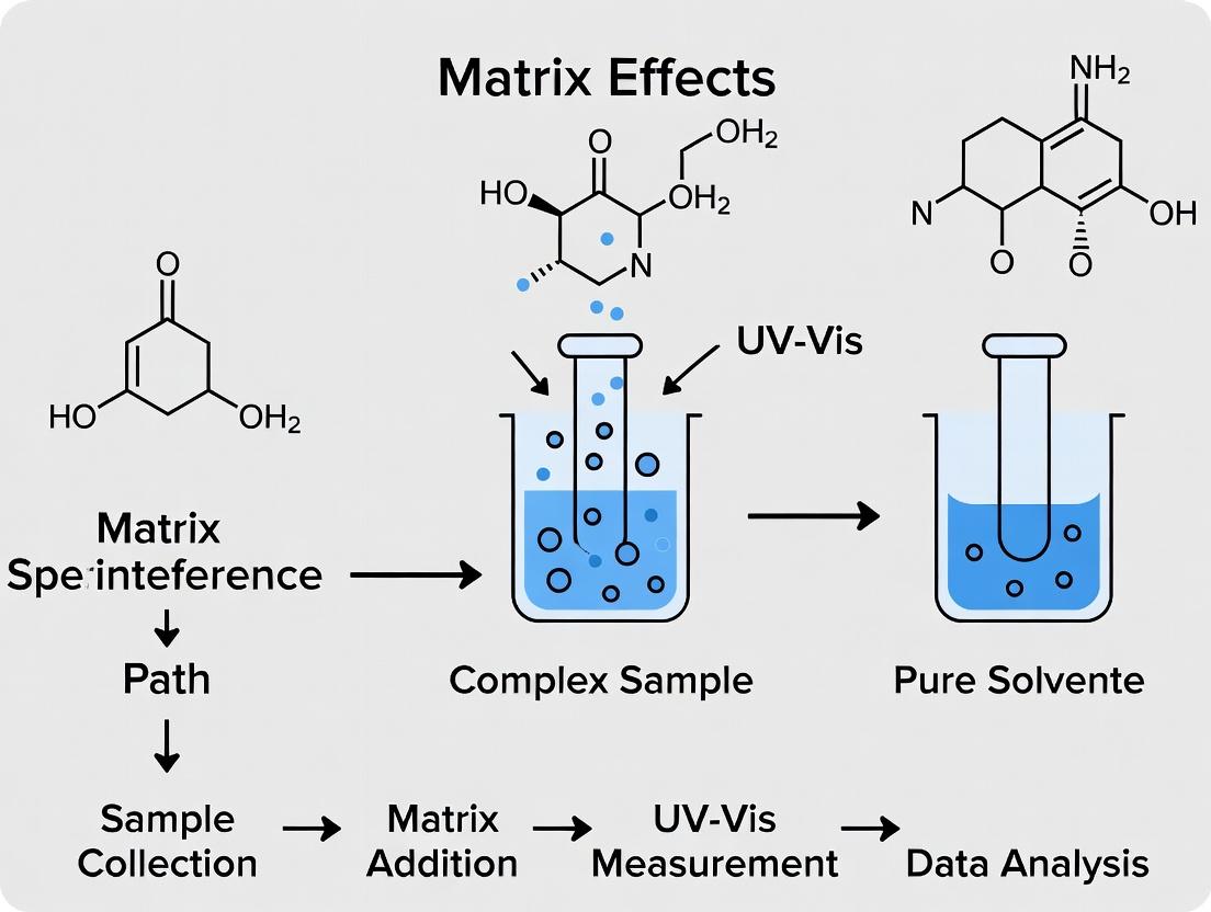
What Are Matrix Effects in UV-Vis? Understanding the Invisible Interference in Complex Samples
This technical support center is framed within a thesis on addressing matrix effects in complex sample UV-Vis analysis. It provides troubleshooting guidance for researchers, scientists, and drug development professionals encountering deviations from ideal Beer-Lambert behavior due to sample matrix interferences.
Troubleshooting Guides & FAQs
Q1: Why does my calibration curve show good linearity with standards but analyte recovery in my biological sample is consistently low? A: This is a classic sign of a matrix-induced suppression effect. Components in the sample (e.g., proteins, lipids, salts) may be binding to your analyte, preventing it from interacting with the incident light, or causing precipitation. The analyte is present but its effective molar absorptivity is reduced.
- Troubleshooting Protocol:
- Perform a standard addition experiment. Add known concentrations of your analyte directly into the sample matrix and measure the absorbance.
- Plot the absorbance vs. concentration added. If the slope of this line is shallower than the slope of your pure solvent calibration curve, it confirms suppression.
- Implement a sample clean-up step such as protein precipitation (using acetonitrile or methanol), solid-phase extraction (SPE), or filtration (0.22 µm or 10 kDa MWCO filter).
Q2: My sample absorbance is higher than expected, and I observe scattering or a sloping baseline. What is happening? A: You are likely experiencing light scattering and non-specific background absorption from particulate matter or colloidal components in the matrix (e.g., cell debris, aggregated proteins). This adds a positive interferent signal across wavelengths.
- Troubleshooting Protocol:
- Centrifuge your sample at high speed (e.g., 10,000-15,000 x g for 10 minutes) or filter it through a 0.2 µm syringe filter.
- Use a matched blank/correction solution. Prepare a matrix blank that is identical to your sample but without the analyte. Measure its absorbance spectrum and subtract it from your sample spectrum.
- For turbid samples, consider using a dual-beam instrument with integrated scattering correction or derivative spectroscopy to minimize baseline effects.
Q3: How can I definitively identify and quantify the magnitude of a matrix effect in my assay? A: Use the "Post-Extraction Spiking" or "Method of Standard Addition" to calculate the Matrix Factor (MF).
- Experimental Protocol for Matrix Factor Calculation:
- Prepare an analyte standard in pure solvent (Solution A).
- Prepare your sample matrix (e.g., plasma extract, buffer solution) without the analyte.
- Spike the same concentration of analyte into the pure solvent and into the processed sample matrix to create Solution B.
- Measure the absorbance of both solutions at your analytical wavelength.
- Calculate the Matrix Factor: MF = (Absorbance of Solution B) / (Absorbance of Solution A).
- Interpret the result: MF = 1 indicates no effect; MF > 1 indicates enhancement; MF < 1 indicates suppression.
Q4: My analyte's absorption spectrum shape changes in the sample matrix compared to standard. What does this indicate? A: This suggests a chemical interaction altering the analyte's electronic environment. Common causes include changes in pH affecting a chromophore's ionization state, complex formation with metal ions, or binding to proteins/carriers.
- Troubleshooting Protocol:
- Record full UV-Vis spectra (not just a single wavelength) for the standard and the sample.
- Check and control the pH of both standard and sample solutions using appropriate buffers (e.g., phosphate, Tris). Ensure pH is within ±0.1 units.
- Investigate potential complexing agents (e.g., EDTA in buffers) and consider their removal or consistent use.
- Use techniques like fluorescence quenching or equilibrium dialysis if protein binding is suspected.
Table 1: Common Matrix Effects and Their Impact on UV-Vis Analysis
| Matrix Effect Type | Primary Cause | Typical Sample | Impact on Absorbance | Common Correction Strategy |
|---|---|---|---|---|
| Light Scattering | Particulates, colloids | Cell lysates, fermentation broths | Increases baseline, slope | Filtration, centrifugation, derivative spectroscopy |
| Chemical Interaction | pH shift, complexation | Biological buffers, saliva | Spectral shift, isosbestic point | pH control, use of chelators/buffers |
| Background Absorption | Co-absorbing interferents | Plant extracts, colored media | Positive bias at λ_analysis | Matrix blank subtraction, diode array detection |
| Suppression (Quenching) | Binding, encapsulation | Serum, plasma, lipid formulations | Negative bias, low recovery | Standard addition, extraction, protein precipitation |
Table 2: Matrix Factor (MF) Interpretation Guide
| MF Range | Effect Magnitude | Interpretation for Quantitative Analysis |
|---|---|---|
| 0.85 - 1.15 | Acceptable | Minimal matrix effect; external calibration may be valid. |
| 0.80 - 0.85 or 1.15 - 1.20 | Moderate | Standard addition or matrix-matched calibration recommended. |
| <0.80 or >1.20 | Severe | Requires extensive sample preparation or internal standard. |
Experimental Protocols
Protocol: Standard Addition for Matrix Effect Compensation
- Sample Preparation: Aliquot equal volumes (e.g., 1.0 mL) of your unknown sample into four separate flasks.
- Spiking: Spike three of the flasks with increasing, known volumes (e.g., 0.1, 0.2, 0.3 mL) of a concentrated analyte standard. Add an equivalent volume of pure solvent to the fourth (unspiked) flask to correct for dilution.
- Dilution: Dilute all flasks to the same final volume with an appropriate solvent.
- Measurement: Measure the absorbance of all four solutions.
- Calculation: Plot absorbance vs. concentration of analyte added. Extrapolate the line backwards to the x-intercept. The absolute value of the x-intercept gives the original concentration of the analyte in the unknown sample.
Protocol: Sample Clean-up via Protein Precipitation for Serum Analysis
- Materials: Serum sample, internal standard (if used), ice-cold acetonitrile or methanol, vortex mixer, centrifuge, 0.22 µm PVDF syringe filter.
- Procedure: Mix 100 µL of serum with 300 µL of ice-cold acetonitrile (1:3 ratio). Vortex vigorously for 60 seconds.
- Centrifuge: Centrifuge at 4°C, 15,000 x g for 10 minutes to pellet precipitated proteins.
- Filter: Carefully decant or pipette the supernatant and pass it through a 0.22 µm syringe filter.
- Analysis: The filtrate can be diluted if necessary and analyzed via UV-Vis. Always prepare a matched serum blank (analyte-free) processed identically.
Visualizations
Light Path Deviations Due to Matrix Effects
Matrix Effect Troubleshooting Decision Workflow
The Scientist's Toolkit: Research Reagent Solutions
Table 3: Essential Materials for Mitigating Matrix Effects
| Item | Function & Rationale |
|---|---|
| Solid-Phase Extraction (SPE) Cartridges (C18, HLB) | Selectively bind analyte or interferents for clean-up and pre-concentration from complex matrices like plasma or urine. |
| Molecular Weight Cut-Off (MWCO) Filters (e.g., 10 kDa) | Remove high molecular weight interferents (proteins, polymers) via centrifugal filtration. |
| Protein Precipitation Agents (Cold ACN, MeOH, TCA) | Denature and precipitate proteins from biological samples to reduce binding and scattering. |
| Buffers (Phosphate, Tris, Acetate) | Maintain constant pH to prevent spectral shifts due to analyte ionization state changes. |
| Internal Standard (Structurally similar analog) | Added in constant amount to all samples and standards; corrects for variability in sample preparation and signal suppression/enhancement. |
| Surfactants (e.g., Triton X-100, SDS) | Solubilize membrane proteins or lipid-bound analytes, preventing light scattering from micelles/aggregates. |
| Derivatization Agents | Chemically modify the analyte to enhance its molar absorptivity, shift its λ_max away from interferents, or reduce matrix interaction. |
Technical Support Center: Troubleshooting Matrix Effects in UV-Vis Analysis
Troubleshooting Guides
Issue: High, Variable Baseline Drift
- Likely Culprit: Particulates or precipitating proteins/lipids causing light scattering.
- Actionable Steps:
- Clarify Sample: Centrifuge at 16,000×g for 10-15 minutes or filter through a 0.22 µm or 0.45 µm low-protein-binding syringe filter.
- Check Cuvette: Inspect for scratches or residue. Clean thoroughly with appropriate solvent.
- Modify Matrix: Dilute sample with a compatible solvent (e.g., buffer with surfactant) to reduce scattering particle concentration.
Issue: Non-Linear or Suppressed Calibration Curves
- Likely Culprit: Excipient or protein binding to the analyte, altering its absorptivity.
- Actionable Steps:
- Matrix-Matched Standards: Prepare calibration standards in the same biological matrix (e.g., plasma, tissue homogenate) as the samples.
- Standard Addition: Perform the method of standard addition to the sample itself to account for binding effects.
- Extraction/Cleanup: Implement a protein precipitation step (e.g., with acetonitrile) or solid-phase extraction (SPE) to isolate the analyte from interfering matrix components.
Issue: Unreplicateable Absorbance Readings
- Likely Culprit: Incomplete solubilization of lipids or adsorption of analyte to vial/protein.
- Actionable Steps:
- Optimize Solubilization: Ensure adequate use of detergents (e.g., Triton X-100) or organic solvents to keep lipids and analyte in solution.
- Use Additives: Add a competitive binding agent (e.g., 1% Bovine Serum Albumin) or use silanized vials to prevent surface adsorption.
- Control Temperature: Use a temperature-controlled cuvette holder to maintain consistent sample environment.
Issue: Unexpected Peaks or Spectral Shoulders
- Likely Culprit: Co-eluting excipients (e.g., preservatives like benzyl alcohol) or metabolites with overlapping absorbance.
- Actionable Steps:
- Spectral Scanning: Run a full UV-Vis scan (e.g., 220-500 nm) to identify characteristic shapes of interfering compounds.
- Chromatographic Separation (if HPLC-UV): Optimize the mobile phase gradient to resolve the analyte peak from interferent peaks.
- Background Subtraction: Use a blank matrix sample for automatic background subtraction if the spectrometer software allows.
Frequently Asked Questions (FAQs)
Q1: How can I quickly assess if particulates are affecting my assay? A: Perform a simple turbidity check by comparing the scattering at a non-absorbing wavelength (e.g., 550 nm or 650 nm) for your sample versus your blank buffer. A significant increase indicates light-scattering interference.
Q2: What is the most effective way to remove proteins from my cell lysate for UV-Vis analysis of a small molecule? A: Protein precipitation using cold acetonitrile (sample:ACN ratio of 1:2 or 1:3) is fast and effective. Vortex, centrifuge at >10,000×g for 10 minutes, and carefully recover the supernatant for analysis.
Q3: I suspect my drug compound is binding to serum albumin. How can I confirm and mitigate this? A: You can confirm by running spectra of the compound in buffer vs. in serum. A spectral shift or broadening suggests binding. Mitigation strategies include using a displacement agent (e.g., fatty acids), diluting the sample, or adding a mild denaturant like urea (if compatible with your assay).
Q4: Why does my buffer blank sometimes show absorbance in the low UV range (<230 nm)? A: Many common buffer components (e.g., TRIS, certain salts, EDTA) and plastic leachates absorb strongly below 230 nm. Use high-purity reagents, water (HPLC-grade), and ensure all glassware/cuvettes are meticulously clean. Consider using phosphate or perchlorate salts for lower UV cutoffs.
| Matrix Component | Typical Concentration Range | Primary Interference Mechanism in UV-Vis | Wavelength Range Most Affected |
|---|---|---|---|
| Proteins (e.g., BSA, IgG) | 30-80 mg/mL (serum) | Light scattering, absorption (<280 nm), analyte binding | < 280 nm (absorption), All (scattering) |
| Lipids (Triglycerides, Lipoproteins) | 1-10 mg/mL (plasma) | Strong light scattering, turbidity | All, especially >500 nm |
| Common Excipient: Polysorbate 80 | 0.01-0.1% (v/v) | Micelle formation, alters analyte partitioning | Minimal direct absorption |
| Common Excipient: Benzyl Alcohol | 0.5-1.0% (v/v) | Direct UV absorption | ~257 nm, ~225 nm |
| Silica Particulates (from SPE) | Variable | Severe light scattering, baseline offset | All wavelengths |
Experimental Protocol: Mitigating Matrix Effects via Protein Precipitation and Filtration
Objective: To isolate a small-molecule analyte from a protein-rich matrix (e.g., plasma) for UV-Vis analysis.
Materials:
- Plasma sample (100 µL)
- Ice-cold Acetonitrile (ACN, 300 µL)
- Vortex mixer
- Microcentrifuge
- 0.22 µm PVDF syringe filter
- 1.5 mL microcentrifuge tubes
- UV-compatible micro-cuvette
Procedure:
- Precipitation: Pipette 100 µL of plasma into a 1.5 mL microcentrifuge tube. Add 300 µL of ice-cold ACN.
- Mix: Vortex the mixture vigorously for 60 seconds.
- Pellet Proteins: Centrifuge the tube at 16,000 × g for 10 minutes at 4°C.
- Clarify: Carefully collect the supernatant without disturbing the protein pellet. Pass the supernatant through a 0.22 µm PVDF syringe filter into a clean tube.
- Analysis: The filtered supernatant can now be diluted as needed and analyzed via UV-Vis spectroscopy. Always run a matrix-processed blank (subject a blank plasma sample to the same protocol) for accurate background subtraction.
Diagram: Workflow for Addressing Matrix Effects
Title: Matrix Effect Troubleshooting Decision Tree
The Scientist's Toolkit: Key Research Reagent Solutions
| Item | Primary Function | Example in This Context |
|---|---|---|
| Low-Protein-Binding Filters (PVDF, PTFE) | To remove sub-micron particulates and aggregates without adsorbing the analyte of interest. | Clarifying plasma supernatants post-protein precipitation (0.22 µm). |
| Solid-Phase Extraction (SPE) Cartridges (C18, HLB) | To selectively isolate and concentrate the analyte from a complex matrix, removing salts, proteins, and polar interferences. | Cleaning up a small molecule from lipid-rich tissue homogenate prior to UV-Vis. |
| Chaotropic Agents (Urea, Guanidine HCl) | To denature proteins, disrupting protein-analyte binding interactions. | Releasing a protein-bound drug in serum to measure its total concentration. |
| Surfactants/Detergents (Triton X-100, CHAPS) | To solubilize membrane proteins and lipids, preventing aggregation and scattering. | Preparing a clear, homogeneous sample from a cell membrane fraction. |
| Protease/Phosphatase Inhibitor Cocktails | To prevent degradation of proteinaceous analytes or modification of phospho-analytes during sample preparation. | Stabilizing protein targets in a cell lysate for subsequent analysis. |
| Matrix-Matched Standard Materials | To provide a background-identical medium for creating calibration curves, compensating for matrix effects. | Creating a calibration curve in charcoal-stripped serum for a serum-based assay. |
Troubleshooting Guides & FAQs
Q1: My UV-Vis absorbance readings for my nanoparticle suspension are anomalously high and increase sharply at lower wavelengths. What is the likely cause and how can I resolve it?
A: This is a classic sign of scattering interference, particularly from large particles or aggregates. Scattering increases as wavelength decreases (λ^-4 dependence, Rayleigh scattering), inflating the apparent absorbance. To resolve:
- Filter or Centrifuge: Pass the sample through a 0.22 µm or 0.45 µm syringe filter, or centrifuge to remove large aggregates.
- Sample Dilution: Sometimes diluting the sample reduces particle-particle interactions that cause aggregation.
- Use an Integrating Sphere: For dedicated quantitative analysis of scattering samples, use a spectrophotometer equipped with an integrating sphere detector to separate scattered light from truly absorbed light.
- Background Correction: Use a non-absorbing, scattering blank (e.g., a blank suspension without the analyte) to correct the baseline.
Q2: I suspect my target analyte's absorption peak is overlapped by a matrix component. How can I confirm and correct for this?
A: Absorption overlap leads to poor selectivity and overestimation. To confirm and correct:
- Scan the Blank Matrix: Obtain a full UV-Vis spectrum of your sample matrix (without the analyte). Compare it to your analyte's spectrum.
- Use Derivative Spectroscopy: Apply 1st or 2nd derivative transformations to your spectra. This technique can enhance resolution of overlapping peaks, allowing for quantification of the analyte despite the interference.
- Employ Chemometrics: Use multivariate calibration methods like Partial Least Squares (PLS) regression. These algorithms can deconvolve the combined signal from multiple absorbers if you have a calibrated set of standards with varying known concentrations of both analyte and interferent.
Q3: My analyte's absorbance decreases over time in the cuvette, or a precipitate forms. What should I do?
A: This indicates a chemical interaction (e.g., complexation, precipitation, oxidation) between the analyte and the matrix. To address:
- Check Chemical Compatibility: Review the stability of your analyte in the solvent/buffer used. Adjust pH or change solvent if necessary.
- Use a Stabilizing Agent: Incorporate agents like chelators (EDTA to sequester metal catalysts) or antioxidants (ascorbic acid) to prevent degradation.
- Minimize Measurement Time: Prepare samples immediately before measurement and use kinetic mode to monitor stability.
- Change Preparation Method: Consider protein precipitation or solvent extraction to isolate the analyte from the reactive matrix components before analysis.
Q4: My calibration curve has good linearity in pure solvent but fails in the spiked matrix. How do I recover accuracy?
A: This is the definitive sign of a matrix effect. Implement the Standard Addition Method:
- Spike known, increasing amounts of your analyte standard directly into aliquots of your sample matrix.
- Measure the absorbance for each spiked aliquot.
- Plot added analyte concentration vs. absorbance. The x-intercept (where absorbance=0) gives the negative of the original analyte concentration in the sample. This method internally corrects for most multiplicative interferences.
Experimental Protocols
Protocol 1: Assessing and Correcting for Scattering via Filtration
- Prepare your nanoparticle or colloidal sample as usual.
- Split into two aliquots.
- Filter one aliquot through a 0.22 µm membrane filter (compatible with your solvent).
- Use the filtered solution as the blank/background correction for scanning the unfiltered aliquot.
- Compare the corrected spectrum of the unfiltered sample to a direct scan of the filtered sample.
Protocol 2: Standard Addition Method for Matrix Effects
- Pipette equal volumes (e.g., 2.0 mL) of your unknown sample into five separate volumetric flasks (or tubes).
- Spike these flasks with increasing, known volumes of your standard analyte solution (e.g., 0, 0.5, 1.0, 1.5, 2.0 mL).
- Dilute all flasks to the same final volume with the appropriate solvent.
- Measure the absorbance for each solution.
- Plot absorbance (y-axis) versus concentration of the added standard (x-axis). Perform linear regression.
- Calculate the original sample concentration (C_o) as:
C_o = |x-intercept| × (Dilution Factor).
Protocol 3: Derivative Spectroscopy for Peak Resolution
- Acquire UV-Vis spectra for your analyte standard, matrix blank, and sample using a narrow spectral bandwidth (e.g., 1 nm) and small data interval (e.g., 0.5 nm).
- Export the digitized absorbance (A) vs. wavelength (λ) data.
- Using spectroscopic software (e.g., built-in tools, Origin, Matlab), calculate the 1st derivative (dA/dλ) or 2nd derivative (d²A/dλ²) spectrum using a Savitzky-Golay smoothing algorithm (e.g., 7-11 point window).
- Quantify the analyte using the peak-to-zero or peak-to-trough amplitude in the derivative spectrum, which is often proportional to concentration and less affected by broad, overlapping backgrounds.
Data Presentation
Table 1: Impact of Scattering Correction Methods on Apparent Absorbance at 400 nm
| Sample Type | Uncorrected Absorbance | After 0.45 µm Filtration | After Baseline Subtraction with Scattering Blank |
|---|---|---|---|
| Nanoparticle Suspension A | 1.254 | 0.873 | 0.901 |
| Aggregated Protein Sample B | 0.987 | 0.601 | 0.622 |
| Cell Lysate (clarified) C | 0.456 | 0.431 | 0.440 |
Table 2: Accuracy Recovery Using Standard Addition vs. External Calibration
| Method | Theoretical Spiked Concentration (µM) | Measured Concentration (µM) | % Recovery |
|---|---|---|---|
| External Calibration | 10.0 | 13.7 ± 0.4 | 137% |
| Standard Addition | 10.0 | 9.8 ± 0.3 | 98% |
| External Calibration | 25.0 | 31.2 ± 0.5 | 125% |
| Standard Addition | 25.0 | 24.5 ± 0.4 | 98% |
The Scientist's Toolkit
Table 3: Key Research Reagent Solutions for Mitigating UV-Vis Interferences
| Item | Function | Example Use Case |
|---|---|---|
| 0.22/0.45 µm PVDF or Nylon Syringe Filter | Removes particulate matter causing scattering. | Clarifying nanoparticle suspensions or biological lysates before reading. |
| Matrix-Matched Calibration Standards | Standards prepared in the same blank matrix as samples to compensate for multiplicative effects. | Quantifying drugs in serum or complex growth media. |
| Chelating Agent (e.g., 0.1 mM EDTA) | Binds metal ions that may catalyze oxidation or form complexes with the analyte. | Stabilizing phenolic compounds or vitamins in buffer. |
| Surfactant (e.g., 0.1% Triton X-100) | Prevents aggregation of hydrophobic molecules or particles. | Maintaining dispersion of lipophilic drugs in aqueous assay buffers. |
| Derivatization Agent | Chemically modifies the analyte to enhance its molar absorptivity or shift its λ_max away from interferents. | Pre-column derivatization of amino acids with ninhydrin for visible detection. |
| Solid Phase Extraction (SPE) Cartridge | Selectively isolates and concentrates the analyte while removing interfering matrix. | Cleaning up environmental water samples prior to contaminant analysis. |
Diagrams
Title: Troubleshooting Scattering in UV-Vis Analysis
Title: Standard Addition Method Workflow
Title: Diagnosing UV-Vis Interference Mechanisms
Technical Support Center
Troubleshooting Guides & FAQs
FAQ: Addressing Common Issues in UV-Vis Analysis of Complex Samples
Q1: My accuracy, as determined by spike-and-recovery experiments, is consistently low (<85%). What is the most likely cause and how can I troubleshoot it? A1: Low recovery is a primary indicator of a matrix effect. This occurs when components in your sample (e.g., proteins, salts, excipients) alter the analyte's absorptivity or cause light scattering.
- Troubleshooting Steps:
- Confirm the Effect: Compare the slope of your calibration curve in neat solvent vs. matrix-matched standard. A significant difference (>10%) confirms a matrix effect.
- Implement Standard Addition: Use the method of standard addition to calibrate directly in the sample matrix. This is the most robust solution for quantitative accuracy in complex samples.
- Improve Sample Cleanup: Introduce or optimize a sample preparation step. For biological fluids, consider protein precipitation (using acetonitrile or perchloric acid) followed by filtration (0.22 µm or 0.45 µm).
- Use a Blank Correction: Ensure you are using an appropriate matrix blank to zero the instrument, correcting for non-specific background absorption.
Q2: My precision (%RSD) is unacceptably high between sample replicates. Where should I focus my investigation? A2: Poor precision often stems from sample handling or instrument instability, exacerbated by complex matrices.
- Troubleshooting Steps:
- Check Homogeneity: Ensure your sample solution is fully dissolved and homogeneous before measurement. Vortex and centrifuge if necessary.
- Verify Cuvette Technique: Clean cuvettes meticulously. Always position the cuvette in the holder with the same orientation (mark the cuvette). Fingerprints on the clear windows are a common source of error.
- Instrument Performance: Run a diagnostic check using a holmium oxide or didymium filter to verify wavelength accuracy. Check the stability of the lamp (replace if >1000 hours old).
- Automate Pipetting: Manual pipetting of viscous or complex samples is a major variability source. Use calibrated, positive-displacement pipettes or automate dilution steps.
Q3: My Limit of Quantification (LOQ) is too high for my intended application. What strategies can I use to lower it? A3: The LOQ is directly impacted by the signal-to-noise ratio (S/N). In complex samples, noise from the matrix is the limiting factor.
- Troubleshooting Steps:
- Pathlength Increase: Use a cuvette with a longer pathlength (e.g., 10 mm to 50 mm) to increase the analyte's absorbance signal (per Beer-Lambert Law).
- Pre-concentration: Employ a technique like solid-phase extraction (SPE) or liquid-liquid extraction (LLE) to concentrate the analyte and dilute the interfering matrix.
- Derivatization: If applicable, use a chromogenic reagent to form a derivative with your analyte that has a higher molar absorptivity (ε) at a wavelength less prone to matrix interference.
- Spectral Processing: Apply Savitzky-Golay smoothing to your absorption spectrum to reduce high-frequency noise. Use a first or second derivative spectrum to resolve overlapping peaks from the background.
Table 1: Impact of Matrix-Matched Calibration on Analytical Figures of Merit for Drug X in Plasma
| Calibration Method | Accuracy (% Recovery) | Precision (%RSD, n=6) | Limit of Quantification (LOQ) |
|---|---|---|---|
| Neat Solvent (Buffer) | 72.5 ± 8.2 | 15.3 | 5.0 µg/mL |
| Matrix-Matched (Plasma) | 98.2 ± 3.1 | 4.7 | 2.1 µg/mL |
| Standard Addition | 99.5 ± 2.8 | 3.9 | 1.8 µg/mL |
Table 2: Effect of Sample Preparation on Signal-to-Noise (S/N) and LOQ
| Sample Prep Method | Avg. S/N at 1 µg/mL | Calculated LOQ (µg/mL) * | Key Interference Removed |
|---|---|---|---|
| None (Dilute-and-Shoot) | 12:1 | 2.5 | None |
| Protein Precipitation | 25:1 | 1.2 | Proteins, Lipids |
| Solid-Phase Extraction | 50:1 | 0.6 | Proteins, Salts, Polar Organics |
*LOQ calculated as concentration giving S/N = 10.
Experimental Protocols
Protocol 1: Standard Addition for Accurate Quantification in Complex Matrices
- Prepare Sample Aliquots: Pipette equal volumes (e.g., 2.0 mL) of your unknown sample solution into four separate 5 mL volumetric flasks.
- Spike Standards: To three of the flasks, add known increasing volumes (e.g., 0.5, 1.0, 1.5 mL) of a standard analyte solution of known concentration. Add no standard to the fourth flask.
- Dilute to Volume: Dilute all flasks to the mark with an appropriate solvent and mix thoroughly.
- Measure Absorbance: Measure the absorbance of all four solutions at your analytical wavelength.
- Plot and Calculate: Plot absorbance (y-axis) vs. concentration of standard added (x-axis). Extrapolate the linear plot to the x-axis (where y=0). The absolute value of the x-intercept is the concentration of the analyte in the original sample solution.
Protocol 2: Evaluating Matrix Effects via Calibration Slope Comparison
- Prepare Neat Solvent Calibrants: Prepare a minimum of 5 standard solutions of your analyte in a clean, pure solvent (e.g., buffer, methanol). Cover the expected concentration range.
- Prepare Matrix-Matched Calibrants: Prepare the same standard concentrations, but add them to your matrix (e.g., blank plasma extract, formulation placebo) that has undergone the exact same sample preparation as your unknowns.
- Acquire Spectra: Measure the absorbance of all calibrants at the target wavelength.
- Analyze Data: Create two calibration curves. Calculate the slope of each linear regression.
- Calculate Matrix Effect (ME %): ME (%) = (SlopeMatrix-Matched / SlopeNeat Solvent - 1) × 100%. An ME > ±10% indicates a significant matrix effect requiring mitigation.
Mandatory Visualizations
Diagram Title: Workflow for Managing Matrix Effects in UV-Vis Analysis
Diagram Title: Matrix Effect Impact and Mitigation Pathways
The Scientist's Toolkit: Key Research Reagent Solutions
Table 3: Essential Materials for Complex Sample UV-Vis Analysis
| Item | Function in Experiment |
|---|---|
| Matrix-Matched Blank | A sample containing all components except the target analyte. Used to zero the instrument, correcting for background absorption/scattering from the matrix. |
| Holmium Oxide Filter | A wavelength accuracy standard. Used to verify and calibrate the wavelength scale of the UV-Vis spectrophotometer. |
| Solid-Phase Extraction (SPE) Cartridges (C18) | Used to selectively bind, clean up, and concentrate the analyte from a complex liquid sample, removing interfering salts, proteins, and polar organics. |
| Chromogenic Derivatization Reagent | A chemical that reacts specifically with the target analyte to produce a strongly absorbing compound, enhancing sensitivity and selectivity. |
| Certified Reference Material (CRM) | A material with a precisely known analyte concentration. Serves as the primary standard for establishing calibration curves and validating method accuracy. |
| Quartz Micro Cuvette (e.g., 50 µL, 10 mm path) | Allows for analysis of small volume samples. Quartz is transparent down to 190 nm, enabling full UV range analysis. |
| 0.22 µm Syringe Filter (Nylon or PVDF) | Used for final clarification of samples after protein precipitation or extraction to remove particulates that cause light scattering. |
Technical Support Center
Troubleshooting Guides & FAQs
Q1: My calibration curve shows excellent linearity in buffer, but the spiked plasma samples show a consistent positive bias. What is the most likely cause and how can I confirm it? A: This is a classic sign of a matrix-induced enhancement effect. Non-volatile plasma components (e.g., phospholipids, salts) can co-elute with your analyte and enhance its ionization efficiency in LC-MS/MS, or alter its spectroscopic properties in UV-Vis. To confirm:
- Perform a post-column infusion test. Continuously infuse your analyte into the mobile post-column while injecting a blank plasma extract. A drift or peak in the baseline at your analyte's retention time indicates ion suppression/enhancement.
- Perform a post-extraction spike experiment. Compare the response of your analyte spiked into blank plasma before extraction versus spiked into the final extract of blank plasma after extraction. A significant difference indicates matrix effects.
Q2: My method validation passes, but patient samples yield erratic, sometimes negative values when re-run. What could be wrong? A: This points to variable matrix effects between individual plasma lots. Your validation likely used a pooled plasma matrix, which averages out effects. Individual samples can have vastly different levels of interfering substances (e.g., from diet, disease state, concomitant medications).
- Troubleshooting Step: Re-assay the problematic samples using the standard addition method. Prepare aliquots of the sample and spike with known increments of the analyte. Plot the response and extrapolate to find the original concentration. If the results differ from your original calibration curve method, variable matrix effects are confirmed.
Q3: How can I distinguish matrix effects from poor extraction recovery in my sample preparation? A: You must design a experiment to decouple the two. Use the following protocol:
- Prepare three sets of samples in triplicate: (A) Analyte in pure solvent (neat solution). (B) Analyte spiked into blank plasma before extraction. (C) Analyte spiked into the final extract of blank plasma after extraction.
- Analyze all samples.
- Calculate: Recovery (%) = (Peak Area B / Peak Area C) x 100. This measures extraction efficiency.
- Calculate: Matrix Effect (%) = (Peak Area C / Peak Area A) x 100. A value of 100% means no effect, >100% is enhancement, <100% is suppression.
Q4: I'm using UV-Vis spectroscopy, not MS. Are matrix effects still a concern? A: Absolutely. While different in mechanism, they are equally problematic. In UV-Vis, background absorbance from plasma pigments (e.g., bilirubin, hemoglobin) or turbidity can cause significant interference, leading to overestimation of concentration.
- Solution: Implement a robust sample clean-up (e.g., solid-phase extraction, protein precipitation with careful centrifugation). Always use a method blank (processed blank plasma) and subtract its absorbance from your sample readings. Second-derivative spectroscopy can also help resolve overlapping absorbance bands.
Key Data on Common Matrix Interferences
Table 1: Common Plasma Interferents and Their Impact on Drug Assays
| Interferent Class | Source | Primary Impact (LC-MS/MS) | Primary Impact (UV-Vis) |
|---|---|---|---|
| Phospholipids | Cell membranes | Severe ion suppression, especially in ESI+ | Minimal direct impact |
| Salts (Na+, K+) | Plasma, sample prep | Ion suppression, source contamination | High background absorbance |
| Proteins | Plasma | Non-specific binding, column fouling | Light scattering, turbidity |
| Hemolysis (Hb) | Poor blood draw | Can alter ionization | Strong absorbance <450 nm |
| Lipids (Chylomicrons) | Non-fasted subjects | Alters extraction efficiency, ion suppression | Severe light scattering/turbidity |
| Bilirubin | Liver function | Minor ion suppression | Strong absorbance ~450-460 nm |
| Endogenous Metabolites | Individual physiology | Variable ion competition | Potential spectral overlap |
Table 2: Efficacy of Common Mitigation Strategies
| Mitigation Strategy | Reduces Ion Suppression | Reduces Background Absorbance | Cost & Complexity | Key Limitation |
|---|---|---|---|---|
| Improved Sample Clean-up (SPE) | High | High | Medium-High | Method development time |
| Stable Isotope Internal Standard | Compensates for effect | No | High | Availability, cost |
| Dilution of Sample | Low-Moderate | Low | Low | May drop analyte below LLOQ |
| Modified Chromatography | High | Low | Medium | Requires method re-development |
| Standard Addition Method | Compensates for effect | Compensates for effect | High | Labor-intensive for batches |
Experimental Protocols
Protocol 1: Quantitative Assessment of Matrix Effect and Recovery
- Objective: To quantitatively determine matrix effect (ME) and extraction recovery (RE) for an analyte in plasma.
- Materials: Blank human plasma, analyte stock solution, internal standard (IS) stock solution, appropriate solvents and materials for extraction (e.g., protein precipitation reagents, SPE cartridges).
- Procedure:
- Prepare three sets of samples (n=6 each):
- Set A (Neat): Analyte + IS in reconstitution solvent.
- Set B (Pre-extraction Spike): Add analyte + IS to blank plasma, then perform extraction.
- Set C (Post-extraction Spike): Extract blank plasma, then add analyte + IS to the final extract.
- Process all samples according to the analytical method.
- Analyze all samples by LC-MS/MS or UV-Vis.
- Calculate:
- ME (%) = (Mean Peak Area of Set C / Mean Peak Area of Set A) × 100
- RE (%) = (Mean Peak Area of Set B / Mean Peak Area of Set C) × 100
- Process Efficiency (PE%) = (Mean Peak Area of Set B / Mean Peak Area of Set A) × 100 = (ME% × RE%)/100
- Prepare three sets of samples (n=6 each):
Protocol 2: Post-Column Infusion Test for LC-MS/MS
- Objective: To visually identify regions of ion suppression/enhancement in a chromatographic run.
- Materials: LC-MS/MS system, syringe pump, analyte solution at constant concentration (e.g., 100 ng/mL in mobile phase), T-connector.
- Procedure:
- Connect a syringe pump with the analyte solution via a T-connector between the HPLC column outlet and the MS ion source.
- Start a constant infusion of the analyte (e.g., 5-10 µL/min).
- Start the MS in selected reaction monitoring (SRM) mode for the analyte.
- Inject a blank plasma extract onto the LC column and start the gradient.
- Monitor the analyte signal. A stable baseline indicates no matrix effect. A dip (suppression) or peak (enhancement) in the baseline indicates co-elution of matrix interferents affecting ionization.
Visualization: Experimental Workflows
Diagram 1: General Workflow Showing Point of Matrix Interference
Diagram 2: Troubleshooting Decision Tree for Matrix Effects
The Scientist's Toolkit: Key Research Reagent Solutions
Table 3: Essential Materials for Mitigating Matrix Effects
| Item | Function & Rationale |
|---|---|
| Stable Isotope-Labeled Internal Standard (SIL-IS) | The gold standard for LC-MS/MS. Co-elutes with the analyte, experiences identical matrix effects and recovery losses, allowing for perfect compensation. |
| Analog Internal Standard | A structurally similar compound used when a SIL-IS is unavailable. Must be chosen to have similar extraction recovery and ionization as the analyte. |
| Phospholipid Removal SPE Plates | Specialized solid-phase extraction sorbents designed to selectively retain phospholipids from plasma, dramatically reducing a major source of ion suppression. |
| Supported Liquid Extraction (SLE) Plates | An alternative to SPE using a diatomaceous earth support. Often provides cleaner extracts than liquid-liquid extraction with better reproducibility. |
| Matrix Matched Calibrators | Calibration standards prepared in the same biological matrix (e.g., pooled plasma) as the samples. Partially accounts for consistent matrix effects. |
| Method Blank (Processed Blank Matrix) | A blank plasma sample taken through the entire sample preparation and analysis process. Critical for identifying and subtracting background signal in UV-Vis and checking for carryover/interference in LC-MS. |
| Post-Column Infusion Kit (T-connector, syringe pump) | Hardware necessary for performing the diagnostic post-column infusion test to visualize ion suppression/enhancement zones. |
Proven Techniques to Combat Matrix Effects: From Sample Prep to Advanced Corrections
Technical Support Center & Troubleshooting Guide
Frequently Asked Questions (FAQs)
Q1: My UV-Vis absorbance readings for plasma samples are abnormally high and non-linear with dilution. What is the most likely cause and solution?
A: This is a classic sign of a significant scattering matrix effect, often from incomplete deproteinization or lipid residues. First, ensure your protein precipitation protocol is rigorous: use a minimum 2:1 ratio of organic solvent (e.g., acetonitrile) to sample, vortex for 2 minutes, and centrifuge at 4°C, >10,000 RCF for 15 minutes. Filter the supernatant through a 0.22 µm PVDF or nylon membrane syringe filter. Re-evaluate the absorbance.
Q2: After liquid-liquid extraction (LLE), my analyte recovery is consistently below 60%. How can I optimize this?
A: Low recovery in LLE typically points to suboptimal solvent choice or pH control. For acidic analytes, ensure the aqueous phase is at least 2 pH units below the analyte's pKa; for basic analytes, 2 pH units above. Increase the extraction efficiency by performing two sequential extractions with fresh organic solvent, pooling the extracts. See Table 1 for solvent selection guidance.
Q3: I used dilution to reduce matrix interference, but now my target analyte concentration is below the detection limit. What are my alternatives?
A: Dilution can compromise sensitivity. Implement a selective extraction or clean-up step before dilution. Consider solid-phase extraction (SPE) using a cartridge selective for your analyte's chemical properties (e.g., C18 for non-polar, WCX for cations). This will preconcentrate the analyte and remove interferents, allowing for a less destructive dilution factor.
Q4: How do I choose between protein precipitation, LLE, and SPE for my specific biological matrix?
A: The choice balances required purity, recovery, and throughput. See Table 2 for a comparative summary based on common research goals within UV-Vis analysis of complex samples.
Troubleshooting Guides
Issue: Inconsistent Absorbance Baselines Across Different Sample Batches
- Check 1: Verify deproteinization reagent freshness and lot consistency. Degraded acetonitrile or methanol can reduce precipitation efficiency.
- Check 2: Standardize sample homogenization and incubation times prior to deproteinization. Variable cell lysis can release different amounts of interfering substances.
- Action Protocol: Run a blank matrix prepared from a pooled control sample through your entire sample prep workflow. This batch-specific "process blank" should be subtracted from sample readings.
Issue: Precipitate Formation During Spectral Scanning
- Cause: Incomplete removal of precipitated protein or solubility changes post-treatment.
- Solution Protocol: After the initial centrifugation and filtration, subject the clarified supernatant to a second, brief "polishing" centrifugation (5 min at >12,000 RCF) in a cooled microcentrifuge immediately before loading into the cuvette. Keep the sample chamber temperature-controlled to prevent condensation or precipitation.
Issue: Poor Resolution of Overlapping Absorption Peaks
- Cause: Co-extraction of matrix components with similar chromophores.
- Solution Protocol: Implement a pH-dependent back-extraction step. For example, extract an acidic analyte into an organic solvent from an acidified aqueous phase, then shake the organic phase with a small volume of a basic aqueous buffer (pH 10). The analyte will transfer back to the aqueous phase, leaving many neutral and acidic interferents in the organic layer.
Table 1: Common LLE Solvents for Matrix Clean-up in UV-Vis Analysis
| Solvent | Polarity Index | Best For Extracting | Immiscible With | Notes for UV-Vis |
|---|---|---|---|---|
| n-Hexane | 0.1 | Non-polar lipids, hydrocarbons | Water, acetonitrile | Very low UV cutoff (~195 nm), excellent for low-wavelength detection. |
| Ethyl Acetate | 4.4 | Medium-polarity analytes, many drugs | Water, saline solutions | Moderate UV cutoff (~256 nm). Evaporates easily for reconstitution. |
| Chloroform | 4.1 | Alkaloids, hormones, peptides | Water, buffers | High density, excellent recovery. UV cutoff (~245 nm). Toxic - use in fume hood. |
| Methyl tert-butyl ether (MTBE) | 2.5 | Medium to low polarity compounds | Water, methanol | Low UV cutoff (~210 nm), lower toxicity than chloroform/ether. |
Table 2: Comparison of Sample Preparation Techniques
| Technique | Typical Recovery (%) | Key Advantage | Primary Limitation | Best Suited For |
|---|---|---|---|---|
| Dilution | ~100 (but dilute) | Simplicity, speed, preserves labile analytes | Severe loss of sensitivity, does not remove interferents | Simple buffers, samples with very high initial analyte concentration. |
| Protein Precipitation | 70-95 | Fast, high-throughput, good for small molecules | Limited clean-up, can clog flow systems, ion suppression possible | Initial step for plasma/serum prior to a secondary method. |
| Liquid-Liquid Extraction | 60-90 | Excellent clean-up, concentration capability, scalable | Emulsion formation, uses large solvent volumes, manual. | Removing lipids, isolating analytes from complex biological fluids. |
| Solid-Phase Extraction | 50-95 (method-dependent) | Superior clean-up, selective, automatable | Method development is complex, cartridges can dry out. | Targeted removal of specific interferences, demanding UV-Vis applications. |
Experimental Protocols
Protocol 1: Optimized Dual-Step Deproteinization for Plasma/Serum
- Purpose: To remove proteins and phospholipids effectively, minimizing scatter and background absorption in the 200-300 nm range.
- Materials: Ice-cold Acetonitrile (ACN), 2% Formic Acid in ACN, 0.22 µm PVDF filter plate or syringe filters, cooled centrifuge.
- Procedure:
- Pipette 100 µL of sample (plasma/serum) into a microcentrifuge tube.
- Add 300 µL of ice-cold 2% Formic Acid in ACN (3:1 ratio).
- Vortex vigorously for 3 minutes.
- Centrifuge at 12,000 RCF for 10 minutes at 4°C.
- Transfer the supernatant to a new tube containing 500 µL of pure ice-cold ACN. Vortex for 1 minute.
- Centrifuge again at 15,000 RCF for 15 minutes at 4°C.
- Filter the final supernatant through a 0.22 µm PVDF membrane.
- The filtrate is ready for dilution (if needed) and UV-Vis analysis.
Protocol 2: pH-Mediated Liquid-Liquid Extraction for Acidic Analytics
- Purpose: To selectively extract and concentrate an acidic target analyte from a biological homogenate.
- Materials: 0.1 M HCl, 0.1 M Phosphate Buffer (pH 7.0), Saturated Sodium Chloride solution, Ethyl Acetate, conical glass tubes.
- Procedure:
- Acidify 1 mL of sample homogenate with 100 µL of 0.1 M HCl (target pH ~2-3).
- Add 2 mL of Ethyl Acetate to the tube.
- Cap and shake vigorously for 5 minutes, venting occasionally.
- Centrifuge at 3000 RCF for 5 minutes for clear phase separation.
- Transfer the upper (organic) layer to a new tube.
- Repeat steps 2-5 with a fresh 2 mL of Ethyl Acetate and pool the organic layers.
- Add 1 mL of the pH 7.0 phosphate buffer to the pooled organic extract.
- Shake for 3 minutes to back-extract the analyte into the aqueous phase.
- Centrifuge and carefully retrieve the lower aqueous layer. This is your cleaned and concentrated extract for analysis.
Visualizations
Title: Sample Preparation Decision Workflow for UV-Vis Analysis
Title: How Sample Preparation Counters Matrix Effects
The Scientist's Toolkit: Key Research Reagent Solutions
| Item | Primary Function in Sample Prep | Key Consideration for UV-Vis |
|---|---|---|
| HPLC-Grade Acetonitrile | Protein precipitant. Strong denaturing power, reduces lipid co-precipitation. | Must have low UV absorbance, especially below 220 nm. |
| Acidified Organic Solvents (e.g., 1-2% FA in ACN) | Enhances protein precipitation efficiency and reproducibility for a wider range of analytes. | Acid type and concentration can affect analyte stability. |
| PVDF Syringe Filters (0.22 µm) | Removal of residual particulates post-precipitation/extraction to prevent light scattering. | Low protein binding, compatible with most organic solvents. |
| Solid-Phase Extraction Cartridges (C18, HLB, Ion-Exchange) | Selective binding and washing to isolate analyte from complex matrices. | Choice of sorbent is critical; must match analyte chemistry. |
| pH-Adjustment Buffers | Critical for controlling ionization state during LLE or SPE to maximize recovery. | Buffer should not absorb in your target wavelength range. |
| Mass Spectrometry-Grade Water | Used for dilution and reconstitution. Minimal ionic/organic impurities. | Essential for a flat, low background baseline in sensitive assays. |
Technical Support Center: Troubleshooting UV-Vis Analysis with Matched Blanks
This support center addresses common challenges in implementing matched blank subtraction to mitigate matrix effects in complex biological and pharmaceutical samples.
FAQs & Troubleshooting Guides
Q1: My sample absorbance after subtraction is negative or near zero. What does this indicate? A: This typically signals an error in blank preparation. The blank's matrix is more optically dense than your sample. Verify that the blank contains all non-analyte components at their exact concentrations in the sample. Common culprits are mismatched excipient, buffer, salt, or stabilizer (e.g., glycerol) concentrations. Re-prepare the blank, ensuring it undergoes the same handling (e.g., vortexing, heating, filtration) as the sample.
Q2: How do I choose between a reagent blank and a matched matrix blank? A: The choice is critical and depends on your sample complexity.
| Blank Type | Composition | Primary Purpose | When to Use |
|---|---|---|---|
| Reagent/Solvent Blank | Pure solvent (e.g., water, buffer). | Corrects for solvent absorbance & cuvette/solvent light scattering. | Simple solutions in a clear, uniform matrix. |
| Matched Matrix Blank | Identical to sample matrix minus the specific analyte(s) of interest. | Corrects for all matrix-derived absorbance, scattering, and interference. | Complex samples: cell lysates, serum, drug formulations, crude extracts. |
Protocol 1: Preparation of a Matched Matrix Blank for Protein-Drug Binding Studies
- Replicate Matrix: Prepare an identical volume of the biological matrix (e.g., serum, assay buffer with proteins) as your sample.
- Omission: Do not add the drug/analyte of interest to this mixture.
- Process Parallelism: Subject this matrix to all identical sample preparation steps: incubation, dilution, addition of quenching agents, filtration, or heating.
- Measurement: Load this processed matrix blank into the spectrophotometer first to establish the baseline (zero absorbance) for the measurement.
- Subtraction: The instrument software subtracts this baseline spectrum from your sample spectrum, revealing the net analyte signal.
Q3: My baseline drifts or is noisy, leading to poor reproducibility. How can I fix this? A: This often stems from instrumental or environmental factors.
- Thermal Equilibrium: Allow the lamp and instrument to warm up for at least 30 minutes before use.
- Cuvette Consistency: Use the same matched pair of cuvettes for blank and sample. Ensure they are meticulously cleaned and positioned identically in the holder.
- Blank Refresh: For long kinetics assays, measure a fresh blank at regular intervals, as the baseline can shift.
Protocol 2: Standard Operating Procedure for Validated Blank Subtraction
- Cuvette Baseline Scan: Scan with clean, dry, air-filled cuvettes to detect any inherent defects.
- Solvent Blank Scan: Fill cuvettes with pure solvent, scan, and store as a reference. This validates cuvette matching and solvent clarity.
- Matched Matrix Blank Scan: Prepare as per Protocol 1 and scan. This spectrum is the crucial background for your sample set.
- Sample Scan: Scan the prepared sample. The software performs:
Corrected Sample Abs = Sample Abs - (Matrix Blank Abs - Solvent Blank Abs). - Verification: Analyze a standard in the matrix to check recovery rates (should be 95-105%).
Q4: How do I handle samples with very high background absorbance (e.g., cell culture media)? A: Use a double-beam instrument if available. For single-beam systems:
- Pre-Dilution: Dilute both sample and its matched blank equally with solvent, ensuring the matrix composition remains proportional.
- Pathlength Reduction: Use a micro-volume cell with a shorter pathlength (e.g., 1 mm instead of 10 mm) to lower the absolute absorbance of both sample and blank.
- Background Peaks: Always scan the full wavelength range. Identify and avoid analyzing analyte peaks that overlap with strong background peaks (e.g., from phenol red in media).
The Scientist's Toolkit: Research Reagent Solutions
| Item | Function in Matched Blank Protocols |
|---|---|
| Synthetic Matrix Formulation | A precisely defined mixture of salts, proteins, and excipients used to simulate a complex biological fluid (e.g., artificial saliva, simulated body fluid) for creating consistent, reproducible matched blanks. |
| Dialysis or Desalting Columns | Used to remove small molecule analytes from a biological matrix to generate a true "analyte-free" matrix blank for macromolecular studies. |
| Ultrapure Water System | Provides water with minimal UV absorbance, essential for preparing low-background solvents and blanks. |
| Matched Quartz Cuvette Pair | A pair of cuvettes with near-identical optical properties, critical for minimizing baseline artifacts in differential measurements. |
| In-Line Filter (0.22 or 0.45 µm) | For clarifying samples and blanks consistently, removing particulates that cause light scattering. |
| Stable Reference Material (e.g., NIST SRM) | A material with known absorbance properties used to validate instrument performance and subtraction accuracy. |
Visualization of Workflows
Title: Matched Blank Subtraction Workflow
Title: Troubleshooting Blank Subtraction Problems
Technical Support Center
FAQs and Troubleshooting Guides
Q1: When performing derivative spectroscopy, I get excessive noise that obscures my peaks. What are the main causes and solutions?
A: Excessive noise is a common artifact of the derivative transformation, which amplifies high-frequency noise. Key causes and mitigations are listed in the table below.
| Cause | Diagnostic Check | Recommended Solution |
|---|---|---|
| Insufficient Signal-to-Noise Ratio (SNR) in raw spectrum | Check baseline flatness in raw absorbance mode. RMS noise > 0.001 AU is problematic. | Increase scan averaging (≥ 4 scans), use a slower scan speed, or increase sample concentration/path length. |
| Overly aggressive smoothing applied before derivation | Compare raw vs. smoothed spectrum; features may be broadened. | Apply mild smoothing (e.g., Savitzky-Golay, 5-13 points) after derivative calculation, not before. |
| Incorrect derivative parameters | Noise spikes coincide with derivative order increase. | Use a lower derivative order (2nd or 3rd). For Savitzky-Golay, increase polynomial order (e.g., 3rd order poly for 2nd derivative). |
| Stray light or instrumental artifacts | Noise is non-random or pattern repeats. | Perform instrument baseline correction with a blank, ensure cuvette is clean, and check for lamp aging. |
Experimental Protocol: Optimizing Derivative Spectrum Acquisition
- Instrument Setup: Use a 1 cm quartz cuvette. Set instrument to high-resolution mode (e.g., 0.5 nm data interval).
- Baseline Correction: Acquire a spectrum of the pure solvent/buffer blank. Store this baseline.
- Sample Scanning: Scan your sample from a wavelength range 50-100 nm wider than your region of interest. Use a slow scan speed (e.g., 60 nm/min) and set averaging to 4 scans.
- Derivative Processing: First, subtract the stored baseline. Then, compute the 2nd derivative using a Savitzky-Golay algorithm (e.g., 11-point window, 3rd-order polynomial). Avoid using instrument-built-in derivative functions if they don't allow parameter control.
- Validation: Compare the derivative peak amplitude (negative peak for 2nd derivative) to the standard deviation of a flat, featureless region of the derivative spectrum. A ratio >10 is desirable.
Q2: In dual-wavelength spectroscopy, how do I accurately select the analytical and reference wavelengths (λ1 and λ2) for an analyte in a turbid or scattering sample?
A: The core principle is that λ1 and λ2 are chosen so that the interfering background (scattering, matrix absorbance) has equal absorbance at both wavelengths, while the analyte has a significant difference.
Troubleshooting Guide:
- Problem: Corrected absorbance (Aλ1 - Aλ2) still correlates with sample turbidity.
- Solution: The isosbestic point of the background may shift. Empirically determine the optimal pair by measuring several turbid blanks (no analyte) and finding the wavelength pair that gives the most consistent near-zero ΔA.
- Problem: Corrected signal is too low for accurate quantification.
- Solution: λ1 should be at or near the analyte's absorbance maximum. λ2 should be on the slope of the analyte's band where its absorptivity is lower, but not where another interferent absorbs. Use the derivative spectrum to help identify a suitable λ2 on the same broad band.
- Problem: Signal is unstable over time.
- Solution: Verify lamp stability and cuvette positioning. Use a dual-beam instrument if available. The protocol below formalizes wavelength selection.
Experimental Protocol: Establishing a Dual-Wavelength Method
- Characterize Analyte: Obtain a clean spectrum of the pure analyte in clear solvent. Identify λmax.
- Characterize Background: Obtain spectra of multiple representative blank matrices (e.g., cell lysates, fermentation broths) that mimic sample turbidity/color.
- Wavelength Pair Identification: Overlay the analyte and background spectra. Search for a region where background spectra are superimposable (constant ΔA). Choose λ1 at the analyte's λmax. Test several candidate λ2 wavelengths on the analyte's flank. The optimal pair yields maximum (Aλ1-Aλ2) for the analyte and near-zero for all background spectra.
- Calibration: Prepare analyte standards in the background matrix. Plot ΔA (Aλ1 - Aλ2) vs. concentration.
Q3: How do I validate that my derivative or dual-wavelength method has successfully corrected for a complex matrix effect compared to a simple direct absorbance measurement?
A: Validation requires comparing figures of merit in the presence of the matrix. Key quantitative data should be summarized as below.
| Validation Metric | Direct Absorbance at λmax | Derivative (2nd) Method | Dual-Wavelength Method |
|---|---|---|---|
| Background Signal (Blank Matrix) | 0.245 ± 0.032 AU | 0.0005 ± 0.0012 ΔAU/Δλ² | 0.002 ± 0.005 ΔAU |
| LOD (3σ) in Buffer | 0.08 µM | 0.15 µM | 0.10 µM |
| LOD (3σ) in Complex Matrix | 0.52 µM | 0.18 µM | 0.12 µM |
| Slope of Calibration in Matrix vs. in Buffer | 68% of buffer slope | 98% of buffer slope | 102% of buffer slope |
| Accuracy (Spike Recovery) in Matrix | 72% | 99% | 101% |
Experimental Protocol: Method Validation for Matrix Effect Correction
- Prepare Calibration Sets: Create two sets of analyte standards: one in pure buffer (Set A) and one in the representative complex matrix (Set B).
- Measure with All Methods: Analyze both sets using (i) direct absorbance at λmax, (ii) your optimized derivative method, and (iii) your optimized dual-wavelength method.
- Calculate Metrics:
- LOD: 3 × (standard deviation of blank matrix measurement) / slope of calibration in matrix.
- Matrix Effect: (Slope of calibration in matrix / Slope of calibration in buffer) × 100%. A value of 100% indicates complete correction.
- Accuracy: Spike a known concentration of analyte into the matrix, measure, and calculate recovery %.
- Statistical Test: Perform a t-test on the slopes of the matrix vs. buffer calibrations. A p-value > 0.05 for the derivative/dual-wavelength methods indicates successful correction.
The Scientist's Toolkit: Research Reagent Solutions
| Item | Function in Spectroscopy |
|---|---|
| High-Purity Spectral Grade Solvents | Minimize baseline UV absorption artifacts, especially below 250 nm, ensuring a flat, low-noise blank. |
| Stable Chromophore or Dye Standard (e.g., Potassium Dichromate) | Used for instrument wavelength accuracy verification and pathlength validation. |
| Scattering Suspension Standard (e.g., polystyrene microspheres, Ludox) | For empirically testing and optimizing dual-wavelength methods against controlled scattering interference. |
| Savitzky-Golay Smoothing & Derivative Software/Toolbox | Essential for performing controlled, reproducible derivative transformations with user-defined polynomial order and window size. |
| Matched Quartz Cuvettes (Pair-Matched) | Critical for dual-wavelength and difference spectroscopy to cancel out minor absorbance differences from cell to cell. |
| Buffer Salts without UV Absorbing Impurities | Certain biological buffers (e.g., HEPES) can contain UV-absorbing contaminants; specially purified grades are needed for low-background work. |
Visualizations
Title: Derivative Spectroscopy Workflow
Title: Logic of Background Correction Methods
Technical Support Center
Troubleshooting Guides & FAQs
Q1: My standard addition calibration curve shows poor linearity (R² < 0.98). What could be the cause and how do I fix it? A: Poor linearity often stems from incorrect spiking volumes or incomplete equilibration. Ensure that:
- The spike volume is ≤ 5% of the total sample volume to avoid dilution matrix changes.
- The sample and standard are fully miscible. Vortex and sonicate for 2 minutes post-spiking.
- You are using a matched solvent for the standard stock. Prepare the standard in a matrix blank if possible.
- Check for analyte stability. Perform the additions and measurements within the analyte's known stability window.
Q2: I suspect my sample matrix causes signal suppression/enhancement. How do I confirm this with the standard addition method? A: Perform a recovery test. Split your sample into two aliquots:
- Aliquot A: Analyze directly.
- Aliquot B: Spike with a known concentration (Cspike) near the expected sample concentration, then analyze. Calculate recovery: % Recovery = [(Cfound in B - Cfound in A) / Cspike] * 100. A recovery significantly different from 100% (e.g., <85% or >115%) confirms a matrix effect. The Standard Addition Method is then required for accurate quantification.
Q3: How do I determine the optimal number and concentration of standard additions? A: A minimum of 3 additions (plus the unspiked sample) is required. Best practice is 5-6 additions. The spike concentrations should:
- Bracket the estimated sample concentration.
- Increase the native signal by approximately 25%, 50%, 100%, 150%, and 200%.
- Avoid exceeding the linear dynamic range of the instrument. Run a preliminary test to define this range.
Q4: After analysis, how do I calculate the original sample concentration from my standard addition data? A: The calculation is based on the x-intercept of the calibration curve. Plot Signal (Absorbance) vs. Concentration of Spike added. Perform a linear regression (y = mx + c). The original sample concentration is given by |x-intercept| = |(-c)/m|.
Table 1: Example Standard Addition Data for Drug Analysis in Serum by UV-Vis
| Sample ID | Volume of Sample (mL) | Volume of Std Spike (µL) | Concentration of Spike Added (µg/mL) | Total Measured Absorbance (λ=275 nm) |
|---|---|---|---|---|
| Blank (Matrix) | 2.0 | 0 | 0.00 | 0.005 |
| Unspiked | 2.0 | 0 | 0.00 | 0.241 |
| SA-1 | 2.0 | 20 | 0.50 | 0.337 |
| SA-2 | 2.0 | 40 | 1.00 | 0.428 |
| SA-3 | 2.0 | 60 | 1.50 | 0.522 |
| SA-4 | 2.0 | 80 | 2.00 | 0.615 |
Table 2: Calculated Results from Linear Regression
| Parameter | Value |
|---|---|
| Linear Equation (y = mx + c) | y = 0.1871x + 0.2409 |
| Correlation Coefficient (R²) | 0.9998 |
| X-Intercept (µg/mL) | -1.287 |
| Original Sample Concentration | 1.29 µg/mL |
Experimental Protocols
Protocol 1: Standard Addition for API Quantification in Herbal Extract
Purpose: To determine the concentration of a target active pharmaceutical ingredient (API) in a complex herbal matrix, correcting for background absorption and interference.
Materials: See "The Scientist's Toolkit" below. Procedure:
- Sample Preparation: Precisely weigh 100 mg of dried, powdered herbal extract. Extract with 10 mL of 70% methanol via sonication (30 min). Centrifuge at 10,000 rpm for 10 min. Filter (0.45 µm nylon) the supernatant. This is your sample stock solution.
- Standard Solution: Prepare a 100 µg/mL primary standard of the pure API in 70% methanol.
- Spiking Series: Into five separate 10 mL volumetric flasks, pipette 1.0 mL of the sample stock solution.
- Flask 0: Dilute to mark with solvent (unspiked).
- Flask 1: Add 0.1 mL of standard, then dilute to mark.
- Flask 2: Add 0.2 mL of standard, then dilute to mark.
- Flask 3: Add 0.4 mL of standard, then dilute to mark.
- Flask 4: Add 0.8 mL of standard, then dilute to mark.
- Analysis: Measure the UV-Vis absorbance at the λ_max for the API for all solutions against a solvent blank.
- Data Analysis: Plot Absorbance vs. Concentration of API added (0, 1, 2, 4, 8 µg/mL in the final flask). Perform linear regression and calculate the original concentration as described in FAQ A4.
Protocol 2: Recovery Test for Matrix Effect Verification
Purpose: To validate the presence of matrix effects before undertaking a full standard addition experiment. Procedure:
- Prepare a neat standard in pure solvent at concentration C_ref.
- Prepare a matrix-matched standard by spiking the blank matrix (e.g., placebo formulation, biological fluid) with the same amount of analyte to achieve concentration C_ref.
- Prepare a sample at the expected concentration.
- Analyze all three solutions under identical instrument conditions.
- Compare the signals: Significant difference between the neat and matrix-matched standard signals indicates a matrix effect. Disagreement between the matrix-matched standard and sample (after correction for dilution) indicates the standard addition method is necessary.
Visualizations
Standard Addition Method Workflow
Decision Tree: When to Use Standard Addition
The Scientist's Toolkit: Research Reagent Solutions
| Item | Function in Standard Addition Method |
|---|---|
| High-Purity Analytical Standard | Provides the known reference for spiking. Must be of known concentration and purity (>98%) to ensure accuracy of additions. |
| Matrix-Matched Blank Solvent | The solvent used to prepare the standard and dilute samples. Should mimic the sample matrix as closely as possible without the analyte (e.g., placebo formulation, blank serum). |
| Certified Volumetric Glassware (Class A) | Ensures precise measurement of sample and standard volumes, which is critical for the accuracy of the spiking process. |
| Syringe Filters (0.2/0.45 µm, Nylon or PTFE) | Removes particulate matter from complex samples (e.g., biological fluids, plant extracts) to prevent light scattering and instrument blockage. |
| UV-Transparent Cuvettes (Quartz or Methacrylate) | Holds the sample for absorbance measurement. Must be compatible with the sample solvent and have a defined pathlength (usually 1 cm). |
| Stable Reference Material (CRM) | Used for ultimate method validation. Analyzing a Certified Reference Material with a known concentration in a similar matrix validates the entire standard addition protocol. |
Technical Support Center
Troubleshooting Guides & FAQs
Q1: During PCA of UV-Vis spectra from complex biological samples, my scores plot shows poor clustering between sample groups. What could be the cause and how can I resolve it? A: Poor clustering often stems from unaccounted matrix effects or inadequate pre-processing. First, ensure consistent background subtraction using a matrix-matched blank. Second, apply Standard Normal Variate (SNV) or Multiplicative Scatter Correction (MSC) to minimize scattering effects from particulates. Third, verify that your spectral range (e.g., 220-800 nm) captures all relevant analyte features. If clustering remains poor, consider derivative spectroscopy (Savitzky-Golay, 2nd polynomial, 15-point window) to enhance resolution of overlapping peaks before PCA.
Q2: My PLS model for API concentration prediction shows high RMSEC but a low RMSECV, indicating overfitting. How should I adjust the model? A: This discrepancy suggests the model is too complex. Follow this protocol:
- Perform cross-validation (e.g., Venetian blinds, 10 splits) to determine the optimal number of latent variables (LVs).
- Limit LVs to the point where the predicted residual error sum of squares (PRESS) curve reaches a minimum and a subsequent LV does not decrease it by more than 5%.
- Apply variable selection methods like Variable Importance in Projection (VIP). Retain only variables with VIP scores >1.0.
- Recalibrate the PLS model with the reduced variable set and LV count.
- Validate with an independent test set not used in calibration.
Q3: I am getting negative loadings in my PCA model for UV-Vis data. Is this normal, and how should I interpret them? A: Yes, negative loadings are normal and meaningful. In UV-Vis, a negative loading vector indicates spectral regions that are inversely correlated with the primary positive loading pattern. For deconvolution, this often points to:
- A Suppressing Matrix Effect: A component in the sample matrix (e.g., protein, excipient) is absorbing in that region and its concentration varies inversely with your analyte of interest.
- A Chemical Interaction: Such as complex formation that alters the molar absorptivity of the analyte.
- A Baseline Artifact: If negative loadings appear at regions of low absorbance, revisit your baseline correction method.
Q4: When applying PLS for deconvolution of overlapping drug peaks, how do I handle non-linear responses due to the matrix? A: Matrix-induced non-linearity requires advanced techniques. Implement one of these protocols:
- Protocol A: Non-Linear PLS (NLPLS): Use a quadratic inner relation. Center your X (spectra) and Y (concentration) data. The model will iteratively fit a polynomial relationship between X-scores and Y-scores.
- Protocol B: Pre-processing with Orthogonal Signal Correction (OSC): Apply OSC to remove spectral variance in X that is orthogonal (uncorrelated) to Y. This strips away matrix-specific variance that causes non-linear distortion.
- Protocol C: Kernel PLS: Transform the input data into a higher-dimensional space where the relationship becomes linear. This is effective for severe non-linearity but requires more computational power.
Q5: How many samples are minimally required to build a robust PLS model for quantitative spectral deconvolution? A: The sample size depends on the complexity of the matrix. Use the following table as a guideline:
| Sample Matrix Complexity | Minimum Recommended Samples (Calibration Set) | Recommended Validation Set | Key Justification |
|---|---|---|---|
| Simple Buffer Solution | 20-30 | 10-15 | Covers expected concentration range and instrument noise. |
| Cell Lysate / Formulation | 40-60 | 15-25 | Accounts for variability in background biomolecules/excipients. |
| Serum/Plasma | 60-100+ | 25-40 | Required to model high variability in proteins, lipids, and endogenous compounds. |
Note: These are minimums. For thesis research, larger sets strengthen statistical significance.
Experimental Protocols
Protocol 1: Standard Workflow for PCA-Based Spectral Deconvolution to Identify Matrix Effects
- Sample Preparation: Prepare a calibration set of your analyte in a simple solvent (e.g., buffer). Prepare a validation set in the full complex matrix (e.g., serum).
- Data Acquisition: Collect UV-Vis spectra (e.g., 220-500 nm) for all samples in triplicate. Use the same quartz cuvette (1 cm pathlength).
- Data Pre-processing: Arrange spectra into a matrix (samples x wavelengths). Apply: a) Savitzky-Golay smoothing (2nd order, 11 points), b) SNV normalization, c) Mean-centering.
- PCA Execution: Perform PCA on the pre-processed data matrix. Use the NIPALS algorithm for missing data tolerance.
- Interpretation: Examine the Scree plot to select PCs. Analyze loadings plots (PC1 vs. PC2) to identify wavelength regions contributing to variance. Compare scores plots of standard vs. matrix samples to visualize matrix-induced clustering.
Protocol 2: Developing and Validating a PLS Model for Quantification in Complex Matrices
- Design of Experiments: Create a calibration set using a central composite design to span expected concentration ranges of the analyte and known interfering matrix components.
- Spectra Collection & Pre-processing: As per Protocol 1, but ensure all spectra are aligned and truncated to the same wavelength axis.
- Model Calibration: Build a PLS1 (for single analyte) or PLS2 (for multiple analytes) model. Use cross-validation (leave-one-out or k-fold) to determine optimal LVs.
- Model Validation: Use an independent test set. Calculate key figures of merit: Root Mean Square Error of Prediction (RMSEP), Relative Error of Prediction (REP%), and the slope/R² of the predicted vs. known reference plot.
- Reporting: Report the regression vector (b-coefficients) and VIP scores for model interpretability in your thesis.
Diagrams
Title: Chemometric Workflow: PCA vs. PLS for Spectral Analysis
Title: Core Concept of Multivariate Spectral Deconvolution
The Scientist's Toolkit: Research Reagent Solutions
| Item / Reagent | Primary Function in Chemometric UV-Vis Analysis |
|---|---|
| High-Purity Solvent (HPLC Grade) | Provides a consistent, low-absorbance background for preparing standards and blanks, crucial for accurate baseline correction. |
| Matrix-Matched Blank | A sample containing all matrix components except the target analyte(s). Essential for correcting for additive matrix effects and scattering. |
| Standard Reference Material (SRM) | Certified materials with known analyte concentrations. Used for instrument performance verification and as anchor points for PLS calibration models. |
| Stable Chemical Derivatization Agent | Used to selectively alter the UV-Vis spectrum of a target analyte, improving its spectral distinction from interferents for better deconvolution. |
| Multicomponent Calibration Mix | A precisely prepared mixture of all expected analytes and key interferents at varying ratios, used for building robust, representative PLS training sets. |
| Quartz Cuvettes (Matched Pair) | Ensure consistent pathlength (e.g., 1.00 cm) across all samples to prevent pathlength variance from being modeled as a chemical signal. |
| Software with NIPALS Algorithm | Handles PCA/PLS calculations on data with missing values or slight spectral misalignments, common in real-world experiments. |
| Savitzky-Golay Filter Parameters | (Polynomial order, window width). Defined settings for consistent spectral smoothing and derivative calculation without distorting peak shapes. |
Diagnosing and Solving UV-Vis Matrix Problems: A Step-by-Step Troubleshooting Guide
Troubleshooting Guides & FAQs
Q1: What are the primary experimental causes of non-linear calibration in UV-Vis analysis of complex samples? A: Non-linear calibration curves often result from matrix effects such as scattering, chemical interactions, or stray light. Key causes include:
- Sample Turbidity: Particulates scatter light, causing deviations from Beer-Lambert law.
- Chemical Matrix Effects: Unwanted analyte-matrix interactions (e.g., protein binding, solvent polarity shifts) alter the molar absorptivity.
- Stray Light: Imperfections in monochromators allow light outside the target wavelength to reach the detector, flattening the curve at high absorbances.
- Analyte Aggregation/Association: Concentration-dependent dimerization or polymerization changes absorption characteristics.
Q2: What are the most effective strategies to reduce high baseline noise in my spectra? A: High baseline noise compromises detection limits. Mitigation strategies are tiered:
- Instrument & Preparation:
- Allow lamp and instrument to warm up for >30 minutes.
- Use high-quality, matched quartz cuvettes. Clean meticulously.
- Ensure samples are fully dissolved and homogeneous.
- Acquisition Parameters:
- Increase scanning bandwidth to improve signal-to-noise ratio (S/N), at the cost of some resolution.
- Use slower scan speeds and employ the instrument's signal averaging function (e.g., 5-10 scans).
- Mathematical Correction:
- Apply Savitzky-Golay smoothing in post-processing (use with caution to avoid data distortion).
Q3: Why is my method showing poor spike recovery for my analyte in a biological matrix, and how can I fix it? A: Poor spike recovery directly indicates significant matrix interference. The issue is likely inadequate calibration or sample preparation.
- Cause: The calibration standards (in pure solvent) behave differently than the analyte spiked into the complex matrix. The matrix suppresses or enhances the analyte's absorbance.
- Solution: Employ matrix-matched calibration. Prepare your calibration standards in a blank matrix that mimics your sample (e.g., serum, cell lysate buffer). This calibrates out the matrix effect. If a true blank matrix is unavailable, use the standard addition method.
Experimental Protocols
Protocol 1: Matrix-Matched Calibration for Complex Samples
Objective: To construct a calibration curve that accounts for matrix-induced signal suppression/enhancement.
- Prepare Blank Matrix: Process the sample matrix without the analyte (e.g., drug-free serum, blank fermentation broth).
- Prepare Stock Solution: Dissolve high-purity analyte in appropriate solvent to create a primary stock.
- Prepare Calibration Standards: Serially dilute the stock solution into the blank matrix to cover the expected concentration range (e.g., 5-7 points).
- Prepare Solvent-Based Standards: In parallel, prepare the same concentration series in pure solvent (e.g., buffer, methanol).
- Analysis: Measure absorbance of all standards at λ_max.
- Data Analysis: Plot absorbance vs. concentration for both sets. Compare slope, linearity (R²), and y-intercept. Use the matrix-matched curve for quantifying unknown samples.
Protocol 2: Standard Addition Method
Objective: To determine analyte concentration in an unknown sample when a blank matrix is unavailable.
- Aliquot Samples: Pipette equal volumes (e.g., 1 mL) of the unknown sample into 5 separate volumetric flasks.
- Spike: Add known, increasing amounts of analyte standard solution (e.g., 0, 10, 20, 30, 40 µL of a stock) to each flask.
- Dilute: Bring all flasks to the same final volume with a compatible diluent.
- Analysis: Measure the absorbance for each spiked sample.
- Data Analysis: Plot absorbance vs. spiked analyte concentration. Extrapolate the linear plot backwards to the x-axis. The absolute value of the x-intercept is the concentration of the analyte in the original unknown sample.
Data Presentation
Table 1: Comparison of Calibration Methods for Analyte X in Serum
| Method | Linear Range (µg/mL) | R² | Slope | Apparent Recovery of 50 µg/mL Spike |
|---|---|---|---|---|
| Solvent-Based | 5-100 | 0.992 | 0.0185 | 72% |
| Matrix-Matched | 5-100 | 0.998 | 0.0221 | 99% |
| Standard Addition | N/A | 0.999 | 0.0218 | 101% |
Table 2: Impact of Signal Averaging on Baseline Noise (Peak at 340 nm)
| Number of Scans Averaged | Signal (Abs) | Noise (Abs, RMS) | Signal-to-Noise (S/N) |
|---|---|---|---|
| 1 | 0.254 | 0.0012 | 212 |
| 5 | 0.253 | 0.0006 | 422 |
| 10 | 0.254 | 0.0004 | 635 |
The Scientist's Toolkit
Table 3: Key Research Reagent Solutions for Mitigating UV-Vis Matrix Effects
| Item | Function |
|---|---|
| Surfactant (e.g., Triton X-100) | Reduces scattering by solubilizing particulates and preventing aggregation. |
| Protein Precipitation Agent (e.g., Acetonitrile, TCA) | Removes proteins that cause turbidity and bind small molecule analytes. |
| Digestion Acid (e.g., HNO₃ for ICP) | For solid samples, digests matrix into a clear, homogeneous liquid. |
| Derivatization Reagent | Chemically modifies analyte to enhance absorptivity and specificity, shifting λ_max away from matrix interference. |
| Matrix-Mimicking Buffer | Provides a consistent ionic and pH environment for preparing matrix-matched standards. |
Diagrams
Diagram Title: UV-Vis Troubleshooting Decision Pathway
Diagram Title: Matrix Effect Correction Workflow
Technical Support Center
Troubleshooting Guides & FAQs
Q1: We are analyzing a drug candidate in plasma using UV-Vis. After a 10-fold dilution, the signal is within range, but the calibration curve shows significant matrix interference (non-parallel slopes). What is the primary issue and how can we troubleshoot it?
A1: The non-parallel slopes indicate that the sample matrix is still exerting a significant effect, even after a 10-fold dilution. The dilution reduced the absolute interference but not its proportional effect. To troubleshoot:
- Confirm Linearity: Run a standard addition calibration in the diluted matrix versus a neat solvent calibration. Slopes differing by >15% confirm a residual matrix effect.
- Investigate Wavelength: Perform a scan (220-350 nm) of a diluted blank matrix. Identify and select an analytical wavelength where the matrix absorbance is minimal (<0.2 AU) but the analyte peak is still distinct.
- Optimize Dilution Factor: Systematically test dilution factors (e.g., 5, 10, 20, 50). Plot Measured Concentration vs. Expected Concentration (from spike-recovery) for each factor. The optimal factor is where the recovery reaches 95-105% and the variance is minimized.
Q2: Our method requires a high dilution factor (100x) to eliminate matrix background in cell lysate samples, but now our target analyte is near the limit of detection (LOD). What strategies can we use to improve detectability without sacrificing matrix reduction?
A2: This is a classic sensitivity vs. matrix trade-off. Strategies include:
- Pre-Analysis Sample Cleanup: Implement a rapid protein precipitation or solid-phase extraction (SPE) step before dilution. This removes interferents, allowing for a lower final dilution factor.
- Pathlength Enhancement: Use a micro-volume cell with an extended pathlength (e.g., 10 mm) for the diluted sample to increase effective absorbance.
- Derivatization: Employ a chromogenic or fluorogenic derivatization reagent that reacts specifically with your analyte to create a complex with a much higher molar absorptivity (ε), boosting the signal.
- Background Subtraction Algorithms: Use instrument software to subtract the spectrum of a diluted blank lysate from all sample spectra, correcting for residual background.
Q3: How do I systematically determine the optimal dilution factor for a new type of complex sample (e.g., plant extract) to minimize both matrix effects and signal loss?
A3: Follow this validated experimental protocol:
Protocol: Optimal Dilution Factor Determination
- Prepare Samples: Create a pooled sample of your plant extract. Spike it with a known concentration of your target analyte to create a "mid-level" QC sample.
- Dilution Series: Prepare a serial dilution of the spiked sample and a matching unspiked (blank) matrix. Test factors (e.g., 2, 5, 10, 20, 50, 100) using an appropriate diluent (e.g., acidified methanol/water for phenolic compounds).
- Analysis: Analyze all dilutions of the blank to track background absorbance. Analyze the spiked dilutions.
- Calculations:
- Calculate Absolute Signal for the analyte at each dilution.
- Calculate Background Signal from the blank at the analyte's λ_max.
- Calculate Signal-to-Background (S/B) Ratio.
- Perform Spike Recovery (%) at each dilution: (Measured Conc. in Spiked Matrix / Expected Conc.) * 100.
- Optimization: Plot Dilution Factor vs. Recovery % and vs. S/B Ratio. The optimal factor is typically at the "knee" of the S/B curve where recovery first enters the 95-105% range.
Data Presentation
Table 1: Impact of Dilution Factor on Key Analytical Parameters in Serum Analysis of Compound X
| Dilution Factor | Analyte Signal (AU) | Matrix Background (AU) | Signal/Background Ratio | Spike Recovery (%) | RSD of Replicates (%) |
|---|---|---|---|---|---|
| 5 | 0.851 | 0.312 | 2.73 | 78.5 | 4.2 |
| 10 | 0.421 | 0.125 | 3.37 | 92.1 | 3.1 |
| 20 | 0.215 | 0.051 | 4.22 | 98.7 | 2.8 |
| 50 | 0.088 | 0.015 | 5.87 | 101.3 | 3.5 |
| 100 | 0.044 | 0.007 | 6.29 | 99.5 | 5.8 |
Table 2: Comparison of Sample Prep Strategies for Complex Matrices
| Strategy | Approx. Matrix Reduction | Typical Analyte Loss | Best For | Key Limitation |
|---|---|---|---|---|
| Simple Dilution | High (proportional to DF) | None (theoretical) | Clear, low-viscosity samples | Can push analyte below LOD |
| Protein Precipitation | Moderate-High | Low-Moderate (10-20%) | Protein-rich biofluids (plasma) | Can cause analyte co-precipitation |
| SPE | Very High | Variable (can be optimized) | All complex matrices | Method development intensive |
| Derivatization | Low (must combine with DF) | None | Analytes with poor chromophores | Adds reaction time & complexity |
Experimental Protocol
Detailed Methodology: Standard Addition with Variable Dilution for Matrix Effect Quantification
Objective: To quantify and correct for matrix effects while determining the optimal dilution factor.
Reagents & Materials: See "The Scientist's Toolkit" below.
Procedure:
- Sample Preparation: Aliquot 1.0 mL of the unknown sample matrix (e.g., homogenized tissue supernatant) into five 2-mL volumetric flasks (S1-S5).
- Standard Spiking: Spike flasks S2-S5 with increasing, known volumes of a primary analyte stock solution (e.g., 0, 25, 50, 75, 100 µL). Spike flask S1 with 0 µL (blank matrix).
- Dilution Series: Dilute each flask to the mark with an appropriate solvent to achieve your test dilution factor (e.g., 20x). This creates a standard addition series in the matrix.
- Neat Calibrators: In parallel, prepare a traditional calibration curve in pure solvent (e.g., 5 concentration points) at the same final dilution.
- UV-Vis Analysis: Measure the absorbance of all solutions at the λ_max of the analyte. Use a matched cuvette and blank (diluent only).
- Data Analysis:
- Plot Absorbance vs. Added Concentration for the standard addition series.
- Perform linear regression. The absolute value of the x-intercept is the apparent concentration of the analyte in the diluted matrix.
- Compare the slope of the standard addition line (mmatrix) to the slope of the neat solvent calibration line (msolvent).
- Calculate the Matrix Effect (ME %): (mmatrix / msolvent - 1) * 100%. An ME% beyond ±15% is significant.
- Repeat the entire experiment at different dilution factors (e.g., 10x, 50x) to find the factor where ME% falls within the acceptable range.
Mandatory Visualizations
Diagram Title: Workflow for Determining Optimal Sample Dilution Factor
Diagram Title: The Dilution Factor Trade-off: Signal vs. Matrix
The Scientist's Toolkit
Key Research Reagent Solutions for Dilution Optimization Studies
| Item | Function & Role in Optimization |
|---|---|
| Matrix-Matched Calibrators | Standards prepared in a processed blank matrix. Essential for evaluating and correcting for proportional matrix effects after dilution. |
| Stable, High-Purity Analyte Stock Solution | Provides the known quantity for spike-recovery experiments, the gold standard for assessing method accuracy at different dilution factors. |
| Appropriate Diluent (e.g., Acidified Solvent, Buffer) | Must solubilize the analyte, quench matrix activity (e.g., enzyme inhibition), and maintain chemical stability. Choice directly impacts background. |
| Protein Precipitation Agents (e.g., ACN, TCA, MeOH) | Used in pre-dilution cleanup to remove proteins, a major source of light scattering and binding interference, allowing for lower final DF. |
| Solid-Phase Extraction (SPE) Cartridges | Provide selective enrichment of analyte and/or removal of interferents, drastically reducing matrix prior to dilution and analysis. |
| Chromogenic Derivatization Reagent | Chemically modifies the analyte to enhance its molar absorptivity (ε), boosting signal strength to counteract losses from high dilution. |
| Certified Cuvettes (e.g., 10 mm, 50 µL micro) | Ensure accurate pathlength. Micro cells conserve sample for testing multiple DFs; extended pathlength cells boost sensitivity for dilute samples. |
Selecting Optimal Wavelengths and Bandwidths to Avoid Interferent Peaks
Troubleshooting Guides & FAQs
Q1: In my UV-Vis analysis of a drug compound in plasma, I observe unexpected shoulders or peaks overlapping my analyte's λ_max. What is the most likely cause and initial step? A1: This is a classic sign of matrix interference from endogenous biomolecules (e.g., proteins, bilirubin) or excipients. The initial step is to perform a blank subtraction scan using a matrix-matched blank (processed plasma without the analyte). Compare the blank's absorbance spectrum to your sample's spectrum to identify the interfering species' spectral contribution.
Q2: After identifying an interferent, how do I systematically select a new analysis wavelength? A2:
- Obtain pure spectra of your analyte and the confirmed interferent.
- Plot them on the same axes (see Diagram 1).
- Identify regions where the analyte has high absorptivity and the interferent has minimal or stable absorbance (isosbestic points can be useful).
- Calculate the Specific Absorbance Ratio or Signal-to-Interference Ratio (SIR) at candidate wavelengths. Choose the wavelength that maximizes SIR while maintaining sufficient analyte sensitivity.
Q3: When should I adjust the spectrometer bandwidth, and what is the trade-off? A3: Adjust bandwidth when dealing with sharp, narrow interferent peaks adjacent to your analyte peak. Narrowing the bandwidth can improve selectivity by excluding light from the interferent's absorption band. Trade-off: Reduced light throughput decreases signal-to-noise ratio (SNR). A wider bandwidth improves SNR but reduces spectral resolution, potentially increasing interference.
Q4: My method validation fails specificity due to interference from a metabolite. What advanced computational approach can help? A4: Implement Derivative Spectroscopy. Calculating the first or second derivative of the absorbance spectrum can suppress broad-band background interference from turbidity or matrix components and resolve overlapping peaks, allowing for more accurate quantification of the analyte. (See Protocol 1).
Q5: How do I validate that my selected wavelength/bandwidth combination is robust? A5: Perform a wavelength robustness test as part of method validation. Measure replicate samples (n=6) at the nominal wavelength and at ±2 nm. The relative standard deviation (RSD) of the concentrations should be <2%. For bandwidth, test ± 20% of the nominal slit width.
Experimental Protocols
Protocol 1: Employing Derivative Spectroscopy to Resolve Overlapping Peaks
Objective: To mathematically enhance spectral resolution and eliminate broad-band interference for accurate analyte quantification.
Materials: See "Scientist's Toolkit" below. Procedure:
- Record the zero-order absorbance spectrum (A vs. λ) of the sample, the pure analyte, and the suspected interferent across a suitable range.
- Export the digitized spectral data (wavelength and absorbance pairs).
- Using spectral analysis software (e.g., MATLAB, Python with SciPy, or instrument software), apply a Savitzky-Golay smoothing filter to reduce high-frequency noise.
- Calculate the second derivative spectrum (d²A/dλ²) of the sample and pure analyte spectra using the same software parameters (e.g., polynomial order, window size).
- In the derivative spectrum, identify a zero-crossing point for the interferent where the analyte shows a strong derivative peak. Quantify the analyte based on the amplitude at this point.
- Construct a calibration curve using the derivative amplitude of standard solutions.
Protocol 2: Systematic Wavelength Selection via Signal-to-Interference Ratio (SIR) Calculation
Objective: To quantitatively compare candidate analysis wavelengths and select the one that maximizes analyte signal relative to interferent signal.
Procedure:
- Prepare standard solutions of the Analyte (A) and the Interferent (I) at concentrations expected in samples.
- Scan both solutions across the relevant wavelength range (e.g., 200-350 nm).
- Tabulate the molar absorptivity (ε) for A and I at 1-2 nm intervals.
- For each wavelength (λi), calculate the SIR using the formula: SIR(λi) = εA(λi) * CA / εI(λi) * CI Where C are the representative concentrations.
- Select the wavelength with the highest SIR that also falls within a region of relatively flat absorbance for the analyte to minimize sensitivity to small instrument wavelength shifts.
Table 1: Example SIR Calculation for Drug X (C=10 µM) vs. Interferent Y (C=15 µM)
| Wavelength (nm) | ε_X (M⁻¹cm⁻¹) | ε_Y (M⁻¹cm⁻¹) | SIR (Calculated) |
|---|---|---|---|
| 260 | 12,500 | 8,200 | 1.02 |
| 262 | 11,800 | 4,500 | 1.75 |
| 264 | 10,200 | 1,100 | 6.18 |
| 266 | 9,500 | 2,300 | 4.13 |
| 268 | 8,900 | 7,800 | 1.14 |
Based on simulated data for illustrative purposes.
Diagrams
Diagram 1: Workflow for Addressing Spectral Interference
Diagram 2: Principle of Derivative Spectroscopy
The Scientist's Toolkit: Key Research Reagent Solutions
Table 2: Essential Materials for Mitigating Matrix Effects in UV-Vis Analysis
| Item | Function & Rationale |
|---|---|
| Matrix-Matched Blank Solvents | Solvent blends matching the sample matrix (e.g., acid-digested plasma, simulated intestinal fluid). Critical for accurate blank subtraction to account for background absorbance and light scattering. |
| High-Purity Chemical Standards | Certified reference materials (CRMs) of the target analyte and suspected interferents (e.g., common metabolites, formulation excipients). Essential for obtaining pure spectral signatures for SIR calculations. |
| Savitzky-Golay Smoothing Filters | Digital filters implemented in spectral software. Reduce high-frequency instrument noise before derivative transformation, preventing noise amplification. |
| Derivatization Agents | Chemical reagents (e.g., chromophores) that selectively react with the analyte to shift its λ_max away from interferent regions, enhancing selectivity. |
| Solid-Phase Extraction (SPE) Cartridges | Used for sample clean-up to physically remove interfering matrix components prior to analysis, simplifying the spectral landscape. |
Validating Sample Homogeneity and Cuvette Compatibility to Reduce Scattering Artifacts
Technical Support Center: Troubleshooting FAQs
Q1: My UV-Vis spectrum shows a steeply sloping baseline that increases sharply at lower wavelengths. What is the most likely cause and how do I fix it?
A: This is a classic sign of light scattering, often due to particulate matter in the sample or an incompatible cuvette. First, centrifuge or filter your sample using a 0.2 µm syringe filter. Ensure you are using the correct cuvette type: use quartz cuvettes for wavelengths below 340 nm, as plastic or glass cuvettes will absorb UV light and cause apparent scattering artifacts. Always run a blank with your sample matrix after homogenization.
Q2: I observe significant signal fluctuation and noise in repeated measurements of the same sample. What steps should I take?
A: This indicates potential sample heterogeneity or cuvette positioning errors.
- Homogeneity Protocol: Vortex the sample for 30 seconds immediately before pipetting. For viscous or complex biological fluids (e.g., cell lysates), use a brief sonication pulse (10 sec, on ice) followed by vortexing.
- Cuvette Protocol: Clean the cuvette with a compatible solvent, dry it using a lint-free tissue, and always position it in the holder with the same orientation (mark the clear side). Ensure the sample volume is sufficient to clear the light path.
- Validation: Perform five consecutive scans. Calculate the Relative Standard Deviation (RSD%) of the absorbance at λ_max. An RSD > 1% suggests unresolved homogeneity or instrumental issues.
Q3: How can I systematically validate if my cuvette is suitable for my specific experiment?
A: Perform a Cuvette Compatibility and Path Length Validation test.
- Method: Fill the cuvette with a certified reference standard (e.g., 0.01% w/v Potassium Dichromate in 0.05 M H₂SO₄). Scan from 200 nm to 800 nm against an air blank (empty instrument) and a solvent blank.
- Acceptance Criteria: Compare the peak positions and absorbances to certified values (e.g., NIST SRM 935a). Significant deviations, especially in the UV range, indicate unsuitable material or path length inaccuracy.
Q4: For biological nanoparticle samples (e.g., exosomes, liposomes), how can I distinguish between true absorbance and scattering artifacts?
A: Employ a combination of physical and analytical methods.
- Protocol - Filtration/Clarification: Split the sample. Pass one aliquot through a 0.1 µm filter. Compare the spectra of filtered vs. unfiltered sample. A reduction in baseline slope indicates scattering from particles >100 nm.
- Protocol - Blank Subtraction with Matrix Matching: Prepare a blank from the supernatant of ultracentrifuged sample (120,000 x g, 2 hours). This blank contains the soluble matrix but not the nanoparticles. Use this for baseline correction to improve the specificity of the nanoparticle signal.
Q5: What are the critical parameters to document for ensuring reproducibility in sample preparation for UV-Vis?
A: Document the following in your lab notebook:
- Homogenization: Method (vortex, sonicate), duration, temperature.
- Clarification: Centrifugation speed/time, filter type and pore size.
- Cuvette Details: Material (quartz, glass, plastic), path length (1 cm, 2 mm, etc.), manufacturer, and cleaning protocol.
- Blank Composition: Exact formulation of the solution used as a reference.
Table 1: Cuvette Material Compatibility & Typical Cut-off Wavelengths
| Cuvette Material | UV Cut-off (nm) | Optimal Range (nm) | Chemical Resistance | Typical Use Case |
|---|---|---|---|---|
| Quartz (SUPRASIL) | ~170 nm | 170 - 2700 | High (except HF) | Far-UV, standard UV-Vis, high precision |
| UV-Transparent Plastic | ~230 nm | 230 - 900 | Low (aqueous buffers) | Routine visible & near-UV, disposable use |
| Optical Glass | ~340 nm | 340 - 2500 | Moderate | Visible & NIR spectroscopy |
| PMMA (Acrylic) | ~300 nm | 300 - 800 | Very Low | Educational/visible only |
Table 2: Troubleshooting Scattering Artifacts: Symptoms & Solutions
| Symptom | Probable Cause | Diagnostic Test | Corrective Action |
|---|---|---|---|
| High, sloping baseline | Large particles or bubbles | Visual inspection, filter test | Centrifuge, filter, degas |
| Signal drift over time | Particle settling | Measure at t=0, t=5, t=10 min | Homogenize before each reading |
| Irreproducible replicates | Cuvette variation/position | Measure std. solution in all cuvettes | Use matched cuvettes, mark orientation |
| Negative Absorbance | Blank error or contaminant | Re-prepare blank, clean cuvette | Use matrix-matched blank |
Experimental Protocols
Protocol 1: Sample Homogeneity Validation for Complex Fluids
Objective: To ensure a uniform sample matrix and obtain reproducible UV-Vis measurements. Materials: Sample, microcentrifuge, 0.22 µm PVDF syringe filter, vortex mixer, quartz cuvette. Steps:
- Pre-treatment: Centrifuge the bulk sample at 10,000 x g for 5 minutes at 4°C.
- Clarification: Carefully extract the middle layer of the supernatant, avoiding the pellet and top lipid layer. Pass it through a 0.22 µm filter.
- Homogenization: Vortex the filtered sample for 30 seconds.
- Measurement: Immediately pipette the sample into a clean quartz cuvette and acquire the spectrum.
- Reproducibility Check: Repeat steps 3-4 three times from the same filtered aliquot. The RSD at λ_max should be ≤ 1%.
Protocol 2: Cuvette Suitability and Path Length Verification
Objective: To confirm the stated optical properties of a cuvette. Materials: Potassium dichromate certified standard (e.g., 0.01% w/v in 0.05M H₂SO₄), cuvette to be tested, UV-Vis spectrometer. Steps:
- Baseline Correction: Perform a baseline correction with the empty, clean cuvette in the holder.
- Standard Measurement: Fill the cuvette with the Potassium Dichromate standard. Acquire a spectrum from 200 to 800 nm.
- Peak Validation: Identify the peaks at ~257 nm and ~350 nm. Compare the observed λ_max to certified values (Δλ > ±2 nm indicates issue).
- Absorbance Validation: Compare the absorbance at 257 nm (A257) to the certified value for a 1 cm pathlength. Calculate effective path length: Path Length (cm) = A257(measured) / A257(certified per cm). A deviation > 2% may warrant cuvette rejection for precise work.
Visualization: Experimental Workflows
Title: Workflow for Validating Sample & Cuvette
Title: Root Cause Analysis of Scattering Artifacts
The Scientist's Toolkit: Key Research Reagent Solutions
| Item | Function in Homogeneity/Cuvette Validation |
|---|---|
| Quartz Cuvettes (1 cm path) | Gold standard for UV-Vis; minimal absorbance down to ~190 nm, essential for reducing low-wavelength scattering artifacts. |
| Potassium Dichromate Certified Standard | NIST-traceable standard for validating cuvette path length accuracy and spectrometer wavelength calibration. |
| 0.22 µm PVDF Syringe Filters | For clarifying samples by removing particulates >0.22 µm that cause Rayleigh scattering. Chemically resistant for most solvents. |
| Micro Centrifuge | For rapid pre-clarification of samples to remove large aggregates or precipitates before filtration or measurement. |
| Sonication Water Bath or Probe | For disrupting aggregates in nanoparticle suspensions or biological samples to improve temporal homogeneity. |
| Lint-Free Cuvette Wipes | For cleaning cuvette optical surfaces without introducing fibers or scratches that can scatter light. |
| Matrix-Matched Blank Solvents | Precisely matched to sample solution (pH, salt, detergent) to minimize differential scattering in blank subtraction. |
This technical support center provides guidance for researchers addressing matrix effects in complex sample UV-Vis analysis, leveraging advanced instrument firmware for automated correction protocols.
Troubleshooting Guides & FAQs
Q1: The firmware's "Auto-Baseline Correction" for turbid samples is yielding inconsistent absorbance baselines. What should I check? A: This is often due to incorrect cuvette positioning or particulate settling. First, ensure the cuvette's clear faces are perfectly aligned in the beam path. For time-sensitive samples, enable the firmware's "Kinetic Baseline Mode" before sample introduction. This mode takes a baseline reading over a user-defined period (e.g., 10 seconds) and uses the average, compensating for minor settling. If the issue persists, verify that your firmware's "Stray Light Correction" is calibrated for the wavelength range used, as particulates cause significant scatter.
Q2: After performing an automatic solvent subtraction for my protein-drug mixture in buffer, I get negative absorbance in some regions. Is this an error? A: Not necessarily. This indicates the reference (pure buffer) and sample buffer matrices are not identical—a classic matrix effect. The firmware subtracts more absorbance than the sample possesses. Protocol: (1) Confirm your buffer for the reference is from the exact same batch as the sample buffer. (2) Use the firmware's "Reference Spectrum Overlay" tool to visually compare the stored reference with a fresh buffer scan. (3) If the problem continues, use the "Manual Reference Lock" feature to fix a verified reference spectrum for all subsequent samples in that batch.
Q3: How do I validate the firmware's internal algorithm for automatic correction for scattering (e.g., Mie correction) in nanoparticle suspensions? A: Perform a simple linearity test with dilution. Experimental Protocol: (1) Prepare a concentrated stock suspension. (2) Use the firmware to create a "Method" that applies the automatic scattering correction across 350-800 nm. (3) Serially dilute the stock with the same dispersant and measure each dilution with the method. (4) Plot the corrected absorbance at the λmax against concentration. A linear fit (R² > 0.995) validates the algorithm's performance for your system. Non-linearity suggests the correction model may be inappropriate, and you may need to use an integrating sphere accessory.
Q4: The instrument's "Multi-Component Analysis" (MCA) module is giving poor recovery rates for my target analyte in plant extract. What are the likely causes?
A: This typically stems from unaccounted-for matrix interference or spectral overlap. Troubleshooting Steps: (1) Spectral Purity Check: Use the firmware's "Library Spectrum Compare" to ensure your standard analyte spectrum matches its spectrum in a simple solution. (2) Matrix Spike Experiment: Follow this protocol: (i) Acquire spectrum of the plant extract (Sample A). (ii) Add a known concentration of your analyte standard to an aliquot of the same extract (Sample B). (iii) Use the MCA module to analyze both, using your pure component library. The recovery rate is calculated as: (Measured Conc. in B - Measured Conc. in A) / Known Spike Conc. * 100%. If recovery is outside 95-105%, you must include a "Matrix Background" spectrum as an additional component in the MCA model.
Q5: Can I automate corrections for path length variation when using non-standard cuvettes via firmware?
A: Yes, if the firmware has an "Advanced Cuvette Factor" setting. Methodology: (1) Using a standard 10 mm cuvette, measure the absorbance of a stable standard (e.g., 0.1 AU potassium dichromate at 350 nm). (2) Measure the same solution in your non-standard cuvette (e.g., a 2 mm micro-cuvette). (3) Manually calculate the pathlength correction factor: Factor = (Absorbance_non-standard / Absorbance_standard) * (10 mm / Theoretical Pathlength_non-standard). (4) Enter this factor into the firmware's cuvette settings. The instrument will now automatically multiply all absorbance readings from that cuvette position by the inverse of this factor to normalize the pathlength.
Table 1: Comparison of Manual vs. Firmware-Assisted Correction Methods for Hemolyzed Serum Analysis (Analyte: Bilirubin, λ = 450 nm)
| Correction Method | Avg. Recovery Rate (%) | RSD (%) | Time per Sample (min) | Key Firmware Feature Used |
|---|---|---|---|---|
| Manual Baseline Point | 88.5 | 4.7 | 8 | N/A |
| Auto-Scatter Correct | 94.2 | 2.1 | 3 | Mie Scattering Algorithm |
| Multi-Component Analysis | 98.7 | 1.5 | 5* | Library + Matrix Background |
*Includes 2 minutes for method setup.
Table 2: Impact of Automatic Stray Light Correction on Limit of Detection (LOD) in Turbid Media
| Sample Matrix | LOD without Correction (ng/mL) | LOD with Firmware Correction (ng/mL) | Improvement Factor |
|---|---|---|---|
| Clear Buffer | 10.0 | 9.8 | 1.02x |
| Cell Lysate | 25.4 | 12.1 | 2.10x |
| Nanoparticle Suspension | 45.2 | 18.7 | 2.42x |
The Scientist's Toolkit: Research Reagent Solutions
Table 3: Essential Materials for Validating Automated Corrections
| Item | Function in Experiment |
|---|---|
| NIST-Traceable Neutral Density Filters | Provides absolute absorbance standards for verifying firmware accuracy post-correction. |
| Potassium Dichromate in Perchloric Acid | Stable, non-hygroscopic primary standard for wavelength accuracy and photometric linearity checks. |
| Holmium Oxide Glass Filter | Validates firmware's wavelength calibration, critical for accurate spectral subtractions. |
| Stray Light Solution (e.g., NaI/KCl) | High-cutoff solution to calibrate and test the instrument's stray light correction function. |
| Certified Reference Material (CRM) for your matrix (e.g., CRM for serum, water) | Ground-truth sample to benchmark the accuracy of automated multi-component analysis results. |
Experimental Workflow & Pathway Diagrams
Title: UV-Vis Auto-Correction Workflow for Complex Samples
Title: Auto-Correction Result Troubleshooting Decision Tree
Validating Your Method: How Does UV-Vis with Matrix Mitigation Compare to Other Techniques?
Technical Support Center
Troubleshooting Guides & FAQs
Q1: During specificity testing in a complex herbal extract, we observed unexpected peaks that co-elute with our analyte. How can we prove the method is still specific? A1: This is a classic matrix interference. First, ensure you are comparing chromatograms of the blank matrix (extract without analyte), matrix spiked with analyte, and a pure analyte standard. Use Diode Array Detector (DAD) to check peak purity (spectral homogeneity). If co-elution persists, you must modify the chromatographic conditions (e.g., change column, gradient, or pH). If modification is impossible, demonstrate that the interfering peak does not quantifiably affect the accuracy and precision of the result, which may require a justification of acceptable tolerance.
Q2: Our accuracy (recovery) results for a drug in plasma are consistently low (~85%). What are the likely causes and solutions? A2: Low recovery in complex matrices like plasma often indicates:
- Protein Binding: The analyte is bound to plasma proteins and not fully extracted. Solution: Modify the sample preparation. Use protein precipitation with a stronger acid (e.g., perchloric acid) or include a step with a displacing agent.
- Degradation During Processing: The analyte is unstable. Solution: Work under controlled temperature, add stabilizers (e.g., antioxidants, enzyme inhibitors like NaF), or minimize processing time.
- Incomplete Extraction: The extraction solvent or solid-phase cartridge is not optimal. Solution: Perform a recovery study during method development testing different solvents, pH, and sorbents.
Q3: When evaluating linearity in a cell lysate matrix, the residual plot shows a distinct curved pattern. What does this mean and how should we proceed? A3: A curved pattern in residuals indicates a fundamental deviation from linearity. Potential causes:
- Saturation of Detector or Column: At high concentrations, the response plateaus. Solution: Dilute samples to stay within the true linear range of the instrument.
- Matrix-Induced Ion Suppression/Enhancement (in LC-MS) or Absorbance Quenching (in UV-Vis): The matrix effect is concentration-dependent. Solution: Use a more selective sample cleanup, employ a standard addition method, or switch to a matrix-matched calibration curve if justified.
- Chemical Interaction: Analyte may self-associate or interact with matrix components at higher concentrations. Solution: Investigate dilution integrity. The validated range must be where the relationship is truly linear.
Summarized Quantitative Data
Table 1: Typical ICH Q2(R1) Acceptance Criteria for Validation Parameters in Complex Matrices
| Parameter | General ICH Recommendation | Typical Acceptance Criteria for Complex Matrices (e.g., Biological Fluids) | Notes for Complex Matrices |
|---|---|---|---|
| Specificity | No interference. | No peak from matrix co-elutes with analyte (Resolution > 1.5). Peak purity index > 0.99. | Must test a minimum of 6 independent sources of blank matrix. |
| Accuracy (Recovery) | Should be established across the range. | Mean recovery 85-115% (100% for LLOQ). Precision (RSD) ≤15% (20% at LLOQ). | Test at minimum 3 concentration levels (Low, Mid, High) with 6 replicates each. |
| Linearity | Correlation coefficient, y-intercept, slope, residual sum of squares. | r > 0.998. Visual inspection of residual plot for random scatter. | Use minimum 5 concentration levels. Weighted regression (e.g., 1/x²) is often necessary for wide ranges. |
Table 2: Example Accuracy Data for a Small Molecule in Rat Plasma
| Spiked Concentration (ng/mL) | Mean Measured Concentration (ng/mL) | Mean Recovery (%) | RSD (%) (n=6) |
|---|---|---|---|
| 5 (LLOQ) | 4.6 | 92.0 | 8.2 |
| 50 (Low QC) | 48.1 | 96.2 | 4.5 |
| 800 (Mid QC) | 832.4 | 104.1 | 3.1 |
| 1600 (High QC) | 1510.4 | 94.4 | 2.8 |
Experimental Protocols
Protocol 1: Specificity Testing for a Drug in Tissue Homogenate
- Sample Preparation: Prepare six independent batches of blank tissue homogenate from different subjects. Process them through the entire analytical procedure.
- Chromatographic Analysis: Inject the six processed blanks, a processed blank spiked with the analyte at the Lower Limit of Quantification (LLOQ), and a pure analyte standard at working concentration.
- Evaluation: Overlay the chromatograms. At the retention time of the analyte, the peak in the blank must be ≤ 20% of the LLOQ peak and ≤ 5% of the working concentration peak. Use DAD to confirm peak purity for the analyte peak in the spiked sample.
Protocol 2: Accuracy (Recovery) Assessment via Spike-and-Recovery
- Preparation of Solutions: Prepare Quality Control (QC) samples at three concentrations (Low, Mid, High) in the target complex matrix (e.g., serum). Prepare corresponding QC samples in a simple solution (e.g., mobile phase) at the same concentrations.
- Sample Processing: Process the matrix-based and solution-based QC samples identically (n=6 per concentration per type).
- Calculation: For each concentration, calculate the mean peak response (Area Under Curve - AUC). Compute %Recovery = (Mean AUC in Matrix / Mean AUC in Solution) * 100.
- Acceptance: Mean recovery and precision (RSD) must meet pre-defined criteria (e.g., Table 1).
Protocol 3: Linearity and Calibration Curve Establishment
- Calibration Standards: Prepare a minimum of 5 non-zero calibration standards in the complex matrix, spanning the intended range (e.g., from LLOQ to ULOQ). A blank (matrix without analyte) and a zero sample (matrix with internal standard only) are also prepared.
- Analysis: Analyze all standards in duplicate or triplicate. Plot the mean analyte response (peak area ratio to IS) against the nominal concentration.
- Regression Analysis: Apply the most appropriate regression model (often weighted least squares, e.g., 1/x or 1/x²). The correlation coefficient (r) is calculated, and a residual plot (residual vs. concentration) is generated.
- Evaluation: The r value should be >0.998. The residual plot must show random scatter around zero, indicating no systematic bias.
Diagrams
Validation Workflow for Complex Matrices
Troubleshooting Path for Matrix Effects
The Scientist's Toolkit: Research Reagent Solutions
Table 3: Essential Materials for Validating Methods in Complex Matrices
| Item | Function & Rationale |
|---|---|
| Matrix from Multiple Sources (e.g., 6+ individual plasma lots) | To account for biological variability and ensure method robustness by testing specificity and accuracy across different matrix compositions. |
| Stable Isotope-Labeled Internal Standard (IS) | Compensates for losses during sample preparation and variability in instrument response. Crucial for accuracy and precision in LC-MS. |
| Protein Precipitation Reagents (e.g., Acetonitrile, Methanol, TCA) | Removes proteins that can cause interference, column fouling, and analyte binding in biological samples. |
| Solid-Phase Extraction (SPE) Cartridges | Provides selective cleanup to remove interfering matrix components, improving specificity and reducing ion suppression/enhancement. |
| Matrix-Matched Calibration Standards | Calibrators prepared in the same blank matrix as samples. Corrects for consistent matrix effects that impact analyte recovery and signal. |
| Diode Array Detector (DAD) or HRMS | Enables peak purity assessment by comparing spectra across a peak. Essential for proving specificity when analytes co-elute with matrix components. |
Technical Support Center: Troubleshooting Matrix Effects in UV-Vis Analysis
Frequently Asked Questions (FAQs)
Q1: Our corrected UV-Vis readings for an API in a herbal extract still deviate significantly from HPLC-UV. What are the primary culprits? A: The most likely cause is unaddressed specific matrix interference. Simple background subtraction corrects for broad scattering or absorption but cannot account for co-eluting compounds in the extract that absorb at the exact same wavelength as your analyte. This is a spectral overlap issue, which only a separation technique like HPLC can resolve. Other culprits include chemical interactions (e.g., analyte binding to matrix components) altering the molar absorptivity, or insufficient calibration model (e.g., using standard addition instead of external standards).
Q2: What statistical metrics should I use to validate that my corrected UV-Vis method is "sufficient" compared to HPLC-UV? A: A comprehensive statistical comparison is required. Key metrics are summarized in the table below.
Table 1: Statistical Metrics for Method Comparison (UV-Vis vs. HPLC-UV)
| Metric | Target Value | Purpose |
|---|---|---|
| Slope of Regression Line | 1.00 ± 0.05 | Indicates proportional agreement. |
| Coefficient of Determination (R²) | >0.98 | Measures strength of linear correlation. |
| Bland-Altman Mean Difference | Near Zero | Assesses average bias between methods. |
| Relative Error (RE) per Sample | ≤ ±5% (for high conc.) ≤ ±15% (for low conc.) | Point-by-point accuracy check. |
| Total Error | Within Acceptable Limits* | Combines systematic and random error. |
_Defined based on the intended use of the method (e.g., screening vs. quantification).
Q3: How do I design a standard addition experiment to correct for matrix effects in a complex sample? A: Standard addition is critical for diagnosing and correcting multiplicative matrix effects (e.g., signal suppression/enhancement). Follow this protocol:
Experimental Protocol: Standard Addition for UV-Vis Matrix Correction
- Sample Preparation: Prepare a minimum of 4 aliquots of your identical, homogenized sample solution.
- Spiking: Spike increasing, known concentrations of your pure analyte standard into each aliquot. Leave one aliquot unspiked (the "zero" addition).
- Dilution: Ensure all aliquots are brought to the same final volume with an appropriate solvent to maintain matrix consistency.
- Measurement: Measure the absorbance of each spiked sample at the target λmax.
- Analysis: Plot Added Analyte Concentration (x-axis) vs. Measured Absorbance (y-axis). Perform linear regression. The absolute value of the x-intercept is the original analyte concentration in the sample.
Q4: When is it definitively NOT sufficient to use corrected UV-Vis, even with advanced chemometrics? A: Corrected UV-Vis is not sufficient when:
- The sample contains an isobaric or isomeric interferent with nearly identical spectrum.
- The analyte concentration is very low (near the LOD/LOQ) in a high-background matrix.
- Regulatory compliance (e.g., ICH Q2(R1)) for final product release requires a validated, specific method like HPLC.
- The goal is to identify or quantify multiple specific compounds in a single run.
Troubleshooting Guides
Issue: High, Variable Background in Biological Fluids (e.g., Plasma)
- Symptom: Unstable baseline, poor reproducibility even after blank subtraction.
- Solution: Implement a protein precipitation or solid-phase extraction (SPE) clean-up step prior to UV-Vis analysis. This physically removes particulates and many interfering macromolecules.
- Protocol: For protein precipitation, mix 100 µL of plasma with 300 µL of acetonitrile (or methanol). Vortex for 1 minute, centrifuge at 10,000 x g for 10 minutes. Carefully pipette the clear supernatant for analysis.
Issue: Non-Linear Calibration After Matrix Correction
- Symptom: Standard addition or matrix-matched calibration curves show poor linearity (R² < 0.98).
- Solution: This suggests chemical interaction or saturation. Dilute the sample to move into the linear Beer-Lambert range. If non-linearity persists, suspect strong analyte-matrix binding (e.g., to proteins or metals), necessitating a separation method.
Issue: Corrected UV-Vis Works for One Formulation Batch but Not Another
- Symptom: Method performance is inconsistent across similar sample types.
- Solution: This indicates variable matrix composition. Develop a more robust sample preparation protocol that normalizes the matrix (e.g., digestion, filtration, dilution in a controlled buffer). If variability remains, UV-Vis alone is not sufficiently robust for your purpose.
The Scientist's Toolkit: Key Research Reagent Solutions
Table 2: Essential Materials for Mitigating Matrix Effects in UV-Vis Analysis
| Item | Function | Example/Note |
|---|---|---|
| Matrix-Matched Calibration Standards | Compensates for multiplicative matrix effects by preparing standards in a blank matrix. | Use placebo formulation or processed blank biological fluid. |
| Internal Standard (for dilution control) | Corrects for volumetric inconsistencies during sample prep, not spectral interference. | A compound with distinct λmax, stable in the matrix. |
| Protein Precipitation Agents | Removes proteins causing light scattering and binding. | Acetonitrile, Methanol, Trichloroacetic Acid. |
| Solid-Phase Extraction (SPE) Cartridges | Selectively cleans up and pre-concentrates analyte from complex matrix. | C18 for non-polar analytes, Ion-Exchange for charged species. |
| Derivatization Reagents | Enhances analyte specificity and molar absorptivity by adding a chromophore. | Dinitrophenylhydrazine for carbonyls; OPA for primary amines. |
| Chemometrics Software | Applies advanced algorithms (MCR, PCR) to resolve spectral overlaps. | Required for multi-analyte determination in unseparated mixtures. |
Visualization: Workflow for Decision-Making
Decision Tree: UV-Vis vs. HPLC Method Selection
Experimental Workflow for Method Benchmarking
Technical Support Center: Troubleshooting Guides & FAQs
Context: This support content is designed for researchers working within a thesis focused on mitigating matrix effects in complex sample UV-Vis analysis. It addresses common pitfalls when choosing between UV-Vis and LC-MS and during method development.
Frequently Asked Questions (FAQs)
Q1: My UV-Vis analysis of a drug compound in serum shows inconsistent recovery and high background. Could this be a matrix effect, and how can I confirm it? A1: Yes, this is a classic symptom of matrix interference. To confirm:
- Perform a standard addition experiment. Spike known concentrations of your analyte into different aliquots of the sample matrix. If the calibration curve from standard addition has a different slope than one prepared in pure buffer, matrix effects are present.
- Analyze a blank matrix sample. A significant background signal, especially if it overlaps your analyte's λ_max, indicates interfering substances.
- Compare to a LC-MS confirmatory test. If available, run a subset of samples by a validated LC-MS method. A consistent positive bias in UV-Vis results often suggests spectral overlap from interferences.
Q2: My LC-MS method is highly specific but very slow. Are there strategies to use UV-Vis for reliable screening to reduce LC-MS workload? A2: Yes, a tiered approach is effective:
- Sample Pre-treatment: Implement robust sample cleanup (e.g., solid-phase extraction, protein precipitation with phospholipid removal filters) designed for your matrix to reduce interferences before UV-Vis.
- Derivatization: Use a chromogenic derivatization reagent that reacts specifically with your analyte's functional group to create a unique, strong chromophore, shifting the measurement away from background absorbance.
- Mathematical Correction: Employ derivative spectroscopy (first or second derivative) to resolve overlapping peaks from the matrix and your analyte. This is effective for constant background interference.
Q3: In LC-MS, I see strong ion suppression for my analyte in post-dose samples but not in calibration standards. How can I troubleshoot this? A3: Ion suppression is caused by co-eluting matrix components. Troubleshoot as follows:
- Post-Column Infusion Test: Infuse your analyte directly into the MS detector while injecting a blank matrix extract via the LC. A drop in the baseline signal at specific retention times pinpoints suppression zones.
- Modify Chromatography: Alter the gradient to shift your analyte's retention time away from the suppression zone. Increase peak separation.
- Improve Sample Cleanup: Enhance your extraction protocol (see Q2).
- Use a More Suitable Internal Standard: Switch to a stable isotope-labeled internal standard (SIL-IS), which co-elutes with the analyte and experiences identical suppression, thereby correcting for it.
Q4: How do I decide whether to invest in method development for UV-Vis with cleanup or default to LC-MS for a new assay? A4: Base your decision on the following criteria, summarized in the table below:
Table 1: Decision Matrix for UV-Vis vs. LC-MS Method Selection
| Criterion | Favor UV-Vis with Sample Cleanup | Favor LC-MS |
|---|---|---|
| Required Detection Limit | High (μg/mL to mg/mL) | Low (ng/mL to pg/mL) |
| Sample Matrix Complexity | Low to Moderate (e.g., buffer, purified product) | High (e.g., serum, plasma, tissue homogenate) |
| Specificity Requirement | Moderate (Analyte has unique λ_max, no known interferents) | High (Known isobaric/interfering compounds present) |
| Sample Throughput Need | Very High (100s/day) | Lower (< 50/day) |
| Instrument Access/Cost | Limited access to LC-MS; cost-sensitive | LC-MS available; operational cost justified |
| Regulatory Requirement | Research use only, early-stage screening | GLP/GMP compliance required for submission |
Detailed Experimental Protocols
Protocol 1: Standard Addition Method to Quantify and Correct for Matrix Effects in UV-Vis Purpose: To construct a calibration curve that accounts for signal changes caused by the sample matrix.
- Prepare a stock solution of the target analyte at a known concentration.
- Aliquot five equal volumes of the unknown sample matrix into separate vials.
- Spike four of the aliquots with increasing, known volumes of the analyte stock solution. Leave one unspiked.
- Dilute all aliquots to the same final volume with an appropriate solvent.
- Perform your standard sample preparation (e.g., protein precipitation) and measure absorbance at λ_max for each solution.
- Plot absorbance (y-axis) vs. concentration of the added standard (x-axis). Extrapolate the line backwards to the x-intercept. The absolute value of the intercept is the concentration of the analyte in the original, unspiked sample.
Protocol 2: Post-Column Infusion Test for LC-MS Ion Suppression/Enhancement Purpose: To visually identify regions of ion suppression/enhancement in an LC-MS chromatographic run.
- Prepare a solution of your analyte at a concentration that yields a stable intermediate signal when infused directly into the MS.
- Connect a T-union between the LC column outlet and the MS inlet. Use a syringe pump to continuously infuse the analyte solution at a constant low flow rate (e.g., 10 μL/min).
- Set the MS to monitor the primary ion(s) for your infused analyte in selected reaction monitoring (SRM) or single ion monitoring (SIM) mode.
- Inject a blank, prepared sample matrix (e.g., precipitated plasma) onto the LC and start the chromatographic method with the MS acquiring data.
- Observe the baseline signal. A dip (suppression) or peak (enhancement) in the otherwise stable signal indicates the elution time of matrix components that affect ionization.
The Scientist's Toolkit: Key Research Reagent Solutions
Table 2: Essential Materials for Mitigating Matrix Effects
| Item | Function & Relevance to Thesis |
|---|---|
| Phospholipid Removal SPE Cartridges (e.g., HybridSPE, Ostro) | Selectively removes phospholipids from biological samples, a major cause of ion suppression in LC-MS and background in UV-Vis. |
| Stable Isotope-Labeled Internal Standards | The gold standard for LC-MS quantitation; corrects for both recovery losses and matrix-induced ionization effects. |
| Chromogenic Derivatization Reagents (e.g., OPA, Dansyl chloride, TNBS) | Reacts with specific functional groups (amines, thiols) to form UV-Vis active products, enhancing specificity and sensitivity against background. |
| Protein Precipitation Plates with Filtration | Enables high-throughput sample cleanup. Filters remove precipitated proteins, reducing particulates and some interferents. |
| Matrix-Matched Calibration Standards | Standards prepared in the same biological matrix as samples; partially accounts for consistent matrix effects but does not correct for variable ones. |
Visualization: Experimental Workflows
Title: UV-Vis Matrix Effect Troubleshooting Decision Tree
Title: Post-Column Infusion Test for LC-MS Ion Suppression
Troubleshooting Guides and FAQs
Q1: During method validation, our recovery rates are inconsistent between different lots of human plasma. What could be the cause and how do we document this for regulators?
A: Inconsistent recovery between plasma lots is a classic sign of a variable matrix effect, often due to differences in phospholipid or protein content. For regulatory documentation, you must:
- Investigate: Test a minimum of 10 individual donor lots (from both normal and diseased populations, if relevant). Use post-column infusion and post-extraction addition experiments to pinpoint the interference.
- Document: In your validation report, create a table of recovery percentages for each lot at LQC and HQC levels. Calculate the mean, standard deviation, and %CV. A %CV > 15% typically indicates unacceptable variability.
- Mitigate & Report: Detail the mitigation strategy applied (e.g., enhanced sample cleanup, use of a stable isotope-labeled internal standard, or modified chromatographic conditions). Justify why the final method is robust despite the observed variability. The investigation must be summarized in the method validation report and referenced in the regulatory submission (e.g., CTD section 2.7.1.3 for FDA).
Q2: How should we present matrix effect data (e.g., ion suppression/enhancement %) from a method validation study in a regulatory submission?
A: Present the data clearly and concisely using summary tables. The following format is recommended by ICH M10 guidelines:
Table 1: Summary of Matrix Effect Assessment for [Analyte Name]
| Matrix Lot | Type | ME at LQC (%) | ME at HQC (%) | IS-Normalized MF |
|---|---|---|---|---|
| Plasma 1 | Normal | 88 | 92 | 1.02 |
| Plasma 2 | Normal | 115 | 108 | 0.98 |
| Plasma 3 | Hemolyzed | 105 | 99 | 1.01 |
| Plasma 4 | Lipemic | 65 | 70 | 0.96 |
| ... | ... | ... | ... | ... |
| Mean | 93.2 | 92.3 | 0.99 | |
| %CV | 18.5 | 16.1 | 2.5 |
ME: Matrix Effect; MF: Matrix Factor; LQC: Low Quality Control; HQC: High Quality Control; IS: Internal Standard. Acceptance Criterion: IS-normalized MF %CV should be ≤ 15%.
Q3: Our validated LC-MS/MS method for a drug candidate fails when applied to tissue homogenate samples. What steps should we take, and how is this escalation documented?
A: Tissue homogenates introduce significantly more complex matrices than plasma. This requires a documented method extension or re-validation.
- Troubleshooting Protocol: Perform a comprehensive matrix effect screening using 6+ different tissue source homogenates. Use a post-column infusion setup to create a "matrix effect map" across the chromatographic run.
- Required Experiments: You must repeat key validation parameters for the new matrix: selectivity, matrix effect, recovery, and accuracy/precision. Compare the data directly to your plasma method results in a table.
- Regulatory Documentation: This is considered a "major change" if submitted after the original validation. Document the rationale for the new matrix, the full experimental data, and a comparative analysis in a technical report. Reference this report in the relevant section of your investigational new drug application (IND) amendment.
Experimental Protocols
Protocol 1: Post-Column Infusion Experiment for Matrix Effect Visualization
Purpose: To visually identify regions of ion suppression or enhancement throughout the chromatographic run.
Materials:
- LC-MS/MS system with post-column tee union.
- Infusion pump (e.g., syringe pump).
- Blank matrix extracts from at least 10 individual sources.
- Solution of analyte at constant concentration (e.g., 100 ng/mL in mobile phase).
Procedure:
- Connect the infusion pump line to the tee union placed between the LC column outlet and the MS source.
- Infuse the analyte solution at a constant flow rate (e.g., 10 µL/min).
- Inject a blank matrix extract onto the LC system and start the gradient method.
- Monitor the selected MRM channel in real-time. A steady signal indicates no matrix effect; a dip indicates suppression; a peak indicates enhancement.
- Repeat with blank extracts from different matrix lots and types.
Protocol 2: Post-Extraction Spiking Experiment for Quantitative Matrix Factor (MF) Calculation
Purpose: To quantitatively calculate the matrix factor (MF) and IS-normalized MF.
Materials:
- Blank matrix from at least 6 individual donors.
- Stock solutions of analyte and internal standard (IS).
- Mobile phase or neat solution for "neat" samples.
Procedure:
- Prepare Set A (Neat): Spike analyte and IS at target concentrations into mobile phase/neat solution (n=5).
- Prepare Set B (Post-extract spikes): Extract 6+ individual blank matrix lots using your validated protocol. After extraction, spike the same amount of analyte and IS into the extracted blank matrix.
- Prepare Set C (Regular QCs): Spike analyte and IS into blank matrix before extraction, then extract (n=5 per concentration).
- Analyze all sets by LC-MS/MS.
- Calculate:
- MF = (Peak area of analyte in post-extract spike / Peak area of analyte in neat solution)
- IS-normalized MF = (MF of analyte / MF of IS)
- Calculate the %CV of the IS-normalized MF across all matrix lots.
Diagrams
Matrix Effect Study Regulatory Workflow
Post-Extraction Spiking Quantitative Protocol
The Scientist's Toolkit: Key Research Reagent Solutions
Table 2: Essential Materials for Matrix Effect Studies
| Item | Function in Matrix Effect Studies |
|---|---|
| Charcoal-Stripped/Blank Matrix | Provides a matrix baseline free of endogenous analytes for preparing calibration standards and assessing selectivity. |
| Individual Donor Matrix Lots (≥10) | Essential for assessing inter-lot variability. Should include normal, hemolyzed, lipemic, and relevant disease-state lots. |
| Stable Isotope-Labeled Internal Standard (SIL-IS) | The gold standard for correcting matrix effects in LC-MS/MS. Co-elutes with the analyte, compensating for extraction and ionization variability. |
| Phospholipid Removal SPE Cartridges (e.g., HybridSPE) | Specialized solid-phase extraction media to selectively remove phospholipids—a major source of ion suppression. |
| Post-Column Infusion Tee Union | A simple PEEK or stainless-steel fitting that allows continuous infusion of analyte into the eluent stream for visual matrix effect mapping. |
| Matrix Effect Monitoring Kit (Commercial) | Some vendors offer kits with pre-prepared mixes of phospholipids or other interferents to spike into blanks for systematic challenge testing. |
Technical Support Center
Troubleshooting Guides & FAQs
Q1: My high-throughput UV-Vis assay shows inconsistent absorbance readings across a 96-well plate for the same standard solution. What could be the cause and how can I resolve it? A: This is commonly due to meniscus formation, pipetting inaccuracy, or plate reader optic inconsistencies. To troubleshoot:
- Protocol: Use a low-binding, skirted microplate. Centrifuge the plate at 500 x g for 1 minute before reading to eliminate bubbles and normalize meniscus.
- Validation: Perform a "water blank" scan across the entire plate. Measure the absorbance of deionized water at 230 nm, 260 nm, and 280 nm in every well. Acceptable CV should be <2%.
- Solution: Implement a liquid handling robot with regular tip calibration. Use an integrated plate reader with a vertical light path and auto-focus to minimize well-to-well variability.
Q2: When analyzing drug compounds in complex biological matrices (e.g., serum), I observe a significant baseline drift or scattering. How can I correct for these matrix effects? A: This is a classic matrix interference issue. Employ the following experimental protocol for correction:
- Sample Preparation: Dilute the sample with a matching blank matrix (e.g., drug-free serum) to reduce scatter. For particulates, centrifuge at 16,000 x g for 10 minutes and filter through a 0.22 µm PVDF syringe filter.
- Protocol - Baseline Subtraction: Run a blank containing only the complex matrix (e.g., serum + buffer, no analyte). Record the spectrum from 220 nm to 800 nm. Subtract this spectrum from your sample spectrum using the instrument software.
- Protocol - Derivative Spectroscopy: Apply a second-derivative transformation to your absorbance spectrum. This mathematical correction can effectively minimize baseline offsets and resolve overlapping peaks from the matrix.
Q3: My microvolume sample (2 µL) is evaporating during measurement, leading to concentration increases and erroneous results. How do I prevent this? A: Evaporation is a critical challenge in microvolume analysis.
- Instrument Check: Ensure the instrument's sample retention system (e.g., pedestal, gap card) is properly engaged and clean. Replace the hydrophobic film or gasket if worn.
- Environmental Control: Perform measurements in a humidity-controlled environment (>40% RH). Use the instrument's built-in humidity chamber if available.
- Protocol: Add a brief, pre-measurement equilibration step of 15 seconds after sample deposition. For highly volatile samples, consider adding a viscous agent (e.g., 0.5% glycerol) to your buffer, but ensure it does not absorb in your wavelength range.
Q4: How do I validate the linearity and limit of detection (LOD) for a new high-throughput UV-Vis assay in the presence of a complex sample matrix? A: Validation must be performed in the presence of the matrix. Follow this detailed methodology:
- Standard Spiking Protocol: Prepare a calibration curve by spiking known concentrations of your pure analyte into the blank matrix (e.g., cell lysate, serum). Use at least six concentration points across the expected range.
- Data Analysis: Plot absorbance vs. spiked concentration. Perform linear regression. Acceptable linearity typically requires R² > 0.995.
- LOD Calculation: Prepare 10 independent replicates of the blank matrix spiked with analyte at the expected lowest concentration. LOD = 3.3 * (Standard Deviation of the replicates) / (Slope of the calibration curve).
Table 1: Performance Comparison of UV-Vis Analysis Modes for Complex Samples
| Analysis Mode | Typical Sample Volume | Key Advantage for Complex Matrices | Recommended Application | Approximate LOD for Protein (BSA) |
|---|---|---|---|---|
| Conventional Cuvette | 500 µL - 1 mL | Easy pathlength adjustment for high absorbances | Turbid samples, kinetic studies | 0.1 mg/mL |
| High-Throughput (96-well) | 100 - 300 µL | High sample throughput, statistical power | Drug screening, ELISA endpoint reads | 0.05 mg/mL |
| Microvolume (Pedestal) | 0.5 - 2 µL | Conserves precious sample, minimal dilution | Nucleic acids, purified proteins, expensive compounds | 0.02 mg/mL |
| Microvolume (Capillary) | 1 - 5 µL | Reduced evaporation, automated fluidics | Repeated measurements, integration with LC systems | 0.01 mg/mL |
Table 2: Impact of Common Matrix Correction Techniques on Assay Parameters
| Correction Technique | Baseline Noise Reduction | Effect on Peak Resolution | Recommended for Matrix Type | Typical Increase in Analysis Time |
|---|---|---|---|---|
| Simple Blank Subtraction | High | Low | Clear buffers, simple salts | Minimal |
| Derivative Spectroscopy (2nd) | Very High | High | Serum, cell lysate, turbid solutions | Moderate (data processing) |
| Scatter Correction (e.g., Rayleigh) | Moderate | Moderate | Suspensions, intact cells | Minimal |
| Standard Addition Method | N/A (Accuracy Focus) | Low | All complex matrices | High (additional replicates) |
Experimental Protocols
Protocol 1: Standard Addition for Quantifying Analyte in Complex Matrix Objective: To accurately determine the concentration of an analyte in an unknown matrix while correcting for matrix-induced signal enhancement/quenching.
- Prepare four aliquots of your unknown sample (e.g., drug in serum).
- Spike three aliquots with known, increasing concentrations of the pure analyte standard. One aliquot remains unspiked.
- Prepare a matched blank matrix (serum without drug) for baseline measurement.
- Measure the absorbance of all five samples at the λ-max of your analyte.
- Plot the measured absorbance against the spiked concentration. Extrapolate the linear plot to the x-axis. The absolute value of the x-intercept is the concentration of the analyte in the original unknown.
Protocol 2: Microvolume Nucleic Acid Purity Assessment (A260/A280 & A260/A230) Objective: To assess the purity and concentration of DNA/RNA samples using 1-2 µL.
- Blank the instrument using the storage buffer (e.g., TE buffer, nuclease-free water).
- Carefully pipette 1 µL of sample onto the microvolume pedestal. Ensure full surface contact using the proprietary sample retention system.
- Initiate a full spectrum scan from 220 nm to 350 nm.
- Record the absorbance peaks at 260 nm (nucleic acids), 280 nm (protein), and 230 nm (salt/organics).
- Calculate concentration (for DNA: A260 * 50 ng/µL * dilution factor). Calculate ratios: A260/A280 (pure DNA ~1.8, pure RNA ~2.0) and A260/A230 (should be >2.0).
Visualizations
Title: UV-Vis Analysis Workflow for Complex Samples
Title: Matrix Effect Problem-Solving Pathways
The Scientist's Toolkit: Research Reagent Solutions
Table 3: Essential Materials for UV-Vis Analysis of Complex Samples
| Item | Function | Key Consideration for Future-Proofing |
|---|---|---|
| Low-Binding, UV-Transparent Microplates (e.g., Cyclo-Olefin Polymer) | Minimizes analyte adsorption, ensures optimal light transmission for high-throughput assays. | Compatible with automation and high-speed readers. |
| Precision Microvolume Pipettes & Tips (0.5-10 µL range) | Enables accurate dispensing of precious samples and reagents for microvolume analysis. | Regular calibration and use of conductive tips for volume verification. |
| Syringe Filters (0.22 µm, PVDF or Nylon) | Removes particulates from complex samples (lysates, serum) to reduce light scattering. | Ensure filter material does not adsorb your target analyte. |
| Matched Blank Matrix (e.g., Charcoal-Stripped Serum, Blank Lysate) | Critical for preparing standards and blanks that mimic the sample environment, correcting for background. | Must be validated as truly free of the target analyte and interferents. |
| High-Purity Reference Standards | Provides accurate calibration for quantification and assay validation. | Traceable certificates of analysis (CoA) with purity >98%. |
| Viscosity-Modifying Agent (e.g., Glycerol, Ficoll PM-400) | Reduces evaporation in microvolume measurements and can mimic intracellular viscosity. | Must be non-absorbing in your wavelength range of interest. |
Conclusion
Successfully addressing matrix effects is not merely a technical step but a fundamental requirement for deriving accurate and reliable quantitative data from UV-Vis analysis of complex biological samples. As outlined, a systematic approach—starting with a deep understanding of interference sources, applying robust methodological corrections, diligently troubleshooting issues, and rigorously validating against standards—transforms UV-Vis from a simple tool into a powerful, compliant technique for biomedical research. The future lies in integrating intelligent software corrections and chemometric models to further automate and enhance accuracy. For researchers in drug development, mastering these strategies ensures that UV-Vis remains a vital, cost-effective, and trustworthy asset in the analytical toolkit, capable of supporting critical decisions from bench to clinic.