Specific vs. Non-Specific Protein Quantification by UV-Vis: A Guide for Accurate Analysis in Biomedical Research
This article provides a comprehensive guide for researchers and drug development professionals on selecting and applying UV-Vis spectroscopy methods for protein quantification.
Specific vs. Non-Specific Protein Quantification by UV-Vis: A Guide for Accurate Analysis in Biomedical Research
Abstract
This article provides a comprehensive guide for researchers and drug development professionals on selecting and applying UV-Vis spectroscopy methods for protein quantification. It covers the foundational principles of specific and non-specific assays, detailed methodologies including BCA, Bradford, and direct UV absorbance, and practical troubleshooting for common interferences. A comparative validation of techniques is presented to guide method selection for diverse applications, from HBOC characterization to biopharmaceutical quality control, ensuring accurate and reliable protein concentration data.
Protein Quantification Fundamentals: Understanding Specificity, Interference, and the Beer-Lambert Law
Core Principles of UV-Vis Spectrometry and the Beer-Lambert Law
Theoretical Foundation
UV-Vis spectrometry is a fundamental analytical technique used to measure the absorption of ultraviolet and visible light by a substance. When light passes through a sample, photons can be absorbed by molecules, promoting electrons to higher energy states. The extent of this absorption provides quantitative information about the sample's concentration and qualitative insights into its molecular properties [1].
The relationship between light absorption and sample properties is mathematically described by the Beer-Lambert Law (also known as Beer's Law). This principle states that the absorbance of light by a solution is directly proportional to the concentration of the absorbing species and the path length the light travels through the sample [2] [3]. The fundamental equation is expressed as:
A = εcl
Where:
- A is the absorbance (unitless)
- ε is the molar absorptivity or molar extinction coefficient (M⁻¹cm⁻¹)
- c is the concentration of the absorbing species (M)
- l is the path length of light through the sample (cm) [3] [1]
The relationship between transmittance and absorbance is logarithmic, defined as:
A = log₁₀(I₀/I)
Where I₀ is the incident light intensity and I is the transmitted light intensity [2] [3]. This logarithmic relationship means that absorbance increases as transmittance decreases, as shown in the table below [2]:
| Absorbance | Transmittance |
|---|---|
| 0 | 100% |
| 1 | 10% |
| 2 | 1% |
| 3 | 0.1% |
| 4 | 0.01% |
| 5 | 0.001% |
The molar absorption coefficient (ε) is a substance-specific property that measures how strongly a compound absorbs light at a particular wavelength. Higher values indicate stronger absorption capabilities [2].
Visual representation of the Beer-Lambert Law principle, showing how light attenuation through a sample relates to concentration and path length.
Protein Quantification Methods: Specific vs. Non-Specific
In protein analysis, UV-Vis spectrometry methods can be categorized as specific or non-specific based on their mechanism of detection and susceptibility to interference.
Direct UV Absorbance Methods
The most direct application of UV-Vis spectrometry for protein quantification utilizes the intrinsic absorbance properties of aromatic amino acids. Proteins absorb UV light primarily at 280 nm due to the presence of tryptophan, tyrosine, and phenylalanine residues. These aromatic amino acids act as intrinsic chromophores, with conjugated double bonds that absorb UV light [4] [1]. The far-UV method (205 nm) measures peptide bond absorption and is less affected by protein amino acid composition [5].
Advantages of direct UV methods include speed, simplicity, non-destructiveness, and no requirement for additional reagents [4] [1]. Limitations include interference from compounds absorbing at similar wavelengths (particularly nucleic acids), dependence on protein aromatic amino acid content, and limited dynamic range [4] [1].
Colorimetric Protein Assays
Colorimetric methods rely on chemical reactions that produce colored complexes proportional to protein concentration:
- BCA (Bicinchoninic Acid) Method: Under alkaline conditions, peptide bonds reduce Cu²⁺ to Cu⁺, which chelates with BCA to form a purple complex measurable at 562 nm [4] [6].
- Bradford (Coomassie Brilliant Blue) Method: The dye shifts from cationic (red) to anionic (blue) form when bound to proteins, particularly basic amino acids (lysine, arginine), with maximum absorption at 595 nm [4].
- Lowry Method: Combines the Biuret reaction (copper binding to peptide bonds) with the Folin-Ciocalteau reagent reaction with tyrosine and tryptophan residues [5].
Comparative Experimental Data
Method Performance Across Different Sample Types
Experimental comparisons reveal significant variations in performance between quantification methods depending on sample composition:
Table 1: Comparison of protein concentration results (μg/μL) for snake venoms using different quantification methods [7]
| Method | Naja ashei (Elapids) | Agkistrodon contortrix (Viperids) |
|---|---|---|
| BCA | 289.27 | 201.38 |
| Bradford | 314.60 | 184.70 |
| 2-D Quant Kit | 180.10 | 184.90 |
| Qubit | 125.67 | 172.33 |
| NanoDrop (Direct UV) | 852.00 | 230.40 |
Table 2: Advantages and limitations of major protein quantification methods [4] [1] [5]
| Method | Mechanism | Detection Range | Pros | Cons |
|---|---|---|---|---|
| Direct UV (280 nm) | Aromatic amino acid absorption | ~0.1-100 mg/mL | Fast, non-destructive, no reagents | Nucleic acid interference, variable extinction coefficients |
| BCA | Cu²⁺ reduction by peptide bonds | 20-2000 μg/mL | High sensitivity, compatible with detergents | Affected by reducing agents, chelators |
| Bradford | Protein-dye binding | 1-100 μg/mL | Rapid, simple protocol | Variable response to different proteins, detergent interference |
| Lowry | Biuret reaction + Folin-Ciocalteau | 1-100 μg/mL | High sensitivity, precision | Multiple steps, reagent instability |
The data demonstrates that method selection critically depends on sample composition. For example, Naja ashei venom shows dramatically different measured concentrations across methods (125.67-852.00 μg/mL), while Agkistrodon contortrix venom results are more consistent (172.33-230.40 μg/mL) [7]. This variability stems from differences in venom protein composition between species, highlighting the importance of matching method to sample characteristics.
Hemoglobin Quantification Study
A 2024 systematic comparison of UV-Vis spectroscopy methods for hemoglobin (Hb) quantification identified the sodium lauryl sulfate Hb (SLS-Hb) method as optimal due to its specificity, ease of use, cost-effectiveness, and safety compared to cyanmethemoglobin-based methods [6]. The study emphasized that method selection is often driven by tradition rather than thorough assessment, potentially compromising accuracy in HBOC (hemoglobin-based oxygen carriers) characterization.
Experimental Protocols
Direct UV Absorption at 280 nm
Principle: Measures intrinsic UV absorption of aromatic amino acids in proteins [1].
Procedure:
- Turn on UV-Vis spectrophotometer and allow lamp to warm up for 15-30 minutes
- Prepare appropriate buffer blank matching sample solvent
- Zero instrument with blank at 280 nm
- Dilute protein sample to fall within linear absorbance range (0.1-1.0 AU)
- Measure sample absorbance at 280 nm
- Calculate concentration using Beer-Lambert Law: c = A/(ε·l)
Critical Considerations:
- Use quartz cuvettes for UV measurements [1]
- Ensure absorbance values remain in linear range (<1.0 AU)
- Apply correction for nucleic acid contamination when necessary [5]
- Use appropriate molar extinction coefficient for target protein
BCA Assay Protocol
Principle: Peptide bonds reduce Cu²⁺ to Cu⁺ under alkaline conditions, forming purple complex with bicinchoninic acid [4] [6].
Procedure:
- Prepare BCA working reagent (50:1 ratio of Reagent A:B)
- Add 25 μL standard or unknown protein samples to microplate wells
- Add 200 μL BCA working reagent to each well
- Mix plate thoroughly on plate shaker for 30 seconds
- Incubate at 37°C for 30 minutes
- Measure absorbance at 562 nm using plate reader
- Generate standard curve and calculate unknown concentrations
Modifications for Different Samples:
- For membrane proteins: Include detergents compatible with assay [4]
- For samples with reducing agents: Use alternative methods or remove interferents
General workflow for protein concentration analysis using UV-Vis spectrometry, applicable to both direct and colorimetric methods.
Limitations and Practical Considerations
Beer-Lambert Law Deviations
The Beer-Lambert Law operates under several assumptions that may not hold in practical applications:
Chemical Deviations:
- High Concentration Effects: At elevated concentrations (>0.01 M), intermolecular distances decrease, altering absorption properties through molecular interactions [8] [9]
- Chemical Equilibria: Sample dissociation, association, or pH-dependent changes affect absorption characteristics [8]
- Solvent Effects: Refractive index changes with concentration violate the law's assumptions at high concentrations [9]
Instrumental Deviations:
- Stray Light: Imperfect monochromators allow non-absorbed wavelengths to reach detector [8]
- Polychromatic Light: Use of bandwidths exceeding absorption peak width violates monochromatic light assumption [8]
- Fluorescence: Emitted light from samples increases apparent transmission [8]
Optical Effects:
- Light Scattering: Particulate matter or turbid samples scatter rather than absorb light [8]
- Reflection Losses: Significant at cuvette interfaces, particularly with high refractive index materials [9]
- Interference Effects: Wave nature of light causes interference patterns in thin films [9]
Protein-Specific Considerations
The accuracy of protein quantification depends heavily on sample composition and properties:
Amino Acid Composition: Proteins with atypical aromatic amino acid content yield inaccurate results with direct UV methods [1] [5].
Buffer Compatibility: Common buffer components (detergents, reducing agents, chelators) interfere with colorimetric assays [4] [5].
Protein Conformation: Denaturation or aggregation alters absorption characteristics and reagent accessibility [5].
Essential Research Reagent Solutions
Table 3: Key reagents and materials for protein quantification by UV-Vis spectrometry
| Reagent/Material | Function | Application Notes |
|---|---|---|
| Quartz Cuvettes | Sample holder for UV measurements | Transparent down to 190 nm; required for direct UV methods [1] |
| BCA Assay Kit | Colorimetric protein quantification | Includes BCA reagents, copper solution, protein standards [6] |
| Bradford Reagent | Coomassie dye-based quantification | Compatible with most salts; incompatible with detergents [4] [7] |
| UV-Transparent Buffers | Sample dissolution and dilution | PBS, Tris-HCl, bicarbonate; avoid UV-absorbing additives [1] |
| Protein Standards | Calibration curve generation | BSA commonly used; match standard to sample when possible [5] |
| Microplate Readers | High-throughput absorbance measurement | Enable multiple sample processing; 96-well and 384-well formats [6] |
UV-Vis spectrometry, grounded in the Beer-Lambert Law, provides versatile approaches for protein quantification, with each method offering distinct advantages for specific applications. Direct UV methods excel for pure protein samples where extinction coefficients are known, while colorimetric assays like BCA and Bradford offer enhanced sensitivity for complex mixtures. Method selection must consider the trade-offs between specificity, sensitivity, and susceptibility to interference. Researchers should validate their chosen method with appropriate standards and be mindful of the Beer-Lambert Law's limitations, particularly with complex biological samples. The continuing development of instrumentation and reagent kits enhances the precision and reliability of these fundamental analytical techniques in both research and development settings.
Defining Specific vs. Non-Specific Protein Quantification Methods
Accurate protein quantification is a cornerstone of biochemical research, molecular biology, and biopharmaceutical development. The ability to precisely determine protein concentration is essential for experiments ranging from enzyme kinetics and drug binding studies to quality control of therapeutic proteins [1] [10]. Protein quantification methods can be broadly categorized into two groups based on their mechanism: specific methods and non-specific methods. Specific quantification methods determine the concentration of a particular protein of interest within a mixture, often by leveraging unique structural or binding characteristics. In contrast, non-specific methods measure the total protein content in a sample without distinguishing between different protein types [11] [12]. The fundamental distinction lies in what is being measured: specific methods identify a target protein based on specific amino acids or immunoaffinity, while non-specific methods respond to general protein properties like peptide bonds or overall composition.
Understanding this dichotomy is crucial for researchers selecting the appropriate analytical technique. The choice between specific and non-specific quantification impacts the accuracy, reliability, and interpretation of experimental results, particularly in complex biological matrices where multiple proteins coexist [13]. This guide provides a comprehensive comparison of these methodologies, focusing on their principles, applications, and performance characteristics to inform method selection in research and development settings.
Principles of Specific Protein Quantification
Specific protein quantification methods target unique molecular features that allow them to distinguish a particular protein from others in a mixture. These techniques are indispensable when researchers need to measure a specific protein's concentration amid a complex background of other proteins, such as in cell lysates, serum, or purification fractions.
UV Absorbance at 280 nm (A280)
The A280 method represents a widely used specific quantification approach that leverages the innate ultraviolet absorption properties of aromatic amino acids. This technique is based on the Beer-Lambert law (A = εcl, where A is absorbance, ε is the molar absorptivity, c is concentration, and l is path length) and depends on the presence of tryptophan and tyrosine residues in the target protein [1] [14]. These aromatic side chains absorb UV light intensely at 280 nm, enabling direct concentration measurement without additional reagents. The molar absorptivity (ε) at 280 nm can be accurately predicted from a protein's amino acid sequence, making this method particularly valuable for purified proteins [14].
The specificity of A280 quantification arises from the varying content of aromatic amino acids in different proteins. Since the extinction coefficient is sequence-dependent, this method effectively provides a specific measurement for a given protein with a known amino acid composition [1] [15]. However, this specificity also represents a limitation, as proteins lacking tryptophan or tyrosine cannot be quantified using standard A280 measurements. For such proteins, alternative specific methods like A205 absorbance (which measures peptide bonds) may be employed [14].
Immunoassay-Based Methods
Immunoassays, particularly Enzyme-Linked Immunosorbent Assays (ELISA), represent another category of specific protein quantification. These methods utilize the high specificity of antibody-antigen interactions to selectively capture and quantify a target protein [10]. In a typical sandwich ELISA, the protein of interest is captured between a surface-immobilized primary antibody and a secondary antibody conjugated to a detection enzyme. This setup provides exceptional specificity, enabling measurement of specific proteins even in highly complex mixtures like blood plasma or cell lysates [10].
The specificity of immunoassays stems from the molecular recognition properties of antibodies, which can be engineered to distinguish subtle differences in protein structure, including post-translational modifications, isoforms, and conformers [11]. This high degree of specificity makes immunoassays particularly valuable in diagnostic applications and biopharmaceutical development where particular protein variants must be quantified amid a background of similar molecules.
Principles of Non-Specific Protein Quantification
Non-specific protein quantification methods measure total protein content in a sample without distinguishing between different protein types. These approaches target chemical features common to most proteins, making them suitable for applications where overall protein concentration rather than specific protein identity is of interest.
Colorimetric Assay Methods
Colorimetric assays represent the most commonly used non-specific quantification approaches, relying on chemical reactions that produce a color change proportional to total protein concentration.
Bradford Assay: The Bradford method utilizes the binding of Coomassie Brilliant Blue G-250 dye to basic and aromatic amino acid residues (primarily arginine, tryptophan, tyrosine, histidine, and phenylalanine) [12] [10]. When the dye binds to protein, it undergoes a metachromatic shift from its cationic red form (λmax = 470 nm) to an anionic blue form (λmax = 595 nm). The amount of blue complex formed is proportional to protein concentration and measured spectrophotometrically. While rapid and simple, the Bradford assay shows significant protein-to-protein variation due to differential dye binding based on amino acid composition [12] [15].
Bicinchoninic Acid (BCA) Assay: The BCA method involves a two-step reaction where proteins first reduce Cu²⁺ to Cu⁺ under alkaline conditions (biuret reaction), followed by Cu⁺ complexation with BCA to form a purple-colored complex (λmax = 562 nm) [13] [10]. This assay responds primarily to the peptide backbone and certain side chains (cysteine, tyrosine, tryptophan), offering greater consistency across different proteins than the Bradford method. The BCA assay is temperature-dependent, with higher incubation temperatures increasing reactivity toward peptide bonds and reducing protein-to-protein variation [12].
Lowry Assay: The Lowry method combines the biuret reaction with the Folin-Ciocalteu reaction, where reduced copper-amide complexes further reduce phosphomolybdate-phosphotungstate reagents to produce a blue color (λmax = 750 nm) [12]. This assay detects tyrosine, tryptophan, cysteine, and peptide bonds, but has been largely superseded by the more robust BCA method due to interference issues and procedural complexity.
Fluorescence-Based Methods
Fluorescence assays provide non-specific quantification through the use of fluorescent dyes that interact with protein components. These methods include amine derivatization using dyes like o-phthalaldehyde (OPA) or fluorescamine, which react with primary amines (N-terminus and lysine side chains), and detergent-based probes like NanoOrange that exhibit enhanced fluorescence at protein-detergent interfaces [12]. Fluorescence methods typically offer improved sensitivity and broader dynamic range compared to colorimetric assays but may require specialized instrumentation and are susceptible to interference from fluorescent compounds [13].
Comparative Analysis of Method Performance
Understanding the relative strengths and limitations of specific versus non-specific protein quantification methods enables informed selection for particular applications. The following comparative analysis examines key performance characteristics across method categories.
Table 1: Performance Comparison of Specific vs. Non-Specific Protein Quantification Methods
| Method | Principle | Specificity | Sensitivity | Dynamic Range | Key Interfering Substances |
|---|---|---|---|---|---|
| A280 (Specific) | UV absorbance of aromatic amino acids [1] | Protein-specific (sequence-dependent) [14] | 0.1-1 mg/mL [10] | ~1.0-100 μg/mL (with dilution) [1] | Nucleic acids, detergents, phenols [10] |
| ELISA (Specific) | Antibody-antigen binding [10] | High (epitope-dependent) | pg/mL-ng/mL [10] | ~4-5 orders of magnitude [10] | Cross-reactive antigens |
| Bradford (Non-specific) | Coomassie dye binding to basic/aromatic residues [12] | Total protein (varies by composition) [15] | 1-20 μg/mL [10] | 1-1500 μg/mL [10] | Detergents (SDS, Triton), alkaline buffers [10] |
| BCA (Non-specific) | Copper reduction & BCA chelation [13] | Total protein (varies by composition) | 0.5-20 μg/mL [10] | 20-2000 μg/mL [10] | Reducing agents, metal chelators [10] |
| Lowry (Non-specific) | Copper reduction & Folin-Ciocalteu reaction [12] | Total protein (varies by composition) | 1-100 μg/mL | 5-500 μg/mL | Detergents, reducing agents, sugars [12] |
Table 2: Applicability and Practical Considerations for Protein Quantification Methods
| Method | Sample Volume | Time Required | Cost Considerations | Best Suited Applications |
|---|---|---|---|---|
| A280 | 1-50 μL (microvolume) [15] | <5 minutes [10] | Low (no reagents) [1] | Purified proteins, in-process monitoring [1] |
| ELISA | 50-100 μL | 2-6 hours [10] | High (antibodies, reagents) [10] | Complex mixtures, specific target quantification [10] |
| Bradford | 10-1000 μL | <10 minutes [10] | Low-moderate [15] | Quick estimates, non-denatured proteins [12] |
| BCA | 10-1000 μL | 45 minutes-1 hour [15] | Low-moderate [15] | Detergent-containing samples, general purpose [13] |
| Lowry | 100-1000 μL | 30-60 minutes | Low-moderate | Historical comparisons, research applications |
The data reveal fundamental trade-offs between specificity, sensitivity, and practical considerations. Specific methods like A280 and ELISA provide targeted quantification but may require purified samples or specialized reagents. Non-specific methods offer broader applicability for total protein measurement but exhibit variable responses across different proteins [12]. The Bradford assay, for instance, shows strong dependence on basic amino acid content, potentially underestimating concentrations for proteins with low arginine or lysine content [12]. Similarly, the BCA assay responds variably to different protein compositions, though high-temperature incubation can reduce this variability [12].
Experimental Protocols for Key Methods
A280 Specific Quantification Protocol
Principle: Direct UV absorbance measurement of aromatic amino acids (tryptophan, tyrosine) at 280 nm based on the Beer-Lambert law [1].
Materials and Reagents:
- UV-transparent cuvettes (quartz) or microvolume spectrophotometer
- Protein sample in appropriate buffer
- Reference buffer (matching sample buffer composition)
- Purified protein standard of known concentration (for verification)
Procedure:
- Equilibrate spectrophotometer and set detection wavelength to 280 nm.
- Blank the instrument using reference buffer.
- For fixed-pathlength measurements (typically 1 cm), ensure sample absorbance falls within the instrument's linear range (0.1-1.0 AU). For concentrated samples, dilute with reference buffer to achieve appropriate absorbance.
- For variable-pathlength systems (e.g., SoloVPE), the instrument automatically adjusts pathlength to maintain optimal absorbance [16].
- Measure sample absorbance at 280 nm.
- Calculate concentration using the Beer-Lambert law: c = A/(ε×l), where ε is the molar extinction coefficient and l is pathlength.
Critical Considerations:
- Ensure the protein contains tryptophan or tyrosine residues for accurate quantification.
- Correct for light scattering if necessary, particularly for turbid samples.
- Verify buffer compatibility; many common buffers (e.g., Tris, imidazole) absorb at 280 nm and may interfere [1].
- For variable-pathlength systems, slope spectroscopy (A/l = εc) eliminates dilution requirements and associated errors [16].
BCA Non-Specific Quantification Protocol
Principle: Two-step reaction involving copper reduction by proteins under alkaline conditions followed by color development with bicinchoninic acid [13] [10].
Materials and Reagents:
- BCA working reagent (50:1 ratio of BCA reagent to 4% CuSO₄)
- Protein standards (BSA typically 0-2000 μg/mL)
- Sample tubes or microplate
- Water bath or incubator (37°C or 60°C)
- Spectrophotometer or plate reader capable of reading 562 nm
Procedure:
- Prepare protein standards in the same buffer as unknown samples.
- Add aliquots of standards and unknowns to tubes or microplate wells.
- Add BCA working reagent (typically 1:8 sample:reagent ratio).
- Mix thoroughly and incubate at 37°C for 30 minutes or 60°C for 15 minutes.
- Cool samples to room temperature and measure absorbance at 562 nm.
- Generate standard curve and interpolate unknown concentrations.
Critical Considerations:
- Incubation temperature affects sensitivity and protein-to-protein variation.
- Compatible with detergents but interfered by reducing agents (DTT, β-mercaptoethanol) and metal chelators (EDTA) [13].
- For dilute samples, use the microplate format with enhanced incubation at 60°C for improved sensitivity [12].
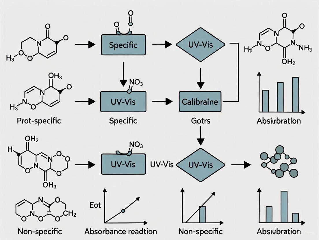
Diagram 1: Method selection framework for protein quantification approaches.
Research Reagent Solutions and Materials
Successful protein quantification requires appropriate selection of reagents and materials tailored to the chosen method. The following table outlines essential solutions and their functions in protein quantification workflows.
Table 3: Essential Research Reagents and Materials for Protein Quantification
| Reagent/Material | Function | Method Applicability | Key Considerations |
|---|---|---|---|
| Quartz Cuvettes | Sample holder for UV measurements | A280 | UV-transparent; required for wavelengths <300 nm [1] |
| BCA Working Reagent | Color development through copper chelation | BCA | Fresh preparation recommended; composition affects sensitivity [13] |
| Coomassie Brilliant Blue G-250 | Protein-binding dye | Bradford | Distinct from R250 used in gel staining [15] |
| Protein Standards (BSA) | Calibration reference | All quantitative methods | Match protein type to unknowns when possible [13] |
| Microplate Readers | Absorbance/fluorescence measurement | BCA, Bradford, ELISA | Enable high-throughput analysis [13] |
| Variable Pathlength Systems | Automated pathlength adjustment | A280 | Eliminate dilution requirements; enhance accuracy [16] |
The distinction between specific and non-specific protein quantification methods represents a fundamental consideration in experimental design across biological research and biopharmaceutical development. Specific methods like A280 absorbance and ELISA provide targeted measurement of particular proteins, leveraging unique structural features or antibody recognition for selective quantification. These approaches are indispensable when the concentration of a specific protein, rather than total protein content, is physiologically or functionally relevant.
Non-specific methods including Bradford, BCA, and Lowry assays offer practical solutions for total protein quantification, responding to general protein characteristics like peptide bonds or particular amino acid side chains. While these methods provide valuable information about overall protein content, their variable response to different protein compositions necessitates careful interpretation and appropriate standard selection.
Method selection should be guided by the specific research question, sample characteristics, and required performance parameters. For purified protein analysis, A280 quantification offers rapid, non-destructive measurement with minimal sample consumption. In complex mixtures requiring target-specific measurement, immunoassays provide the necessary specificity despite greater complexity and cost. For general laboratory applications where total protein content is sufficient, colorimetric assays like BCA and Bradford balance practicality with adequate performance for most applications. Understanding the principles, capabilities, and limitations of each method enables researchers to select the optimal approach for their specific protein quantification needs.
The Critical Role of Aromatic Amino Acids and Protein Composition
The accurate determination of protein concentration is a fundamental requirement in biochemical, pharmaceutical, and biomedical research. However, the diverse physicochemical properties of proteins and the complexity of biological samples present significant analytical challenges. Protein quantification methods can be broadly categorized into specific techniques that rely on the unique aromatic amino acid composition of proteins and nonspecific techniques that measure general protein properties or reactions. Specific methods, such as UV absorbance at 280 nm (A280) and aromatic amino acid analysis (AAAA), exploit the characteristic UV absorption of tryptophan, tyrosine, and phenylalanine residues. These methods provide absolute quantification but require knowledge of the protein's aromatic amino acid composition. In contrast, nonspecific methods like colorimetric assays (Bradford, BCA) measure protein content through dye-binding or chemical reduction reactions, which are highly dependent on the amino acid composition of standard proteins used for calibration and are susceptible to interference from complex matrices [17] [18].
The composition of aromatic amino acids in a protein directly influences the selection and performance of quantification methods. Proteins with challenging properties, chemical modifications, or those in complex matrices like air particulate matter and pollen extracts further complicate accurate quantification [17]. This guide objectively compares the performance of specific and nonspecific protein quantification methods, providing experimental data and protocols to inform method selection for research and development applications.
Comparative Performance of Protein Quantification Methods
Quantitative Comparison of Method Performance
Table 1: Performance characteristics of different protein quantification methods
| Method | Principle | Detection Range | Precision & Recovery | Matrix Effects | Sample Requirements |
|---|---|---|---|---|---|
| A280 (Direct UV) | Specific absorption of aromatic amino acids [17] | Varies with aromatic content; extended range with variable pathlength (0.005-15 mm) [16] | High accuracy when extinction coefficient is known; ~2% instrumental error [16] | Susceptible to interfering UV-absorbing compounds [17] | 10-300 μL (for variable pathlength) [16] |
| Aromatic Amino Acid Analysis (AAAA) | Quantification of Phe and Tyr after hydrolysis [17] [19] | Wide range after hydrolysis; LOD improved with fluorescence detection [17] | 97% recovery for Phe and Tyr; 98% ± 2% (Phe) and 88% ± 4% (Tyr) for BSA CRM [19] | Robust to complex matrices; hydrolysis required [17] | Requires hydrolysis (1h with 8M HBr at 150°C) [19] |
| Bradford Assay | Nonspecific dye binding to basic and aromatic residues [17] [20] | Limited linear range; superior to A280 but surpassed by TCE fluorescence [20] | Protein-dependent response; affected by Arg, Lys, His, Trp, Tyr, Phe content [17] | Highly susceptible to interference from detergents and chemicals [17] | Small volumes (2-5 μL typically used) [20] |
| BCA Assay | Nonspecific copper reduction by proteins [17] | Standard range | Protein-dependent response; affected by Cys, Cystine, Met, Tyr, Trp [17] | Susceptible to interfering reducing agents [17] | Standard volumes |
| TCE Fluorescence | Specific UV-induced modification of Trp and Tyr residues [20] | 10.5-200 μg (100μL assay); 8.7-100 μg (20μL assay) [20] | Superior sensitivity vs. A280; extends beyond Bradford linear range [20] | Specific to Trp/Tyr content; minimal chemical interference [20] | 10-100 μL; compatible with downstream SDS-PAGE [20] |
| qNMR of Aromatic AAs | Quantitative NMR of Phe and Tyr after hydrolysis [19] | Limited by NMR sensitivity | 97% recovery for His, Phe, Tyr at ~1 mM [19] | Minimal interference due to clean aromatic region [19] | Requires hydrolysis and specialized NMR equipment |
Advanced and Emerging Techniques
Table 2: Advanced methodologies for protein quantification
| Technique | Principle | Throughput & Applications | Advantages | Limitations |
|---|---|---|---|---|
| Label-Free LC-MS | LC separation with MS detection of tryptic peptides [21] [22] | High-throughput; discovery proteomics [21] | No isotope labeling required; unlimited sample number [21] | Complex data analysis; requires advanced instrumentation [22] |
| LC-UV (220 nm) | Peptide bond absorption at 220 nm [17] | Medium-throughput; complex samples | Eliminates many interfering substances via separation [17] | Many substances absorb at 220 nm; protein-to-protein variability [17] |
| Raman Spectroscopy | Inelastic scattering providing molecular fingerprints [23] | Real-time bioprocess monitoring | Non-destructive; rich molecular information [23] | Cost and technical implementation hurdles [23] |
| Variable Pathlength UV | Slope spectroscopy using multiple pathlengths [16] | High-throughput biopharmaceutical processing | Eliminates dilution requirements; rapid results (minutes) [16] | Specialized equipment required |
Experimental Protocols for Key Methodologies
2,2,2-Trichloroethanol (TCE) Fluorescence Assay
The TCE fluorescence method represents a specific quantification technique that exploits the photochemical modification of aromatic amino acids. The protocol below enables highly sensitive protein quantification with visualization capabilities [20].
Reagents and Solutions:
- 2,2,2-trichloroethanol (TCE) stock solution: 0.56% (v/v) TCE in phosphate-buffered saline (PBS) for standard assay
- TCE Ultra Reagent: 5% (v/v) TCE in PBS for low-volume assay
- Protein standards: Bovine Serum Albumin (BSA) in PBS (0-20 μg/μL)
- Phosphate-buffered saline (PBS): 137 mM NaCl, 2.7 mM KCl, 10 mM Na₂HPO₄, 1.8 mM KH₂PO₄
Standard Assay Protocol (100 μL):
- Combine 10 μL of protein sample with 90 μL of TCE reagent (0.56% v/v final concentration)
- Incubate mixture under a 15 W UV lamp for 15 minutes
- Measure fluorescence with excitation at 310 nm and emission at 450 nm
- Generate standard curve using BSA standards (0-20 μg/μL)
Low-Volume Assay Protocol (20 μL):
- Combine 10 μL of protein sample with 10 μL of TCE Ultra Reagent (5% v/v final concentration)
- Incubate under UV light for 0-15 minutes while monitoring fluorescence
- Measure fluorescence (excitation 310 nm, emission 450 nm)
- Use remaining sample for SDS-PAGE by dilution with 2X Laemmli Sample Buffer
Key Optimization Parameters:
- Optimal TCE concentration: 0.5% (v/v) final assay concentration
- Optimal UV-exposure time: 15 minutes
- Linear range: 10.5-200 μg (100 μL assay); 8.7-100 μg (20 μL assay)
- Post-assay visualization: Direct fluorescence visualization on UV transilluminator after SDS-PAGE
Aromatic Amino Acid Analysis (AAAA) with Fluorescence Detection
AAAA represents a gold-standard specific method that quantifies proteins based on their phenylalanine and tyrosine content after acid hydrolysis, providing robust quantification even for complex samples [17] [19].
Reagents and Equipment:
- Hydrolysis acid: 8 M hydrobromic acid (HBr) or 6 M hydrochloric acid (HCl)
- Reducing agent: Cysteine hydrochloride
- Internal standards: Terephthalic acid or sulfoisophthalic acid
- Mobile phase: Reversed-phase HPLC solvents
- HPLC system with fluorescence detection
Hydrolysis Protocol:
- Transfer protein sample to hydrolysis vial
- Add 8 M HBr containing 0.1% phenol as a protective agent
- Seal vial under vacuum or nitrogen atmosphere
- Heat at 150°C for 1 hour (HBr) or 6 M HCl at 107°C for 24 hours
- Cool and evaporate acid under vacuum
- Reconstitute hydrolysate in appropriate buffer for analysis
Liquid Chromatography with Fluorescence Detection:
- Column: Reversed-phase C18 column (e.g., 100 × 4.6 mm, 2.7 μm)
- Mobile phase A: 10 mM sodium phosphate, pH 7.0
- Mobile phase B: Methanol
- Gradient: 0-60% B over 5 minutes
- Fluorescence detection: Excitation 210 nm, Emission 305 nm
- Quantification: Peak areas of Phe and Tyr compared to internal standard
Quantification Calculations:
- Protein concentration = (moles Phe + moles Tyr) × MW protein / number of aromatic residues per protein
- Recovery: Typically 97% for standard amino acids; 98% ± 2% for Phe and 88% ± 4% for Tyr in BSA certified reference material
Variable Pathlength UV Spectroscopy (Slope Spectroscopy)
This advanced specific method enables accurate A280 measurements without sample dilution by leveraging variable pathlength technology, particularly valuable for concentrated biopharmaceutical products [16].
Instrumentation and Reagents:
- Solo VPE system or equivalent variable pathlength spectrophotometer
- Quartz sample cups (large, small, micro sizes)
- Protein sample without dilution
- Reference buffer matching sample composition
Protocol:
- Place sample in appropriate cup based on expected concentration
- Instrument automatically selects optimal pathlength range (0.005-15 mm)
- System collects 5-10 absorbance measurements at different pathlengths
- Software plots linear regression of absorbance versus pathlength
- Slope value (m) is used to calculate concentration: c = m/α, where α is molar absorption coefficient
Key Advantages:
- Eliminates dilution-related errors (~2% instrumental error only)
- Rapid analysis (<1 minute per sample)
- Handles concentrated samples (up to 300 mg/mL)
- No baseline correction required when buffer absorbance is pathlength-independent
Visualizing Protein Quantification Methodologies
Specific vs. Nonspecific Quantification Pathways
TCE Fluorescence Assay Workflow
Aromatic Amino Acid Analysis (AAAA) Workflow
Essential Research Reagent Solutions
Table 3: Key reagents and materials for protein quantification experiments
| Reagent/Material | Function & Application | Key Considerations |
|---|---|---|
| 2,2,2-Trichloroethanol (TCE) | Fluorescent labeling of tryptophan and tyrosine residues [20] | UV-induced covalent modification; enables sensitive detection (8.7-200 μg range) [20] |
| Bovine Serum Albumin (BSA) | Standard reference protein for calibration curves [17] [20] | Widely available; consistent aromatic amino acid composition; used in AAAA validation [19] |
| Coomassie Blue G-250 | Dye for Bradford colorimetric assay [17] [20] | Binds basic (Arg, Lys) and aromatic amino acids; susceptible to detergent interference [17] |
| Bicinchoninic Acid (BCA) | Copper reduction detection in BCA assay [17] | Detects Cu+ ions produced by protein reduction; affected by Cys, Tyr, Trp content [17] |
| Hydrobromic Acid (HBr) | Protein hydrolysis for AAAA [19] | Enables rapid hydrolysis (1h at 150°C); preserves aromatic amino acids [19] |
| Deuterated Solvents | NMR analysis for qNMR quantification [19] | Enables quantitative NMR of aromatic amino acids after hydrolysis [19] |
| Internal Standards (e.g., terephthalic acid) | qNMR quantification reference [19] | Provides compound-independent calibration for absolute quantification [19] |
The selection between specific and nonspecific protein quantification methods represents a critical decision point in experimental design, with significant implications for data accuracy and reliability. Specific methods leveraging aromatic amino acid composition—particularly AAAA with fluorescence detection, TCE fluorescence, and advanced A280 with variable pathlength technology—provide superior accuracy and robustness for complex samples. These methods enable absolute quantification traceable to fundamental chemical properties, with AAAA demonstrating remarkable resilience to matrix effects in challenging environmental and biological samples [17].
Nonspecific methods including colorimetric assays offer practical convenience and sensitivity for controlled applications with known protein composition but demonstrate significant vulnerability to matrix interference and protein-to-protein variability. The experimental data presented in this comparison underscores the necessity of method matching to specific application requirements, with aromatic amino acid-based methods providing the foundation for reliable quantification in drug development and complex biomedical research.
For researchers navigating the critical role of aromatic amino acids and protein composition in quantification assays, the emerging methodologies of TCE fluorescence and variable pathlength spectroscopy present particularly valuable tools that balance sensitivity, specificity, and practical implementation for 21st-century protein science.
Accurate protein quantification is foundational to biomedical research and drug development. However, the choice between specific and non-specific ultraviolet-visible (UV-Vis) spectroscopy methods is critically influenced by common laboratory substances that can interfere with measurements, potentially compromising data reliability. This guide objectively compares the performance of various protein quantification assays in the presence of nucleic acids, detergents, and buffer components, providing a framework for selecting the optimal method.
Mechanisms of Interference in UV-Vis-Based Protein Assays
Protein quantification assays function on distinct chemical principles, making them differentially susceptible to interference. Understanding these mechanisms is the first step in mitigating analytical error.
Direct UV Absorbance at 280 nm: This method relies on the innate absorbance of aromatic amino acids (tryptophan and tyrosine) in the protein backbone [24] [25]. Its primary weakness is a lack of specificity; any substance that absorbs light around 280 nm will cause a positive interference, leading to overestimation of protein concentration. Key interferents include nucleic acids (which absorb strongly at 260 nm, with significant scatter into the 280 nm range) and specific buffer components like Tris-hydrochloride and reducing agents (e.g., DTT), whose oxidized forms also absorb at this wavelength [24] [25].
Colorimetric Assays (Bradford, BCA): These methods use a color change reaction that is subsequently measured by UV-Vis spectroscopy.
- The Bradford Assay depends on the binding of Coomassie dye primarily to basic (arginine) and aromatic residues in proteins [24]. The formation of this protein-dye complex can be disrupted by ionic and non-ionic detergents at certain concentrations, which may cause the dye to precipitate [24] [25]. The assay also exhibits significant protein-to-protein variation due to its dependence on amino acid composition.
- The Bicinchoninic Acid (BCA) Assay involves a two-step process: the reduction of Cu²⁺ to Cu⁺ by the protein backbone (biuret reaction), followed by the chelation of Cu⁺ by BCA to form a purple-colored complex [25]. While more tolerant of detergents than the Bradford assay, the BCA method is susceptible to interference from any substance that also reduces copper, including reducing agents like DTT and β-mercaptoethanol [25]. Chelating agents such as EDTA can also interfere by binding the copper ions required for the reaction.
The relationships between these core mechanisms and their primary interferents are visualized below.
Quantitative Comparison of Interference Effects
The following tables synthesize experimental data on the susceptibility of major protein quantification methods to specific interfering substances, providing a clear basis for comparison.
Table 1: Susceptibility of Protein Assays to Common Interferings Substances
| Interfering Substance | A280 (Direct UV) | Bradford Assay | BCA Assay | SLS-Hb (Hb-Specific) |
|---|---|---|---|---|
| Nucleic Acids | High Interference (Absorb at ~260 nm) [24] [25] | Generally Compatible [24] | Generally Compatible [24] | Specific for hemoglobin, avoids interference [6] |
| Detergents | Varies by type and concentration | High Interference (e.g., SDS causes precipitation) [24] [25] | Low Interference (Tolerant of many detergents) [25] | Tolerant due to SLS detergent base [6] |
| Reducing Agents (DTT, BME) | High Interference (Oxidized forms absorb at 280 nm) [25] | Low Interference (Stable in reducing conditions) [25] | High Interference (Reduce Cu²⁺ independently) [25] | Not specifically reported |
| Chaotropic Salts | Can affect baseline absorbance | Can interfere with dye binding | Can interfere with copper reduction | Used in the method itself (enhances specificity) [6] |
| Chelators (EDTA) | Generally Compatible | Generally Compatible | High Interference (Chelates copper ions) [25] | Generally Compatible |
Table 2: Key Performance Characteristics of Quantification Methods
| Method | Chemical Basis | Key Advantage | Key Disadvantage | Optimal Use Case |
|---|---|---|---|---|
| A280 (Direct UV) | Aromatic amino acids [25] | Quick; no special reagents [25] | Non-specific; many interferents [25] | Pure protein solutions, no contaminants |
| Bradford Assay | Shift upon binding basic/aromatic residues [24] [25] | Fast, stable, compatible with reducing agents [25] | Protein-to-protein variation; interfered by detergents [24] [25] | Quick screens of non-denatured proteins |
| BCA Assay | Cu²⁺ reduction by protein backbone [25] | Tolerant of detergents; more uniform protein response [25] | Slow; interfered by reducing agents and chelators [25] | Samples containing detergents |
| SLS-Hb | Hb-specific in SLS buffer [6] | High specificity for hemoglobin; safe and cost-effective [6] | Applicable only to hemoglobin-based samples [6] | Characterization of hemoglobin-based oxygen carriers (HBOCs) |
Detailed Experimental Protocols for Key Assays
To ensure reproducibility and highlight best practices for mitigating interference, detailed protocols for three core assays are provided below.
Bicinchoninic Acid (BCA) Assay Protocol
The BCA assay is known for its tolerance to detergents, making it suitable for many sample types, though it is sensitive to reducing agents [25].
- Standard Curve Preparation: Prepare a series of dilutions from a standard protein stock (e.g., Bovine Serum Albumin) in the same buffer as your unknown samples to create a concentration series covering 0–1.5 mg/mL [6].
- Sample Preparation: Dilute unknown protein samples to an estimated concentration that falls within the standard curve's range. Use triplicates for accuracy [6].
- Working Reagent Preparation: Combine BCA Reagent A and BCA Reagent B in a 50:1 ratio to create the BCA working reagent [6].
- Reaction:
- Absorbance Measurement: Using a plate reader, measure the absorbance of each well at 562 nm [6].
- Data Analysis: Generate a standard curve by plotting the average absorbance of the standards against their known concentrations. Use the linear equation from this curve to calculate the concentration of the unknown samples.
Bradford Assay Protocol
The Bradford assay is prized for its speed and compatibility with reducing agents, but is incompatible with many detergents [25].
- Standard Curve Preparation: Prepare standard protein dilutions (e.g., BSA or Bovine γ-Globulin) in a compatible buffer. A range of 0–1 mg/mL is typical [6].
- Sample Preparation: Dilute unknown samples appropriately. Use triplicates.
- Reagent Preparation: Allow the Coomassie Plus reagent to equilibrate to room temperature for at least 30 minutes before use [6].
- Reaction:
- Pipette 10 µL of each standard and unknown into a 96-well plate.
- Add 300 µL of Coomassie Plus reagent to each well.
- Cover the plate, mix, and incubate at room temperature for 10 minutes [6].
- Absorbance Measurement: Measure the absorbance at 595 nm using a plate reader [6].
- Data Analysis: Create a standard curve from the absorbance of the standards and calculate the unknown concentrations. Note that the standard curve may be non-linear at higher concentrations.
Direct UV Absorbance at 280 nm Protocol
This method is best reserved for purified protein samples where contaminating interferents are absent [25].
- Blank Measurement: Using a UV-Vis spectrophotometer, blank with the appropriate buffer solution that the protein is dissolved in. This is critical for accurate background subtraction [25].
- Spectrum Measurement:
- For microvolume systems (e.g., NanoDrop), place a 1-2 µL droplet of the purified protein sample on the pedestal and obtain a full UV-Vis spectrum, typically from 220 to 320 nm [26].
- For cuvette-based systems, load the sample into a quartz cuvette and measure.
- Concentration Calculation: The instrument software will typically calculate concentration based on the absorbance at 280 nm (A280), the pathlength, and the input extinction coefficient. The extinction coefficient is protein-specific and must be known beforehand [25]. A scan of the spectrum allows for assessment of purity; a pure protein sample typically shows a peak at ~280 nm, while nucleic acid contamination is indicated by a elevated peak at ~260 nm.
The workflow for selecting and executing an appropriate quantification assay, from sample preparation to data interpretation, is summarized below.
The Scientist's Toolkit: Essential Research Reagents & Materials
Successful and reproducible protein quantification requires more than just a protocol. The following table details key reagents and materials, along with their critical functions and considerations.
Table 3: Essential Reagents and Materials for Protein Quantification
| Item | Function/Role in Quantification | Key Considerations |
|---|---|---|
| Standard Protein (BSA or BGG) | Creates a standard curve for colorimetric assays to interpolate unknown sample concentrations. | Protein-to-protein variation in colorimetric assays means the standard should be matched to the sample protein if possible [25]. |
| Coomassie Brilliant Blue G-250 | The active dye in the Bradford assay; binds proteins causing a spectral shift [24]. | Susceptible to precipitation in the presence of detergents; modified commercial kits offer better compatibility [24]. |
| Bicinchoninic Acid (BCA) & Cu²⁺ | The key reagents in the BCA assay. Cu²⁺ is reduced by proteins, and BCA chelates the resulting Cu⁺ [25]. | The assay is sensitive to chelators (e.g., EDTA) which bind Cu²⁺, and reducing agents which reduce Cu²⁺ independently [25]. |
| Sodium Lauryl Sulfate (SLS) | A detergent used in Hb-specific methods to create a uniform, stable complex for accurate spectrophotometric measurement [6]. | Its use in a dedicated kit enhances specificity and tolerability for hemoglobin-based samples [6]. |
| Chaotropic Salts (e.g., Guanidine HCl) | Disrupt cell structures, inactivate nucleases, and facilitate binding of nucleic acids to silica in purification kits [27]. | Essential for nucleic acid removal prior to A280 measurement to prevent interference [27]. |
| Microplate Reader | Instrument for high-throughput absorbance measurement in 96-well plate formats for BCA and Bradford assays [6]. | Enables rapid, multiplexed analysis of many samples and replicates, improving efficiency and statistical power. |
| UV-Vis Spectrophotometer | Instrument for measuring absorbance of samples at specific wavelengths (e.g., 280 nm, 562 nm, 595 nm) [28]. | Modern systems (e.g., NanoDrop) require only 1-2 µL of sample, conserving valuable material [26] [28]. |
In conclusion, the pervasive challenge of interference from nucleic acids, detergents, and buffers in protein quantification demands a strategic and informed approach. The data and protocols presented herein demonstrate that there is no universal "best" method, but rather an optimal choice dictated by sample composition and analytical requirements. For non-specific total protein analysis, the BCA assay offers robust performance in the presence of detergents, while the Bradford assay is preferable with reducing agents. For purified systems, direct A280 measurement provides a rapid solution. Crucially, in the development of complex biologics such as HBOCs, specific methods like SLS-Hb are indispensable for obtaining accurate and reliable quantitative data, ensuring both product efficacy and safety.
In both biomedical research and drug development, the accurate quantification of protein concentration is not merely a preliminary step but a foundational one. The reliability of virtually all subsequent data, from enzymatic studies to the validation of drug targets, hinges on the precision of this initial measurement [10]. The choice between specific and non-specific quantification methods carries profound implications for research accuracy, experimental reproducibility, and ultimately, the success or failure of therapeutic development programs [29].
This guide provides an objective comparison of mainstream protein quantification techniques, framing them within the context of this specific versus non-specific dichotomy. It synthesizes experimental data to illustrate how method selection influences outcomes in critical areas like the characterization of Hemoglobin-Based Oxygen Carriers (HBOCs) and the development of biopharmaceuticals, offering detailed protocols to support rigorous laboratory practice.
Classification of Protein Quantification Methods
Protein quantification methods can be fundamentally categorized based on what they measure. Non-specific methods determine total protein content by reacting with general chemical motifs, such as peptide bonds or specific amino acid side chains common to many proteins. In contrast, specific methods rely on unique, high-affinity interactions, such as antibody-antigen binding, to quantify a particular protein of interest within a complex mixture [29].
The diagram below illustrates the logical decision pathway for selecting an appropriate quantification method based on key experimental parameters.
Comparative Analysis of Key Protein Quantification Methods
Non-Specific Methods: Principles and Trade-offs
Non-specific methods are prized for their speed, low cost, and general applicability for measuring total protein content, but they are susceptible to interference from common buffer components [10] [25].
Ultraviolet-Visible (UV-Vis) Spectroscopy (Absorbance at 280 nm)
- Principle: This technique leverages the intrinsic absorbance of ultraviolet light (at 280 nm) by the aromatic amino acids tryptophan and tyrosine in a protein's sequence. The concentration is calculated using the Beer-Lambert law (A = εcl) [1] [30].
- Experimental Protocol:
- Blank the spectrophotometer with the protein's buffer solution.
- Measure the absorbance of the purified protein solution at 280 nm.
- Calculate concentration using the formula: c = A / (ε × l), where
cis concentration,Ais absorbance,εis the protein's molar extinction coefficient, andlis the path length of the cuvette [30].
Bicinchoninic Acid (BCA) Assay
- Principle: A two-step colorimetric reaction. First, proteins reduce Cu²⁺ to Cu⁺ under alkaline conditions (biuret reaction). Second, BCA chelates the Cu⁺ ions to form a purple complex with strong absorbance at 562 nm [10] [31].
- Experimental Protocol:
- Prepare a series of standard protein solutions (e.g., BSA) of known concentration.
- Mix the BCA working reagent (50 parts Reagent A with 1 part Reagent B) with standards and unknown samples in a microplate [29].
- Incubate at 37°C for 30 minutes [10] [29].
- Measure absorbance at 562 nm and determine sample concentration from the standard curve [10].
Bradford Assay
- Principle: This is a single-step assay where the Coomassie Brilliant Blue G-250 dye binds primarily to basic (arginine, lysine) and aromatic amino acids in proteins. This binding causes a shift in the dye's absorbance maximum from 470 nm (red) to 595 nm (blue) [10] [25].
- Experimental Protocol:
Specific Methods: The Power of Immunological Recognition
Enzyme-Linked Immunosorbent Assay (ELISA)
- Principle: ELISA uses the high specificity of antibodies to capture and detect a target protein. In a common "sandwich" format, a capture antibody is immobilized on a plate. The sample is added, and the target protein binds. A second, enzyme-conjugated detection antibody is then added. After adding a substrate, the resulting color change, proportional to the target protein concentration, is measured [10].
- Experimental Protocol:
- Coat a microplate with a capture antibody specific to the target protein and block to prevent non-specific binding.
- Add samples and standards, incubate, and wash.
- Add an enzyme-linked detection antibody, incubate, and wash.
- Add a substrate solution and measure the resulting absorbance. Concentration is determined from a standard curve [10].
The following table synthesizes experimental data from the literature, providing a direct comparison of the key performance characteristics of each method [10] [29] [31].
Table 1: Comparative Performance of Common Protein Quantification Assays
| Method | Principle | Dynamic Range | Sensitivity | Key Interfering Substances | Assay Time |
|---|---|---|---|---|---|
| UV-Vis (A280) | Aromatic amino acid absorbance [1] | ~0.1 - 1 mg/mL (limited) [10] | Low (< 1 µg/mL problematic) [10] | Nucleic acids, detergents, phenols [10] [25] | ~5 minutes [10] |
| BCA Assay | Cu²⁺ reduction by protein/BCA chelation [10] | 20 - 2000 µg/mL [10] | Moderate (20 µg/mL) [10] | Reducing agents (DTT, β-Me), metal chelators (EDTA) [10] [31] | ~45 minutes [31] |
| Bradford Assay | Coomassie dye binding to basic/aromatic residues [10] | Varies with standard | Moderate | Detergents (SDS, Triton), alkaline buffers [10] [25] | < 10 minutes [10] |
| ELISA | Antibody-antigen specificity [10] | Varies with target | High (pg/mL range) [10] | Minimal due to antibody specificity [10] | Several hours [10] |
Table 2: Suitability for Different Sample Types and Workflows
| Method | Best For | Compatible With Detergents? | Sample Consumption | Cost |
|---|---|---|---|---|
| UV-Vis (A280) | Purified proteins, quick checks [1] | Low compatibility [10] | Very low (1-2 µL) [1] | Low (no reagents) [1] |
| BCA Assay | Samples with detergents, general lab use [10] [25] | Good compatibility [10] [25] | Moderate (25 µL in microplate) [29] | Low to Moderate [31] |
| Bradford Assay | Quick, high-throughput screens without detergents [10] | Poor compatibility [10] [25] | Low (10 µL in microplate) [29] | Low [31] |
| ELISA | Quantifying specific proteins in complex mixtures (serum, lysate) [10] | High compatibility [10] | Moderate | High (antibodies, reagents) [10] |
Case Study: Method Selection in Hemoglobin-Based Oxygen Carrier (HBOC) Development
The critical impact of method choice is powerfully illustrated in the development of HBOCs, where accurately measuring hemoglobin (Hb) content, encapsulation efficiency, and free Hb is essential for both efficacy and safety [29].
A 2024 comparative study evaluated UV-Vis-based methods for quantifying Hb extracted from bovine red blood cells. The research compared non-specific methods (BCA, Bradford, Abs280) with Hb-specific methods (Cyanmethemoglobin, SLS-Hb) [29].
- Key Experimental Findings:
- The study identified the SLS-Hb method as the preferred choice due to its specificity, ease of use, cost-effectiveness, and safety compared to cyanmethemoglobin-based methods [29].
- It highlighted a crucial pitfall: using a non-specific method like BCA or Bradford for HBOC characterization without confirming the absence of other proteins (e.g., from the RBC membrane) can lead to significant inaccuracies in calculating encapsulation efficiency and yield [29].
- An overestimation of free Hb could halt the development of a viable product, while an underestimation could allow a toxic product with high levels of free Hb to advance, posing serious clinical risks [29].
This case underscores the necessity of aligning the method's specificity with the scientific question, especially in a regulated development environment.
The Scientist's Toolkit: Essential Research Reagent Solutions
Successful protein quantification requires more than just a protocol. The following table lists key reagents and materials, along with their critical functions in ensuring accurate results.
Table 3: Essential Reagents and Materials for Protein Quantification
| Item | Function & Importance |
|---|---|
| Standard Protein (e.g., BSA, IgG) | Serves as the reference for generating a calibration curve. The choice of standard should closely match the composition of the sample protein for non-specific assays [10] [25]. |
| High-Purity Buffers & Water | Used for diluting standards and samples. Impurities can absorb light at critical wavelengths, leading to high background and inaccurate readings [1]. |
| Compatible Microplates & Cuvettes | Sample holders must be appropriate for the wavelength. Quartz is required for UV measurements, while specialized plastic is used for visible range assays [30]. |
| Detector-Compatible Reagents (BCA, Dye) | The core chemistries of the assays (e.g., BCA working reagent, Coomassie dye) must be formulated for use with standard laboratory spectrophotometers and plate readers [10] [29]. |
| Wash & Blocking Buffers (for ELISA) | Critical for removing unbound material and preventing non-specific antibody binding, which reduces background noise and improves assay sensitivity and specificity [10]. |
The selection of a protein quantification method is a consequential decision that directly impacts data integrity in drug development and research. No single method is universally superior; the optimal choice is dictated by the experimental context.
- For purified proteins of known sequence and extinction coefficient, UV-Vis at 280 nm offers a rapid, non-destructive option [1].
- For general total protein estimation in complex buffers, the BCA assay often provides a robust balance of compatibility and sensitivity [10] [25].
- For high-throughput screening where speed is critical and detergents are absent, the Bradford assay is highly efficient [10].
- For quantifying a specific protein within a complex biological matrix like serum or cell lysate, ELISA is the definitive choice due to its unparalleled specificity and sensitivity [10].
A growing trend in the field is the use of orthogonal methods—using two different quantification principles to validate results, particularly for critical applications like lot release of biologics [1]. Furthermore, emerging technologies like Microfluidic Diffusional Sizing (MDS) and RF biosensors offer promising, calibration-free alternatives for determining both concentration and affinity simultaneously, potentially overcoming key limitations of traditional assays [10] [11]. By understanding the principles, advantages, and limitations of each technique, scientists can make informed choices that enhance the accuracy, reproducibility, and success of their research and development efforts.
Methodologies in Practice: Protocols for BCA, Bradford, Direct UV, and Emerging Techniques
Accurate protein quantification is a cornerstone of biological research and biopharmaceutical development, forming the critical link between sample processing and data interpretation. Within the context of specific versus non-specific protein quantification methods, the Bicinchoninic Acid (BCA) Assay occupies a crucial position as a robust, colorimetric method that balances practical convenience with reliable performance across diverse sample types. Unlike specific methods like ELISA that target unique protein epitopes through antibody recognition, the BCA assay belongs to the category of non-specific methods that measure total protein content based on general chemical properties common to most proteins [32]. This non-specific approach provides distinct advantages for preliminary protein screening, total protein concentration assessment in complex mixtures, and situations where antibody development for specific detection would be impractical or cost-prohibitive.
The fundamental strength of the BCA assay lies in its unique chemical mechanism that leverages the reducing power of peptide bonds and specific amino acid residues, creating a detection system that is both sensitive and remarkably tolerant to substances that typically interfere with other colorimetric methods, particularly detergents commonly used in protein extraction and solubilization [12] [33]. This detergent compatibility makes the BCA assay indispensable for workflows involving membrane proteins, cellular lysates, and other challenging sample types where complete protein solubilization requires disruptive agents that would compromise other quantification methods.
Mechanistic Principles of the BCA Assay
The BCA assay operates through a two-step, temperature-dependent chemical reaction that converts protein presence into a measurable colorimetric signal. Understanding this mechanism is essential for appreciating both the strengths and limitations of this method compared to alternative protein quantification approaches.
The Biuret Reduction Step
In the initial reaction phase, proteins act as reducing agents in an alkaline environment (pH 11.25), facilitating the reduction of cupric ions (Cu²⁺) to cuprous ions (Cu⁺). This process, known as the biuret reaction, primarily occurs through the coordination of copper ions with peptide bonds that form coordination complexes under alkaline conditions [12] [34]. The reduction efficiency depends on two key factors: the number of peptide bonds present and the concentration of specific amino acid residues including cysteine, tyrosine, and tryptophan, which possess particularly strong reducing capabilities in alkaline environments [35] [25]. This dual pathway for copper reduction contributes to the method's broader protein-to-protein consistency compared to assays like Bradford, which relies heavily on specific amino acid interactions.
BCA Chelation and Color Development
The second stage involves the highly specific chelation of the newly reduced cuprous ions (Cu⁺) by two molecules of bicinchoninic acid (BCA), forming a stable, purple-colored complex [33] [35]. This BCA-Cu⁺ complex exhibits strong light absorption at 562 nm, with the intensity of this purple color being directly proportional to the protein concentration in the sample [34]. The reaction is not a true endpoint assay, as color development continues slowly over time, but reaches sufficient stability following incubation to allow reproducible measurement across multiple samples [35].
The following diagram illustrates the complete BCA assay mechanism:
Figure 1: BCA Assay Reaction Mechanism. The two-step process shows protein-mediated copper reduction followed by BCA-chelate formation resulting in measurable color development.
Experimental Protocol and Methodology
The BCA assay protocol follows a systematic approach to ensure accurate and reproducible protein quantification. The following section outlines the standard procedure adapted for microplate format, which enables high-throughput processing of multiple samples simultaneously [35].
Reagent and Standard Preparation
Working Reagent Preparation: The BCA working reagent is prepared immediately before use by combining Reagent A (containing sodium carbonate, sodium bicarbonate, bicinchoninic acid, and sodium tartrate in an alkaline medium) with Reagent B (containing copper sulfate) at a 50:1 ratio (50 mL Reagent A + 1 mL Reagent B) [35]. The mixed reagent is stable for approximately one week when stored properly.
Protein Standard Preparation: Bovine Serum Albumin (BSA) serves as the reference protein for creating a standard curve. Prepare a dilution series covering the expected concentration range of unknown samples, typically from 25 μg/mL to 2000 μg/mL [35]. Precise standard preparation is critical for assay accuracy, as all unknown sample concentrations will be extrapolated from this standard curve.
Table 1: Example BSA Standard Preparation for Microplate BCA Assay
| Tube | Homogenization Buffer (μL) | BSA Stock (μL) | Final Concentration (μg/mL) |
|---|---|---|---|
| A | 0 | 100 stock | 2000 |
| B | 42 | 125 stock | 1500 |
| C | 110 | 110 stock | 1000 |
| D | 60 | 60 of B | 750 |
| E | 110 | 110 of C | 500 |
| F | 110 | 110 of E | 250 |
| G | 110 | 110 of F | 125 |
| H | 135 | 35 of G | 25 |
| I | 135 | 0 | 0 |
Sample Preparation and Assay Procedure
Sample Preparation: Unknown experimental samples typically require dilution (often 10-fold) in an appropriate buffer such as 1% homogenization buffer to fall within the detection range of the standard curve [35]. For tissues or cells, initial homogenization in detergent-containing buffer (e.g., 1% Triton X-100) followed by centrifugation is necessary to obtain clear supernatants for analysis.
Assay Procedure:
- Plate Setup: Transfer 25 μL of each standard and prepared unknown sample to appropriate wells of a 96-well microtiter plate, in duplicate or triplicate.
- Reagent Addition: Add 200 μL of pre-mixed BCA working reagent to each well containing standard or sample.
- Incubation: Cover the plate and incubate at 37°C for 30 minutes in the dark. The incubation time and temperature directly impact color development; consistent conditions are essential for reproducibility.
- Absorbance Measurement: Following incubation, check for and remove any bubbles that may interfere with reading. Measure the absorbance of each well at 562 nm using a plate reader [35].
Data Analysis and Calculation
Standard Curve Generation: Plot the average absorbance values for each BSA standard against their known concentrations. The relationship typically follows a near-linear pattern, particularly in the mid-range of concentrations, though a polynomial trend line may provide better fit across the entire range [35].
Concentration Calculation: Using the standard curve equation, calculate the protein concentration of unknown samples based on their measured absorbance values. Apply appropriate dilution factors to determine the original sample concentration, typically expressed as μg/μL or mg/mL.
The complete workflow is summarized below:
Figure 2: BCA Assay Workflow. The complete experimental procedure from reagent preparation to concentration calculation.
Compatibility with Detergents: Comparative Analysis
A defining characteristic of the BCA assay is its exceptional tolerance to detergents commonly used in protein extraction and solubilization, setting it apart from many other protein quantification methods. This compatibility makes it particularly valuable for research involving membrane proteins and cellular lysates where detergents are essential for protein solubility.
Mechanism of Detergent Tolerance
The BCA assay's detergent resistance stems from its two-step reaction mechanism. Unlike the Bradford assay, where detergents compete with the dye for binding sites on proteins, the BCA copper reduction reaction occurs effectively even in the presence of various detergents [32] [33]. The alkaline conditions of the assay (pH ~11.25) contribute to this tolerance by maintaining protein-detergeant complexes in solution without interfering with the copper reduction process. This compatibility extends to both ionic and non-ionic detergents, though at varying concentration thresholds.
Comparative Detergent Compatibility
When compared to alternative protein quantification methods, the BCA assay demonstrates superior performance in detergent-containing samples:
Table 2: Detergent Compatibility Across Protein Quantification Methods
| Method | Principle | Detergent Compatibility | Key Limitations with Detergents |
|---|---|---|---|
| BCA Assay | Copper reduction & BCA chelation | High tolerance to a wide range of detergents [33] | Sensitive to copper chelators (EDTA) and reducing agents |
| Bradford Assay | Coomassie dye binding to basic & aromatic residues | Low tolerance; detergents interfere with dye binding [32] [33] | Significant interference from ionic and non-ionic detergents |
| Lowry Assay | Copper reduction & Folin-Ciocalteu reaction | Moderate tolerance | Detergents can cause precipitate formation [12] |
| UV Absorbance (A280) | Aromatic amino acid absorption | Variable interference | Detergents may absorb at 280 nm, creating background interference |
| Fluorescence-Based | Fluorescent dye binding | Sensitive to detergents and high salt concentrations [12] | Quantum yield affected by detergent concentration |
This detergent compatibility is particularly crucial when working with transmembrane proteins, which require solubilization from lipid membranes for accurate quantification. Research has demonstrated that conventional methods like Bradford significantly underestimate membrane protein concentration due to limited dye accessibility to hydrophobic regions, whereas the BCA assay provides more reliable quantification in such challenging samples [32].
Comparative Performance with Alternative Methods
Understanding how the BCA assay performs relative to other protein quantification methods enables researchers to select the most appropriate technique for their specific application. The following comparative analysis highlights key performance metrics across multiple dimensions.
Quantitative Method Comparison
Direct comparison of protein quantification methods reveals distinct performance characteristics that guide method selection:
Table 3: Comprehensive Comparison of Protein Quantification Methods
| Assay Method | Detection Mechanism | Detection Range (μg/mL) | Sensitivity | Compatible with Detergents | Assay Time | Key Interfering Substances |
|---|---|---|---|---|---|---|
| BCA | Copper reduction & BCA chelation | 20-2000 [34] | Moderate (25-2000 μg/mL) [33] | Yes [33] | ~30-120 min [33] [35] | Reducing agents, copper chelators (EDTA) [12] [34] |
| Bradford | Coomassie dye binding | 1-100 [25] | High (1-20 μg/mL) [33] | No [32] [33] | 5-10 min [33] | Detergents, alkaline conditions [32] [12] |
| Lowry | Copper reduction & Folin-Ciocalteu | 1-100 [12] | High | Moderate | ~30-60 min | Detergents, potassium ions, reducing agents [12] |
| UV A280 | Aromatic amino acid absorption | 100-1000 | Low | Variable | Immediate | Nucleic acids, alcohols, buffer ions [34] [25] |
| Qubit | Fluorescent dye binding | 0.25-5 | Very High | No [7] | ~10-15 min | Detergents, high salt [12] |
Application-Specific Performance
The optimal choice of protein quantification method depends heavily on the specific research context and sample characteristics:
Membrane Protein Studies: For transmembrane proteins like Na,K-ATPase (NKA), the BCA assay demonstrates superior performance compared to Bradford, Lowry, and direct UV methods. Research shows conventional methods significantly overestimate concentrations of specific transmembrane proteins compared to ELISA, with variation in resulting data being consistently lower when reactions are prepared based on BCA-determined concentrations [32].
Venom Proteomics: Studies on snake venoms from different species (Viperids vs. Elapids) reveal method-dependent variation in protein quantification. While multiple methods produced similar results for Viperid venoms (Agkistrodon contortrix), significant method-dependent variation was observed for Elapid venoms (Naja ashei), highlighting how protein composition affects quantification accuracy across methods [7].
High-Throughput Applications: The BCA assay adapts well to microplate formats, enabling processing of numerous samples simultaneously. Although requiring longer incubation times than Bradford assays, its broader dynamic range and detergent compatibility make it preferable for automated high-throughput applications, particularly when using robotic liquid handling systems [33].
Essential Research Reagent Solutions
Successful implementation of the BCA assay requires specific reagents and materials optimized for protein quantification. The following table outlines essential components and their functions within the assay system.
Table 4: Essential Research Reagents for BCA Assay
| Reagent/Material | Function/Application | Specifications |
|---|---|---|
| BCA Assay Kit | Complete reagent system for protein quantification | Typically contains BCA Reagent A and Copper Reagent B [35] |
| Bovine Serum Albumin (BSA) | Protein standard for calibration curve | High purity, fatty acid-free recommended [35] |
| Microplate Reader | Absorbance measurement at 562 nm | Capable of reading 96-well or 384-well plates |
| Triton X-100 | Non-ionic detergent for membrane protein extraction | Used in homogenization buffers (typically 0.1-1%) [35] |
| SDS | Ionic detergent for protein solubilization | Compatible with BCA at low concentrations [35] |
| Protease Inhibitors | Prevent protein degradation during processing | Added to homogenization buffers for tissue samples [35] |
The BCA assay represents a robust, versatile solution for total protein quantification, particularly valuable in research scenarios requiring detergent compatibility for membrane protein studies or cellular lysate analysis. Its unique two-step mechanism, combining the biuret reaction with bicinchoninic acid chelation, provides a balanced approach with moderate sensitivity, broad dynamic range, and consistent performance across diverse protein types. While method selection should always consider specific research needs—opting for Bradford when speed is paramount with purified samples, or specialized assays like ELISA for specific protein targeting—the BCA assay remains the method of choice for challenging sample types containing detergents or exhibiting variable amino acid composition. Its established protocol, adaptability to high-throughput formats, and reliable performance in complex biological matrices ensure its continued relevance in both basic research and biopharmaceutical applications where accurate protein quantification forms the foundation for reproducible scientific discovery.
In the realm of protein analytics, researchers navigate a fundamental divide between specific and non-specific quantification methods. Specific methods, such as immunoassays, target unique protein epitopes but require specialized reagents for each protein of interest. In contrast, non-specific methods measure total protein concentration, serving as essential preliminary tools in virtually every molecular biology workflow. These non-specific techniques primarily operate on two principles: absorbance of ultraviolet light by aromatic amino acids (UV-Vis at 280 nm) and colorimetric reactions. Among colorimetric assays, the Bradford method stands out for its rapid dye-binding mechanism, while alternatives like the Bicinchoninic Acid (BCA) and Lowry assays rely on copper-ion reduction by proteins under alkaline conditions [36] [37]. Each method exhibits distinct responses to experimental variables, with surfactant compatibility representing a critical differentiator that profoundly impacts measurement accuracy. This guide objectively examines the Bradford assay's performance relative to alternatives, focusing on its operational principles, surfactant sensitivities, and optimal applications in research and drug development contexts.
Methodological Principles and Mechanisms
The Bradford Assay: A Protein-Dye Binding Mechanism
The Bradford assay, developed by Marion M. Bradford in 1976, utilizes the distinctive properties of Coomassie Brilliant Blue G-250 dye to determine protein concentration [38]. Under acidic conditions, the free dye exists predominantly in a cationic, red form with maximum absorbance at 465 nm. Upon binding to proteins, the dye undergoes a structural transition to a stable, anionic blue form that absorbs light at 595 nm [37] [38]. This binding event occurs through multiple intermolecular forces: the dye's sulfonic acid groups form ionic bonds with positively charged amino acid residues (primarily arginine, lysine, and histidine), while van der Waals forces and hydrophobic interactions stabilize the complex through the dye's aromatic backbone [38]. The resultant color shift from brownish-red to intense blue provides the quantitative basis for the assay, with the absorbance at 595 nm being directly proportional to protein concentration within the linear range of 1-200 μg/mL [38].
The following diagram illustrates the core mechanism of the Bradford assay and the primary sources of surfactant interference:
Alternative Protein Quantification Mechanisms
Alternative protein quantification methods operate on fundamentally different principles. The BCA assay involves a two-step process where peptide bonds and specific amino acids (cysteine, tyrosine, and tryptophan) reduce copper ions from Cu²⁺ to Cu⁺ under alkaline conditions, followed by chelation of Cu⁺ by bicinchoninic acid to form a purple complex with strong absorbance at 562 nm [37] [39]. The traditional Lowry method enhances the Biuret reaction (copper-protein complex formation) with Folin-Ciocalteu reagent, which reacts with reduced copper complexes to produce an intense blue color measurable between 650-750 nm [36]. In contrast, direct UV absorbance at 280 nm exploits the natural ultraviolet absorption of aromatic amino acids (tryptophan, tyrosine, and phenylalanine) without additional reagents, though this method suffers from variable extinction coefficients between different proteins and interference from nucleic acids [37] [40].
Comparative Performance Analysis
Quantitative Comparison of Protein Assay Methods
The following table summarizes the key technical parameters and surfactant compatibilities of major protein quantification methods:
Table 1: Comprehensive Comparison of Protein Quantification Methods
| Parameter | Bradford Assay | BCA Assay | Lowry Assay | UV Absorbance (A₂₈₀) |
|---|---|---|---|---|
| Principle | Coomassie dye binding to basic amino acids [38] | Copper reduction & BCA chelation [37] | Copper reduction & Folin-Ciocalteu reaction [36] | Aromatic amino acid absorbance [37] |
| Detection Range | 1-200 μg/mL [38] | 20-2000 μg/mL [37] | 5-150 μg/mL [36] | Varies by protein [40] |
| Absorbance Wavelength | 595 nm [38] | 562 nm [37] | 650-750 nm [36] | 280 nm [37] |
| Assay Time | <10 minutes [37] [38] | ~45 minutes [37] | Protocol-dependent [36] | Immediate [40] |
| Ionic Detergent Compatibility | Poor (e.g., SDS causes interference) [36] [38] | Compatible with most ionic and non-ionic detergents [36] [39] | Compatible with detergents [36] | Generally compatible |
| Non-Ionic Detergent Compatibility | Variable (Triton X-100 may enhance sensitivity) [41] | Compatible [36] [39] | Compatible [36] | Generally compatible |
| Reducing Agent Compatibility | Compatible (DTT, BME) [36] | Incompatible (interferes with copper reduction) [36] [39] | Incompatible (interferes with copper reduction) [36] | Compatible |
| Protein-to-Protein Variation | High (dependent on basic amino acid content) [42] | Moderate [39] | Moderate [36] | High (dependent on aromatic amino acid content) [42] |
Experimental Data on Surfactant Effects
Research specifically investigating detergent effects on the Bradford assay reveals complex interactions. Friedenauer et al. (1989) demonstrated that Triton X-100 at low concentrations (0.008% v/v) could increase assay sensitivity toward various proteins by an average of 33%, with enhancements ranging from +11% for myelin basic protein to +128% for aprotinin [41]. The study suggested that nonionic detergents might facilitate nonionic interactions of the dye with proteins having limited capacity for ionic binding. In contrast, sodium dodecyl sulfate (SDS) at 0.004% (w/v) decreased assay sensitivity by approximately 75%, likely due to disruption of the ionic binding between the dye and protein [41].
A 2020 comparative study analyzing snake venoms from Agkistrodon contortrix (Viperidae) and Naja ashei (Elapidae) highlighted how protein composition affects different assays. For A. contortrix venom, Bradford, BCA, and 2-D Quant kits produced similar concentration values, while each method yielded significantly different results for N. ashei venom, underscoring the impact of variable protein composition on assay accuracy [7]. This protein-dependent variability represents a fundamental limitation of dye-binding assays compared to copper-based methods that primarily respond to peptide bonds [36].
Experimental Protocols
Standard Bradford Assay Protocol
The following workflow details the optimized procedure for performing the Bradford protein assay in standard laboratory conditions:
Step-by-Step Procedure:
Reagent Preparation: Commercial Bradford reagent is typically ready-to-use. Alternatively, prepare Coomassie dye by dissolving 100 mg Coomassie Brilliant Blue G-250 in 50 mL 95% ethanol, adding 100 mL 85% phosphoric acid, and diluting to 1 L with distilled water [38]. Filter through Whatman No. 1 paper if necessary.
Protein Standards: Prepare bovine serum albumin (BSA) standards in the range of 0-1.0 mg/mL using the same buffer as your unknown samples. Typical standard concentrations include 0, 0.1, 0.2, 0.3, 0.4, 0.5, and 1.0 mg/mL [38].
Sample Preparation: Dilute unknown samples to fall within the assay's linear range (1-200 μg/mL). For concentrated samples, preliminary dilutions of 1:50 or 1:100 in PBS or assay buffer are appropriate [38]. Centrifuge samples to remove particulate matter.
Assay Setup: Pipette 20 μL of each standard and unknown sample into separate cuvettes or microplate wells. Add 1 mL of Bradford reagent to each, mixing thoroughly by gentle inversion or pipetting [38]. Include a blank containing only buffer and reagent.
Incubation: Allow samples to incubate at room temperature for 5-20 minutes. Color development is largely complete within 5 minutes, though some proteins may benefit from longer incubation [38]. Protect from light during incubation.
Absorbance Measurement: Measure absorbance at 595 nm using a spectrophotometer or microplate reader. Blank the instrument with the reagent blank before reading standards and unknowns [38].
Data Analysis: Generate a standard curve by plotting absorbance versus known standard concentrations. Determine unknown sample concentrations by interpolating from the linear regression equation of the standard curve (typically y = mx + c, where y is absorbance, x is concentration) [38].
Interference Testing Protocol
To evaluate surfactant effects on the Bradford assay, incorporate these modifications:
- Prepare standard curves containing identical surfactant concentrations as test samples
- For ionic detergents like SDS, test concentrations ranging from 0.001-0.1%
- For non-ionic detergents like Triton X-100, test concentrations from 0.001-0.1%
- Include surfactant-only controls to assess background absorbance
- Consider sample dialysis or precipitation for surfactant removal if interference is significant [40]
The Researcher's Toolkit: Essential Reagents and Equipment
Table 2: Essential Materials for Bradford Assay Implementation
| Category | Specific Items | Purpose & Specifications | Considerations |
|---|---|---|---|
| Core Reagents | Coomassie Brilliant Blue G-250 | Primary dye for protein detection | Distinct from R250 used in gel staining; ensure G250 for quantification [37] |
| Bovine Serum Albumin (BSA) | Protein standard for calibration curve | High purity (>98%), lack of enzymatic activity, consistent lot-to-lot response [38] | |
| Phosphoric Acid, Ethanol | Reagent components for dye preparation | Maintain acidic conditions for proper dye function [38] | |
| Buffers & Solutions | PBS or Tris-HCl Buffer | Sample dilution and standard preparation | Low ionic strength buffers minimize interference [38] |
| Compatible Detergents (if required) | Sample solubilization while minimizing interference | Non-ionic detergents like Triton X-100 at <0.01% may be tolerable [41] | |
| Equipment | Spectrophotometer/Plate Reader | Absorbance measurement at 595 nm | Capable of reading at 595 nm with <0.005 OD accuracy recommended [36] |
| Cuvettes or Microplates | Sample containment during measurement | Disposable plastic preferred to prevent dye adsorption to quartz [39] | |
| Precision Pipettes | Accurate liquid handling | Critical for reproducible standard curve preparation [38] | |
| Interference Management | Dialysis Membranes | Removal of small interfering substances | Effective for detergents, reducing agents <3 kDa [40] |
| Precipitation Kits (e.g., TCA) | Protein concentration and interference removal | Useful for incompatible surfactants at high concentrations [40] |
The Bradford assay remains a cornerstone technique in protein science, offering unparalleled speed and simplicity for total protein quantification. Its distinctive surfactant sensitivities—severe interference from ionic detergents like SDS but potential enhancement from non-ionic detergents like Triton X-100—dictate specific application boundaries [36] [41]. When working with samples containing reducing agents or requiring rapid analysis, the Bradford method provides distinct advantages over copper-based assays. However, in detergent-rich environments typical of membrane protein preparations, copper-based methods like BCA deliver superior reliability [36] [39].
The experimental evidence confirms that method selection must be guided by both sample composition and research objectives. For drug development professionals and researchers, maintaining multiple quantification platforms enables appropriate method matching to experimental conditions. The Bradford assay's particular sensitivity to basic amino acid composition further necessitates careful standard selection, with BSA serving as a general purpose standard but potentially introducing inaccuracies when analyzing proteins with markedly different amino acid profiles [42]. Through understanding these principles and limitations, researchers can strategically employ the Bradford assay within a comprehensive protein analytics workflow, leveraging its unique strengths while mitigating its vulnerabilities through methodological compensation or alternative technique selection.
- Core Principle: Utilizes intrinsic UV absorption by aromatic amino acids for rapid, non-destructive protein quantification.
- Primary Advantage: Exceptional speed and simplicity for purified protein samples.
- Key Limitation: Susceptible to significant interference from nucleic acids and other UV-absorbing contaminants.
- Method Classification: A non-specific method, ideal for quick assessments but less suited for complex biological mixtures.
In the landscape of protein quantification methodologies, techniques are broadly categorized into specific and non-specific methods. Specific methods, such as immunoassays or mass spectrometry, identify and quantify a target protein based on its unique structural or amino acid sequence. In contrast, non-specific methods, like Direct UV Absorbance at 280 nm (A280), measure the total protein content in a sample based on general chemical properties, without distinguishing between individual protein types [24].
The A280 method is a foundational technique in biochemical research and biopharmaceutical development. Its position as a rapid, non-destructive, and reagent-free assay makes it a first-choice tool for routine protein quantification during purification processes, formulation, and quality control, particularly when working with well-characterized, purified proteins [1] [43]. This guide objectively evaluates its performance against alternative methods, with a focus on the critical challenge of nucleic acid interference.
Principles and Mechanisms of A280 Quantification
The A280 method is predicated on the Beer-Lambert Law (A = ε * c * l), which establishes a linear relationship between absorbance (A) and protein concentration (c), given a known molar absorptivity (ε) and path length (l) [1] [43].
The primary chromophores responsible for absorbance at 280 nm are the aromatic amino acids tryptophan and tyrosine, with a minor contribution from phenylalanine [44] [1] [43]. The peptide bonds within the protein backbone absorb light at lower wavelengths (around 200-220 nm) but do not contribute significantly at 280 nm. Because the concentration of these aromatic residues varies between different proteins, each protein has a unique extinction coefficient (ε). This is a fundamental source of variability; a protein standard with a dissimilar aromatic amino acid composition to the analyte protein will yield inaccurate concentration estimates [45] [43].
Table: Chromophores in UV-Based Protein Quantification
| Chromophore | Absorption Maximum | Contribution to A280 | Notes |
|---|---|---|---|
| Tryptophan | ~280 nm | Strong | Primary contributor |
| Tyrosine | ~275 nm | Moderate | Significant contributor |
| Phenylalanine | ~257 nm | Weak | Minor contributor |
| Peptide Bonds | ~190-220 nm | None | Not a factor at 280 nm |
The Interference Problem: Nucleic Acids and Other Factors
The simplicity of the A280 method is counterbalanced by its susceptibility to interference from common sample contaminants.
Nucleic Acid Interference
Nucleic acids (DNA and RNA) represent the most significant interferent. They exhibit a strong absorption peak at or near 260 nm [46]. The proximity of this peak to the 280 nm measurement wavelength means that the presence of nucleic acids in a protein sample causes a substantial elevation in the A280 reading, leading to a marked overestimation of the true protein concentration [44] [24]. The severity of this overestimation can be profound, rendering the raw A280 value useless for samples containing even modest amounts of nucleic acids.
Other Common Interfering Substances
- Buffer Components: Certain buffers, such as Tris and glycine, contain functional groups that absorb UV light and can interfere if used at high concentrations [45].
- Detergents and Lipids: Ionic and non-ionic detergents used for protein solubilization can scatter light or absorb UV radiation, compromising accuracy [12].
- Small Molecules: Reducing agents like dithiothreitol (DTT) and chelating agents like EDTA can also interfere with the absorbance reading [12].
Comparative Analysis of Protein Quantification Methods
No single protein quantification method is universally superior; each has distinct strengths and weaknesses. The table below provides a structured comparison of A280 with other common techniques.
Table: Comparison of Common Protein Quantification Methods
| Method | Principle | Key Advantage | Key Disadvantage | Nucleic Acid Interference |
|---|---|---|---|---|
| Direct UV A280 | Absorbance of aromatic amino acids [1] | Fast, non-destructive, no reagents [1] | Protein-to-protein variability [43] | High - causes overestimation [24] |
| Bradford Assay | Dye-binding to basic residues (Arg, Lys) [24] | Sensitive, easy to perform [45] | Variable response based on amino acid composition [45] | Low |
| BCA Assay | Cu²⁺ reduction and BCA chelation [45] | Compatible with detergents [45] | Sensitive to reducing agents [45] | Low |
| Amino Acid Analysis (AAA) | Hydrolysis and quantitation of individual amino acids [45] | Considered a gold standard; high accuracy [12] | Expensive, slow, complex [45] | None (after hydrolysis) |
| FTIR Spectrometry | Absorption by amide bonds in the polypeptide backbone [45] | Universal standard curve possible; not biased by sequence [45] | Interference from some buffer components [45] | Low |
Quantitative Performance Comparison
Experimental data highlights the practical impact of these methodological differences. A study comparing quantification methods for snake venoms—complex protein mixtures—found that the A280 method (measured via NanoDrop) yielded significantly different results from colorimetric assays like BCA and Bradford. This underscores that the optimal method depends heavily on sample composition, and A280 results can vary substantially from other techniques [7].
Furthermore, a study on quantifying proteins within nucleoprotein complexes demonstrated that a modified A260/A280 formula (Cmg/mL = 1.55A280 – 0.76A260) could be used to accurately determine protein concentration even in the presence of nucleic acids, provided the formula's coefficients were adjusted using known standards [47].
Table: Experimental Protein Concentration Results from Venom Study (μg/μL) [7]
| Method | Agkistrodon contortrix Venom | Naja ashei Venom |
|---|---|---|
| Pierce BCA | ~201 | ~289 |
| Bradford Assay | ~184 | ~69 |
| 2-D Quant Kit | ~187 | ~139 |
| NanoDrop (A280) | ~154 | ~537 |
Experimental Protocols
Standard Protocol for Direct A280 Measurement
This protocol is suitable for purified protein samples in a compatible buffer [43].
- Sample Preparation: Dilute the protein sample in a UV-transparent buffer (e.g., PBS, Tris-HCl). Ensure the expected concentration falls within the instrument's linear range (typically 0.1-1.0 mg/mL). Centrifuge the sample to remove any particulate matter that could cause light scattering.
- Blank Measurement: Fill a quartz cuvette with the buffer used to dilute the protein. This is your blank solution.
- Instrument Calibration: Place the blank cuvette in the spectrophotometer and calibrate (zero) the absorbance at 280 nm.
- Sample Measurement: Replace the blank with the protein sample and record the absorbance at 280 nm.
- Concentration Calculation: Calculate the concentration using the Beer-Lambert law and the protein's known extinction coefficient. If the coefficient is unknown, a general estimate (e.g., ε1% = 10) can be used, recognizing this introduces error.
Protocol for A280 Measurement with Nucleic Acid Correction
For samples suspected of containing nucleic acids, the following correction method can be applied [47].
- Dual-Wavelength Measurement: Measure the absorbance of the sample at both 260 nm and 280 nm.
- Apply Correction Formula: Use an empirical formula to calculate the corrected protein concentration. The Warburg-Christian formula is a common example:
Cprotein (mg/mL) = (1.55 * A280) - (0.76 * A260) - Formula Refinement (Optional): For greater accuracy, the coefficients in the formula (1.55 and 0.76) can be modified experimentally using the specific protein and nucleic acid in the sample, provided their pure concentrations are known [47].
Decision Workflow for A280 Protein Quantification
The Scientist's Toolkit: Essential Research Reagents and Materials
Successful protein quantification requires not only the right method but also the correct materials and reagents.
Table: Essential Materials for UV-Based Protein Quantification
| Item | Function/Description | Key Considerations |
|---|---|---|
| UV-Transparent Buffer (e.g., PBS, Tris-HCl) | Dissolves and dilutes the protein sample without absorbing significantly at 280 nm. | Avoid buffers containing amines (e.g., glycine) or other UV-absorbing molecules at high concentrations [45]. |
| Quartz Cuvette | Holds the sample for measurement in a standard spectrophotometer. | Essential for UV range measurements; plastic cuvettes are only suitable for visible light [1]. |
| UV/Vis Spectrophotometer | Instrument that measures the intensity of light absorbed by the sample at specific wavelengths. | Requires calibration and a deuterium lamp for UV light generation [1]. Microvolume instruments (e.g., NanoDrop) conserve sample. |
| Protein Standard (BSA) | A protein of known concentration used to validate the method or estimate an unknown extinction coefficient. | Be aware that the standard's amino acid composition will affect the result if used for a standard curve in A280 [45]. |
| Correction Formula | An empirical equation (e.g., Warburg-Christian) to correct for nucleic acid contamination. | Provides an estimate; accuracy improves if coefficients are refined for the specific sample [47]. |
Sodium Lauryl Sulfate (SLS-Hb) and Cyanmethemoglobin for Oxygen Carriers
The accurate quantification of hemoglobin (Hb) is a cornerstone in the development and characterization of Hemoglobin-Based Oxygen Carriers (HBOCs), a class of therapeutic agents designed to mimic the oxygen-carrying function of red blood cells [48] [49]. The selection of an appropriate quantification method is critical, as it directly influences the assessment of key parameters such as Hb content, encapsulation efficiency, and product yield, which in turn are vital for confirming oxygen delivery capability and economic viability [6]. This guide provides a objective comparison between two principal Hb-specific methods used in HBOC research: the Sodium Lauryl Sulfate (SLS-Hb) method and the Cyanmethemoglobin method. Within the broader context of protein quantification, these methods represent a specific, targeted approach compared to non-specific general protein assays, a distinction crucial for obtaining accurate and reliable data in the rigorous characterization of HBOCs [6].
Methodological Principles and Protocols
Sodium Lauryl Sulfate (SLS-Hb) Method
The SLS-Hb method operates on the principle of detergent-mediated hemolysis and heme-chromogen formation. Sodium lauryl sulfate, an anionic detergent, lyses red blood cells and denatures hemoglobin, releasing the heme groups. The SLS then binds to the globin chains and coordinates with the iron in the heme, forming a stable chromogen complex. This complex exhibits a characteristic absorption peak in the 540-550 nm range and another strong peak in the Soret band around 410-420 nm, the intensity of which is directly proportional to hemoglobin concentration [6] [50].
Detailed Experimental Protocol for SLS-Hb:
- Reagent Preparation: Prepare a working SLS reagent by dissolving sodium lauryl sulfate in a suitable buffer, typically phosphate buffer, to a concentration sufficient for complete hemolysis and complex formation (e.g., 0.1-0.2% w/v).
- Sample Preparation: Dilute the HBOC sample or whole blood appropriately with a saline solution (e.g., 0.9% NaCl) to bring the expected hemoglobin concentration within the linear range of the assay (typically 0-200 g/L).
- Reaction: Mix a precise volume of the diluted sample (e.g., 20 µL) with a larger volume of the SLS working reagent (e.g., 5 mL) in a cuvette. The dilution factor (DF) must be recorded.
- Incubation: Allow the mixture to incubate at room temperature for a short period (typically 1-5 minutes) to ensure complete lysis and chromogen formation.
- Spectrophotometric Measurement: Measure the absorbance of the solution against a reagent blank at the primary wavelength, usually 539 nm or 550 nm [51] [50]. Some protocols also recommend a bichromatic measurement (e.g., 540/504 nm) to correct for turbidity.
- Calculation: Calculate the hemoglobin concentration using the formula derived from the Beer-Lambert law:
- Hemoglobin (g/L) = (ASample / ε) × DF × MW / (d × 1000)
- Where ASample is the measured absorbance, ε is the millimolar absorptivity of the SLS-Hb complex (consult reagent supplier or established literature for the value, e.g., ~26.5 L • mmol⁻¹ • cm⁻¹ at 539 nm), DF is the dilution factor, MW is the molecular weight of hemoglobin (~64,500 g/mol), and d is the pathlength of the cuvette in cm. In practice, concentration is often determined by comparison to a calibration curve prepared from a certified hemoglobin standard.
Cyanmethemoglobin Method
The cyanmethemoglobin (HiCN) method is internationally recognized as the reference standard for hemoglobin quantification [51]. Its principle involves the oxidation of hemoglobin and its derivatives (except sulfhemoglobin) to methemoglobin by ferricyanide. The methemoglobin then reacts with cyanide ions present in the Drabkin's reagent to form cyanmethemoglobin, a stable, brown-colored complex.
Detailed Experimental Protocol for Cyanmethemoglobin:
- Reagent Preparation: Prepare Drabkin's reagent, which typically contains potassium ferricyanide [K3Fe(CN)6], potassium cyanide (KCN), and sodium bicarbonate (NaHCO3) in water.
- Sample Preparation: Dilute the HBOC or blood sample appropriately with saline.
- Reaction: Mix a precise volume of the diluted sample (e.g., 20 µL) with a known volume of Drabkin's reagent (e.g., 5 mL). The dilution factor (DF) is 251 in this example [52].
- Incubation: Allow the reaction to proceed for at least 10-15 minutes at room temperature, protected from light, to ensure complete color development.
- Spectrophotometric Measurement: Measure the absorbance of the solution against a reagent blank at 540 nm, the wavelength of maximum absorption for the HiCN complex.
- Calculation: The hemoglobin concentration is calculated using the specific absorptivity of the HiCN complex, which is internationally defined as 11.0 L • mmol⁻¹ • cm⁻¹ at 540 nm [52].
- Hemoglobin (g/L) = (ASample / AStandard) × Concentration of Standard × DF
- Alternatively, using the formula from the search results for the filter paper adaptation: Hb (g/dL) = [(A540 of test sample / A540 of standard) × Concentration of standard (mg/dL) × 251] / 1000 [52]. The factor of 1000 converts mg/dL to g/dL.
Figure 1: Comparative Workflow of SLS-Hb and Cyanmethemoglobin Methods. The SLS-Hb method involves a simpler and faster procedure, while the Cyanmethemoglobin method requires careful handling of reagents containing cyanide and a longer incubation period.
Performance Comparison and Experimental Data
The following tables summarize the key characteristics and experimental performance data for the SLS-Hb and Cyanmethemoglobin methods, based on published literature.
Table 1: Direct Comparison of SLS-Hb and Cyanmethemoglobin Methodologies
| Parameter | Sodium Lauryl Sulfate (SLS-Hb) Method | Cyanmethemoglobin (HiCN) Method |
|---|---|---|
| Basic Principle | Detergent binding to globin and heme, forming a SLS-Hb chromogen [6] | Oxidation and cyanidation of heme, forming stable HiCN complex [52] |
| Key Reagents | Sodium lauryl sulfate, buffer | Potassium ferricyanide, potassium cyanide, sodium bicarbonate [52] |
| Primary λ (nm) | 539-550 nm & Soret band (~418 nm) [6] [50] | 540 nm [52] |
| Hazard Profile | Low; non-toxic reagents [6] [51] | High; requires careful disposal due to toxic cyanide [6] [51] |
| Incubation Time | Short (1-5 minutes) [51] | Longer (10-30 minutes) [52] |
Table 2: Summary of Experimental Performance Data from Literature
| Study Focus | Correlation Coefficient (r) | Difference in Means | Key Conclusion | Source |
|---|---|---|---|---|
| Evaluation of SLS vs. HiCN | r² = 0.996 | Not specified | SLS-Hb is an appropriate alternative to the reference HiCN method. | [51] |
| Comparison for HBOC Characterization | Not specified | Not specified | SLS-Hb is identified as the preferred choice due to specificity, ease of use, cost-effectiveness, and safety. | [6] |
| Filter Paper HiCN vs. Analyzer | r = 0.976 (P <0.001) | 0.1 g/dL | The filter paper HiCN method is comparable to automated analyzers (many of which use SLS). | [52] |
The data in Table 2 demonstrates a strong correlation between the SLS-Hb and reference HiCN methods, validating SLS-Hb as a reliable quantitative technique [51]. Furthermore, recent research specifically within the HBOC field identifies SLS-Hb as the preferred method when considering a balance of analytical performance and practical factors like safety and cost [6].
The Scientist's Toolkit: Essential Research Reagents
Successful quantification of hemoglobin for HBOC development relies on a set of key materials and reagents. The following table details these essential items and their specific functions in the experimental workflow.
Table 3: Essential Research Reagents and Materials for Hb Quantification
| Item | Function/Application |
|---|---|
| Sodium Lauryl Sulfate (SLS) | Anionic detergent; lyses RBCs, denatures Hb, and forms a stable chromogen for spectrophotometric detection [6]. |
| Drabkin's Reagent | Contains potassium ferricyanide and potassium cyanide; converts all forms of Hb (except sulfHb) to cyanmethemoglobin (HiCN) for reference method analysis [52]. |
| Hemoglobin Standard | Certified standard of known Hb concentration; essential for constructing a calibration curve and ensuring quantitative accuracy in both methods. |
| Spectrophotometer / Microplate Reader | Instrument for measuring the absorbance of the SLS-Hb or HiCN complex at its specific wavelength (~540 nm) [52] [6]. |
| Cuvettes / Microplates | Disposable or reusable containers for holding samples during absorbance measurement. |
| Buffer Salts (e.g., Phosphate) | Used to prepare reagent solutions and maintain a stable pH, which is critical for consistent chromogen formation and stability. |
Both the SLS-Hb and Cyanmethemoglobin methods provide specific and reliable quantification of hemoglobin, a critical requirement in the development of Hemoglobin-Based Oxygen Carriers. The Cyanmethemoglobin method remains the validated reference standard against which new methods are often compared. However, comprehensive evaluation of experimental data and practical considerations positions the SLS-Hb method as a superior choice for routine analysis in HBOC research. Its excellent correlation with the reference method, combined with significant advantages in safety, cost, speed, and ease of use, makes it highly suitable for the rigorous and iterative characterization processes required in pharmaceutical development [6] [51]. The selection of a specific Hb quantification method over a non-specific protein assay is paramount for obtaining accurate data on Hb content and encapsulation efficiency, thereby de-risking the development pathway for these complex therapeutic products.
Ultraviolet-Visible (UV-Vis) spectrometry serves as a fundamental tool for protein quantification, utilizing the intrinsic absorbance of aromatic amino acids at 280 nm. This method operates on the well-established Beer-Lambert Law (A = ε · c · l), where absorbance is proportional to concentration [1]. While valued for its speed, cost-effectiveness, and non-destructive nature, UV-Vis spectrometry faces significant limitations, including interference from contaminants like nucleic acids, low sensitivity for dilute samples, and dependence on protein composition, which can compromise accuracy in complex mixtures [1]. These challenges are particularly acute in distinguishing specific from non-specific interactions, a critical requirement in modern drug development and basic research.
The limitations of traditional techniques have driven the development of advanced methods that offer greater specificity, sensitivity, and the ability to analyze proteins in near-native states. This guide objectively compares three emerging and orthogonal techniques—Radio-Frequency (RF) Sensing, Fluorescence Assays, and Microfluidic Diffusional Sizing (MDS)—against traditional UV-Vis and each other. These methods provide researchers with powerful tools to move beyond simple concentration measurement toward a detailed understanding of protein size, interactions, and assembly states in complex biological environments.
Technique Deep Dive: Principles and Applications
Microfluidic Diffusional Sizing (MDS)
Principle: MDS quantifies the hydrodynamic radius (Rh) of biomolecules directly in solution by analyzing their diffusional behavior within microfluidic channels. A sample stream, flanked by buffer streams, flows laminarly through a microchannel. Analyte molecules diffuse laterally, and the resulting concentration profile is measured downstream. Larger molecules diffuse more slowly, leading to a narrower profile, from which the hydrodynamic size is calculated without need for calibration [53] [54].
Key Advancements:
- Single-Molecule MDS (smMDS): A recent breakthrough integrates confocal fluorescence microscopy to detect and size individual protein molecules and complexes. This digital approach allows for the resolution of heterogeneous mixtures and sizing at ultra-low concentrations down to the femtomolar range, providing unprecedented detail for polydisperse samples [53].
- FULL-MDS: This variant uses non-covalent fluorescent lipid labels (e.g., Nile blue) to characterize crude lipid nanoparticles and native nanodiscs directly from cellular membranes, a task difficult for traditional covalent labeling methods [55].
Advantages for Specificity: MDS directly reports on hydrodynamic size, a parameter that changes upon specific binding or assembly. This allows it to distinguish specific complexes (which have a defined, larger size) from non-specific aggregates or unbound components in a mixture, even without purification [55] [53].
Fluorescence Assays
Principle: This category encompasses techniques that exploit the properties of fluorophores. Upon excitation with light of a specific wavelength, these molecules emit light at a longer wavelength. The detected fluorescence signal can report on concentration, binding events, and conformational changes [56] [57].
Key Modalities and Applications:
- Fluorescence Polarization (FP): Measures the change in the polarization of emitted light when a small, fluorescently-labeled molecule binds to a larger protein, indicating binding.
- Förster Resonance Energy Transfer (FRET): Detects molecular interactions by measuring energy transfer between two fluorophores in close proximity.
- Bioluminescence Resonance Energy Transfer (BRET): A version of FRET that uses a bioluminescent luciferase (rather than an external light source) as the energy donor, leading to exceptionally low background [57].
Advantages for Specificity: Fluorescence assays can be engineered for high specificity by targeting unique protein epitopes with labeled antibodies or by designing biosensors that only produce a signal upon a specific interaction or enzymatic activity. Bioluminescence assays, in particular, offer a very high signal-to-noise ratio due to the absence of autofluorescence from the sample, making them ideal for detecting specific, low-abundance targets in live cells [57].
RF Sensing with Interpretable Machine Learning
Principle: RF sensing for biomolecules is an emerging field. In one advanced approach, RF signals probe the environment, and changes in these signals, caused by the presence and properties of biomolecules, are recorded. The key innovation lies in using interpretable machine learning models to decode these signals into meaningful biological information.
Key Advancements:
- RF-CRATE: This is a mathematically interpretable ("white-box") deep learning model designed specifically for complex-valued RF signals. Unlike typical "black-box" neural networks, RF-CRATE's architecture and decision-making process are grounded in the principle of complex sparse rate reduction, making its predictions transparent and trustworthy [58]. It is designed to extract discriminative features from limited wireless data to identify and characterize biological targets.
Potential for Specificity: While less established for direct protein quantification compared to the other techniques, interpretable RF sensing's potential lies in its label-free nature and ability to potentially distinguish molecular targets based on their inferred physical properties, all through a non-contact and potentially non-invasive modality [58].
Comparative Performance Analysis
The table below provides a direct, data-driven comparison of the key performance metrics for the featured emerging techniques alongside traditional UV-Vis spectrometry.
Table 1: Comparative Analysis of Protein Analysis Techniques
| Technique | Measured Parameter | Effective Concentration Range | Key Strength | Key Limitation | Specific vs. Non-Specific Discrimination |
|---|---|---|---|---|---|
| UV-Vis Spectrometry | Absorbance at 280 nm | Micromolar to millimolar [1] | Fast, cost-effective, non-destructive [1] | Interference from contaminants, low sensitivity [1] | Low; relies on purified samples. |
| Microfluidic Diffusional Sizing (MDS) | Hydrodynamic Radius (Rh) | Femtomolar to micromolar [53] [54] | Label-free or sparse labeling; analysis in complex mixtures [55] [53] | Requires fluorescent labeling for low-concentration smMDS [53] | High; directly measures complex formation via size change. |
| Fluorescence/Bioluminescence Assays | Fluorescence intensity, FRET/BRET efficiency | Picomolar to nanomolar (Bioluminescence) [57] | Extremely high sensitivity and signal-to-noise (Bioluminescence) [57] | May require genetic engineering or labeling [56] | High; can be engineered for target-specific recognition. |
| RF Sensing (RF-CRATE) | Inferred physical properties via RF signal | Information Not Available in Search Results | Label-free, non-contact, interpretable AI model [58] | Emerging technology; not yet established for protein quantification [58] | Theoretical; requires further validation. |
Experimental Protocols
Protocol: Single-Molecule Microfluidic Diffusional Sizing (smMDS)
This protocol outlines the key steps for characterizing protein hydrodynamics at the single-molecule level [53].
- Sample Preparation: Fluorescently label the protein of interest using a standard covalent labeling chemistry (e.g., NHS-ester dyes). For lipid nanoparticles, use the FULL-MDS approach with non-covalent dyes like Nile blue [55]. Purify the labeled sample if necessary to remove free dye.
- Microfluidic Setup: Use a Polydimethylsiloxane (PDMS)-based microfluidic device fabricated via soft lithography. The device features a main channel with a flow-focusing nozzle. Apply a controlled negative pressure at the outlet using a syringe pump to drive flow.
- Data Acquisition: Introduce the sample into the device. Use a confocal fluorescence microscope, scanning its laser focus across the width of the microchannel at several points along its length (covering different diffusion times). Record fluorescence bursts from individual molecules passing through the confocal volume.
- Data Analysis: For each detection point in the channel, build a spatial distribution profile of the fluorescence events. Fit these profiles to an advection-diffusion model to calculate the diffusion coefficient (D) of the molecules. Calculate the hydrodynamic radius (Rh) using the Stokes-Einstein equation.
Protocol: Bioluminescence-Based Reporter Assay for Protein-Protein Interaction
This protocol uses the NanoBiT system to detect specific interactions in live cells [57].
- Construct Design: Genetically fuse the protein of interest to the small fragment (SmBiT) of NanoLuc luciferase. Fuse the potential interacting protein to the large fragment (LgBiT).
- Cell Transfection: Co-transfect the two constructs into a relevant mammalian cell line (e.g., HEK293).
- Signal Detection: 24-48 hours post-transfection, add a cell-permeable luciferase substrate to the culture medium. If the two target proteins interact, the SmBiT and LgBiT fragments are brought into proximity, forming an active luciferase enzyme that produces light.
- Measurement and Quantification: Measure the emitted luminescence using a luminometer or a bioluminescence imager (e.g., GloMax Galaxy). The signal intensity is proportional to the strength of the protein-protein interaction.
Signaling Pathways and Workflow Visualizations
The following diagrams illustrate the core workflows and conceptual frameworks of the discussed techniques.
MDS Workflow for Protein Sizing
Specific vs. Non-Specific Binding Landscape
The Scientist's Toolkit: Essential Research Reagents and Materials
Successful implementation of these advanced techniques requires specific reagents and instrumentation.
Table 2: Key Research Reagent Solutions
| Item | Function | Example Use Cases |
|---|---|---|
| Nile Blue Dye | A non-covalent, fluorescent lipid label for sparse labeling [55]. | FULL-MDS of native nanodiscs and lipid nanoparticles in crude cell extracts [55]. |
| NanoLuc Luciferase | A small, bright engineered luciferase for bioluminescent reporter assays [57]. | Live-cell protein-protein interaction assays (e.g., NanoBiT), high-throughput screening [57]. |
| BODIPY Dyes | Synthetic fluorophores with high quantum yield and photostability [56]. | Fluorescent labeling for smMDS and fluorescence microscopy; can be conjugated to targeting moieties [56]. |
| PDMS Microfluidic Chip | The core device for MDS, providing a controlled environment for diffusional sizing [53] [54]. | All MDS and smMDS experiments for protein sizing and interaction analysis [53]. |
| ChIP-Validated Antibodies | Antibodies verified for specificity and efficacy in Chromatin Immunoprecipitation [59]. | Identifying specific protein-DNA interactions in complex genomic samples [59]. |
| Biotinylated DNA Probes | Oligonucleotides tagged with biotin for pull-down assays [59]. | Isolating specific protein-DNA complexes for downstream analysis (WB, MS) [59]. |
Troubleshooting UV-Vis Analysis: Overcoming Interference, Matrix Effects, and Instrument Limitations
In the pursuit of accurate protein quantification, researchers consistently encounter three formidable adversaries: turbidity, solvent absorption, and buffer incompatibilities. These sample-related challenges represent critical bottlenecks in biopharmaceutical development, analytical research, and diagnostic applications, potentially compromising data integrity and leading to erroneous conclusions. Within the broader thesis contrasting specific versus non-specific protein quantification methods in UV-Vis research, these interference factors take on heightened significance, as they differentially affect various quantification platforms.
The fundamental principle of UV-Vis spectroscopy for protein quantification relies on the Beer-Lambert law, which states that absorbance is proportional to concentration [1]. However, this relationship assumes ideal conditions that are frequently violated in complex biological matrices. Turbidity, caused by light-scattering particulates, introduces deviations from this linear relationship by scattering incident light rather than absorbing it [60]. Solvent absorption creates competing chromophores that obscure the target signal, while buffer components can chemically interfere with assay reagents or alter protein-dye binding characteristics [13] [24]. Understanding these interference mechanisms is paramount for selecting appropriate quantification strategies and implementing effective countermeasures to ensure data reliability across diverse experimental conditions.
Understanding the Fundamental Challenges
Turbidity: Beyond Simple Cloudiness
Turbidity in protein samples arises from suspended particles that scatter light, including protein aggregates, cellular debris, or insoluble complexes [60]. This scattering effect poses a particular problem for UV-Vis spectroscopy, as it leads to artificially inflated absorbance readings that do not correspond to actual protein concentration. The nephelometric method, which specifically measures scattered light, confirms that the more particles present in suspension, the greater the interference [60].
In industrial contexts and aquatic ecosystems, high turbidity indicates compromised quality, and similarly in protein analytics, it signifies sample impurities that must be addressed [60]. The impact extends beyond mere measurement inaccuracy; turbid samples can mask the presence of harmful microorganisms or interfere with downstream applications [60]. For drug development professionals, this is particularly critical when characterizing therapeutic proteins, where aggregation state directly influences efficacy and safety profiles.
Solvent Absorption: The Competing Chromophores
Solvent-related interference represents a more insidious challenge because the absorption signal from the solvent itself competes with the target protein measurement. Numerous chemical reagents exhibit strong absorbance in the UV range, creating significant background interference. Nucleic acids, for instance, absorb strongly at 280nm, directly competing with aromatic amino acids [24]. Other interfering substances include reducing agents, detergents, and various buffer components commonly used in protein extraction and stabilization [13].
The absorption method's simplicity becomes its liability when analyzing protein mixtures or complex biological samples containing non-protein elements that absorb UV light [24]. This limitation is particularly problematic in drug development when analyzing proteins in biological fluids, where endogenous compounds create substantial background interference that must be accounted for through appropriate controls and sample preparation techniques [24].
Buffer Incompatibilities: Chemical Interference Mechanisms
Buffer incompatibilities present multifaceted challenges that vary by detection method. For colorimetric assays, specific chemical interactions between buffer components and assay reagents can inhibit color development or produce precipitate. The Bradford assay, for example, is incompatible with various ionic and non-ionic surfactants, which cause precipitation of the Coomassie dye reagent [24]. Similarly, copper-chelation-based methods like BCA and Lowry assays are compromised by reducing agents that interfere with the copper reduction step [13].
These incompatibilities necessitate careful assay selection based on sample composition. As summarized in Table 1, each quantification method exhibits distinct susceptibility profiles to common interfering substances found in biological buffers. Understanding these interactions is essential for developing robust quantification protocols, particularly in pharmaceutical applications where excipients and stabilizers are routinely incorporated into protein formulations.
Comparative Analysis of Protein Quantification Methods
Methodologies and Experimental Protocols
To objectively compare quantification performance under challenging conditions, researchers employ standardized experimental protocols. The following section details methodologies for major protein assay types, with particular emphasis on interference handling.
UV Absorption Method at 280 nm The direct UV absorption method capitalizes on the intrinsic absorbance of aromatic amino acids (tryptophan, tyrosine, and phenylalanine) without additional reagents [1]. The standard protocol involves: (1) blanking the spectrophotometer with an appropriate reference solution matching the sample buffer; (2) measuring sample absorbance at 280 nm; (3) calculating concentration using the Beer-Lambert law (A = ε × c × l) with the protein's extinction coefficient [1]. For samples with potential nucleic acid contamination, the method requires correction formulas or alternative wavelength pairs [24]. Modern microvolume spectrophotometers like the Unchained Labs Lunatic system enable measurements with just 2 μL samples while minimizing solvent interference through fixed pathlength microcuvettes [28].
Bradford (Coomassie Dye-Binding) Assay The Bradford assay protocol involves: (1) preparing protein standards in a composition matching the unknown samples; (2) adding Coomassie Brilliant Blue G-250 dye to both standards and samples; (3) incubating for 5-10 minutes at room temperature; (4) measuring absorbance at 595 nm [13] [24] [29]. The binding mechanism primarily involves electrostatic interactions with basic amino acid residues (particularly arginine) and hydrophobic interactions [24]. The assay is fast, simple, and compatible with reducing agents, but exhibits significant protein-to-protein variation and interference from detergents [13] [24].
BCA (Bicinchoninic Acid) Assay The BCA method employs a two-step reaction: (1) protein chelation with copper ions in alkaline conditions (biuret reaction), reducing Cu²⁺ to Cu⁺; (2) bicinchoninic acid complexes with Cu⁺, producing a purple color with strong absorbance at 562 nm [13] [29]. Standard microplate procedure involves: (1) adding 25 μL sample to 96-well plate; (2) adding 200 μL BCA working reagent (50:1 reagent A:B); (3) incubating 30 minutes at 37°C; (4) measuring absorbance at 562 nm [29]. This method shows greater compatibility with detergents but is compromised by reducing agents that short-circuit the copper reduction step [13].
Chromatographic and Orthogonal Methods Liquid chromatography-tandem mass spectrometry (LC-MS/MS) provides an orthogonal approach that effectively bypasses many interference issues [24]. The bottom-up protocol involves: (1) enzymatic digestion of proteins into peptides; (2) chromatographic separation; (3) mass spectrometric analysis of unique signature peptides [24]. This method offers high specificity and sensitivity in complex biological matrices, though it requires sophisticated instrumentation and method optimization [24].
Performance Comparison Under Challenging Conditions
Table 1 summarizes the comparative performance of major protein quantification methods when confronted with common interference challenges, synthesizing data from multiple experimental studies.
Table 1: Performance Comparison of Protein Quantification Methods Under Interference Conditions
| Method | Turbidity Interference | Solvent Absorption Issues | Detergent Compatibility | Reducing Agent Compatibility | Protein-to-Protein Variation |
|---|---|---|---|---|---|
| Direct UV 280 nm | High interference; scattering increases apparent absorbance | Severe interference; any UV-absorbing compound affects accuracy | Compatible with most | Compatible | High; depends on aromatic amino acid content |
| Bradford Assay | Moderate interference | Minimal for most solvents | Incompatible with many detergents | Compatible | High; response varies with basic residue content |
| BCA Assay | Moderate interference | Minimal for most solvents | Compatible with most | Incompatible | Moderate; more consistent than Bradford |
| Lowry Assay | Moderate interference | Minimal for most solvents | Compatible with most | Incompatible | Moderate |
| LC-MS/MS | Minimal interference after sample preparation | Minimal with proper separation | Compatible with most | Compatible | Low when using signature peptides |
Experimental data from a 2024 comparative study highlights how these methodological differences translate to practical performance variations [29]. When quantifying hemoglobin in the presence of potential contaminants, method selection significantly impacted accuracy. The SLS-Hb method emerged as superior due to its specificity, ease of use, and reliability in complex matrices [29].
Table 2 presents quantitative recovery data from this study, demonstrating how different methods perform with complex biological samples.
Table 2: Quantitative Recovery Data from Comparative Method Study [29]
| Method | Low Concentration Sample | Medium Concentration Sample | High Concentration Sample | Remarks |
|---|---|---|---|---|
| BCA Assay | 92% recovery | 95% recovery | 98% recovery | Consistent performance across concentrations |
| Bradford Assay | 85% recovery | 88% recovery | 91% recovery | Lower recovery, especially at low concentrations |
| Absorbance 280 nm | 115% recovery | 108% recovery | 102% recovery | Overestimation due to contaminants |
| SLS-Hb Method | 99% recovery | 101% recovery | 100% recovery | Superior accuracy and precision |
The Scientist's Toolkit: Research Reagent Solutions
Navigating interference challenges requires both methodological selection and appropriate reagent solutions. The following toolkit highlights essential materials for overcoming specific interference issues in protein quantification.
Table 3: Research Reagent Solutions for Addressing Interference Challenges
| Reagent/Material | Function | Application Context |
|---|---|---|
| Compatible Detergents | Solubilize membrane proteins without assay interference | Use with copper-based assays (BCA) instead of dye-binding assays |
| Dialysis Cassettes | Remove small interfering substances via membrane separation | Eliminate reducing agents, salts, or small molecule contaminants |
| Protein Precipitation Reagents | Concentrate proteins while removing soluble interferents | TCA precipitation effectively removes many interfering compounds |
| Affinity Purification Resins | Selectively capture target proteins from complex mixtures | Remove contaminants prior to quantification |
| Alternative Protein Standards | Match standard response to target protein | BGG instead of BSA for antibody quantification improves accuracy |
| Specialized Assay Kits | Modified formulations for challenging samples | Surfactant-compatible Bradford or reducing agent-tolerant BCA kits |
Strategic Workflow for Method Selection
The following decision pathway provides a systematic approach for selecting optimal quantification methods based on sample characteristics and research objectives, integrating insights from comparative studies and methodological principles.
Method Selection Decision Pathway
This workflow emphasizes systematic assessment of sample characteristics before method selection, highlighting critical decision points where interference risks necessitate specific countermeasures. The pathway incorporates both methodological selection and sample preparation requirements, acknowledging that most real-world samples require some form of pre-processing before accurate quantification.
Turbidity, solvent absorption, and buffer incompatibilities present persistent challenges in protein quantification, but strategic methodological selection and appropriate sample preparation can effectively mitigate these issues. The comparative data presented reveals that no single method excels in all scenarios; rather, each approach offers distinct advantages for specific interference profiles.
For the researcher navigating this complex landscape, the key recommendations emerge clearly: prioritize direct UV methods for pure, known proteins in compatible buffers; select copper-chelation assays like BCA for detergent-containing samples; utilize dye-binding methods like Bradford when reducing agents are present; and implement orthogonal LC-MS/MS approaches for complex matrices requiring high specificity. Furthermore, the growing availability of modified assay formulations specifically designed to tolerate common interferents expands the toolbox available to today's scientists.
Within the broader thesis of specific versus non-specific protein quantification, these interference considerations highlight a fundamental trade-off: non-specific methods (UV absorption, general colorimetric assays) offer simplicity and convenience but heightened vulnerability to matrix effects, while specific methods (SLS-Hb, LC-MS/MS, immunoassays) provide robustness in complex samples at the cost of greater methodological complexity. By understanding these relationships and implementing the systematic selection strategy outlined herein, researchers and drug development professionals can confidently navigate interference challenges to generate reliable, reproducible protein quantification data across diverse applications.
In the field of protein research, accurate quantification is a fundamental prerequisite for reliable results in downstream applications, from basic characterization to drug development. This process relies heavily on spectrophotometric techniques, primarily UV-Vis spectroscopy, which can be broadly categorized into specific methods like direct UV absorbance at 280 nm and 205 nm, and non-specific colorimetric assays such as Bradford, BCA, and Lowry. A common challenge across all these methods is the non-linear response outside optimal absorbance ranges, leading to significant quantification errors. This guide objectively compares the performance of these techniques, focusing on their linear dynamic ranges and the practical dilution strategies required to maintain accuracy, providing researchers with a framework for robust experimental design.
Theoretical Foundation: Principles of Protein Absorbance and Non-Linearity
Specific Methods: Direct UV Absorbance
Specific quantification methods leverage the innate light-absorbing properties of proteins.
- A280 Method: This approach relies on the absorbance of ultraviolet light at 280 nm by the aromatic amino acids tryptophan and tyrosine. The concentration is determined using the Beer-Lambert law (A = ε × c × l), where the molar absorptivity (ε) can be predicted from the protein's amino acid sequence [14]. Its main advantage is that it is reagent-free and non-destructive, allowing for sample recovery [1].
- A205 Method: For proteins lacking tryptophan and tyrosine, absorbance at 205 nm provides an alternative. This signal arises primarily from the peptide bonds, with additional contributions from other amino acid side chains. The absorptivity at 205 nm can also be predicted from the protein sequence, offering a sequence-specific method even for proteins without aromatic residues [14].
Non-Specific Methods: Colorimetric Assays
Non-specific methods involve a chemical reaction that produces a color change measured by absorbance.
- Bradford Assay: This method is based on the binding of Coomassie Brilliant Blue G-250 dye to proteins, causing a shift in its absorbance maximum from 465 nm to 595 nm [61] [13].
- BCA Assay: The Bicinchoninic Acid (BCA) assay relies on the reduction of Cu²⁺ to Cu⁺ by peptide bonds under alkaline conditions. Two BCA molecules then chelate each Cu⁺ ion, forming a purple complex with a strong absorbance at 562 nm [32] [13].
- Lowry Assay: Similarly to the BCA assay, the Lowry method involves the reduction of copper ions by proteins. The reduced copper subsequently reacts with the Folin-Ciocalteu reagent, resulting in a chromophore with a broad absorption spectrum, typically measured at or near 750 nm [61] [13].
The non-linearity in these assays arises from instrumentation limitations at high absorbance (leading to stray light effects) and chemical factors, such as saturation of the dye-binding sites or deviation from the stoichiometry of the underlying reduction reaction.
Comparative Performance Data: Linearity and Dynamic Range
The following tables summarize the key operational parameters and performance characteristics of the primary protein quantification methods, providing a direct comparison of their optimal linear ranges.
Table 1: Operational Comparison of Protein Quantification Methods
| Method | Principle | Wavelength (nm) | Incubation Time | Sample Volume |
|---|---|---|---|---|
| Direct A280 | Absorbance of aromatic amino acids [1] | 280 | Immediate (no reagents) [61] | 2-3 µL [61] |
| Bradford | Protein-dye binding shift [61] | 595 | 5-45 minutes [61] | 5 µL [61] |
| BCA | Copper reduction & BCA chelation [32] [61] | 562 | 30 minutes at 37°C [61] | 30 µL [61] |
| Lowry | Copper reduction & Folin-Ciocalteu reaction [61] | 750 | 50 minutes total [61] | 100 µL [61] |
Table 2: Optimal Linear Ranges and Sensitivity of Quantification Methods
| Method | Optimal Linear Range (µg/mL) | Minimum Protein per Reaction (µg) | Key Interfering Substances |
|---|---|---|---|
| Direct A280 | 125 - 1000 [61] | 0.38 [61] | Nucleic acids, detergents, aromatic compounds [13] |
| Bradford | 62.5 - 1000 [61] | 0.31 [61] | Detergents [13] |
| BCA | 15.63 - 1500 [61] | 0.47 [61] | Reducing agents (e.g., DTT) [13] |
| Lowry | 15.63 - 1000 [61] | 1.56 [61] | Reducing agents, chelating agents [13] |
The data reveals a clear trade-off between sensitivity, dynamic range, and operational convenience. The BCA assay offers the widest dynamic range, making it suitable for samples with unknown or highly variable concentrations. In contrast, the direct A280 method is the fastest and requires the smallest sample volume but has a more limited range and is susceptible to interference from common buffer components.
Experimental Protocols for Linearity Assessment and Correction
Protocol 1: Establishing a Standard Curve and Validating Linearity
This foundational protocol is critical for determining the effective linear range of any method in a specific laboratory setting.
- Stock Solution Preparation: Prepare a 2 mg/mL stock solution of Bovine Serum Albumin (BSA) in purified water [61].
- Sample Dilution Series: Perform a serial dilution of the BSA stock to create standards covering the entire potential range of the method (e.g., from 15 µg/mL to 2000 µg/mL).
- Absorbance Measurement:
- For colorimetric assays (Bradford, BCA, Lowry), add the specified volume of each standard to the corresponding reagent, incubate under the required conditions (time, temperature), and measure the absorbance at the appropriate wavelength [61].
- For direct A280, simply pipette 2-3 µL of each standard onto the measurement surface of a microvolume spectrophotometer and record the absorbance [61].
- Data Analysis: Plot the measured absorbance values against the known protein concentrations. Perform linear regression analysis on the data points that fall within the visually linear portion of the curve. The acceptable linear range is typically defined by an R² value ≥ 0.98.
Protocol 2: Strategic Sample Dilution for Linearity Correction
When a sample's initial absorbance falls outside the linear range, a systematic dilution is required.
- Initial Screening: Measure the absorbance of the undiluted sample.
- Dilution Factor Calculation: If the absorbance is above the upper limit of the linear range, calculate the necessary dilution factor (D) to bring it to the midpoint of the range. For example, if the absorbance is 1.2 but the linear range only extends to 0.8, a dilution factor of at least 1.5 is needed (1.2 / 0.8 = 1.5).
- Sample Dilution: Dilute the sample using the same buffer it is dissolved in. This controls for the background absorbance of the buffer components, which is especially crucial for A205 measurements [14].
- Corrected Concentration Measurement: Re-measure the absorbance of the diluted sample. The true concentration of the original sample is calculated as: Concentration = (Calculated concentration from standard curve) × D.
The logical workflow for addressing non-linearity through dilution is summarized in the following diagram:
The Scientist's Toolkit: Essential Research Reagent Solutions
Successful protein quantification requires not just skill but also the right materials. The table below lists key reagents and their functions.
Table 3: Essential Reagents and Materials for Protein Quantification
| Item | Function & Importance |
|---|---|
| BSA (Bovine Serum Albumin) | A widely used, high-purity standard for creating calibration curves due to its stability and affordability [13]. |
| Bradford Reagent | Contains Coomassie dye for the Bradford assay; compatible with salts and solvents but not detergents [13]. |
| BCA Working Reagent | Contains bicinchoninic acid and copper sulfate for the BCA assay; compatible with detergents but not reducing agents [61] [13]. |
| Lowry Reagent & Folin-Ciocalteu | A two-component system for the modified Lowry assay; sensitive to various interfering substances [61]. |
| High-Purity Buffer (e.g., PBS) | Used for sample dilution and standard preparation; high purity is critical to minimize background absorbance, especially at low UV wavelengths [1] [14]. |
| Quartz Cuvettes / Microplates | Essential for holding samples during measurement. Quartz is necessary for UV range (A280); specialized microplates enable low-volume measurements [1] [61]. |
Orthogonal Validation and Advanced Applications
For critical applications, particularly in pharmaceutical development, relying on a single quantification method is insufficient. Orthogonal validation using a second, independent method is a best practice. For instance, a concentration determined via A280 can be cross-verified with a BCA assay. Furthermore, advanced UV-Vis instruments like the Unchained Labs Lunatic system offer high-throughput quantification of 96 samples in 10 minutes using only 2 µL per sample, demonstrating accuracy within 2% of NIST reference materials [28]. This level of precision is essential for complying with regulatory guidelines in drug development.
For specialized proteins, method selection is crucial. When working with large transmembrane proteins, conventional assays like Bradford can significantly underestimate concentration due to poor dye accessibility [32]. In such cases, target-specific methods like ELISA provide superior accuracy, as they are less influenced by protein composition and the presence of non-target proteins in heterogeneous mixtures [32].
Navigating the non-linearity of protein absorbance measurements is a fundamental skill in biophysical analysis. The optimal strategy involves a clear understanding of the strengths and limitations of each quantification method. The direct A280 method offers speed and simplicity for purified proteins, while colorimetric assays like BCA provide broader dynamic ranges and can be more suitable for complex mixtures, provided their specific interference profiles are respected. Ultimately, the most robust approach combines the use of a method-appropriate standard curve, strategic sample dilution to remain within the validated linear range, and orthogonal validation for critical results. This disciplined methodology ensures the accuracy and reproducibility of protein concentration data, forming a reliable foundation for all subsequent research and development.
In the field of protein research, the choice between specific methods (like ELISA) and non-specific methods (like colorimetric assays and direct UV absorbance) is fundamental. However, the accuracy of any method is fundamentally dependent on the proper functioning and calibration of the UV-Vis spectrophotometer used for analysis. Instrumental artifacts can introduce significant errors, leading to inaccurate protein concentration readings, wasted resources, and unreliable scientific conclusions. For researchers and drug development professionals, understanding these pitfalls is not merely a technical exercise but a critical component of ensuring data integrity. This guide objectively compares the effects of three common instrumental pitfalls—stray light, baseline drift, and wavelength inaccuracy—across different protein quantification methods, providing experimental data and protocols to diagnose and mitigate these issues within the broader context of methodological specificity.
Understanding the Pitfalls and Their Mechanisms
Stray Light
Definition and Causes: Stray light is defined as any light that reaches the detector in a spectrophotometer but lies outside the wavelength bandwidth selected for analysis by the monochromator [62]. It arises from light scatter, diffraction by optical components, imperfections in the instrument, or even from the sample itself [62].
Impact on Absorbance: Stray light introduces a significant error in the measured absorption signal because the detector responds to the total light energy it receives without being able to discriminate between the intended analytical wavelength and stray light [62]. The effect becomes particularly pronounced at high absorbance values, as the stray light component constitutes a larger fraction of the total transmitted light, leading to a negative deviation from the Beer-Lambert law and a reduction in the instrument's linear dynamic range [62]. This is especially problematic in the UV range where the energy throughput of the instrument is naturally lower [62].
Baseline Drift
Definition: Baseline drift refers to an unplanned, steady rise or fall of the spectrophotometer's baseline absorbance over time or during a measurement.
Causes in Protein Analysis: A common cause is a change in the UV absorption properties of the solvent or buffer during a run, such as in gradient elution chromatography [63]. Other reasons include changes in solvent refractive index, inadequate column equilibration, a dirty stationary phase, or the presence of UV-absorbing pH modifiers [63]. Particulates in the sample that cause light scattering can also result in a baseline offset if not properly corrected [64].
Wavelength Inaccuracy
Definition: Wavelength inaccuracy refers to a discrepancy between the wavelength selected (or reported) by the spectrophotometer and the actual wavelength of light passing through the sample.
Impact on Specific Methods: This pitfall is particularly detrimental for methods that rely on precise absorbance maxima. For instance, the Bradford assay depends on a metachromatic shift in the dye's absorption maximum from 465 nm to 595 nm upon protein binding [12]. An inaccurate wavelength setting would miscalculate the degree of this shift, leading to erroneous concentration values. Similarly, direct UV quantification at 280 nm would be compromised if the instrument is not calibrated correctly.
Comparative Analysis of Pitfalls Across Quantification Methods
The following table summarizes the impact of these instrumental pitfalls on various protein quantification methods, highlighting the vulnerability of non-specific techniques.
Table 1: Impact of Instrumental Pitfalls on Common Protein Quantification Methods
| Quantification Method | Principle & Specificity | Impact of Stray Light | Impact of Baseline Drift | Impact of Wavelength Inaccuracy |
|---|---|---|---|---|
| Direct UV (A280) [1] | Non-specific; relies on absorbance of aromatic amino acids (Tyr, Trp). | High impact at high concentrations; reduces linear range and underestimates concentration [62]. | High impact; uncorrected drift directly affects the A280 reading [64]. | Critical impact; a small shift from 280 nm can cause a large error in the calculated concentration. |
| Bradford Assay [12] | Non-specific; dye binding to basic and aromatic residues. | High impact at the 595 nm measurement; can lead to underestimation. | Moderate impact; can be mitigated with proper blanking, but affects baseline of measurement. | Severe impact; the assay relies on a precise 595 nm measurement after a spectral shift [12]. |
| BCA Assay [12] | Non-specific; reduction of Cu²⁺ to Cu⁺ by proteins in an alkaline medium. | High impact at the 562 nm measurement; can lead to underestimation. | Moderate impact; can be mitigated with proper blanking. | High impact; an inaccurate 562 nm setting will affect the color intensity measurement. |
| Lowry Assay [12] | Non-specific; combination of biuret reaction and Folin-Ciocalteu reduction. | High impact at the 750 nm measurement. | Moderate impact; can be mitigated with proper blanking. | High impact; relies on precise measurement at 750 nm. |
| ELISA [32] | Highly specific; antibody-antigen interaction. | Typically lower impact, as the colorimetric or fluorescent readout is often at a less susceptible wavelength and compared to a standard curve. | Lower impact, as the baseline is established for each well independently, and the readout is relative to controls. | Lower impact, as the method is less dependent on the exact wavelength for a single chromophore and uses a standard curve for relative quantification. |
The data reveals a clear trend: non-specific colorimetric methods are highly vulnerable to all three instrumental pitfalls because they depend on the accurate measurement of a chromogenic reaction at a single, specific wavelength. In contrast, specific methods like ELISA are more robust because their quantification is based on a relative standard curve, making them less sensitive to minor instrumental aberrations. A recent 2024 study underscores this by demonstrating that conventional methods (Lowry, BCA, Bradford) significantly overestimated the concentration of the transmembrane protein Na, K-ATPase compared to a newly developed ELISA, partly due to such methodological limitations [32].
Experimental Protocols for Detection and Mitigation
Protocol 1: Stray Light Verification (ASTM Procedure)
Stray light is measured using cut-off filters that transmit light only above a certain wavelength. Any signal detected below this cut-off is stray light [62].
Key Materials:
Procedure:
- Turn on the spectrophotometer and allow it to warm up for the recommended time.
- Set the wavelength for testing (e.g., 220 nm for NaI).
- Perform a blank measurement with an empty or water-filled cuvette to establish a 0%T baseline.
- Place the sealed NaI cuvette in the sample holder and measure the transmittance at 220 nm.
- Interpretation: The NaI solution should absorb all light at 220 nm. According to the ASTM procedure, any measured transmittance above 0% is due to stray light. The European Pharmacopoeia recommends using a 12 g/L potassium chloride solution at 198 nm, where an absorbance of greater than 2A indicates acceptably low stray light [62].
Protocol 2: Baseline Correction and Validation
Proper baseline correction is essential for accurate absorbance readings, particularly in direct UV methods.
Key Materials:
- High-purity buffer or solvent matching the sample solution.
- Deionized water.
- Dry lab wipes.
Procedure (adapted from DeNovix protocols) [64]:
- System Cleanliness: Ensure all measurement surfaces are clean.
- Establish a Flat Baseline: Using the UV-Vis application, set the baseline correction to 'None'. Measure a blank with 1 µL of deionized water (dH₂O). The resulting spectrum should be a relatively flat line along the baseline; if not, clean the surfaces and repeat.
- Test the Sample Buffer: Pipette 1 µL of the sample buffer onto the measurement surface and measure the spectrum. The buffer should not show significant absorbance at the wavelength planned for baseline correction (e.g., 340 nm for UV methods).
- Apply and Verify Correction: In the software, set the appropriate baseline correction wavelength (commonly 340 nm for UV/Protein A280 apps, or 750 nm for methods extending into the visible range) [64]. Re-measure the sample. The baseline should now be flat, correcting for offsets caused by instrument noise or light-scattering particulates.
Protocol 3: Wavelength Accuracy Verification
Wavelength accuracy can be checked using standard solutions with known and sharp absorption peaks.
Key Materials:
- Holmium oxide filter or holmium oxide solution (e.g., 4% holmium oxide in perchloric acid).
- Didymium filter.
Procedure:
- Consult the instrument manufacturer's manual for the recommended procedure and certified wavelength standards.
- Scan the holmium oxide filter or solution over its specified range (e.g., 200 nm to 650 nm).
- Record the wavelengths of the observed absorption peaks. Key holmium oxide peaks include 241.0 nm, 279.4 nm, 287.5 nm, 360.9 nm, 418.5 nm, 453.2 nm, 536.2 nm, and 637.5 nm.
- Interpretation: Compare the measured peak wavelengths to the certified values. The difference should fall within the manufacturer's specification for wavelength accuracy (typically ±0.5 nm or better for a well-calibrated instrument). If the values are outside the tolerance, perform a wavelength calibration as per the instrument's manual.
The Scientist's Toolkit: Essential Research Reagents and Materials
Table 2: Key Research Reagent Solutions for Instrument Validation
| Item | Function in Pitfall Management |
|---|---|
| Potassium Chloride (KCl) Solution (12 g/L) [62] | A standard solution for verifying low stray light levels at 198 nm according to pharmacopoeial procedures. |
| Holmium Oxide Filter | A stable reference material with sharp, known absorption peaks for validating wavelength accuracy across the UV-Vis spectrum. |
| Sealed Cuvettes with Cut-off Solutions (e.g., NaI, NaNO₂) [62] | Essential for standardized measurement of stray light transmittance at specific wavelengths (220 nm, 340 nm, 370 nm). |
| High-Purity Buffers & Solvents [1] | Used for sample preparation, blank measurement, and baseline correction to minimize interference from contaminants. |
| BSA (Bovine Serum Albumin) Standard [12] [65] | A ubiquitous protein standard for generating calibration curves in colorimetric and direct UV assays, helping to assess overall system performance. |
Logical Workflow for Diagnostic and Mitigation
The following diagram illustrates a systematic workflow for diagnosing and addressing the instrumental pitfalls discussed in this guide.
Instrumental pitfalls pose a significant threat to the accuracy of protein quantification, with effects that vary dramatically between specific and non-specific methods. As the comparative data shows, non-specific colorimetric assays are profoundly susceptible to errors from stray light, baseline drift, and wavelength inaccuracy. The robustness of specific methods like ELISA, as evidenced in recent research, provides a compelling argument for their use in critical applications, particularly with complex proteins such as transmembrane targets [32]. A rigorous, protocol-driven approach to instrument validation and maintenance, as outlined in this guide, is not merely a best practice but a fundamental requirement for generating reliable, reproducible data in scientific research and biopharmaceutical development.
Accurate protein quantification is a cornerstone of reliable research and development in fields ranging from basic molecular biology to biopharmaceutical production [10] [12]. The fundamental question, "How clean is clean?" epitomizes the critical need for precision, especially when validating processes like medical device cleaning or quantifying key therapeutic proteins [66]. Ultraviolet-Visible (UV-Vis) spectrophotometry provides a suite of accessible methods for this task, which can be broadly categorized into specific methods, such as immunoassays that target a single protein, and non-specific methods, like the BCA or Bradford assays, which measure total protein content based on general chemical principles [32] [13] [12].
A significant challenge with non-specific colorimetric assays is their susceptibility to interference from substances commonly found in sample buffers, such as detergents, reducing agents, and salts, which can lead to inaccurate concentration readings [66] [13] [10]. Furthermore, these assays can exhibit poor sensitivity at the lower end of their detection range, making it difficult to quantify proteins near critical thresholds, such as the 6.4 µg/cm² cleanliness acceptance criterion for medical devices [66]. This article explores three powerful experimental optimization strategies—Standard Addition, Working Reagent Ratios, and Sample Volume Adjustments—to overcome these limitations, enhance data reliability, and provide a rigorous comparison of protein assay performance within UV-Vis research.
Core Principles and Comparison of Protein Assay Methods
Understanding the basic chemistries of common assays is essential for diagnosing issues and applying the correct optimization strategy. Non-specific methods rely on reactions with protein backbone structures or particular amino acid side chains, while specific methods use antibodies for unique identification [32] [12].
Table 1: Fundamental Principles of Common Protein Quantification Assays
| Assay Method | Chemical Principle | Detection Wavelength | Key Interfering Substances |
|---|---|---|---|
| UV Absorbance (A280) | Absorption by aromatic amino acids (Tryptophan, Tyrosine) [13] [25] | 280 nm [65] [25] | Nucleic acids, lipids, detergents, buffer ions [13] [25] |
| BCA | Copper reduction (Biuret reaction) followed by bicinchoninic acid (BCA) complexation [10] [12] | 562 nm [65] [12] | Reducing agents (e.g., DTT), metal chelators (e.g., EDTA) [13] [10] |
| Bradford | Shift in Coomassie dye absorption upon binding to arginine/aromatic residues [10] [12] | 595 nm [10] [25] | Detergents (e.g., SDS, Triton X-100) [13] [10] |
| Lowry | Copper reduction followed by Folin-Ciocalteu reagent reaction [12] [25] | 750 nm [65] [67] | Ammonium ions, carbohydrates, reducing agents [65] [12] |
| ELISA | Antigen-antibody binding detected via an enzyme-linked colorimetric or fluorescent reaction [32] [10] | Varies (colorimetric/fluor.) | Fewer, due to high antibody specificity [32] [10] |
Table 2: Performance Comparison of Protein Quantification Methods
| Assay Method | Dynamic Range (BSA) | Advantages | Disadvantages |
|---|---|---|---|
| UV Absorbance | 50 - 2000 µg/mL [65] | Quick, no reagents, sample recovery possible [10] [65] | High interference, requires aromatic amino acids [13] [25] |
| BCA | 20 - 2000 µg/mL [65] | Compatible with detergents, wide dynamic range [13] [10] | Interference from reducing agents, requires incubation [10] [25] |
| Bradford | 10 - 2000 µg/mL [65] [67] | Rapid, simple, compatible with reducing agents [10] [25] | High protein-to-protein variation, interference from detergents [13] [12] |
| Lowry | 5 - 200 µg/mL [65] [67] | High sensitivity [65] [67] | Many interfering substances, lengthy procedure [65] [25] |
| ELISA | pg/mL to ng/mL [10] | High specificity and sensitivity, minimal buffer interference [32] [10] | Time-consuming, costly, requires specific antibodies [32] [10] |
Optimization Strategy 1: Standard Addition Method
Principle and Application
The Standard Addition Method (SAM) is a powerful technique to compensate for matrix effects that can skew analytical results. Matrix effects occur when components in the sample buffer (e.g., salts, detergents, impurities) alter the analytical signal, leading to inaccurate quantification [66]. This method is particularly valuable for analyzing proteins in complex lysis buffers or other challenging matrices where interference is suspected.
The core principle involves "spiking" the unknown sample with known quantities of the analyte of interest. By measuring the signal before and after these additions, you can construct a standard curve that inherently accounts for the matrix's influence, thus yielding a more accurate concentration reading for the original, unspiked sample [66].
Experimental Protocol
The following workflow outlines the standard addition method for a protein assay in a microplate format. This protocol is adaptable to assays like BCA or Bradford.
Procedure Details:
- Preparation: Prepare a stock solution of a known concentration of your reference protein (e.g., BSA).
- Aliquoting: Pipette equal volumes of your unknown protein sample into at least 4-5 wells of a microplate.
- Spiking: Add increasing volumes of the BSA stock solution to each well. Add an equal volume of buffer (the BSA diluent) to one well to serve as the "zero spike" sample. Bring all wells to the same total volume with an appropriate buffer.
- Assay Execution: Add the colorimetric working reagent (e.g., BCA) to all wells as per the manufacturer's instructions.
- Incubation and Measurement: Incubate the plate under the recommended conditions (e.g., 37°C for 30 minutes) and measure the absorbance.
- Data Analysis: Plot the measured absorbance (y-axis) against the concentration of protein added via spiking (x-axis). Perform linear regression. The absolute value of the x-intercept (where y=0) represents the concentration of the protein in the original, unspiked sample [66].
Optimization Strategy 2 & 3: Working Reagent Ratios and Sample Volume
Rationale and Combined Workflow
Assay sensitivity is fundamentally limited by the Beer-Lambert law and the efficiency of the color-forming reaction. At low protein concentrations, the signal-to-noise ratio can be poor, leading to imprecise and inaccurate data. Modifying the working reagent ratio and increasing the sample volume within the optical path are two complementary strategies to enhance signal strength in microplate-based assays. Increasing the sample volume improves the effective pathlength and the total amount of analyte being probed. Optimizing the reagent ratio ensures the chemical reaction proceeds to completion efficiently, maximizing color development [66].
The experiment below tests these two variables simultaneously to achieve optimal sensitivity.
Experimental Protocol for Enhanced Sensitivity
This protocol uses the micro-BCA assay as an example but can be adapted to other colorimetric methods.
Procedure Details:
- Parameter Selection: Choose variables to test. For example, challenge the manufacturer's recommended ratio (e.g., 1:8 sample-to-reagent volume for BCA) against an optimized ratio (e.g., 1:1.24) [66]. Simultaneously, test a standard low volume (25 µL) against a higher volume (150 µL).
- Standard Curve Preparation: Create a serial dilution of a protein standard (e.g., BSA from 0.5 to 40 µg/mL) [66].
- Plate Setup: For each combination of parameters (e.g., Low Volume/Old Ratio, High Volume/New Ratio), pipette the designated sample volume into the microplate wells, add the corresponding volume of working reagent, and mix.
- Incubation and Reading: Incubate the plate under controlled conditions (e.g., 60 minutes at 37°C) and read the absorbance.
- Analysis: Construct standard curves for each parameter set. The optimal conditions will be indicated by a calibration curve with a higher slope (greater sensitivity), a linear fit that more closely approaches the origin (better accuracy at low concentrations), and an improved coefficient of determination (R²) [66].
Data from Optimization Studies
Research demonstrates the efficacy of these optimizations. A 2023 study systematically improved the micro-BCA assay by increasing the sample volume from 25 µL to 150 µL and optimizing the working reagent ratio from 1:8 to 1:1.24 (sample-to-reagent) [66].
Table 3: Impact of Reagent and Volume Optimization on Micro-BCA Assay Performance [66]
| Parameter | Standard Conditions | Optimized Conditions | Impact on Performance |
|---|---|---|---|
| Sample Volume | 25 µL | 150 µL | Increases protein per well, enhancing signal at low concentrations. |
| Reagent Ratio (Sample:Reagent) | 1:8 | 1:1.24 | Optimizes reaction chemistry and optical pathlength for microplate reader. |
| Resulting Sensitivity | Lower, with potential inaccuracy near the limit of quantification (LOQ). | Increased sensitivity and accuracy, especially in the 0.5 - 6.4 µg/mL range. |
The Scientist's Toolkit: Essential Research Reagent Solutions
Successful implementation of these optimization strategies requires high-quality materials and careful preparation.
Table 4: Essential Reagents and Materials for Protein Assay Optimization
| Item | Function / Purpose | Example / Notes |
|---|---|---|
| Reference Protein | Serves as the standard for calibration curves. | Bovine Serum Albumin (BSA) is most common; consider Bovine Gamma Globulin (BGG) for antibody samples [13]. |
| Colorimetric Assay Kit | Provides the optimized reagents for the specific assay chemistry. | Pierce BCA Protein Assay Kit [66]; compatibility with sample buffers is key [13]. |
| UV-Transparent Microplates | Holds samples for absorbance measurement in plate readers. | 96-well plates are standard for high-throughput workflows [10]. |
| Microplate Spectrophotometer | Measures the absorbance of samples in microplates. | Instrument capable of reading at relevant wavelengths (e.g., 562 nm for BCA) [66] [65]. |
| Precision Pipettes and Tips | Ensures accurate and reproducible liquid handling. | Critical for volume adjustments and standard addition serial dilutions. |
| Compatible Buffer | Diluent for standards and samples. | Must be protein-free and verified for non-interference with the assay (e.g., ACS grade water) [66]. |
The choice between specific and non-specific protein quantification methods is not merely binary but a strategic decision based on the research question. Non-specific methods like BCA and Bradford are excellent for estimating total protein content in relatively pure or simple mixtures and are cost-effective for routine work [13] [10]. However, as demonstrated in a 2024 study, they can significantly overestimate the concentration of a target transmembrane protein (Na, K-ATPase) in a heterogeneous mixture compared to a specific method like ELISA [32]. This highlights a critical limitation: non-specific assays measure all proteins in a sample and are influenced by the variable amino acid composition of different proteins [32] [12].
Conversely, specific methods like ELISA provide unparalleled accuracy for quantifying a particular protein of interest, even in complex backgrounds like cell lysates, because they rely on highly specific antibody-antigen interactions [32] [10]. The trade-off is that they are often more time-consuming, expensive, and require specialized reagents [32] [10].
The optimization strategies detailed herein—Standard Addition, and adjustments to Working Reagent Ratios and Sample Volume—powerfully bridge this gap. They enhance the accuracy, precision, and sensitivity of non-specific UV-Vis methods, bringing their performance closer to that of specific methods in challenging scenarios. By systematically employing standard addition to negate matrix effects and fine-tuning the assay chemistry and physics, researchers can generate data of higher quality and reliability. These optimized protocols make non-specific assays more robust for applications like cleaning validation in medical device manufacturing [66] or for quantifying proteins in complex biological samples, ensuring that downstream analyses and conclusions are built upon a solid experimental foundation.
Best Practices for Sample Preparation, Cuvette Selection, and Regular Calibration
In UV-Vis research, the fundamental challenge lies in choosing between specific and non-specific protein quantification methods. Non-specific techniques, such as colorimetric assays (Bradford, BCA, Lowry), measure total protein concentration but are prone to significant interference from non-target proteins and chemicals [32]. In contrast, specific methods like ELISA and innovative tag-based assays quantify only the target protein, even in complex mixtures, providing superior accuracy for critical applications [32] [68]. This guide objectively compares these methodologies, providing supporting experimental data and detailed protocols to empower researchers in making informed decisions.
Cuvette Selection: The Foundation of Accurate UV-Vis Measurement
The cuvette serves as the primary interface between your sample and the spectrophotometer. Selecting the appropriate cuvette is critical, as an incorrect choice can introduce significant errors, undermining even the most careful sample preparation.
Cuvette Materials and Optical Properties
The material determines the wavelength range of your analysis and its suitability for different solvents and applications.
| Material | Wavelength Range | Best For | Pros | Cons |
|---|---|---|---|---|
| UV-Grade Quartz | ~190 nm to 2,500 nm [69] | UV-Vis spectroscopy, nucleic acid/protein quantification [69] | Excellent UV transmission, chemically resistant, reusable | Expensive, fragile |
| Optical Glass | ~340 nm to 2,500 nm [69] | Colorimetric assays (e.g., BCA, Bradford) in visible range [69] | Affordable, good optical clarity in visible light, reusable | Not suitable for UV measurements (<340 nm) |
| Standard Plastic | ~380 nm to 780 nm [69] | Teaching labs, bacterial OD measurements, visible-light assays [69] | Inexpensive, disposable, unbreakable | Not UV-transparent, lower optical quality, limited chemical resistance |
| UV-Transparent Plastic | ~220 nm to 900 nm [69] | Disposable option for UV work (e.g., DNA quantification) | Usable in UV range, disposable, convenient | More expensive than standard plastic, lower optical quality than quartz |
Cuvette Sizes and Sample Volume
Cuvettes are categorized based on the sample volume they require, which is determined by their internal dimensions.
| Size Type | Typical Volume | Use Case |
|---|---|---|
| Macro | >3.5 mL [69] | Abundant samples, applications requiring thermal stability |
| Standard | 3.0 - 3.5 mL [69] | General use with ample sample volume |
| Semi-Micro | 0.7 - 1.5 mL [69] | Limited sample availability |
| Micro | 50 - 700 µL [69] | Very scarce or precious samples |
| Ultra-Micro | 5 - 150 µL [69] | High-throughput systems, minimal sample volumes |
Best Practices for Cuvette Use:
- Cleanliness: Always use clean cuvettes. Rinse them thoroughly with a compatible solvent or rinsing agent like acetone or deionized water before use [70].
- Handling: Avoid touching the clear optical windows; hold cuvettes by the frosted or opaque sides [69].
- Orientation: Always place the cuvette in the holder in the same orientation. Ensure the clear sides are facing the light path [70].
- Pathlength Consistency: For concentration calculations using Beer-Lambert law, a consistent and known pathlength (typically 1 cm) is essential [28].
Sample Preparation: Optimizing for Specificity and Accuracy
Proper sample preparation is paramount for reliable and reproducible results, whether using specific or non-specific methods.
Preparing Liquid Samples for UV-Vis Spectroscopy
For solution-based measurements, follow this workflow to ensure accuracy.
Key Steps for Liquid Sample Preparation:
- Solvent Selection: Choose a solvent that fully dissolves your sample and is transparent in the wavelength range of interest. For protein quantification at 280 nm, aqueous buffers are standard [71] [70].
- Concentration Optimization: The sample concentration must be within the dynamic range of the instrument. For absorbance measurements, if the sample is too concentrated, no light will reach the detector. If it is too dilute, the signal will be too weak [70]. Modern systems can measure IgG concentrations from 0.02 to 200 mg/mL without dilution [28].
- Baseline/Reference Measurement: Always acquire a baseline spectrum using the cuvette filled only with the solvent or buffer. This accounts for absorbance from the cuvette and solvent, ensuring your sample measurement is accurate [71] [70].
- Filtration: To remove contaminants or aggregates that can scatter light, filter solutions before measurement if possible [70].
Sample Preparation for Specific Quantification Methods
- ELISA: This method requires a specific antibody against your target protein. The sample is typically used in a complex mixture (e.g., cell lysate) without extensive purification. The protocol involves immobilizing the sample, blocking to prevent non-specific binding, and then incubating with primary and secondary antibodies for detection [32].
- In-Gel Fluorescence (Connectase-based): This method requires a target protein genetically fused to an N-terminal CnTag. The sample is mixed with a pre-formed fluorophore-Connectase conjugate (N-Cnt) for 5-30 minutes at room temperature. The reaction works in physiological buffers and even in denaturing RIPA buffer, often with increased labeling efficiency. The samples are then directly analyzed on a polyacrylamide gel [68].
Experimental Comparison: Specific vs. Non-Specific Methods
To illustrate the practical differences, the following data compares the performance of various quantification techniques.
Quantitative Comparison of Protein Quantification Methods
Table: Performance comparison of different protein quantification methods. Data adapted from [32] and [68].
| Method | Principle | Specificity | Reported Sensitivity | Key Advantages | Key Limitations |
|---|---|---|---|---|---|
| Bradford / BCA / Lowry | Colorimetric change | Non-specific | Varies with protein | Inexpensive, simple, high-throughput | Overestimates target protein in mixtures [32], sensitive to interferents |
| Direct UV (A280) | Tryptophan/Tyrosine Absorbance | Semi-specific | ~0.03-275 OD [28] | Fast, no reagents, preserves sample | Requires pure protein, buffer interference |
| ELISA | Antigen-Antibody Binding | Highly Specific | High (protein-dependent) | Highly specific and sensitive, works in complex mixtures | Requires specific antibody, labor-intensive [32] |
| In-Gel Fluorescence (Connectase) | Enzymatic Tag Ligation | Highly Specific | ~0.1 fmol (3 pg) [68] | Extreme sensitivity, high signal-to-noise, quantitative | Requires genetic tagging (CnTag) [68] |
Supporting Experimental Data
- Overestimation by Non-Specific Assays: A 2024 study quantifying the transmembrane protein Na,K-ATPase (NKA) revealed that conventional methods (Lowry, BCA, Bradford) significantly overestimated the protein concentration compared to a specific ELISA. This overestimation was due to the presence of non-target proteins in the sample, which contributed to the colorimetric signal [32].
- Sensitivity of Specific Methods: A 2023 study demonstrated that the in-gel fluorescence Connectase assay is orders of magnitude more sensitive than Western blotting. When comparing serial dilutions of purified proteins, the detection limit was ~0.1 fmol (3 pg) for the in-gel method versus ~100 fmol (3 ng) for the Western blot using a fluorescent secondary antibody [68].
Regular Calibration and Instrument Best Practices
Consistent calibration and proper instrument setup are essential for maintaining data integrity over time.
Performing Accurate Baseline Measurements
The baseline measurement corrects for the absorbance of the cuvette and solvent.
- Standard Procedure: Place a cuvette filled with your solvent in the sample beam and acquire the baseline. Replace this cuvette with your sample for measurement [71].
- Reference Beam Attenuation: For samples with very strong absorbance (>3 AU), place a neutral density filter in the reference beam. This balances the light intensity between the sample and reference beams, reducing noise and allowing for accurate measurement of highly absorbing samples [71].
Optimizing Instrument Parameters
- Response Time: This is the time the detector collects light. A longer response time improves the signal-to-noise ratio, which is particularly beneficial when measuring samples with low signal [71].
- Scanning Speed: The speed at which the monochromator moves must be paired with an appropriate response time to prevent spectral distortion. A general guideline is: Response × Scanning speed < FWHM/10 (where FWHM is the full width at half the height of your target peak) [71].
Calibration and Maintenance
- Regular Calibration: Perform regular calibration checks according to the manufacturer's specifications to ensure photometric and wavelength accuracy [28].
- Validation with Standards: Use reference materials (RMs), such as those from the National Institute of Standards and Technology (NIST), to validate instrument performance. For example, measuring NISTmAb and comparing the result to its certified value confirms the accuracy of your quantification [28].
The Scientist's Toolkit: Essential Research Reagent Solutions
Table: Key reagents and materials for protein quantification experiments.
| Item | Function / Description | Example Use Case |
|---|---|---|
| UV-Grade Quartz Cuvette | Holds sample for analysis; transparent down to ~190 nm. | Essential for direct protein quantification at A280. |
| ELISA Kit | Contains plates, capture/detection antibodies, and substrates for specific immunoassay. | Quantifying a specific protein (e.g., a cytokine) in a complex cell culture supernatant [32]. |
| Connectase Enzyme & CnTag Peptide | Components for the highly specific in-gel fluorescence detection method. | Sensitive, quantitative detection of a recombinant CnTagged protein directly in a polyacrylamide gel [68]. |
| NIST Reference Material (RM) | Certified standard with a known concentration (e.g., NISTmAb). | Validating the accuracy and precision of your UV-Vis instrument and method [28]. |
| Compatible Buffer/Solvent | Liquid in which the sample is dissolved; must be transparent at measured wavelengths. | Preparing protein samples for A280 measurement without introducing interfering absorbance. |
The choice between specific and non-specific protein quantification methods in UV-Vis research is a strategic one, with a direct impact on data reliability. Non-specific colorimetric assays offer speed and cost-effectiveness for estimating total protein but can be misleading in impure samples. For accurate quantification of a target protein, specific methods like ELISA and innovative techniques such as Connectase-based in-gel fluorescence are superior, providing the sensitivity and specificity required for critical drug development work. By integrating the best practices outlined for sample preparation, informed cuvette selection, and regular calibration, researchers can ensure their UV-Vis spectroscopy generates robust, reproducible, and meaningful data.
Validation and Comparative Analysis: Selecting the Right Assay for Your Application
Accurate protein quantification is a prerequisite for a vast array of biological and pharmaceutical research applications, from ensuring consistent loading in Western blots to calculating enzymatic kinetics and purifying biopharmaceuticals [12]. The choice of quantification method can significantly impact the reliability and reproducibility of experimental results. This guide provides a objective, data-driven comparison of three foundational techniques: the BCA (bicinchoninic acid) Assay, the Bradford (Coomassie dye-binding) Assay, and Direct UV Absorbance at 280 nm.
Framed within the broader context of specific versus non-specific protein quantification methods in UV-Vis research, this analysis will dissect the principles, performance, and practical considerations of each method to aid researchers, scientists, and drug development professionals in selecting the most appropriate tool for their specific application.
Methodologies and Underlying Principles
Understanding the fundamental chemical principles of each assay is key to interpreting their performance differences and susceptibility to interference.
Bicinchoninic Acid (BCA) Assay
The BCA assay is a two-step, colorimetric, copper-based method. The first step involves the biuret reaction, where peptide bonds in proteins reduce cupric ions (Cu²⁺) to cuprous ions (Cu⁺) under alkaline conditions [12] [72]. In the second step, the bicinchoninic acid reagent selectively chelates the Cu⁺ ions, forming a stable purple-colored complex [72]. The absorbance of this complex at 562 nm is proportional to the protein concentration [12] [72]. The reaction is temperature-dependent, and higher incubation temperatures can increase sensitivity and reduce variability between different proteins [12].
Bradford (Coomassie Dye-Binding) Assay
The Bradford assay is a single-step, colorimetric method based on the binding of Coomassie Brilliant Blue G-250 dye to protein [12] [73]. In its free, cationic form, the dye is reddish-brown with a peak absorbance at 465 nm. Upon binding to primarily basic (e.g., arginine, lysine) and aromatic amino acids (e.g., tryptophan, tyrosine) in proteins, the dye stabilizes in its blue, anionic form, which has a peak absorbance at 595 nm [12] [74] [75]. The shift in absorbance is the basis for quantification.
Direct UV Absorbance at 280 nm
The Direct UV method is a native spectroscopic technique that does not require additional reagents. It relies on the innate ability of aromatic amino acids—tryptophan, tyrosine, and to a lesser extent, phenylalanine—to absorb ultraviolet light [75]. The maximum absorbance for this interaction occurs at 280 nm. According to the Beer-Lambert Law (A = εcl), the absorbance (A) is directly proportional to the protein's concentration (c), its molar extinction coefficient (ε), and the pathlength (l) [75]. This method is simple and rapid but requires a known extinction coefficient for accurate quantification.
Performance Comparison: Sensitivity, Range, and Precision
Systematic studies and manufacturer data provide clear metrics for comparing the core performance characteristics of these three assays. The following table summarizes key quantitative data from the literature, including a comprehensive comparative study [76].
Table 1: Performance Characteristics of BCA, Bradford, and Direct UV Protein Assays
| Feature | BCA Assay | Bradford Assay | Direct UV (A280) |
|---|---|---|---|
| Principle | Copper reduction & BCA chelation [72] | Coomassie dye shift [73] | UV absorption by aromatic amino acids [75] |
| Detection Wavelength | 562 nm [72] | 595 nm [73] | 280 nm [75] |
| Sensitivity (Lower Limit of Detection) | ~0.1 mg/mL [76] | ~0.006 mg/mL [76] | ~0.1 mg/mL (varies by protein) [12] |
| Dynamic Range (General) | 20–2000 μg/mL [72] [75] | 0.006–100 mg/mL [76] | 0.1–1 mg/mL (pathlength dependent) [12] |
| Linear Dynamic Range (for proteins) | Wide and linear [72] | Narrower than BCA [74] | Wide, but dependent on ε [77] |
| Protein-to-Protein Uniformity | Good; response proportional to peptide bonds [72] | Poor; biased by arginine/lysine content [12] [74] | Poor; dependent on aromatic amino acid content [12] |
| Sample Volume Required | Low to moderate (e.g., 10-25 μL) [72] | Low (e.g., 5-10 μL) [74] | Very low (0.5-2 μL) [75] |
| Assay Time | 30 min - 2 hours (temperature dependent) [72] | ~5-10 minutes [74] | < 1 minute [75] |
| Key Interfering Substances | Reducing agents (DTT, β-mercaptoethanol), copper chelators (EDTA) [12] [72] | Detergents (SDS, Triton X-100) [12] [74] | Nucleic acids, turbidity, other UV-absorbing compounds [75] |
Experimental Protocols for Key Assays
Standard Microplate BCA Assay Protocol
This is a common protocol for the Pierce BCA assay, which can be adapted to tube formats [72].
- Preparation: Dilute the protein standard (e.g., BSA) to a known concentration series.
- Working Reagent: Mix Reagent A (sodium carbonate, BCA) with Reagent B (cupric sulfate) at a 50:1 ratio.
- Reaction: Add a small volume of sample or standard (e.g., 25 μL) to the working reagent (e.g., 200 μL) in a microplate well.
- Incubation: Incubate the plate at 37°C for 30 minutes.
- Measurement: Cool the plate to room temperature and measure the absorbance at 562 nm using a microplate reader.
- Analysis: Generate a standard curve and calculate unknown sample concentrations.
Standard Bradford Assay Protocol
The Bradford assay is notably rapid and simple [74] [75].
- Preparation: Prepare a series of protein standard dilutions.
- Reaction: Combine a small volume of sample or standard (e.g., 5-10 μL) with the Bradford reagent (e.g., 250-300 μL). For commercial kits like the NativePro, this may involve mixing with two ready-to-use reagents [78].
- Incubation: Incubate the mixture at room temperature for ~5-10 minutes.
- Measurement: Measure the absorbance at 595 nm.
- Analysis: Generate a standard curve and interpolate unknown concentrations.
Direct UV Absorbance Protocol
This non-destructive method is ideal for purified protein samples [75].
- Blanking: Use the protein's buffer solution as a blank to zero the spectrophotometer.
- Measurement: Pipette a small volume (1-2 μL) of the purified protein sample onto the pedestal of a microvolume spectrophotometer (e.g., Nanodrop) or use a quartz cuvette. Measure the absorbance at 280 nm.
- Calculation: Calculate the concentration using the Beer-Lambert law: Concentration (mg/mL) = A280 / (ε * l), where ε is the mass extinction coefficient (mL·mg⁻¹·cm⁻¹) and l is the pathlength in cm.
Interference and Compatibility: A Critical Consideration
The chemical basis of each assay dictates its vulnerabilities to common substances found in sample buffers.
- BCA Compatibility: The BCA assay is tolerant of up to 5% surfactants (detergents), making it suitable for samples solubilized in SDS or Triton X-100 [72]. However, it is highly sensitive to reducing agents like DTT and β-mercaptoethanol, which also reduce Cu²⁺, and chelators like EDTA, which sequester copper ions [12] [72]. Specialized kits are available for compatibility with reducing agents [72].
- Bradford Compatibility: The Bradford assay is highly sensitive to detergents, which can cause precipitation and inaccurate readings [12] [74]. It is, however, generally compatible with reducing agents, making it a good choice for samples containing DTT or β-mercaptoethanol [74].
- Direct UV Limitations: The Direct UV method is susceptible to contamination from any compound that absorbs at or near 280 nm. Significant interferents include nucleic acids (peak at 260 nm), tryptophan, and tyrosine in buffers, and particulate matter that causes light scattering [75]. It is best used with highly purified proteins.
Cost and Practicality Analysis
Beyond pure performance, practical considerations often drive method selection in the lab.
- Unit Cost and Reagents: Direct UV absorbance is the most economical per sample, as it requires no consumable reagents beyond the buffer [76]. Among colorimetric assays, the Bradford method typically has the lowest unit cost, followed by the BCA assay [76].
- Equipment and Automation: The BCA and Bradford assays require a plate reader or spectrophotometer capable of measuring absorbance at their respective wavelengths. The Bradford assay, with its simple one-step protocol, is often cited as ideal for educational labs and high-throughput screening [74]. Several commercial kits, such as the NativePro Bradford Kit, are now designed for full automation on robotic liquid handlers and clinical chemistry analyzers [78]. Direct UV requires a dedicated UV spectrophotometer, with microvolume instruments like the Nanodrop being particularly popular for their minimal sample requirements [75].
- Workflow Integration: For pure protein solutions where concentration needs to be verified quickly without destruction, Direct UV is unmatched. For complex mixtures like cell lysates, the detergent-compatible BCA assay is often preferred. For rapid, sensitive quantification of samples in compatible buffers, the Bradford assay is highly efficient.
Research Reagent Solutions
Selecting the right kit and standards is essential for reproducible results. The following table lists examples of key reagents and their functions.
Table 2: Essential Reagents and Kits for Protein Quantification
| Item | Function | Example Product / Specification |
|---|---|---|
| BCA Assay Kit | A two-component reagent system for colorimetric total protein quantification. | Pierce BCA Protein Assay Kit [72] |
| Bradford Assay Kit | A ready-to-use reagent for fast, dye-based protein quantification. | NativePro Bradford Protein Assay Kit (Automated) [78] |
| Protein Standard (BSA) | A purified protein of known concentration used to generate a calibration curve. | Bovine Serum Albumin (BSA), often supplied with kits or prepared separately [72] [78] |
| Dilution-Free Standards | Pre-diluted, multi-concentration standards that save time and improve accuracy. | Pierce Dilution-Free BSA Protein Standards [72] |
| Microplate Reader | An instrument for measuring absorbance in 96-well or 384-well plate formats. | Spectrophotometer with capability for 562 nm (BCA) and 595 nm (Bradford) [73] |
| Microvolume Spectrophotometer | An instrument for direct UV measurement of 0.5-2 μL samples without a cuvette. | Instruments such as the Nanodrop [75] |
Workflow and Interference Analysis
The following diagram illustrates the decision-making workflow for selecting a protein quantification method based on sample type and key interference factors, integrating the comparative data discussed.
The BCA, Bradford, and Direct UV protein assays each occupy a distinct niche in the researcher's toolkit, with performance and cost profiles that make them optimal for different scenarios.
- For the highest sensitivity and speed with compatible samples: The Bradford Assay is superior, provided detergents are absent [74] [76].
- For robust quantification of complex samples containing detergents: The BCA Assay offers the best combination of uniformity and tolerance, despite a longer protocol [72] [74].
- For rapid verification of concentration for purified proteins: Direct UV Absorbance is the most efficient and cost-effective method, assuming the absence of significant UV-absorbing contaminants [75] [76].
Ultimately, the best method is the one that is most compatible with a given sample's composition and the experiment's requirements for accuracy, throughput, and cost. This head-to-head comparison provides the necessary framework to make that critical choice with confidence.
Protein quantification is a fundamental requirement in biochemical research and biopharmaceutical development, serving as a critical parameter for evaluating protein purity, determining drug loading in biotherapeutics, and ensuring accurate dosing in clinical applications. The choice between specific and non-specific quantification methods presents a significant consideration for researchers, with each approach offering distinct advantages and limitations that impact measurement accuracy and precision. Non-specific methods, including colorimetric assays like BCA, Bradford, and Lowry, detect total protein content based on general chemical reactions with peptide bonds or specific amino acids, but they vary in their susceptibility to interference from common buffer components and demonstrate different reactivity profiles across diverse protein types [12] [32]. In contrast, specific quantification methods such as direct UV-Vis spectroscopy at 280 nm (A280), sodium lauryl sulfate hemoglobin (SLS-Hb) assay, and enzyme-linked immunosorbent assay (ELISA) target intrinsic protein properties or unique structural elements, offering enhanced selectivity but often requiring specialized instrumentation or protocol optimization [29] [32].
The validation of analytical methods for protein quantification requires careful assessment of multiple performance characteristics, including linearity, accuracy, precision, and sensitivity, all of which must be established across the expected concentration range of the target application. As noted in studies evaluating protein assays for medical device cleaning validation, method validation should demonstrate a linearity R² value greater than 0.9900, with accuracy within ±15% of the nominal value and precision achieving ≤15% RSD (relative standard deviation) [66]. Furthermore, the limit of detection (LOD) and limit of quantification (LOQ) must be established to define the working range of the method, with particular attention to ensuring that critical decision points (such as cleanliness acceptance criteria of 6.4 μg/cm² for medical devices) fall within the optimally accurate and precise portion of the calibration curve [66]. This comprehensive comparison guide examines the performance characteristics of major protein quantification techniques, providing researchers with experimental data and validation criteria to support method selection for specific applications in drug development and biopharmaceutical characterization.
Fundamental Principles and Methodologies
Spectroscopic Protein Quantification
Ultraviolet-Visible (UV-Vis) spectroscopy represents one of the most established techniques for direct protein quantification, operating on the fundamental principle of the Beer-Lambert law (A = εcl, where A is absorbance, ε is the molar extinction coefficient, c is concentration, and l is pathlength) [16] [1]. Proteins inherently absorb UV light at 280 nm primarily due to the presence of aromatic amino acids (tryptophan, tyrosine, and phenylalanine) within their structure, enabling direct concentration measurement without the need for additional reagents or complex sample preparation [1]. This approach offers significant advantages including rapid analysis, minimal sample consumption, and non-destructive characterization, allowing for subsequent analysis of the same sample [1] [28]. The accuracy of direct UV measurements depends heavily on the accuracy of the extinction coefficient and the absence of interfering substances that may also absorb at similar wavelengths, such as nucleic acids or specific buffer components [1] [79].
Advanced implementations of UV-Vis spectroscopy have emerged to address traditional limitations and enhance measurement reliability. Variable pathlength technology, as exemplified by systems like the Solo VPE, enables automated absorbance measurements across multiple pathlengths (from 0.005 mm to 15 mm) and applies slope spectroscopy (A/l = αc) to determine protein concentration from the linear relationship between absorbance and pathlength, effectively eliminating the need for manual sample dilutions and reducing associated errors [16]. This approach demonstrates particular utility for concentrated protein solutions up to 300 mg/mL, which would normally require extensive dilution with fixed pathlength instruments [16]. Furthermore, specialized microvolume spectrophotometers like the Lunatic and Stunner systems address the challenge of measuring limited sample volumes by incorporating fixed pathlength microcuvettes (0.1 mm and 0.7 mm) that require only 2 μL of sample while maintaining accuracy within 2% of NIST-certified reference materials [28]. These technological advancements have significantly expanded the practical application range of UV-Vis spectroscopy for protein quantification across diverse biopharmaceutical contexts.
Colorimetric Protein Assays
Colorimetric protein assays represent the most widely utilized category of non-specific quantification methods, employing biochemical reactions that generate colored products in proportion to protein concentration. The bicinchoninic acid (BCA) assay operates through a two-step mechanism involving the reduction of cupric ions (Cu²⁺) to cuprous ions (Cu⁺) by proteins under alkaline conditions, followed by the formation of a purple-colored complex with bicinchoninic acid that is measured at 562 nm [66] [12]. This assay demonstrates temperature-dependent reactivity, with elevated temperatures (37-60°C) increasing color formation from tryptophan, tyrosine, and peptide bonds, thereby reducing protein-to-protein variability [12]. The Bradford assay (Coomassie Blue) utilizes the differential color shift of Coomassie Brilliant Blue G-250 dye from 465 nm to 595 nm upon binding primarily to arginine residues but also histidine, phenylalanine, tryptophan, and tyrosine residues in proteins [12]. While offering rapid analysis and minimal interference from carbohydrates, the Bradford assay exhibits significant protein-to-protein variation due to its predominant response to basic amino acids and susceptibility to interference from detergents [12] [32].
The Lowry assay combines the biuret reaction (reduction of copper ions by peptide bonds) with an enhancement step involving the reduction of Folin-Ciocalteu reagent by tyrosine and tryptophan residues, producing a characteristic blue color measurable at 750 nm [12]. Although largely superseded by the more sensitive BCA assay, the Lowry method remains historically significant and demonstrates particular sensitivity to interfering substances including detergents, lipids, and certain buffer components [12]. More specialized colorimetric approaches include the SLS-Hb method, which has been identified as the preferred choice for hemoglobin quantification due to its specificity, safety, and cost-effectiveness compared to cyanmethemoglobin-based methods [29]. A critical limitation common to all colorimetric assays is their variable response to different protein types, particularly challenging for transmembrane proteins which may be underestimated due to limited accessibility of reactive amino acid residues [32].
Advanced and Specialized Detection Methods
Recent technological innovations have introduced sophisticated alternatives for protein detection that address specific challenges in quantification. Radio frequency (RF)-based sensors utilize interdigitated capacitor designs that detect changes in resonance frequency resulting from alterations in the dielectric properties of analytes upon protein binding [11]. This label-free approach enables differentiation between specific and non-specific binding interactions, as demonstrated by the invariant resonance frequency observed for the high-affinity biotin-streptavidin complex compared to significant frequency variations with non-specific interactions like biotin-cytochrome C and biotin-lysozyme [11]. RF sensors offer advantages of reusability, portability, and minimal sample preparation, making them particularly suitable for point-of-care diagnostic applications where traditional laboratory infrastructure may be limited [11].
Enzyme-linked immunosorbent assays (ELISA) represent the gold standard for specific protein quantification in complex mixtures, leveraging the precise antigen-antibody recognition to achieve exceptional selectivity [32]. Both direct and indirect ELISA formats provide high specificity and sensitivity for target proteins, with sandwich ELISA offering enhanced precision through dual antibody recognition of different epitopes [32]. For comprehensive protein interactome studies, mass spectrometry-based techniques including affinity purification-MS, proximity labeling-MS, and cross-linking-MS have enabled unprecedented insights into protein-protein, protein-metabolite, and protein-nucleic acid interactions [80]. These advanced approaches facilitate system-level understanding of biological processes but require specialized expertise and instrumentation, making them most suitable for specialized research applications rather than routine quantification.
Figure 1: Classification of major protein quantification methodologies with primary technique categories and representative examples.
Comparative Performance Analysis
Accuracy and Precision Across Methods
The quantitative performance of protein assays varies significantly based on the nature of the target protein, sample matrix composition, and the specificity of the detection mechanism. A comprehensive comparison of conventional protein assays for transmembrane protein quantification revealed substantial differences in accuracy, with colorimetric methods consistently overestimating Na,K-ATPase (NKA) concentration compared to ELISA [32]. The Bradford, BCA, and Lowry assays demonstrated higher apparent protein concentrations by 1.5- to 2-fold relative to the specific ELISA measurement, attributed to the presence of non-target proteins in the samples that contributed to the colorimetric signal but were not detected by the immunoassay [32]. This discrepancy highlights a critical limitation of non-specific methods in complex biological samples containing heterogeneous protein mixtures, where they can only provide estimates of specific protein concentrations without additional purification steps.
Evaluation of UV-Vis spectroscopy performance for antibody quantification using variable pathlength technology demonstrates exceptional accuracy with errors of approximately 2% attributed primarily to the instrument itself rather than sample preparation variables [16]. This enhanced precision results from the elimination of manual dilution steps, which typically introduce significant analyst-dependent variability in traditional fixed-pathlength approaches [16]. Similarly, microvolume UV-Vis systems with fixed pathlength microcuvettes have demonstrated accuracy within 2% of NIST-certified reference materials for monoclonal antibodies, meeting both USP and Ph. Eur. guidelines for analytical validation [28]. For colorimetric assays, method optimization through increased sample volume and adjusted working reagent ratios (from 1:8 to 1:1.24 sample-to-reagent ratio) has been shown to improve sensitivity at low protein concentrations (0.5-5 μg/mL), addressing a recognized limitation of standard BCA assay protocols in the critical range near cleaning validation acceptance criteria [66].
Table 1: Comparative Analysis of Protein Quantification Method Performance Characteristics
| Method | Principle | Dynamic Range | Accuracy (vs Reference) | Precision (RSD) | Key Limitations |
|---|---|---|---|---|---|
| Direct A280 | UV absorbance by aromatic amino acids | 0.1-100 mg/mL (with variable pathlength) [16] [28] | ±2% (NIST mAb) [28] | <1% [28] | Interference from nucleic acids, requires accurate ε [1] |
| BCA Assay | Copper reduction by peptide bonds | 0.5-40 μg/mL (standard), 0.5-1,500 μg/mL (microplate) [66] [12] | ±15% (acceptable range) [66] | ≤15% [66] | Interference by reducing agents, temperature sensitivity [12] |
| Bradford Assay | Coomassie dye binding to basic residues | 1-100 μg/mL [12] | Varies by protein (overestimates transmembrane proteins) [32] | ≤15% (with optimization) [66] | Protein-to-protein variation, detergent interference [12] [32] |
| SLS-Hb Method | Specific hemoglobin detection | Not specified | High accuracy for hemoglobin [29] | High precision [29] | Specific to hemoglobin applications [29] |
| ELISA | Antigen-antibody binding | Depends on target and antibodies | High specificity for target protein [32] | Low variability in downstream applications [32] | Requires specific antibodies, method development [32] |
Sensitivity and Dynamic Range
The sensitivity and usable concentration range of protein quantification methods present practical considerations for method selection based on sample characteristics and application requirements. Direct UV-Vis spectroscopy at 280 nm typically demonstrates a limited dynamic range (approximately 0.1-2 AU) for fixed pathlength instruments, necessitating sample dilution to maintain linearity within the Beer-Lambert law assumptions [1]. This limitation has been effectively addressed through technological innovations including variable pathlength systems that automatically adjust pathlength from 0.005 mm to 15 mm to maintain optimal absorbance, enabling accurate quantification of protein concentrations ranging from 10-300 mg/mL without dilution [16]. Similarly, microvolume systems with dual pathlength microcuvettes (0.1 mm and 0.7 mm) extend the measurable concentration range to 0.03-275 OD, accommodating IgG quantification from 0.02-200 mg/mL in 2 μL sample volumes [28].
Colorimetric assays exhibit varying dynamic ranges optimized through protocol modifications and detection formats. The standard BCA assay demonstrates a usable range of 0.5-40 μg/mL, while the microplate adaptation extends the upper limit to 1,500 μg/mL through enhanced detection sensitivity [66] [12]. A significant limitation of conventional colorimetric methods is their reduced sensitivity and accuracy at low protein concentrations (<2 μg/mL), with the lower quantification limit typically approximately 2 μg/mL, creating challenges for applications requiring precise measurement near threshold values such as medical device cleaning validation (6.4 μg/cm²) [66]. The implementation of standard addition methodology, where known concentrations of protein standard are added to unknown samples, has demonstrated improved accuracy in this critical low concentration range by mitigating matrix effects and extending the reliable quantification limit [66]. For specialized applications involving transmembrane proteins, ELISA demonstrates superior sensitivity and reliability compared to colorimetric methods, producing consistently low variation in downstream functional assays when used for sample concentration normalization [32].
Experimental Protocols and Methodologies
UV-Vis Spectroscopy with Variable Pathlength Technology
The implementation of slope spectroscopy using variable pathlength technology represents a significant advancement in direct protein quantification, reducing analytical error by eliminating dilution requirements. The Solo VPE system integrates with UV-Vis spectrophotometers to automate pathlength adjustment and measurement according to the following workflow: First, the instrument performs an initial absorbance scan to determine the approximate sample concentration and identify the optimal pathlength range that will maintain absorbance measurements within the linear range of the Beer-Lambert law (typically 0.1-2 AU) [16]. The system then automatically generates 5-10 absorbance measurements at successively increasing or decreasing pathlengths within the identified linear range, employing a highly precise computer-controlled linear stage to adjust pathlength with minimal analyst intervention [16].
The collected absorbance-pathlength data pairs are subjected to linear regression analysis (A = ml + b, where m is slope, l is pathlength, and b is y-intercept), generating slope, intercept, and R² values that quantify the linearity of the relationship [16]. According to the slope spectroscopy equation derived from the Beer-Lambert law (m = αc, where α is the molar absorption coefficient and c is concentration), the protein concentration is calculated as c = m/α, using a user-supplied extinction coefficient specific to the target protein [16]. This approach demonstrates particular utility for biopharmaceutical applications including in-process testing and product release, where rapid turnaround (minutes versus hours) and minimal sample manipulation provide significant advantages over traditional methodologies requiring extensive dilution and associated error propagation [16]. Method validation should include verification of linearity (R² > 0.9900), accuracy (±2%), and precision (<1% RSD) using certified reference materials where available [66] [28].
Figure 2: Experimental workflow for protein quantification using variable pathlength slope spectroscopy, illustrating the automated measurement and calculation process.
BCA Assay Optimization for Enhanced Sensitivity
The micro-BCA assay provides a sensitive approach for protein quantification in the low μg/mL range, but requires optimization to achieve reliable performance near detection limits. Begin by preparing protein standards in the concentration range of 0.5-40 μg/mL using bovine serum albumin (BSA) or a relevant reference protein dissolved in the same matrix as the unknown samples to minimize matrix effects [66]. For improved sensitivity at low concentrations, modify the standard working reagent ratio by increasing the sample volume to 150 μL and adding 187 μL of BCA working reagent (1:1.24 ratio) instead of the manufacturer-recommended 25 μL sample to 200 μL reagent (1:8 ratio) [66]. This adjustment enhances signal detection while maintaining the total reaction volume appropriate for standard 96-well plates.
Pipette standards and unknown samples in triplicate into the microplate wells, including appropriate blank solutions containing only buffer to correct for background absorbance [66]. Add the prepared BCA working reagent to each well, cover the plate to prevent evaporation, and incubate at 37°C for 30 minutes or 60°C for 30 minutes for enhanced color development from peptide bonds and aromatic residues [12]. Following incubation, measure the absorbance at 562 nm using a plate-reading spectrophotometer, ensuring that sample values fall within the linear range of the standard curve [66]. For samples exhibiting potential matrix interference, implement the standard addition method by spiking known concentrations of protein standard into separate aliquots of the unknown sample, measuring the response, and extrapolating to determine the original concentration through linear regression [66]. Method validation should confirm linearity (R² > 0.9900) across the working range, accuracy (±15%) at critical decision points, and precision (≤15% RSD) for replicate measurements [66].
ELISA Development for Transmembrane Protein Quantification
The development of an indirect ELISA for specific transmembrane protein quantification addresses significant limitations of conventional colorimetric assays when applied to complex membrane protein samples. Begin with plate coating by adsorbing a commercially available primary antibody specific to the target protein (e.g., Na,K-ATPase α-subunit) to the wells of a 96-well microplate through overnight incubation in carbonate-bicarbonate buffer (pH 9.6) at 4°C [32]. Following washing with PBS-Tween (0.05%), block non-specific binding sites by incubating with a protein-based blocking buffer (e.g., 1-5% BSA or non-fat dry milk) for 1-2 hours at room temperature [32]. Prepare relative standard curves using serial dilutions of a well-characterized preparation of the target protein, ideally a lyophilized aliquot quantified through amino acid analysis to establish traceability [32].
Add standards and unknown samples to appropriate wells in triplicate, incubate to allow antigen-antibody binding (typically 2 hours at room temperature or overnight at 4°C), then wash thoroughly to remove unbound protein [32]. Incubate with an enzyme-conjugated secondary antibody (e.g., horseradish peroxidase- or alkaline phosphatase-conjugated) specific to the host species of the primary antibody, followed by additional washing to remove unbound conjugate [32]. Develop the colorimetric signal by adding an appropriate enzyme substrate (e.g., TMB for HRP or pNPP for AP), stop the reaction after optimal color development, and measure the absorbance at the appropriate wavelength (e.g., 450 nm for TMB) [32]. Calculate protein concentrations in unknown samples by interpolation from the standard curve, validating assay performance through parallel analysis with conventional methods to demonstrate improved accuracy and reduced variability in downstream applications [32].
Research Reagent Solutions and Materials
Table 2: Essential Research Reagents and Materials for Protein Quantification Experiments
| Item | Function/Application | Key Specifications |
|---|---|---|
| Quartz Cuvettes | Sample holder for UV-Vis measurements (190-1100 nm range) | High UV transparency, precise pathlength (typically 1 cm) [1] |
| Microvolume Plates | Sample holder for microvolume spectrophotometers (e.g., Lunatic) | Integrated microcuvettes with fixed pathlengths (0.1 mm, 0.7 mm) [28] |
| BCA Working Reagent | Colorimetric detection of total protein | Pre-mixed bicinchoninic acid and copper sulfate solutions [66] [12] |
| Coomassie Blue Reagent | Colorimetric detection of total protein | Coomassie Brilliant Blue G-250 in methanol/phosphoric acid [29] [12] |
| Primary Antibodies | Specific detection for ELISA | Target-specific, appropriate host species, validated for application [32] |
| Enzyme-Conjugated Secondary Antibodies | Signal generation in ELISA | HRP or AP conjugates, specific to primary antibody host species [32] |
| Protein Standards (BSA) | Calibration curve generation | High purity, accurately quantified, matrix-matched to samples [66] [12] |
| NIST Reference Materials | Method validation and verification | Certified values for mAbs, amino acids, nucleotides [28] |
| Buffer Components (PBS, Tris, etc.) | Sample dilution and preparation | High purity, low UV absorbance, compatible with detection method [1] |
Orthogonal Method Verification and Troubleshooting
The implementation of orthogonal verification methods provides critical validation for protein quantification results, particularly when employing non-specific assays for complex samples. Research demonstrates significant discrepancies between method types when applied to transmembrane proteins, with BCA, Bradford, and Lowry assays overestimating Na,K-ATPase concentration by 1.5- to 2-fold compared to ELISA [32]. This systematic bias underscores the importance of method selection based on sample characteristics and the limitations of general protein assays for specific protein quantification in heterogeneous mixtures. Orthogonal verification becomes particularly crucial when quantifying proteins in complex matrices such as membrane preparations, biological fluids, or partially purified extracts, where interfering substances may artificially inflate or suppress apparent protein concentrations [12] [32].
Common interference issues require specific mitigation strategies to ensure accurate quantification. Nucleic acid contamination represents a significant challenge for direct A280 measurements, with absorption at 260 nm potentially leading to substantial overestimation of protein content; this interference can be corrected by applying algorithms such as the Warburg-Christian equation (protein concentration (mg/mL) = 1.55 × A280 - 0.76 × A260) or through sample purification [1]. Detergent interference presents particular challenges for Bradford assays and fluorescence-based methods, while reducing agents (e.g., DTT, β-mercaptoethanol) and metal chelators (e.g., EDTA) interfere with BCA and Lowry assays [12]. Light scattering from particulate matter or protein aggregates can significantly impact UV absorbance measurements, requiring correction approaches based on Rayleigh and Mie scattering principles or sample clarification through centrifugation or filtration [79]. For colorimetric assays, dilutional nonlinearity may indicate matrix effects, necessitating the use of standard addition methodology rather than simple external calibration to achieve accurate quantification [66].
The selection of appropriate protein quantification methods requires careful consideration of accuracy, precision, specificity, and practical constraints across diverse research and development contexts. Direct UV-Vis spectroscopy approaches, particularly those incorporating variable pathlength technology, offer exceptional accuracy (within 2%) and precision (<1% RSD) for purified protein solutions while minimizing sample manipulation and analysis time [16] [28]. Colorimetric assays including BCA and Bradford provide practical solutions for total protein quantification in relatively pure samples but demonstrate significant limitations for complex mixtures and transmembrane proteins due to variable reactivity with different protein classes and susceptibility to interference from common buffer components [12] [32]. Immunoassays such as ELISA deliver superior specificity for target proteins in complex matrices but require extensive method development and specific reagent availability [32].
A structured framework for method selection should prioritize fitness-for-purpose validation based on the specific application requirements and sample characteristics. For biopharmaceutical development and quality control, where regulatory compliance and product characterization are paramount, direct UV-Vis methods with NIST-traceable reference materials provide the necessary accuracy and precision [28]. For research applications involving complex biological samples or transmembrane proteins, specific detection methods such as ELISA or advanced techniques including RF sensors may be necessary to overcome the limitations of conventional colorimetric assays [11] [32]. Method validation should always include assessment of linearity, accuracy, precision, and sensitivity across the expected concentration range, with verification against orthogonal methods when appropriate to ensure reliable results [66]. Through careful method selection, optimization, and validation, researchers can achieve the accurate and precise protein quantification necessary to support robust scientific conclusions and biopharmaceutical product development.
The development of Hemoglobin-Based Oxygen Carriers (HBOCs) represents a promising frontier in addressing blood supply shortages and transfusion medicine challenges. These oxygen-carrying biologics are designed to serve as temporary "oxygen bridges" in scenarios where red blood cell transfusions are contraindicated, unavailable, or refused [81]. Accurate characterization of HBOCs—including precise quantification of hemoglobin content, encapsulation efficiency, and overall yield—is paramount for ensuring both therapeutic efficacy and safety profiles [82]. The quantification methodology selected directly influences the reliability of experimental data and the validity of subsequent conclusions regarding HBOC performance.
Within the broader context of protein quantification methods in UV-Vis research, HBOC characterization presents unique challenges that distinguish it from general protein analysis. The presence of carrier components, potential interference from formulation excipients, and the necessity to differentiate between various hemoglobin states (oxyHb, deoxyHb, metHb) necessitate careful method selection [82]. This case study provides a comprehensive comparative evaluation of UV-Vis spectroscopy-based approaches for hemoglobin quantification specific to HBOC applications, offering researchers evidence-based guidance for method selection.
Method Comparison: Analytical Approaches for HBOC Quantification
UV-Vis spectroscopy offers multiple pathways for hemoglobin quantification, each with distinct mechanisms, advantages, and limitations. These methods can be broadly categorized into direct quantification (based on intrinsic hemoglobin absorbance) and indirect quantification (utilizing chromogenic reactions). The table below summarizes the key characteristics of each method:
Table 1: Comparison of Protein Quantification Methods Relevant to HBOC Research
| Method | Principle | Concentration Range | Key Advantages | Key Limitations |
|---|---|---|---|---|
| Direct UV Absorbance at 280 nm [83] [65] | Measures aromatic amino acids (Tyr, Trp) absorbance | 50-2000 µg/mL (BSA) | Rapid, minimal sample consumption, non-destructive | Variable extinction coefficients, nucleic acid interference, buffer component interference |
| Direct SLS-Hemoglobin Method [82] [84] | Sodium lauryl sulfate enhances specificity for Hb | Not specified | High specificity for Hb, cost-effective, avoids toxic reagents | Limited characterization in complex HBOC formulations |
| Bradford Assay [65] [25] | Coomassie dye binding to basic/aromatic residues | 10-2000 µg/mL (BSA) | Rapid, compatible with reducing agents, simple protocol | Variable response across proteins, detergent interference |
| BCA Assay [65] [25] | Biuret reaction + bicinchoninic acid detection | 20-2000 µg/mL (BSA) | Less protein-dependent, detergent compatible, high sensitivity | Reducing agent interference, longer incubation required |
| Biuret Method [65] | Copper chelation by peptide bonds | 150-9000 µg/mL (BSA) | Consistent chromogenesis, simple procedure | Low sensitivity, interference by certain buffers |
| Folin-Lowry Assay [25] | Copper complexation + Folin-Ciocalteu reduction | 5-200 µg/mL (BSA) | High sensitivity, stable endpoint | Multiple interference, precise timing required, complex procedure |
Comparative Experimental Data for HBOC Quantification
Recent research has systematically evaluated these methods specifically for HBOC applications. A 2024 comparative study examined UV-Vis spectroscopy-based approaches for quantifying hemoglobin extracted from bovine red blood cells, mirroring common HBOC source material [82] [84]. The findings demonstrated that method performance varies significantly based on the specific HBOC formulation and the required parameters (total Hb, encapsulation efficiency, etc.).
Table 2: Performance Metrics of Selected Quantification Methods for HBOC Research
| Method | Specificity for HBOC Context | Precision/Reproducibility | Implementation Practicality | Recommended Application in HBOC Pipeline |
|---|---|---|---|---|
| SLS-Hemoglobin Method [82] | High (identified as preferred for HBOC) | High correlation coefficients | Excellent (simple, cost-effective, safe) | Routine quality control, encapsulation efficiency |
| Direct A280 [65] | Low to moderate (requires pure samples) | Variable (R² 0.9983-0.9999 in calibration) | Excellent (rapid, no reagents) | Purified HBOC solutions, quick estimates |
| BCA Assay [65] | Moderate | High (R² >0.99 typically) | Good (compatible with detergents) | Total protein in complex formulations |
| Bradford Assay [65] [25] | Moderate (sequence-dependent) | High (with appropriate standards) | Excellent (rapid, simple protocol) | Screening during purification |
The SLS-hemoglobin method emerged as the preferred approach for HBOC applications due to its optimal balance of specificity, ease of use, cost-effectiveness, and safety—particularly when compared to cyanmethemoglobin-based methods that require toxic reagents [82]. This method specifically addresses the unique requirements of HBOC characterization, where distinguishing hemoglobin from other protein components and carrier materials is essential.
Experimental Protocols for HBOC Quantification
Direct UV Absorbance Method at 280 nm
Principle: This method leverages the intrinsic absorbance of aromatic amino acids (tryptophan and tyrosine) in hemoglobin at 280 nm [83] [65]. The absorbance measurement correlates with protein concentration through the Beer-Lambert law.
Procedure:
- Prepare appropriate blank matching the buffer composition.
- Set UV-Vis spectrophotometer to 280 nm wavelength.
- Use quartz cuvettes with 1 cm path length (standard).
- Measure absorbance of HBOC samples.
- Calculate concentration using formula: C = A × F Where: C = protein concentration, A = measured absorbance, F = specific protein factor (extinction coefficient) [83].
Critical Considerations for HBOC Research:
- Requires prior knowledge of hemoglobin extinction coefficient
- Significant nucleic acid contamination causes overestimation
- Buffer components absorbing at 280 nm interfere (e.g., Tris, EDTA)
- Not suitable for HBOCs with carrier materials absorbing in UV range
- Best applied to purified hemoglobin solutions before encapsulation/formulation
SLS-Hemoglobin Method for HBOC Characterization
Principle: This approach utilizes sodium lauryl sulfate to enhance specificity toward hemoglobin, minimizing interference from other proteins and HBOC matrix components [82].
Procedure:
- Prepare SLS reagent solution (concentration optimized for target HBOC).
- Mix HBOC sample with SLS reagent following established protocols.
- Incubate to ensure complete reaction (time and temperature standardized).
- Measure absorbance at appropriate wavelength (protocol-specific).
- Compare against hemoglobin standard curve prepared identically.
Advantages Specific to HBOC Applications:
- Enhanced specificity for hemoglobin versus other proteins
- Reduced interference from polymeric HBOC carrier materials
- Compatibility with various HBOC formulation buffers
- Avoids cyanide-based reagents, improving laboratory safety
- Cost-effective for high-throughput HBOC screening
BCA Assay Protocol for HBOC Analysis
Principle: The Bicinchoninic Acid (BCA) method involves two sequential reactions: (1) reduction of Cu²⁺ to Cu⁺ by protein peptide bonds in alkaline conditions (biuret reaction), and (2) chelation of Cu⁺ by BCA to form a purple-colored complex with intense absorbance at 562 nm [65] [25].
Procedure:
- Prepare BCA working reagent according to manufacturer instructions.
- Mix HBOC sample with BCA reagent (typical ratio 1:8).
- Incubate at 37°C for 30 minutes (temperature and time critical).
- Measure absorbance at 562 nm against appropriate blank.
- Determine concentration from standard curve using BSA or pure hemoglobin.
Modifications for HBOC Applications:
- Account for potential interference from HBOC stabilizers
- Validate with hemoglobin standards rather than BSA when possible
- Consider surfactant-compatible BCA variants for encapsulated HBOCs
- Adapt microplate format for high-throughput HBOC screening
The Scientist's Toolkit: Essential Reagents and Materials for HBOC Quantification
Successful quantification of hemoglobin in HBOC formulations requires specific research reagents and laboratory materials. The following table outlines essential components of the HBOC researcher's toolkit:
Table 3: Essential Research Reagent Solutions for HBOC Quantification
| Category | Specific Items | Function in HBOC Quantification | Application Notes |
|---|---|---|---|
| Spectrophotometry Equipment | UV-Vis spectrophotometer with microvolume capability [65] | Absorbance measurement across UV and visible spectra | Enables low-volume sampling precious HBOC candidates |
| Cuvettes & Consumables | Quartz cuvettes (1 cm path), disposable microcuvettes [65] | Sample containment for absorbance measurement | Quartz essential for UV measurements, disposables reduce cross-contamination |
| Hemoglobin Standards | Purified human or bovine hemoglobin [82] | Standard curve generation for quantitative analysis | Must match HBOC source material when possible |
| Chemical Reagents | Sodium lauryl sulfate (SLS) [82], BCA reagents [65] | Specific hemoglobin detection, total protein quantification | SLS preferred for specificity to hemoglobin in HBOCs |
| Buffer Systems | Phosphate buffers, saline solutions | Sample dilution and matrix matching | Must not interfere with absorbance at target wavelengths |
| Specialized Kits | Commercial hemoglobin quantification kits | Standardized protocols for specific applications | Validate for HBOC matrices before implementation |
Impact of Quantification Methods on HBOC Research and Development
Method Selection Influences Characterization Outcomes
The choice of quantification methodology directly impacts critical HBOC characterization parameters. Inaccurate hemoglobin quantification propagates through subsequent calculations of encapsulation efficiency, oxygen-carrying capacity, and dosage determinations in preclinical studies [82]. Research indicates that method-driven inaccuracies can obscure structure-function relationships in HBOC design, potentially leading to suboptimal formulation decisions.
The growing emphasis on HBOC safety profiles—particularly regarding vasoactivity and oxidative stress—further elevates the importance of precise quantification [85] [86]. Inconsistent methodology across research groups complicates comparative analysis of HBOC candidates and may contribute to the variable clinical outcomes observed in the field [87] [81].
Future Directions in HBOC Quantification
Emerging approaches address specific limitations of current methods. Multi-wavelength algorithms that account for methemoglobin contamination and assay combinations that cross-validate results show promise for enhanced accuracy [82]. Additionally, method standardization initiatives across HBOC research laboratories would significantly improve data comparability and accelerate field advancement.
As novel HBOC formulations incorporate increasingly complex design features—including antioxidant coatings [88], nanoparticle encapsulation [82], and copolymerization strategies [86]—quantification methods must evolve correspondingly. The ideal future method would simultaneously quantify total hemoglobin, differentiate oxidation states, and account for carrier matrix effects without requiring extensive sample preparation.
Based on comprehensive methodological evaluation, the following recommendations emerge for hemoglobin quantification in HBOC research:
For routine HBOC characterization, the SLS-hemoglobin method provides the optimal balance of specificity, practicality, and safety, particularly for bovine-derived HBOCs [82].
During early screening of multiple HBOC candidates, BCA assay offers practical advantages for general protein quantification with moderate throughput.
For purified hemoglobin solutions before formulation, direct A280 measurement provides rapid, non-destructive quantification when extinction coefficients are established.
Method validation against orthogonal approaches is essential when analyzing novel HBOC formulations with complex matrices or structural modifications.
The selection of quantification methodology should align with both the specific HBOC formulation characteristics and the research context—from early discovery through preclinical development. As the HBOC field advances toward clinical application, robust, standardized quantification methods will play an increasingly critical role in ensuring product quality, safety, and efficacy.
Accurate protein quantification is a foundational step in biopharmaceutical research and development, impacting everything from initial discovery to final product manufacturing. The choice between specific and non-specific quantification methods can significantly influence experimental outcomes and therapeutic efficacy. For researchers working with biopharmaceuticals, complex biomaterials, and precious clinical samples, selecting the appropriate analytical method is crucial for obtaining reliable, reproducible data that meets regulatory standards.
This guide provides a comprehensive comparison of protein quantification technologies, focusing on their performance characteristics across different application scenarios. The content is framed within the broader thesis that method selection must be driven by the specific sample composition and analytical requirements rather than tradition or convenience. We present experimental data, detailed methodologies, and practical selection criteria to empower scientists in making informed decisions for their unique research contexts.
Fundamental Protein Quantification Methods: Principles and Mechanisms
Methodological Classification and Underlying Principles
Protein quantification methods can be broadly categorized into two classes: specific methods that detect particular protein components or attributes, and non-specific methods that measure general protein content through indirect assays. Understanding the fundamental mechanisms underlying each approach is essential for proper method selection.
UV Absorption Methods rely on the natural ultraviolet light absorption properties of aromatic amino acids in proteins. The primary mechanism involves measuring absorbance at 280 nm, where tryptophan and tyrosine residues exhibit strong absorption [89] [5]. The concentration is calculated using the Beer-Lambert law (A = εcl), where absorbance depends on the molar extinction coefficient (ε), concentration (c), and path length (l) [89]. This method is direct, requires no additional reagents, and preserves protein activity, but its accuracy varies with the aromatic amino acid composition of the target protein [89] [5].
Colorimetric Assays employ chemical reactions that produce measurable color changes proportional to protein concentration:
- BCA (Bicinchoninic Acid) Assay: Under alkaline conditions, peptide bonds reduce Cu²⁺ to Cu⁺, which then chelates with BCA to form a purple complex measurable at 562 nm [6] [89]. The reduction reaction is proportional to protein concentration, with sensitivity enhanced by incubation at elevated temperatures (37-60°C) [89] [12].
- Bradford (Coomassie Blue) Assay: Coomassie Brilliant Blue G-250 dye exists in a cationic red form that converts to an anionic blue form when bound to proteins through van der Waals interactions and ionic bonding, primarily with basic amino acids (arginine, lysine) and aromatic residues [6] [89]. The color shift from red (λmax = 465 nm) to blue (λmax = 595 nm) provides the quantitative signal [89] [12].
- Lowry Assay: This two-step method combines the biuret reaction (copper reduction by peptide bonds under alkaline conditions) with Folin-Ciocalteu reagent reduction by tyrosine and tryptophan residues, producing a blue color measurable at 750 nm [5] [12].
The diagram below illustrates the fundamental decision process for selecting between specific and non-specific quantification methods:
Advanced Technology Platforms for Specialized Applications
Beyond fundamental laboratory methods, advanced technology platforms offer enhanced capabilities for specific biopharmaceutical applications:
- Electrochemiluminescence (MesoScale Discovery): ECL technology provides wide dynamic range (3-4 logs), low matrix effects, high sensitivity, and flexible multiplexing (up to 10-plex) with sample volumes as low as 25μL [90]. This platform is widely used for biomarker profiling, pharmacokinetics, and immunogenicity assays.
- Proximity Extension Assay (Olink): Olink's PEA technology uses antibody-based probing with DNA tags that generate amplifiable sequences when in proximity, enabling high-throughput multiplexing (96- or 48-plex) with extremely low sample volume (1μL) requirements [90]. This technology is particularly valuable for exploratory biomarker profiling in early-phase clinical trials.
- Single Molecule Array (Quanterix): Simoa technology employs paramagnetic beads and signal amplification to achieve femtogram-level sensitivity, making it indispensable for detecting ultra-low abundance biomarkers, particularly in neurological disorders [90].
- Bead-Based Multiplexing (Luminex): xMAP technology uses color-coded microspheres to simultaneously detect up to 500 analytes in a single run, offering high-throughput capabilities with pg/mL sensitivity and 3-4 log dynamic range [90].
Comparative Performance Analysis of Protein Quantification Methods
Quantitative Comparison of Fundamental Methods
The following table summarizes the performance characteristics of the most commonly used protein quantification methods based on comparative studies:
Table 1: Performance comparison of fundamental protein quantification methods
| Method | Detection Mechanism | Detection Range | Key Interfering Substances | Protein-to-Protein Variability | Sample Volume |
|---|---|---|---|---|---|
| UV-280 | Aromatic amino acid absorption | 0.1-100 mg/mL [91] | Nucleic acids, turbidity, buffers absorbing at 280 nm [89] [5] | High (depends on Tyr/Trp content) [5] | 1-2 μL (microvolume) [91] |
| BCA | Copper reduction & BCA chelation | 20-2000 μg/mL [89] | Reducing agents, chelators (EDTA) [89] [12] | Moderate (improves at higher temperatures) [12] | 25 μL (microplate) [6] |
| Bradford | Coomassie dye binding | 1-100 μg/mL [12] | Detergents (SDS, Triton X) [89] [5] | High (depends on basic amino acids) [5] [12] | 10 μL (microplate) [6] |
| Lowry | Biuret reaction + Folin-Ciocalteu | 1-100 μg/mL [12] | Ammonium ions, lipids, detergents, carbohydrates [5] [12] | Moderate (depends on Tyr/Trp content) [5] | Not specified |
Application-Specific Method Performance
Recent comparative studies have demonstrated how method selection significantly impacts quantification accuracy in complex samples:
Hemoglobin-Based Oxygen Carriers Research A 2024 systematic comparison of UV-vis spectroscopy-based methods for hemoglobin quantification in HBOC development identified the sodium lauryl sulfate hemoglobin (SLS-Hb) method as superior due to its specificity, ease of use, cost-effectiveness, and safety compared to cyanmethemoglobin-based methods [6]. The study emphasized that method choice is often driven by tradition rather than rigorous assessment, potentially leading to inaccurate characterization of critical parameters like encapsulation efficiency and yield [6].
Snake Venom Proteomics A 2020 methodological comparison revealed significant variability in protein concentration measurements between different snake venoms [7]. For Agkistrodon contortrix venom (Viperidae family), four methods (BCA, Bradford, 2-D Quant, Qubit) provided similar concentration values, while for Naja ashei venom (Elapidae family), each method yielded significantly different results due to substantial differences in protein composition between the two venoms [7]. This highlights the profound impact of sample composition on method accuracy.
Whey Protein Fractionation Research on ultrafiltration fractionation of whey proteins demonstrated that different protein concentration analysis methods do not yield equivalent results [5]. The study found that UV absorption and Lowry methods provided the most accurate and sensitive measurements with highest precision for major whey proteins, while other methods showed varying responses to different proteins and measured smaller protein-based fragments, potentially leading to misleading membrane retention and fractionation results [5].
Comparison of Advanced Technology Platforms
Table 2: Performance comparison of advanced protein quantification platforms
| Platform | Multiplexing Capacity | Sensitivity | Dynamic Range | Sample Volume | Best Applications |
|---|---|---|---|---|---|
| MSD | Up to 10-plex [90] | fg/mL (S-PLEX) to ng/mL (V-PLEX) [90] | 3-5 logs [90] | ≥25 μL [90] | Biomarker profiling, PK/ADA assays [90] |
| Olink | 48- or 96-plex [90] | fg/mL [90] | 3-4 logs [90] | 1 μL [90] | Exploratory biomarker discovery [90] |
| Quanterix | Up to 4-plex [90] | fg/mL [90] | >4 logs [90] | Not specified | Neurological biomarkers, ultra-sensitive applications [90] |
| Luminex | Up to 500-plex [90] | pg/mL [90] | 3-4 logs [90] | 50 μL [90] | High-throughput cytokine profiling [90] |
| Jess | Up to 3-plex [90] | pg-level [90] | 2-3 logs [90] | 3 μL [90] | Size-based protein quantification, phospho/total protein [90] |
Experimental Protocols and Methodologies
Detailed Methodologies from Cited Studies
Hemoglobin Quantification Comparative Study [6] Objective: Compare accuracy of UV-vis spectroscopy-based methods for hemoglobin quantification in HBOC development. Hb Extraction Protocol: Bovine blood was washed three times with 0.9% NaCl solution (2000× g, 20 min, 4°C). The RBC pellet was mixed with distilled water and toluene (1:1:0.4 volume ratio), stored overnight at 4°C in a separation funnel, after which the stroma-free Hb solution was collected, centrifuged (8000× g, 20 min, 4°C), filtered, and stored at -80°C. BCA Assay Protocol: 25 µL of Hb standards or sample dilutions were loaded in triplicate in 96-well plates, mixed with 200 µL BCA working reagent (50:1 reagent A:B), shaken for 30s, incubated at 37°C for 30 min, and absorbance measured at 562 nm. Bradford Assay Protocol: 10 µL of samples/standards were loaded in triplicate, mixed with 300 µL Coomassie Plus reagent, incubated 10 min at room temperature, and absorbance measured at 595 nm. Key Finding: SLS-Hb method was identified as preferred due to specificity, ease of use, cost-effectiveness, and safety.
Snake Venom Protein Quantification Study [7] Objective: Compare five protein concentration measurement techniques for venoms from Agkistrodon contortrix and Naja ashei. BCA Protocol: Nine-point calibration curve (0-2000 µg/mL BSA), venoms diluted 500× (N. ashei) or 100× (A. contortrix), incubated 30 min at 37°C, absorbance measured at 562 nm. Bradford Protocol: Six-point calibration curve (0.2-2 µg/µL BSA), venoms diluted 200×, incubated 20 min with Bradford reagent, absorbance measured at 595 nm. 2-D Quant Kit Protocol: Protein precipitation followed by copper solution addition and color development, absorbance measured at 480 nm. NanoDrop Protocol: Direct measurement at 280 nm with 10× diluted venoms, using extinction coefficient of 10. Key Finding: Significant method-dependent variation was observed, especially for N. ashei venom, highlighting profound impact of protein composition on quantification accuracy.
Standard Operating Procedures for Key Methods
BCA Microplate Procedure [6] [89]
- Prepare BSA standards in the range of 0-2000 μg/mL.
- Add 25 μL of standards or unknown samples to a 96-well plate in triplicate.
- Add 200 μL of BCA working reagent (50:1 mixture of Reagent A:B) to each well.
- Mix thoroughly on a plate shaker for 30 seconds.
- Incubate at 37°C for 30 minutes.
- Measure absorbance at 562 nm using a plate reader.
- Generate standard curve and calculate sample concentrations.
Bradford Microplate Procedure [6] [89]
- Prepare BSA standards in the range of 0.2-2 μg/μL.
- Add 10 μL of standards or unknown samples to a 96-well plate in triplicate.
- Add 300 μL of Coomassie Plus reagent (pre-equilibrated to room temperature) to each well.
- Mix thoroughly and incubate for 10 minutes at room temperature.
- Measure absorbance at 595 nm using a plate reader.
- Generate standard curve and calculate sample concentrations.
Direct UV Absorbance Measurement [91]
- Blank the spectrophotometer with an appropriate buffer solution.
- Apply 1-2 μL of protein sample to the measurement pedestal.
- Measure absorbance at 280 nm.
- Calculate concentration using the Beer-Lambert law: Concentration (mg/mL) = A280 / (ε × path length in cm), where ε is the protein-specific extinction coefficient.
Method Selection Framework for Specific Applications
Decision Framework for Biopharmaceutical Applications
The following workflow provides a systematic approach for selecting the optimal protein quantification method based on sample characteristics and research objectives:
Application-Specific Recommendations
Biopharmaceutical Production and Quality Control For purified protein therapeutics during manufacturing and quality control, direct UV absorbance at 280 nm is typically preferred due to its non-destructive nature, precision, and minimal sample preparation [91] [12]. The accuracy of this method depends on knowing the exact extinction coefficient for the specific protein, which should be experimentally determined [5] [12]. When analyzing formulations with excipients that absorb at 280 nm, the BCA assay provides a suitable alternative with similar accuracy and wider dynamic range [89] [12].
Complex Biomaterial Analysis For protein quantification in complex biomaterials such as polymer-based drug delivery systems, hydrogels, or tissue engineering scaffolds, the BCA assay is generally recommended due to its relative insensitivity to many common biomaterial components [92] [93]. When working with collagen-based materials or other biomaterials with unusual amino acid compositions, standard curves should be prepared using the same protein constituent to minimize matrix effects [93].
Clinical Sample Analysis For clinical samples such as serum, plasma, or tissue homogenates, platform-specific technologies like MSD, Olink, or Quanterix often provide the required sensitivity and multiplexing capability for biomarker quantification [90]. When resources are limited, the Bradford assay offers reasonable sensitivity for total protein measurement, though researchers should be aware of its significant protein-to-protein variability, especially with complex samples like blood products [7] [12].
Essential Research Reagent Solutions
The following table details key reagents and materials essential for implementing the protein quantification methods discussed in this guide:
Table 3: Essential research reagents for protein quantification methodologies
| Reagent/Material | Primary Function | Application Examples | Key Considerations |
|---|---|---|---|
| BCA Assay Kit | Colorimetric detection via copper reduction and BCA chelation | Total protein quantification in biopharmaceuticals [6] [89] | Sensitive to reducing agents and chelators; temperature-dependent reaction [89] [12] |
| Coomassie Plus Reagent | Dye-binding detection through metachromatic shift | Rapid protein estimation in biomaterial extracts [6] [89] | Sensitive to detergents; variable response to different proteins [89] [5] |
| SLS-Hb Reagent | Specific hemoglobin quantification via surfactant interaction | HBOC characterization [6] | Higher specificity for hemoglobin vs. general protein assays [6] |
| NanoDrop Spectrophotometer | Microvolume UV absorbance measurement | Concentrated protein solutions in biomanufacturing [91] | Requires minimal sample volume; accurate only with known extinction coefficients [89] [91] |
| MSD Multi-Spot Plates | Electrochemiluminescence-based multiplex detection | Cytokine profiling in clinical samples [90] | Wide dynamic range; low sample volume requirements; multiplexing capability [90] |
| Olink Panels | Proximity extension assay for high-plex quantification | Exploratory biomarker discovery [90] | Extremely low sample volume; high multiplexing capacity (48- or 96-plex) [90] |
| Quanterix Simoa Kits | Single molecule array for ultra-sensitive detection | Neurological biomarkers in serum/CSF [90] | Femtogram-level sensitivity; suitable for low-abundance biomarkers [90] |
The selection of appropriate protein quantification methods for biopharmaceuticals, complex biomaterials, and clinical samples requires careful consideration of method principles, limitations, and application-specific requirements. No single method is universally superior; rather, the optimal choice depends on the sample matrix, required precision, available resources, and downstream applications.
Specific methods like direct UV absorbance provide accurate quantification for purified proteins with known characteristics, while non-specific colorimetric assays offer practical solutions for complex mixtures at the potential cost of protein-to-protein variability. Advanced platforms enable unprecedented sensitivity and multiplexing for specialized applications but require significant infrastructure investment.
By applying the systematic selection framework presented in this guide and understanding the fundamental principles underlying each methodology, researchers can make informed decisions that enhance data reliability and accelerate biopharmaceutical development. As the field evolves with increasingly complex therapeutic modalities, continued method evaluation and refinement will remain essential for accurate protein characterization across the development pipeline.
Leveraging Orthogonal Methods for Data Validation and Enhanced Reliability
In the field of protein research, the reliability of analytical data is paramount, particularly when developing biopharmaceuticals where inaccuracies can have significant clinical and economic consequences. Orthogonal validation refers to the practice of verifying results using multiple, independent analytical methods that operate on different physical or chemical principles. This approach provides enhanced confidence in data by ensuring that findings are not artifacts of a single methodological approach. According to the International Working Group on Antibody Validation's widely accepted proposal, orthogonal strategies represent one of the five conceptual pillars for antibody validation, alongside binary, ranged, complementary, and heterologous methodologies [94].
The fundamental principle behind orthogonal validation is that when results from two or more fundamentally different analytical techniques converge, the likelihood of methodological artifacts or systematic errors diminishes significantly. As Katherine Crosby, Senior Director of Antibody Applications & Validation at Cell Signaling Technology, explains: "Orthogonal validation is similar in principle to using a reference standard to verify a measurement. Just as you need a different, calibrated weight to check if a scale is working correctly, you need antibody-independent data to cross-reference and verify the results of an antibody-driven experiment" [94]. This cross-referencing approach controls for methodological bias and provides more conclusive evidence of specificity and accuracy.
Within protein quantification research, orthogonal methods are particularly valuable for addressing the limitations inherent in any single analytical technique. UV-Vis spectroscopy, while valuable for its speed and accessibility, faces challenges including interference from contaminants, dependence on protein composition, and limited dynamic range [1]. By combining UV-Vis with orthogonal approaches, researchers can overcome these limitations and generate more reliable, publication-quality data that stands up to rigorous scientific scrutiny.
Orthogonal Methodologies for Protein Analysis
Classification of Protein Quantification Methods
Protein quantification methods can be broadly categorized into specific and non-specific techniques based on their recognition mechanisms. Non-specific methods, including many UV-Vis spectroscopy approaches, measure total protein content based on general chemical properties or reactions common to most proteins. In contrast, specific methods quantify individual proteins based on unique structural or binding characteristics, such as antibody-antigen interactions or unique peptide sequences.
UV-Vis spectrometry represents a widely used non-specific approach that determines protein concentration by measuring absorbance at 280 nm, primarily due to the presence of aromatic amino acids (tryptophan, tyrosine, and phenylalanine) [1]. This technique operates on the Beer-Lambert principle, where absorbance is proportional to concentration. While valued for its speed, simplicity, and non-destructive nature, direct UV absorbance is susceptible to interference from nucleic acids, salts, and other impurities that absorb at similar wavelengths [1] [29]. Additionally, variations in aromatic amino acid content between different proteins can lead to inaccurate concentration determinations when using standardized calibration curves.
Other common non-specific methods include colorimetric assays such as BCA (bicinchoninic acid), Bradford (Coomassie Blue), and Lowry assays, each with distinct mechanisms and limitations. The BCA method involves protein reduction of Cu²⁺ to Cu⁺ in an alkaline medium, followed by Cu⁺ complexation with BCA to form a purple product measurable at 562 nm [65]. The Bradford method relies on the binding of Coomassie Brilliant Blue G-250 dye to proteins, causing a shift in absorbance maximum from 465 nm to 595 nm [65] [29]. The Lowry method combines the biuret reaction with the Folin-Ciocalteu phenol reagent reduction, producing a blue color measurable at 750 nm [65].
Specific Protein Detection Methods
Specific protein quantification techniques provide targeted measurement of individual proteins within complex mixtures. Immunoassays, such as enzyme-linked immunosorbent assays (ELISA), utilize antibody-antigen recognition to achieve high specificity [95]. These methods employ antibodies specifically generated against target proteins, enabling quantification even in complex biological samples like serum or cell lysates. However, immunoassays can be susceptible to cross-reactivity with similar proteins and require extensive validation [95].
Mass spectrometry-based methods, particularly parallel reaction monitoring (PRM-MS), offer an alternative specific approach by quantifying unique peptide sequences derived from target proteins through proteolytic digestion [95]. This technique provides exceptional specificity and can simultaneously monitor multiple proteins, though it requires specialized instrumentation and expertise.
Protein A chromatography represents another specific method that leverages the affinity between Protein A and the Fc region of antibodies, making it particularly useful for monoclonal antibody quantification during biopharmaceutical production [96]. This method allows for selective antibody quantification in complex mixtures based on specific binding characteristics rather than general protein properties.
Comparative Analysis of Protein Quantification Methods
Performance Characteristics of UV-Vis-Based Methods
The table below summarizes the key performance characteristics of common UV-Vis-based protein quantification methods, highlighting their advantages, limitations, and optimal use cases:
Table 1: Comparison of UV-Vis-Based Protein Quantification Methods
| Method | Principle | Detection Range | Advantages | Disadvantages |
|---|---|---|---|---|
| Direct UV Absorbance | Absorbance at 280 nm by aromatic amino acids | 50-2000 μg/mL (BSA) [65] | Simple, non-destructive, rapid | Interference from nucleic acids, variable extinction coefficients [65] [1] |
| BCA Assay | Reduction of Cu²⁺ to Cu⁺ by proteins in alkaline medium | 20-2000 μg/mL (BSA) [65] | Less susceptible to detergents, good sensitivity | Interference by reducing agents, variable response between proteins [65] |
| Bradford Assay | Coomassie Blue G-250 dye binding to proteins | 10-2000 μg/mL (BSA) [65] | Rapid, minimal interference from buffers | Variable dye response, detergent interference [65] [29] |
| Lowry Assay | Biuret reaction with Folin-Ciocalteu reduction | 5-200 μg/mL (BSA) [65] | High sensitivity | Time-consuming, interference from many compounds [65] |
| Biuret Method | Copper ion chelation by polypeptide chains | 150-9000 μg/mL (BSA) [65] | Simple procedure, consistent chromogenic rate | Low sensitivity, interference by ammonium ions [65] |
The selection of an appropriate method depends on multiple factors, including required sensitivity, sample composition, potential interferents, and protein characteristics. For instance, while direct UV absorbance offers simplicity and preservation of samples for further analysis, its accuracy diminishes significantly in the presence of nucleic acid contamination or when analyzing proteins with atypical aromatic amino acid composition [1].
Specific vs. Non-Specific Method Performance
The table below compares the performance of specific and non-specific protein quantification methods for hemoglobin analysis, demonstrating how method selection influences results:
Table 2: Comparison of Specific and Non-Specific Methods for Hemoglobin Quantification
| Method | Specificity | Principle | Accuracy Concerns | Best Applications |
|---|---|---|---|---|
| SLS-Hb Method | High (Hb-specific) | SLS binding to Hb forming a complex with characteristic spectrum [29] | Minimal interference | Specific Hb quantification in complex mixtures [29] |
| CyanmetHb Method | High (Hb-specific) | Hb oxidation and cyanide complex formation [29] | Uses toxic cyanide reagents | Standardized clinical Hb measurements [29] |
| BCA Assay | Non-specific | Cu²⁺ reduction by peptide bonds [29] | Overestimation if other proteins present | Total protein in purified systems [29] |
| Coomassie Blue | Non-specific | dye binding to basic and aromatic residues [29] | Variable response between proteins | Rapid total protein estimation [29] |
| Absorbance at 280 nm | Non-specific | Aromatic amino acid absorption [29] | Nucleic acid contamination, variable extinction coefficients | Purified protein systems without contaminants [29] |
A comparative study evaluating hemoglobin quantification methods identified the sodium lauryl sulfate hemoglobin (SLS-Hb) method as particularly advantageous due to its specificity, ease of use, cost-effectiveness, and safety compared to cyanmethemoglobin-based methods [29]. This highlights how method specificity significantly impacts accuracy in complex biological samples where multiple proteins are present.
Experimental Protocols for Orthogonal Validation
Orthogonal Validation Using Transcriptomics Data
The protocol below outlines a robust approach for orthogonal validation of antibody-based protein detection using transcriptomics data:
Table 3: Protocol for Orthogonal Validation with Transcriptomics
| Step | Procedure | Purpose | Considerations |
|---|---|---|---|
| 1. RNA-Seq Data Collection | Obtain RNA-Seq data for 37 normal tissues from databases like Human Protein Atlas [97] | Identify tissues with high and low expression of target | Ensure data quality and normalization (e.g., TPM values) [97] |
| 2. Tissue Selection | Select two tissues: one with high RNA expression and one with low/no expression (≥5-fold difference) [97] | Create binary experimental model | Avoid housekeeping genes with uniform expression [97] |
| 3. IHC Staining | Perform IHC on both tissues simultaneously using standardized protocol [97] | Generate protein-level data | Maintain consistent processing conditions [94] |
| 4. Data Correlation | Compare staining intensity with corresponding mRNA levels [97] | Verify antibody specificity | Consider spatial distribution in tissue sections [94] [97] |
| 5. Interpretation | Confirm correlation between high RNA expression and strong IHC staining [97] | Validate antibody performance | Account for secreted proteins with displaced localization [97] |
This approach was successfully implemented by Cell Signaling Technology during validation of their Nectin-2/CD112 antibody (clone D8D3F). Researchers first consulted RNA expression data from the Human Protein Atlas to identify cell lines with high (RT4 and MCF7) and low (HDLM-2 and MOLT-4) expression of Nectin-2, then confirmed that western blot results correlated with the transcriptomics data [94].
Mass Spectrometry-Based Orthogonal Validation
For mass spectrometry-based orthogonal validation, the following protocol provides a framework for corroborating immunoassay results:
Table 4: Protocol for MS-Based Orthogonal Validation
| Step | Procedure | Purpose | Considerations |
|---|---|---|---|
| 1. Sample Preparation | Aliquot serum samples, measure total protein with BCA assay [95] | Standardize protein input | Use high-purity reagents to minimize interference [95] |
| 2. SIS-PrEST Preparation | Express stable isotope-labeled standards, purify and quantify using QTag [95] | Create internal standards for absolute quantification | Ensure 3-5 unique peptides per protein for reliable detection [95] |
| 3. Liquid Chromatography | Load peptides onto trap column, separate with 90-min gradient [95] | Peptide separation | Optimize gradient for resolution of target peptides [95] |
| 4. Mass Spectrometry | Analyze using Parallel Reaction Monitoring (PRM-MS) [95] | Target protein quantification | Monitor multiple peptides per protein for confidence [95] |
| 5. Data Correlation | Compare MS results with immunoassay data [95] | Method verification | Pearson correlation >0.9 indicates excellent agreement [95] |
This methodology was employed in a study of Duchenne muscular dystrophy biomarkers, where carbonic anhydrase III and lactate dehydrogenase B were quantified using both sandwich immunoassays and PRM-MS, demonstrating exceptional correlation (Pearson correlations of 0.92 and 0.946, respectively) [95].
Visualizing Orthogonal Validation Workflows
Orthogonal Validation Decision Pathway
Orthogonal Validation Decision Pathway
Method Selection Based on Sample Characteristics
Method Selection Based on Sample Characteristics
Essential Research Reagent Solutions
The table below outlines key research reagents and materials essential for implementing orthogonal validation strategies in protein quantification studies:
Table 5: Essential Research Reagents for Orthogonal Validation
| Reagent/Material | Function | Application Examples | Considerations |
|---|---|---|---|
| Protein A Chromatography Resin | Selective antibody binding via Fc region [96] | mAb titer determination during bioprocessing [96] | Binding capacity, resin lifetime, cleaning protocols |
| Stable Isotope-Labeled Standards (SIS-PrESTs) | Internal standards for absolute quantification by MS [95] | PRM-MS assays for biomarker verification [95] | Incorporation efficiency, purification quality |
| High-Affinity Antibody Pairs | Target capture and detection in immunoassays [95] | Sandwich ELISA for specific protein quantification [95] | Cross-reactivity testing, epitope non-overlap |
| Chromogenic Assay Kits | Total protein quantification via colorimetric reaction [65] | BCA, Bradford, Lowry assays [65] | Compatibility with buffer components, detection range |
| RNA-Seq Databases | Transcriptomics data for expression level assessment [94] [97] | Orthogonal validation of IHC antibodies [97] | Data normalization, tissue-specific expression patterns |
| Planar RF/Microwave Sensors | Label-free protein detection via dielectric properties [11] | Specific vs. non-specific binding differentiation [11] | Reusability, functionalization requirements |
Orthogonal validation represents a paradigm shift in protein analytics, moving beyond single-method verification to a comprehensive approach that leverages multiple independent methodologies. As demonstrated throughout this comparison, while UV-Vis spectroscopy offers valuable advantages in speed, cost-effectiveness, and operational simplicity, its limitations necessitate confirmation through orthogonal methods, particularly for critical applications in drug development and diagnostic biomarker verification.
The strategic integration of specific and non-specific methods, guided by the workflows and decision pathways outlined in this article, enables researchers to achieve unprecedented reliability in protein quantification. By implementing these orthogonal validation frameworks and selecting appropriate research reagents, scientists can generate data with enhanced credibility, ultimately accelerating therapeutic development and strengthening scientific discovery.
Conclusion
Selecting between specific and non-specific UV-Vis protein quantification methods is not a matter of habit but a critical decision that directly impacts research validity and product safety. The foundational principles highlight that non-specific methods like BCA and Bradford are efficient but susceptible to protein-to-protein variability and buffer interference, while specific methods like SLS-Hb offer superior accuracy for defined analytes. Methodological and troubleshooting insights empower scientists to optimize protocols and mitigate common pitfalls. Finally, comparative validation underscores that no single method is universally perfect; the optimal choice is dictated by the sample matrix, required specificity, and application context, from fundamental research to stringent biopharmaceutical quality control. Future directions will likely see greater integration of novel, label-free techniques like RF sensing to provide rapid, calibration-free analysis, further enhancing the precision and scope of protein analytics in biomedical science.
References
- 1. UV-VIS Spectrometry for Protein Concentration Analysis [https://www.mabion.eu/science-hub/articles/uv-vis-spectrometry-for-protein-concentration-analysis/]
- 2. Beer-Lambert Law | Transmittance & Absorbance [https://www.edinst.com/resource/the-beer-lambert-law/]
- 3. The Beer-Lambert Law [https://chem.libretexts.org/Bookshelves/Physical_and_Theoretical_Chemistry_Textbook_Maps/Supplemental_Modules_(Physical_and_Theoretical_Chemistry)/Spectroscopy/Electronic_Spectroscopy/Electronic_Spectroscopy_Basics/The_Beer-Lambert_Law]
- 4. Comparing BCA, Bradford, and UV protein quantification ... [https://www.ptglab.com/news/blog/comparing-bca-bradford-and-uv-protein-quantification-methods-for-scientists/?srsltid=AfmBOopbn8PlO1JLn9oAsBUgOgFQ-rYvHe8xRQCat9jbYjwKiz3J2nrH]
- 5. Comparison of analysis methods for protein concentration ... [https://www.sciencedirect.com/science/article/abs/pii/S001191641100138X]
- 6. Comparative Evaluation of UV-Vis Spectroscopy-Based ... [https://pmc.ncbi.nlm.nih.gov/articles/PMC11430504/]
- 7. Comparison of Methods for Measuring Protein ... [https://pmc.ncbi.nlm.nih.gov/articles/PMC7142616/]
- 8. Understanding the Limits of the Bouguer-Beer-Lambert Law [https://www.spectroscopyonline.com/view/understanding-the-limits-of-the-bouguer-beer-lambert-law]
- 9. The Bouguer‐Beer‐Lambert Law: Shining Light on the ... [https://pmc.ncbi.nlm.nih.gov/articles/PMC7540309/]
- 10. Common Protein Concentration Assays: Pros & Cons [https://fluidic.com/insight/choosing-the-right-protein-concentration-assay-pros-and-cons-of-common-techniques/]
- 11. Radio frequency based detection of specific and ... [https://www.frontiersin.org/journals/sensors/articles/10.3389/fsens.2025.1511104/full]
- 12. Differences in Protein Assays and Their Results [https://absbio.com/white-papers/differences-in-protein-assays-and-their-results/]
- 13. Overview of Protein Assays Methods [https://www.thermofisher.com/us/en/home/life-science/protein-biology/protein-biology-learning-center/protein-biology-resource-library/pierce-protein-methods/overview-protein-assays.html]
- 14. Sequence-specific determination of protein and peptide ... [https://pmc.ncbi.nlm.nih.gov/articles/PMC3690723/]
- 15. Comparing BCA, Bradford, and UV protein quantification ... [https://www.ptglab.com/news/blog/comparing-bca-bradford-and-uv-protein-quantification-methods-for-scientists/?srsltid=AfmBOoow7KdRHo4JWbScyoOs_--C5kG6YVsLUwxfQQ1HmjbAvXxf3_nA]
- 16. UV-Vis Based Determination of Protein Concentration [https://www.bioprocessintl.com/monoclonal-antibodies/uv-vis-based-determination-of-protein-concentration-validating-and-implementing-slope-measurements-using-variable-pathlength-technology]
- 17. Determination of the protein content of complex samples by ... [https://pmc.ncbi.nlm.nih.gov/articles/PMC9142416/]
- 18. Determination of the protein content of complex samples by ... [https://pubmed.ncbi.nlm.nih.gov/35320366/]
- 19. Quantitative 1H Nuclear Magnetic Resonance (qNMR) of ... [https://www.mdpi.com/2409-9279/6/1/11]
- 20. Protein quantification and visualization via ultraviolet ... [https://www.nature.com/articles/s41598-019-50385-9]
- 21. | Labeling and Label-Free Protein Identification Quantification Protein [https://www.bioinfor.com/protein-quantification/]
- 22. Statistical protein and significance analysis in label-free... quantification [https://bmcbioinformatics.biomedcentral.com/articles/10.1186/1471-2105-13-S16-S6]
- 23. Real-Time Protein Monitoring: The Power of UV-VIS and ... [https://www.spectroscopyonline.com/view/real-time-protein-monitoring-the-power-of-uv-vis-and-raman-spectroscopy-in-bioprocessing]
- 24. Quantitative analysis of therapeutic proteins in biological fluids [https://pmc.ncbi.nlm.nih.gov/articles/PMC10003146/]
- 25. Top 5 Protein Quantification Assays [https://bitesizebio.com/23824/top-5-protein-quantification-assays/]
- 26. UV-Vis Spectrophotometric Analysis of DNA Retrieval for ... [https://www.mdpi.com/2076-0825/10/10/246]
- 27. DNA Purification | DNA Extraction Methods [https://www.promega.com/resources/guides/nucleic-acid-analysis/dna-purification/]
- 28. UV/Vis Spectroscopy for DNA & Protein Analysis [https://www.unchainedlabs.com/uv-vis-spectroscopy/]
- 29. Comparative Evaluation of UV-Vis Spectroscopy-Based ... [https://www.mdpi.com/2218-273X/14/9/1046]
- 30. UV-Vis Spectroscopy: Principle, Strengths and Limitations ... [https://www.technologynetworks.com/analysis/articles/uv-vis-spectroscopy-principle-strengths-and-limitations-and-applications-349865]
- 31. Comparing BCA, Bradford, and UV protein quantification ... [https://www.ptglab.com/news/blog/comparing-bca-bradford-and-uv-protein-quantification-methods-for-scientists/?srsltid=AfmBOoobGju6IffVpWwI_NW3jBEZjh2L2I02Bu1x4pI8NAN_5ddyEkXA]
- 32. Evaluating the efficacy of protein quantification methods on ... [https://pmc.ncbi.nlm.nih.gov/articles/PMC11614547/]
- 33. Bradford vs BCA Assay: Which to Choose? [https://opentrons.com/applications/bradford-assay/bradford-vs-bca?srsltid=AfmBOooq8UxgazP_EEGmwEG4lT7XX7_E8kkOjqwRpo3Xjb4-uisN0n0B]
- 34. Comparing BCA, Bradford, and UV protein quantification ... [https://www.ptglab.com/news/blog/comparing-bca-bradford-and-uv-protein-quantification-methods-for-scientists/?srsltid=AfmBOoriJqX9EA7GyqJvLaEP8Rn5AkxGqGwxuzblvy8WB9tGZiCSdqxB]
- 35. Pierce BCA Protein Assay Protocol [https://www.protocols.io/view/pierce-bca-protein-assay-protocol-dyti7wke.html]
- 36. Which protein assay is best for you? [https://azurebiosystems.com/blog/protein-assay-best/]
- 37. Comparing BCA, Bradford, and UV protein quantification ... [https://www.ptglab.com/news/blog/comparing-bca-bradford-and-uv-protein-quantification-methods-for-scientists/?srsltid=AfmBOorJdyYey6cK0wAKkkZrQ4PhbwGptDGCwfErcPD5y0Xkz2g2aj5O]
- 38. Bradford protein assay [https://www.abcam.com/en-us/knowledge-center/western-blot/bradford-assay]
- 39. BCA or Bradford Protein Assay: Choosing Between the Two [https://info.gbiosciences.com/blog/bca-or-bradford-protein-assay-choosing-between-the-two]
- 40. Overview of Protein Assays Methods [https://www.thermofisher.com/ar/en/home/life-science/protein-biology/protein-biology-learning-center/protein-biology-resource-library/pierce-protein-methods/overview-protein-assays.html]
- 41. Sensitivity and variability of the Bradford protein assay in ... [https://pubmed.ncbi.nlm.nih.gov/2473666/]
- 42. Comparison to Other Quantitation Methods [https://www.merckmillipore.com/HK/en/life-science-research/protein-detection-quantification/direct-detect-spectrometer/Comparison-to-Other-Methods/H6Sb.qB.06QAAAFPqLJFgZOm,nav]
- 43. How to Measure Protein Concentration Using Absorbance ... [https://synapse.patsnap.com/article/how-to-measure-protein-concentration-using-absorbance-at-280-nm]
- 44. When to Use UV Absorbance for Protein Measurement [https://synapse.patsnap.com/article/understanding-a280-when-to-use-uv-absorbance-for-protein-measurement]
- 45. Comparison to Other Quantitation Methods [https://www.merckmillipore.com/IN/en/life-science-research/protein-detection-quantification/direct-detect-spectrometer/Comparison-to-Other-Methods/H6Sb.qB.06QAAAFPqLJFgZOm,nav]
- 46. UV absorbance DNA quantitation [https://www.bmglabtech.com/en/application-notes/uv-absorbance-dna-quantitation/]
- 47. Quantitation of nucleoprotein complexes by UV ... [https://pmc.ncbi.nlm.nih.gov/articles/PMC10210059/]
- 48. HEMOGLOBIN-BASED OXYGEN CARRIERS: CURRENT ... [https://pmc.ncbi.nlm.nih.gov/articles/PMC6874912/]
- 49. Hemoglobin-Based Oxygen Carriers: Selected Advances ... [https://www.mdpi.com/1422-0067/26/19/9775]
- 50. Quantitative analysis of heme and hemoglobin for the ... [https://www.sciencedirect.com/science/article/pii/S0003267024005671]
- 51. An evaluation of hemoglobin determination using sodium lauryl sulfate - PubMed [https://pubmed.ncbi.nlm.nih.gov/8356944/]
- 52. Comparison of efficacy of filter paper cyanmethemoglobin method with automated hematology analyzer for estimation of hemoglobin - PMC [https://pmc.ncbi.nlm.nih.gov/articles/PMC9528559/]
- 53. Single-molecule digital sizing of proteins in solution [https://www.nature.com/articles/s41467-024-50825-9]
- 54. Microfluidic Diffusional Sizing of Single-Molecule Proteins [https://www.ufluidix.com/blog/advancements-in-protein-sizing-with-single-molecule-microfluidic-diffusional-sizing/]
- 55. FULL-MDS: Fluorescent Universal Lipid Labeling for ... [https://pubmed.ncbi.nlm.nih.gov/36574263/]
- 56. An update on recent advances in fluorescent materials for ... [https://pubs.rsc.org/en/content/articlehtml/2025/ra/d5ra03102h]
- 57. Bioluminescence vs. Fluorescence: Choosing the Right ... [https://www.promegaconnections.com/bioluminescence-fluorescence-choosing-the-right-assay-for-your-experiment/]
- 58. [2507.21799] Unlocking Interpretability for RF Sensing [https://arxiv.org/abs/2507.21799]
- 59. Methods for Detecting Protein-DNA Interactions [https://www.thermofisher.com/us/en/home/life-science/protein-biology/protein-biology-learning-center/protein-biology-resource-library/pierce-protein-methods/methods-detecting-protein-dna-interactions.html]
- 60. Turbidity: Understanding its Impact and Measurement [https://www.winsen-sensor.com/knowledge/turbidity.html]
- 61. BSA Protein quantification by absorbance [https://www.bmglabtech.com/en/application-notes/absorbance-based-methods-for-protein-quantification-on-bmg-labtech-instruments/]
- 62. What is Stray light and how it is monitored? [https://lab-training.com/what-is-stray-light-and-how-it-is-monitored/]
- 63. Why is my UV baseline changing during flash column ... [https://www.biotage.com/blog/why-is-my-uv-baseline-changing-during-flash-column-chromatography]
- 64. Baseline Correction | Technical Note 119 [https://www.denovix.com/tn-119-baseline-correction/]
- 65. Protein quantitation using a UV-visible spectrophotometer [https://jascoinc.com/applications/protein-quantitation-spectrophotometer/]
- 66. Improving Protein Assay Methods to More Accurately Assess ... [https://pmc.ncbi.nlm.nih.gov/articles/PMC10764063/]
- 67. Quantitation using a Protein -Visible UV - Biuret... Spectrophotometer [https://www.jasco-global.com/solutions/protein-quantitation-using-a-uv-visible-spectrophotometer-biuret-method/]
- 68. Specific, sensitive and quantitative protein detection by in- ... [https://www.nature.com/articles/s41467-023-38147-8]
- 69. Selecting The Right Cuvette: A Comprehensive Guide To ... [https://cuvet.co/selecting-the-right-cuvette-a-comprehensive-guide-to-types-materials-and-usage/]
- 70. Sample Preparation for UV-Vis Spectroscopy [https://www.ossila.com/pages/preparing-samples-uv-vis-spectroscopy]
- 71. Tips and Tricks for - JASCO Inc [https://jascoinc.com/learning-center/best-practices/tips-and-tricks-for-uv-visible-spectroscopy/]
- 72. BCA Assay and Lowry Assays [https://www.thermofisher.com/us/en/home/life-science/protein-biology/protein-assays-analysis/protein-assays/bca-protein-assays.html]
- 73. Bradford Protein Colorimetric Assay Kit [https://www.msesupplies.com/products/bradford-protein-colorimetric-assay-kit-1?srsltid=AfmBOoqNanJv1fkxT22nZWlZonF_QY-8RX_Zk_KJsgZYtvllVLgcu7oA]
- 74. Bradford vs BCA Assay: Which to Choose? [https://opentrons.com/applications/bradford-assay/bradford-vs-bca?srsltid=AfmBOooU1ngmuVgovakyyG7p3VAv5m82-332-vk_qQ1em-MX8Y20ITJb]
- 75. Comparing BCA, Bradford, and UV protein quantification ... [https://www.ptglab.com/news/blog/comparing-bca-bradford-and-uv-protein-quantification-methods-for-scientists/?srsltid=AfmBOorqzU1KcNR69BkS6gPC8MaxlFXhjtfjiFgw0V-G-EqN3RCDt3tB]
- 76. Detectable limits, linear dynamic ranges, interferences ... [https://www.sciencedirect.com/science/article/abs/pii/S0039914012005255]
- 77. Ultraviolet Detectors: Perspectives, Principles, and Practices [https://www.chromatographyonline.com/view/ultraviolet-detectors-perspectives-principles-and-practices]
- 78. NativePro Bradford Protein Assay Kit (Automated) [https://moleculardepot.com/product/nativepro-bradford-protein-assay-kit-automated/?srsltid=AfmBOoqY9b2jVOOkba2mu1f8WbHAuA2L3qc7JBraB-ad9MBzr5fwnfNb]
- 79. Correcting Ultraviolet-Visible Spectra for Baseline Artifacts [https://www.sciencedirect.com/science/article/pii/S0022354923003350]
- 80. Recent Advances in Mass Spectrometry-Based Protein ... [https://www.sciencedirect.com/science/article/pii/S1535947624001774]
- 81. New Applications of HBOC-201: A 25-Year Review ... [https://www.frontiersin.org/journals/medicine/articles/10.3389/fmed.2021.794561/full]
- 82. Comparative Evaluation of UV-Vis Spectroscopy-Based ... [https://pubmed.ncbi.nlm.nih.gov/39334815/]
- 83. Photometric Quantification of Proteins in... - Eppendorf Malaysia [https://www.eppendorf.com/my-en/lab-academy/lab-solutions/other/photometric-quantification-of-proteins-in-aqueous-solutions-via-uv-vis-spectroscopy/]
- 84. Comparative Evaluation of UV-Vis Spectroscopy-Based ... [https://www.semanticscholar.org/paper/Comparative-Evaluation-of-UV-Vis-Spectroscopy-Based-Coll-Satue-Jansman/d6318fce56207d62c87437a7f9b37359aa7d9549]
- 85. Blood Substitutes: Why Haven't We Been More Successful? [https://pmc.ncbi.nlm.nih.gov/articles/PMC4418436/]
- 86. Comparative In Vivo Effects of Hemoglobin-Based Oxygen ... [https://journals.plos.org/plosone/article?id=10.1371/journal.pone.0153909]
- 87. Tracking Research on Hemoglobin-Based Oxygen Carriers [https://pmc.ncbi.nlm.nih.gov/articles/PMC10461757/]
- 88. Comprehensive biophysical and biochemical ... [https://www.sciencedirect.com/science/article/pii/S0141813025072150]
- 89. Comparing BCA, Bradford, and UV protein quantification ... [https://www.ptglab.com/news/blog/comparing-bca-bradford-and-uv-protein-quantification-methods-for-scientists/?srsltid=AfmBOorsL3ymQBjLKGOQ84-EUBOHNioszEJyycA044MXyU7eI13b3rsG]
- 90. Protein Assays: Choosing the Most Effective Method ... [https://www.precisionformedicine.com/blog/protein-assays-choosing-the-most-effective-method-for-biopharmaceutical-development]
- 91. Protein Quantification - UV-Vis Spectrophotometry [https://www.thermofisher.com/blog/materials/protein-quantification-with-uv-vis-spectrophotometry/]
- 92. Biomaterials for Drug Delivery and Human Applications - PMC [https://pmc.ncbi.nlm.nih.gov/articles/PMC10817481/]
- 93. Polymer-based biomaterials for pharmaceutical and ... [https://www.sciencedirect.com/science/article/pii/S0014305723000514]
- 94. Antibody Validation Essentials: Orthogonal Strategy - CST Blog [https://blog.cellsignal.com/hallmarks-of-validation-orthogonal-strategy]
- 95. Orthogonal proteomics methods warrant the development ... [https://pmc.ncbi.nlm.nih.gov/articles/PMC10258980/]
- 96. Validation of Protein A chromatography: orthogonal method... [https://www.degruyterbrill.com/document/doi/10.1515/tjb-2024-0186/html?lang=en&srsltid=AfmBOop3NxOmcuz_N1rDwKkcnyyIRjz0neIuVn56EXdDSTnC3UJ-Xpvv]
- 97. Orthogonal Validation in IHC [https://www.atlasantibodies.com/antibody-validation-g484/enhanced-validation/orthogonal-validation-in-ihc/]