Strategic Approaches to Minimize Analyte Degradation During Sample Extraction
This article provides a comprehensive guide for researchers and drug development professionals on preserving analyte integrity throughout the extraction process. It covers the foundational science of degradation pathways, details practical methodological strategies for extraction and stabilization, offers troubleshooting for common optimization challenges, and outlines validation protocols to ensure data reliability. By integrating these principles, scientists can significantly enhance the accuracy and reproducibility of their analytical results, from early research to quality control.
Strategic Approaches to Minimize Analyte Degradation During Sample Extraction
Abstract
This article provides a comprehensive guide for researchers and drug development professionals on preserving analyte integrity throughout the extraction process. It covers the foundational science of degradation pathways, details practical methodological strategies for extraction and stabilization, offers troubleshooting for common optimization challenges, and outlines validation protocols to ensure data reliability. By integrating these principles, scientists can significantly enhance the accuracy and reproducibility of their analytical results, from early research to quality control.
Understanding Analyte Degradation: Mechanisms and Impact on Data Integrity
For researchers in drug development, managing analyte stability is a fundamental aspect of successful extraction and analysis. Degradation during sample preparation can compromise data integrity, leading to inaccurate quantification and flawed scientific conclusions. This guide provides a focused troubleshooting resource to help you identify, prevent, and mitigate the three most common chemical degradation pathways—hydrolysis, oxidation, and photolysis—within the context of extraction process research.
FAQs: Understanding the Core Concepts
1. What are the primary chemical degradation pathways that can affect analytes during extraction? The three primary chemical degradation pathways are:
- Hydrolysis: Splitting of a molecule by water. It predominantly affects compounds with functional groups like esters, amides, lactones, and lactams [1] [2].
- Oxidation: The removal of an electropositive atom, radical, or electron, or the addition of an electronegative atom or radical. It can occur via auto-oxidation (with atmospheric oxygen) or photo-oxidation (initiated by light without oxygen) [1].
- Photolysis: The decomposition of a molecule by light, often resulting in bond cleavage and the formation of free radicals [1] [2].
2. Why is it crucial to control the pH of the extraction solvent? The rate of hydrolytic degradation is highly dependent on pH, as the reaction can be catalyzed by both hydrogen (H⁺) and hydroxide (OH⁻) ions [2]. For example, the hydrolysis of esters is often fastest at extreme pH levels. Therefore, buffering your extraction solvent to a pH where the analyte is most stable is a critical preventive measure.
3. How does dissolved oxygen in solvents lead to analyte degradation? Dissolved oxygen can drive oxidative degradation through a process called auto-oxidation [1]. This process can be catalyzed by trace metal ions or light, leading to the formation of peroxides and hydroperoxides that propagate the degradation chain reaction. This is a particular concern for compounds like oils, fats, and unsaturated molecules [3] [1].
4. What is the difference between direct and indirect photodegradation?
- Direct Photodegradation: Occurs when the analyte itself absorbs light energy, becomes excited, and undergoes a chemical reaction [2].
- Indirect Photodegradation: Occurs when another light-absorbing molecule (a sensitizer) in the solution transfers energy to the analyte, causing its degradation. This means even analytes that do not directly absorb light can be at risk if sensitizers are present [2].
Troubleshooting Guide: Identifying and Resolving Degradation Issues
Use this guide to diagnose signs of degradation in your samples and implement corrective actions.
| Observation | Potential Degradation Pathway | Investigation Steps | Corrective Actions |
|---|---|---|---|
| Precipitation or crystal growth in suspensions or porous tablets [1]. | Physical instability often linked to polymorphic changes or moisture loss/gain [1]. | 1. Check storage conditions (humidity, temperature).2. Analyze crystal form (e.g., XRPD). | 1. Use well-closed containers.2. Control storage humidity and temperature.3. Select stable polymorphic form during formulation. |
| Change in color (loss or development of color) [1]. | Oxidation or Photolysis [1]. | 1. Correlate color change with exposure to light or air.2. Test samples stored under inert gas vs. air. | 1. Use amber glass or opaque containers.2. Purge solutions with inert gas (N₂/Ar).3. Add antioxidants (e.g., BHT, ascorbic acid). |
| Loss of volatile components (e.g., aroma, potency) from solutions or tablets [1]. | Physical degradation via evaporation [1]. | 1. Check container seal integrity.2. Analyze headspace for volatile loss. | 1. Store in well-closed or airtight containers.2. Use a co-solvent to reduce volatility.3. Lower storage temperature. |
| Drop in assay results or appearance of new peaks in chromatograms. | Hydrolysis, Oxidation, or Photolysis [3] [1]. | 1. Use LC-MS/MS to identify degradation intermediates [3].2. Review chemical structure for labile groups (esters, amides).3. Correlate degradation rate with pH, light exposure, or dissolved O₂. | 1. Adjust solvent pH away from catalytic maxima.2. Use antioxidant and chelating agents.3. Protect from light and use inert atmosphere. |
Quantitative Comparison of Degradation Pathways
The table below summarizes key characteristics of the major degradation pathways to aid in experimental design and risk assessment.
| Pathway | Susceptible Functional Groups | Common Degradation Products | Key Influencing Factors |
|---|---|---|---|
| Hydrolysis [1] [2] | Esters, amides, lactams, lactones, halogenated organics [2]. | Acids, alcohols, amines [2]. Low molecular weight organic acids (oxalic, glyoxylic) [3]. | pH, temperature, solvent ionic strength, buffer species [2]. |
| Oxidation [3] [1] | Phenols, catechols, unsaturated bonds (C=C), heterocycles. | Hydroperoxides, epoxides, quinones. Short-chain carboxylic acids (oxalic, oxamic) [3]. | Oxygen concentration, light, trace metal ions (catalysts), pH [3] [1]. |
| Photolysis [1] [2] | Aromatic rings, carbonyls, nitro groups, compounds with conjugated systems. | Radical species, isomers, dimers/polymers. Varies by compound [1]. | Light intensity/wavelength, clarity of solution, presence of photosensitizers, container material/color [2]. |
Experimental Protocols for Stability Assessment
Protocol 1: Forced Hydrolysis Study
- Objective: To determine the susceptibility of an analyte to hydrolysis and identify the pH of maximum stability.
- Methodology:
- Prepare a stock solution of the analyte in a water-miscible organic solvent (e.g., acetonitrile).
- Spike this solution into a series of standard buffers (e.g., pH 2, 4, 7, 9, 12) to achieve a target concentration.
- Incubate the solutions at a controlled temperature (e.g., 40°C, 60°C) for a set period (e.g., 24-72 hours).
- Analyze samples at predetermined time points using a stability-indicating method (e.g., HPLC-UV/MS).
- Data Analysis: Plot the degradation rate constant (k) against pH to identify the pH region of minimum degradation.
Protocol 2: Photostability Testing
- Objective: To evaluate the compound's stability under defined light conditions.
- Methodology:
- Prepare analyte solutions and spread them as a thin layer in quartz or borosilicate glass vessels.
- Expose samples to a calibrated light source that meets ICH Q1B guidelines (e.g., cool white fluorescent and near-UV lamps).
- Include control samples wrapped in aluminum foil to protect from light.
- Analyze exposed and control samples at various time intervals for potency and related substances.
- Data Analysis: Compare the chromatographic profiles of exposed and protected samples to quantify photodegradation products.
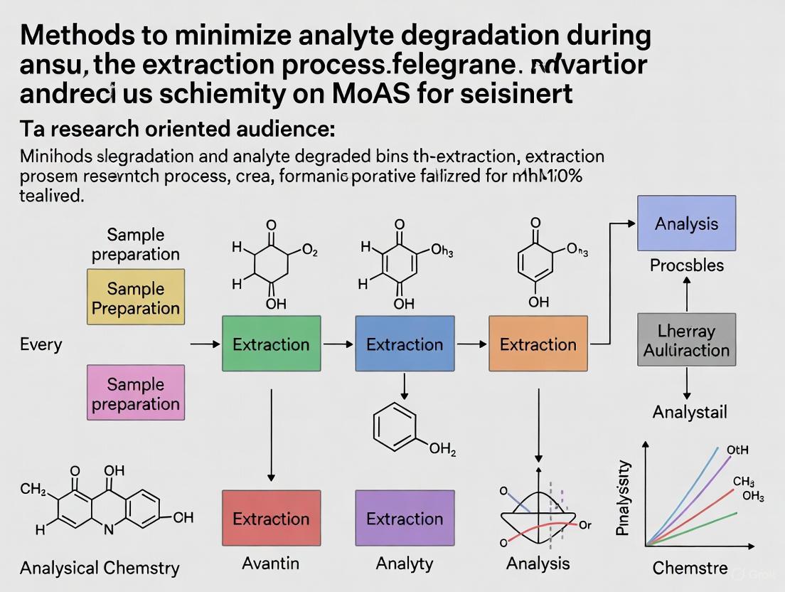
Workflow Visualization
Diagram: A systematic workflow for identifying degradation risks and implementing preventive strategies during extraction.
The Scientist's Toolkit: Essential Reagents for Stability
This table lists key reagents used to prevent analyte degradation in experimental workflows.
| Reagent / Material | Function in Preventing Degradation | Example Applications |
|---|---|---|
| Antioxidants (e.g., BHT, Ascorbic Acid) | Scavenge free radicals and reactive oxygen species (ROS), interrupting the oxidation chain reaction [3]. | Added to extraction solvents for phenolic compounds or unsaturated lipids. |
| Metal Chelators (e.g., EDTA, Citric Acid) | Bind to trace metal ions (e.g., Co²⁺, Fe²⁺) that catalyze oxidation reactions, such as the Haber-Weiss cycle for hydroxyl radical generation [3]. | Purifying buffers and solvents used for compounds susceptible to metal-ion oxidation. |
| Acid/Base Buffers | Maintain a constant pH to minimize acid- or base-catalyzed hydrolysis [2]. | Creating a stable pH environment for hydrolytically labile compounds (esters, amides). |
| Amber Glassware | Filters out ultraviolet and high-energy visible light to prevent photodegradation [1]. | Storing stock solutions and standard preparations of light-sensitive analytes. |
| Inert Gas (e.g., Nitrogen, Argon) | Displaces dissolved oxygen from solvents and creates an anaerobic atmosphere in sample vials [1]. | Purging solutions prior to and during the extraction of easily oxidized analytes. |
How Degradation Compromises Sensitivity, Reproducibility, and Results
Analyte degradation is a critical challenge in pharmaceutical analysis, directly compromising the sensitivity, reproducibility, and accuracy of your experimental results. Degradation products can co-elute with your target analyte, suppress ionization in mass spectrometry, cause peak broadening or tailing, and shift retention times, leading to inaccurate quantification and unreliable data. Understanding these mechanisms and implementing robust preventive strategies is essential for generating valid, reproducible results in drug development.
Frequently Asked Questions (FAQs)
1. How does analyte degradation specifically reduce method sensitivity? Degradation reduces sensitivity through several mechanisms. In LC-MS, degradants can cause ion suppression, reducing the ionization efficiency of your target analyte [4]. Furthermore, when degradation occurs during sample preparation or storage, the actual concentration of your intact analyte decreases, resulting in a lower detector response. Degradants co-eluting with the main peak can also lead to inaccurate integration and inflated impurity profiles, masking the true assay value [5].
2. What are the most common functional groups prone to degradation, and how do they affect my chromatogram? Common labile functional groups and their degradation outcomes are summarized in the table below.
Table 1: Common Degradation-Prone Functional Groups and Their Effects
| Functional Group | Common Degradation Pathway | Potential Impact on Chromatography |
|---|---|---|
| Esters, Amides, Lactams | Hydrolysis [6] | Appearance of new peaks; decrease in main peak area. |
| Amines, Sulfides | Oxidation (to N-oxides, sulfoxides, sulfones) [6] | Appearance of new peaks; peak tailing. |
| Compounds with labile hydrogen (e.g., benzylic, allylic carbon) | Oxidation (to hydroperoxides, ketones) [6] | Appearance of new peaks; poor mass balance. |
| Aromatics, Heterocyclics | Photolysis [5] | Appearance of new peaks upon light exposure. |
3. My method was validated, but I'm now seeing peak tailing/broadening. Could degradation be the cause? Yes. While method validation ensures initial performance, degradation can occur over time and cause these issues. Chemical degradation of the analyte can create products that interact differently with the stationary phase, leading to tailing [7]. Furthermore, mechanically induced peak distortion from precipitated salts or contaminants accumulated from degraded samples can create flow path obstructions, causing broadening and inconsistent retention [7]. A brief, cautious reverse-flow rinse of the HPLC column (if permitted by the manufacturer) can sometimes dislodge such debris and restore performance [7].
4. Why is my mass balance consistently below 95% in forced degradation studies? A low mass balance (the sum of the assay value and degradant levels) indicates that not all degradation products are being accounted for. Common reasons include [5]:
- Degradants are not eluting from the column or are not detected by your detector (e.g., they lack a UV chromophore).
- Degradants or the analyte are lost due to volatility, adsorption, or poor solubility during sample preparation.
- Co-elution of a degradant with the parent analyte peak or another impurity.
- Significant differences in the response factors of the degradants compared to the parent analyte.
Troubleshooting Guides
Problem: Inconsistent Results and Poor Reproducibility Between Runs
Potential Cause 1: Uncontrolled Hydrolytic Degradation During Sample Prep
- Explanation: Your analyte may be degrading due to the pH or aqueous content of the extraction solvent over time, with the rate varying between preparations.
- Solution:
- Optimize pH: Determine the pH stability profile of your analyte. Avoid storing samples in extreme pH conditions for extended periods [6] [5].
- Use Fresh Solvents: Prepare fresh aqueous mobile phases and extraction buffers weekly to prevent bacterial growth and pH drift [8].
- Neutralize Immediately: After acidic or basic hydrolysis steps in forced degradation studies, neutralize the solution promptly to prevent ongoing degradation [5].
Potential Cause 2: Oxidative Degradation in the Sample Vial
- Explanation: Oxygen in the headspace of the sample vial or oxidants in the solvent can slowly degrade your analyte, leading to decreasing peak areas over an analytical sequence.
- Solution:
- Use Inert Gases: Sparge your solvents with helium or argon and blanket sample vials with an inert gas.
- Add Antioxidants: In some cases, adding low concentrations of antioxidants like butylated hydroxytoluene (BHT) can be effective (ensure it doesn't interfere).
- Prepare Fresh Oxidants: For oxidative stress testing, use freshly prepared hydrogen peroxide solutions [6].
Problem: Loss of Sensitivity and High Background in LC-MS
Potential Cause 1: Source Contamination from Degradation Products
- Explanation: Non-volatile degradants or matrix components can accumulate on the ion source and ion transfer components, reducing ionization efficiency and increasing chemical noise.
- Solution:
- Improve Sample Cleanup: Enhance sample preparation with techniques like solid-phase extraction (SPE) or protein precipitation to remove more matrix components before injection [8] [9].
- Use a Divert Valve: Employ a switching valve to direct the LC effluent away from the MS source during the void volume and high organic wash periods, sending only the regions containing your analytes to the MS [8] [9].
- Implement a Flush Method: Use a shutdown method with a long, isocratic high-organic flush at the end of each batch to cleanse the system. Some evidence suggests using a flush in the opposite polarity can be more effective [8].
Potential Cause 2: Use of Non-Volatile Mobile Phase Additives
- Explanation: Phosphate buffers and other non-volatile salts precipitate and coat the ion source, severely suppressing ionization.
- Solution:
Experimental Protocols
Protocol 1: Conducting a Forced Degradation Study for Method Development
Forced degradation studies help establish the stability-indicating nature of an analytical method by intentionally degrading the sample to identify potential degradants [11] [5].
1. Objective: To investigate possible degradation products and pathways, providing a foundation for developing a suitable stability-indicating method [5].
2. Sample Preparation:
- Use drug substance, drug product, and placebo in these studies to discriminate the source of degradants [5].
- Use a mass-compatible method or a Photo-diode Array (PDA) detector to facilitate degradant identification [5].
3. Stress Conditions: Table 2: Recommended Conditions for Forced Degradation Studies
| Stress Condition | Recommended Parameters | Endpoint |
|---|---|---|
| Acid Hydrolysis | 0.1 - 1 M HCl at ~70°C [6] [5] | 5-20% degradation [5] |
| Base Hydrolysis | 0.1 - 1 M NaOH at ~70°C [6] [5] | 5-20% degradation [5] |
| Oxidation | 0.1-3% H₂O₂ at room temperature [6] [5] | 5-20% degradation [5] |
| Thermal | 70°C (mp <150°C) or 105°C (mp >150°C) [5] | 5-20% degradation [5] |
| Photolysis | 1.2 million lux-hr visible & 200 W-hr/m² UV [5] | As per ICH criteria |
4. Evaluation:
- Peak Purity: Use a PDA detector to ensure the main peak is pure and free from co-eluting degradants [5].
- Mass Balance: Calculate the mass balance by adding the assay value and the levels of degradation products. Aim for a mass balance of at least 95%; investigate and justify if lower [5].
Forced Degradation Workflow for Method Development
Protocol 2: Sample Preparation to Minimize Degradation During Extraction
This protocol outlines a general approach for handling samples with degradation-prone analytes.
1. Work in a Controlled Environment:
- Prepare mobile phases and extraction buffers in a clean part of the lab, separate from where samples are handled [8].
- Use high-quality, LC-MS grade solvents and reagents to minimize contamination [8].
2. Minimize Exposure to Degradation Triggers:
- Light: Use amber vials or work in low-light conditions if the analyte is photosensitive.
- Heat: Keep samples on an autosampler cooler set to 4-15°C [10].
- Oxygen: Sparge solvents with inert gas and seal vials tightly.
3. Use Appropriate Sample Cleanup:
- Protein Precipitation: For biological fluids, centrifuge samples at high speed (e.g., 21,000 x g for 15 min) to pellet particulate matter [8].
- Solid-Phase Extraction (SPE): Use SPE to isolate the analyte from a complex matrix and transfer it into a stable, MS-compatible solvent [8] [9].
- Filtration: Use centrifugal filters to remove particulates and, for small molecules, some high molecular weight interferences [12].
4. Automate and Standardize:
- Use an autosampler with a controlled temperature tray.
- Ensure the autosampler needle depth is set correctly to avoid aspirating from the pellet at the bottom of a vial after centrifugation [8].
The Scientist's Toolkit: Key Research Reagent Solutions
Table 3: Essential Materials for Preventing Analyte Degradation
| Reagent / Material | Function / Purpose | Key Considerations |
|---|---|---|
| Ammonium Acetate/Formate | Volatile buffer for LC-MS mobile phases [10] [9]. | Provides pH control without source contamination. Start with 10 mM concentration [9]. |
| Formic Acid / Acetic Acid | Volatile acidic mobile phase additive [9]. | Preferred over TFA for better MS sensitivity. Use at ~0.1% v/v [9]. |
| LC-MS Grade Solvents | High-purity water, acetonitrile, methanol [8]. | Minimizes background contamination and unintended chemical reactions. |
| Hydrogen Peroxide (3%) | Oxidizing agent for forced degradation studies [5]. | Use freshly prepared and store appropriately. |
| Hydrochloric Acid (0.1-1 M) | Acid catalyst for hydrolytic forced degradation [6] [5]. | |
| Sodium Hydroxide (0.1-1 M) | Base catalyst for hydrolytic forced degradation [6] [5]. | |
| Core-Shell Particle C18 Column | High-efficiency analytical column for separation [10]. | Provides good resolution of analytes from degradants. Example: Ascentis Express F5 [10]. |
| SPE Cartridges (e.g., C18) | Solid-phase extraction for sample cleanup and concentration [8] [12]. | Removes matrix interferents and transfers analyte to a stable solvent. |
Degradation Impacts and Solutions Relationship
The Role of Sample Matrix and Container Interactions in Stability
Frequently Asked Questions (FAQs)
1. How can I confirm if an unexpected peak in my chromatogram is a degradant from my standard? You can confirm the peak is a degradant through a time-course study. Inject a freshly prepared standard and then the same standard after it has aged. If the area of the suspected analyte peak decreases over time while the area of the new, unexpected peak increases, this strongly indicates degradation. The degradant peak is typically not present in a freshly prepared standard using the same solvent lot [13]. Mass spectrometry (MS) can provide structural analysis to confirm the new peak is a degradation product of your analyte [14].
2. How does standard degradation affect the quantitation of my samples? If a standard degrades, its peak area decreases relative to its nominal concentration. When this degraded standard is used to calibrate an instrument, the calculated concentration for a fresh sample will be overestimated, creating a positive bias or high recovery error [13]. This assumes the samples themselves are stable. If both standards and samples degrade at the same rate, the error may be masked, but the risk of inaccurate results remains high.
3. My method was validated with stable standards, but degradation has suddenly appeared. What should I check? First, systematically troubleshoot your process [14]:
- Mobile Phase: Prepare a fresh batch of mobile phase to rule out contamination or errors in formulation (e.g., inadvertent addition of acid) [14].
- Diluent: Assess if the diluent itself is causing degradation. The chemical compatibility between your analyte and the solvent is critical.
- Container Interactions: Evaluate if the sample is degrading due to interaction with the container walls or septa [15].
- Environmental Controls: Verify that storage conditions (light, temperature) have not changed and are sufficient for your analyte [15].
4. What are the best practices for storing volatile analytical standards to prevent degradation? Volatile standards require meticulous handling to prevent loss through evaporation or degradation [15]:
- Containers: Use sealed glass vials with PTFE-lined septa. Amber glass is preferred to protect from light. Avoid plastic containers, which can be permeable or introduce contaminants.
- Temperature: Store in a cool, dry environment (e.g., 2°C to 8°C). Avoid freezing for compounds like methanol and acetone, as this can make them difficult to use and potentially alter concentrations upon thawing.
- Handling: Minimize open-container time and exposure to air. Use gas-tight syringes for measurement. Work quickly and re-cap containers immediately.
5. Can the HPLC column itself cause sample degradation? Yes, on-column degradation can occur, especially for small molecules with specific functional groups. One documented cause is interaction with exposed silanol groups on "lightly loaded" C18 columns, which have lower bonded phase coverage. This was observed for a compound with an aniline group. Switching to a "fully bonded" (high-coverage) C18 column from the same manufacturer eliminated the degradation. Modifying the mobile phase pH can also stabilize the analyte and prevent this interaction [14].
Troubleshooting Guides
Problem: On-Column Degradation of Small Molecules
Observed Symptom: Unexpected peaks (degradants) appear in the chromatogram during a purity method, but orthogonal techniques (e.g., NMR) confirm the sample is pure.
Diagnosis and Solution: This is often linked to specific column chemistry. Follow this logical path to diagnose and resolve the issue:
Experimental Protocol to Isolate the Cause:
- Modify Gradient to Reduce On-Column Time: Increase the starting percentage of organic solvent (e.g., acetonitrile) in the gradient while keeping the slope constant. A reduction in degradant peaks with shorter retention times suggests the sample is degrading while interacting with the column [14].
- Change Column Chemistry: Replace the current "lightly loaded" C18 column with a "fully bonded" or "high-coverage" C18 column (bonded phase coverage >3 μmol/m²). The higher surface coverage reduces exposure to reactive silanol groups [14].
- Modify Mobile Phase pH: If the analyte is stable in acid, add 0.1% acetic acid to the aqueous mobile phase. This can protonate the analyte or the silanol groups, minimizing deleterious interactions [14].
- Confirm with Degraded Sample: To ensure your new method can still detect degradants if present, inject a force-degraded sample (e.g., exposed to heat or light) to confirm the degradant peaks are visible with the new conditions [14].
Problem: Standard Solution Degradation
Observed Symptom: The peak area of the standard decreases over time, often accompanied by the appearance of new peaks (degradants) or a noisy baseline.
Diagnosis and Solution: Standard degradation can be caused by multiple factors. Use this guide to identify and address the root cause:
Experimental Protocol for Solution Stability Testing:
- Preparation: Prepare a fresh standard solution. Divide it into several aliquots in different types of vials (clear glass, amber glass, and with different septa if suspected).
- Storage Conditions: Store the aliquots under different stress conditions:
- On the bench exposed to lab light.
- In a refrigerator (4°C) in amber vials.
- In the autosampler tray (with and without temperature control, and with trays covered in foil).
- Analysis: Inject the samples over a period of time (e.g., 24, 48, 72 hours) against a freshly prepared standard.
- Evaluation: Monitor the peak area of the main analyte and the appearance of new peaks. The conditions that show the least change in area and no new peaks indicate the optimal storage conditions.
Table 1. Impact of Container Material on Light-Induced Off-Flavors in Milk
This study demonstrates how container material acts as a part of the sample matrix, influencing analyte stability by blocking or transmitting degrading UV light [16].
| Container Material | Wall Thickness (mm) | Relative Formation of Lipid Oxidation Products (e.g., Hexanal) | Key Finding |
|---|---|---|---|
| Glass (clear) | ~5.0 | Very Low | Best barrier to UV light; serves as the control. |
| HDPE (with white pigment) | Not Specified | Low | Good barrier; pigmentation improves UV protection. |
| PETE | ~0.36 | High | Poor barrier; performance likely affected by thin walls. |
| Polypropylene (PP) | ~1.27 | High | Poorest barrier, despite having the thickest walls. |
Methodology Summary: Milk was spiked with an internal standard (hexanal-d12) and dispensed into various containers. Containers were exposed to a fluorescent UV light source for 2 hours. After cooling, volatile compounds were extracted using Solid-Phase Microextraction (SPME) with a CAR/PDMS fiber and analyzed by GC-MS. Concentrations of off-flavor analytes were calculated using a calibration curve [16].
Table 2. Effect of Extraction Technique on Bioactive Compound Stability
The choice of extraction method directly affects the stability of analytes within the sample matrix, primarily through thermal and mechanical stress [17] [18].
| Extraction Technique | Typical Conditions | Impact on Phytochemical Stability & Yield | Advantage | Disadvantage |
|---|---|---|---|---|
| Maceration | Room temperature, prolonged time | Lower yields for thermolabile compounds; longer exposure can lead to degradation. | Simple, low cost, good for thermolabile compounds. | Low efficiency, long extraction time, high solvent use. |
| Decoction | High-temperature boiling in water | Can cause hydrolysis, dehydration, or degradation of thermolabile/volatile components. | Enhanced dissolution of some compounds. | Unsuitable for thermolabile compounds; extracts many water-soluble impurities. |
| Microwave-Assisted (MAE) | Elevated temperature, short time | High yield and efficiency; short exposure time minimizes degradation. | Rapid, reduced solvent consumption, high efficiency. | Potential thermal degradation if not controlled. |
| Ultrasound-Assisted (UAE) | Lower temperature, mechanical cavitation | High yield; avoids thermal degradation; preserves bioactivity (e.g., antioxidants). | Effective cell disruption, improved yield, low temperature. | Can generate free radicals if not controlled. |
The Scientist's Toolkit: Key Research Reagent Solutions
Table 3. Essential Materials for Minimizing Analyte Degradation
| Item | Function & Rationale |
|---|---|
| Amber Glass Vials | Protects light-sensitive analytes from photodegradation by blocking UV and visible light [13] [15]. |
| PTFE-Lined Septa | Provides an inert, non-absorbing barrier between the sample and the septum, preventing adsorption or leaching of contaminants [15]. |
| "Fully Bonded" C18 HPLC Columns | Columns with high bonded-phase coverage (>3 μmol/m²) minimize interactions with exposed acidic silanol groups on the silica surface, reducing on-column degradation for basic compounds [14]. |
| Chilled Autosampler | Maintaining samples at low temperatures (e.g., 4°C) during analysis slows down chemical degradation and evaporation processes [13]. |
| CAR/PDMS SPME Fiber | Effectively extracts and pre-concentrates small, volatile analytes (like degradation products) for sensitive GC-MS analysis, crucial for identifying and quantifying degradation [16]. |
| Stabilizing Diluents | Using a diluent that mimics a stable sample formulation (e.g., with antioxidants or buffers) can protect the standard from degradation. Always check for solubility and compatibility [13]. |
Experimental Protocol: Evaluating Container Compatibility
Aim: To systematically evaluate the compatibility of a volatile analytical standard with different storage containers and conditions to minimize degradation.
Materials:
- Volatile analytical standard
- Different vials (clear glass, amber glass, with various PTFE/other septa)
- Gas-tight syringes
- GC-MS system with chilled autosampler
- Refrigerator (4°C), Freezer (-20°C)
Procedure:
- Preparation: Prepare a single, large batch of the standard solution in an appropriate solvent.
- Aliquoting: Dispense equal volumes of the standard into the different vials to be tested. Crimp or cap them securely.
- Storage Groups: Create groups for each vial type and store them under different conditions:
- Condition A: Room temperature, on bench, exposed to light.
- Condition B: Room temperature, in drawer or covered with foil.
- Condition C: Refrigerated (4°C), amber vial.
- Condition D: In a chilled autosampler (e.g., 4°C).
- Time-Course Analysis: At predetermined time points (e.g., 0, 6, 24, 72 hours), inject each sample in duplicate using the GC-MS method.
- Data Analysis:
- Plot the peak area of the main analyte versus time for each condition.
- Note the appearance and growth of any new peaks in the chromatogram.
- The condition that maintains the most consistent analyte area and shows the fewest new peaks over time is the optimal storage condition.
Expected Outcome: This protocol will provide empirical data to select the best container and storage environment to ensure standard stability, which is foundational for obtaining accurate and reproducible analytical results.
Practical Strategies for Stabilization and Extraction
In analytical research, particularly during the extraction of delicate compounds, the chemical integrity of target analytes is paramount. Uncontrolled pH, oxidative stress, and enzymatic activity are primary drivers of analyte degradation, leading to inaccurate quantification and loss of critical sample information. This technical support center outlines proven strategies using buffers, antioxidants, and enzyme inhibitors to mitigate these risks. Implementing these chemical stabilization methods is essential for ensuring data reliability in fields from pharmaceutical development to environmental analysis [19] [20].
Troubleshooting Guides & FAQs
Buffer-Related Issues
Problem: Inconsistent analytical results between laboratories using the same "25 mM phosphate pH 7.0" buffer method.
- Cause: The description "25 mM phosphate pH 7.0" is ambiguous. It can be prepared using different salts (e.g., NaH₂PO₄, Na₂HPO₄) or adjustment methods, resulting in solutions with different ionic strengths and buffering capacities [21].
- Solution: The exact buffer preparation procedure must be meticulously documented and standardized. Specify the specific salt used (e.g., disodium hydrogen orthophosphate), the concentration of the acid or base used for pH adjustment, and the order of operations [21].
- Preventive Protocol:
- Weigh the specified mass of the documented salt.
- Dissolve in purified water.
- Adjust pH to the specified value using an acid or base of a defined concentration and type (e.g., 1M NaOH or concentrated H₃PO₄).
- Do not dilute a pH-adjusted stock solution, as this alters the final pH. Always prepare the buffer at its final working concentration [21].
Problem: Pressure fluctuations or erratic retention times in HPLC, especially in gradient methods.
- Cause: Precipitation of buffer salts in the HPLC system when the organic solvent content becomes too high. For example, potassium phosphate buffers can precipitate at acetonitrile concentrations above 70% [22].
- Solution:
- Know the solubility limits of your buffer. Avoid reaching the precipitation point of the buffer in the method gradient [22].
- For low-pressure gradient (LPG) pumps, a best practice is to add a percentage of aqueous buffer (e.g., 5-10%) to the organic solvent channel. This prevents the buffer from encountering 100% organic solvent during pump proportioning, which can cause crystallization in the valve and check valves [22].
- Always flush the HPLC system and column with a high-water-content mobile phase (e.g., 95% water, 5% organic) after using buffers to remove all salt residues before storage [22].
Antioxidant-Related Issues
Problem: An extracted plant or pharmaceutical sample shows declining potency over time, suspected due to oxidative degradation.
- Cause: Reactive Oxygen Species (ROS) in the sample or extraction solvent are degrading the target analytes. This is common with compounds like phenolics or sensitive APIs [20].
- Solution: Incorporate antioxidants into the extraction solvent or sample storage medium. The choice of antioxidant should be guided by the specific ROS present and the nature of your analyte [20].
- Experimental Protocol for Antioxidant Capacity Assessment (DPPH Assay):
- Principle: The stable DPPH• (1,1-diphenyl-2-picrylhydrazyl) radical is purple and absorbs at 517 nm. Antioxidants reduce it to a yellow-colored product, leading to a loss of absorbance [20].
- Procedure: Prepare a 0.1 mM DPPH solution in methanol. Mix 2 mL of this solution with 1 mL of your sample extract (dissolved in a suitable solvent) or a standard antioxidant solution (e.g., Trolox) [23].
- Incubation: Keep the mixture in the dark at room temperature for 30 minutes.
- Measurement: Measure the absorbance of the mixture at 517 nm against a methanol blank. A control sample contains DPPH solution and solvent without the antioxidant.
- Calculation: Calculate the percentage of radical scavenging activity:
[(A_control - A_sample) / A_control] * 100. The results can be expressed as Trolox Equivalents (TE) per gram of extract for standardization [23].
Enzyme Inhibitor-Related Issues
Problem: During a 2D-LC analysis for peak purity, new peaks appear that are identified as degradation products of the main analyte.
- Cause: In-loop analyte degradation. The target compound can be unstable in the first dimension's elution solvent (e.g., acidic mobile phase with TFA) during the extended storage time in the sampling loop while waiting for the 2D analysis [24].
- Solution:
- Test 1 (In-Solution Stability): Dilute the pure analyte in the 1D mobile phase composition and let it stand at the method temperature for the estimated loop storage time. Re-analyze with the 1D method to check for new degradation peaks [24].
- Test 2 (1D-LC Simulation): Analyze the sample using a 1D-LC method that mimics the 2D separation conditions to see if the "new" impurity appears under those chromatographic conditions [24].
- Remediation: If degradation is confirmed, consider using a different 1D mobile phase, reducing the loop storage time, or using a "multi-inject" approach to analyze fractions faster [24].
Problem: Need to screen natural plant extracts for inhibitory activity against a target enzyme like acetylcholinesterase (AChE).
- Cause: Standard colorimetric or fluorescent assays can be interfered with by background absorption or auto-fluorescence from complex plant matrices [25].
- Solution: Use an Immobilized Enzyme Reactor (IMER) coupled with liquid chromatography (LC). The target enzyme is immobilized on a solid support within a capillary or column, which can be used repeatedly in an on-flow system [25].
- Experimental Protocol (On-Flow IMER Activity Screening):
- IMER Preparation: The enzyme (e.g., AChE) is immobilized onto a suitable solid support (e.g., silica monolith, magnetic particles) and packed into a cartridge or capillary [25].
- On-Flow Assay: The IMER is placed in-line with an LC system. A solution of the enzyme's substrate is pumped through the IMER, and the product formation is monitored online by a detector (e.g., UV or MS).
- Inhibition Screening: A plant extract or potential inhibitor is introduced into the system (e.g., via injection). A decrease in the product peak area indicates inhibitory activity.
- Kinetic Analysis: The IC₅₀ value (concentration causing 50% inhibition) can be determined by testing a range of inhibitor concentrations. For reversible inhibition, the inhibition constant (Kᵢ) can be calculated from the IC₅₀ using the Cheng-Prusoff equation, which requires knowledge of the substrate concentration and its Kₘ for the enzyme [26].
Data Presentation Tables
Table 1: Common In Vitro Antioxidant Assessment Methods
| Method | Measured Principle | Key Application Notes |
|---|---|---|
| DPPH• Scavenging [20] [23] | Reduction of a stable radical, measured by absorbance loss at 517 nm. | Simple, rapid; useful for initial screening of pure compounds and extracts. |
| ABTS•+ Scavenging [20] [23] | Scavenging of the pre-formed ABTS radical cation, measured by absorbance loss at 734 nm. | Faster than DPPH; allows analysis of both hydrophilic and lipophilic antioxidants. |
| FRAP (Ferric Reducing Antioxidant Power) [20] [23] | Reduction of Fe³⁺-TPTZ complex to a blue Fe²⁺ form, measured at 593 nm. | Measures reducing capacity; does not involve radical scavenging. |
| CUPRAC (Cupric Reducing Antioxidant Power) [20] [23] | Reduction of Cu²⁺ to Cu⁺, measured by complex formation at 450 nm. | Comparable to FRAP, often more selective and efficient. |
Table 2: Example Inhibitor Potency (IC₅₀) for Reference Drugs
| Drug / Compound | Target Enzyme | IC₅₀ Value | Mode of Action & Notes |
|---|---|---|---|
| Rosuvastatin [25] | HMG-CoA Reductase | 5 nmol/L | Competitive inhibitor; used to lower cholesterol. |
| Atorvastatin [25] | HMG-CoA Reductase | 8 nmol/L | Competitive inhibitor; used to lower cholesterol. |
| Simvastatin [25] | HMG-CoA Reductase | 11 nmol/L | Competitive inhibitor; used to lower cholesterol. |
| Donepezil [25] [26] | Acetylcholinesterase (AChE) | Varies (nM range) | Mixed competitive/non-competitive inhibitor; used for Alzheimer's disease. |
| Luteolin 7-glucoside [23] | α-Glucosidase, Tyrosinase | Strong binding affinity shown via docking | Natural product; molecular docking reveals high potential as a mixed-type inhibitor. |
Stabilization Workflow and Pathways
Antioxidant Defense Network
Analyte Degradation Troubleshooting
The Scientist's Toolkit: Research Reagent Solutions
Table 3: Essential Reagents for Chemical Stabilization
| Reagent / Material | Primary Function | Example Application & Notes |
|---|---|---|
| Phosphate Buffers [21] [22] | Maintain pH during extraction and analysis to prevent acid/base-catalyzed degradation. | Use in HPLC mobile phases; beware of precipitation at high organic solvent content (>70% ACN). |
| Ammonium Acetate / Formate | Volatile buffers for LC-MS compatibility. | Ideal for mass spectrometric detection as they do not leave residue on the ion source. |
| DPPH (1,1-Diphenyl-2-picrylhydrazyl) [20] [23] | Stable radical for measuring free radical scavenging activity of antioxidants. | Used for rapid screening of antioxidant capacity in plant extracts or synthetic compounds. |
| Trolox | Water-soluble vitamin E analog used as a standard in antioxidant assays. | Allows quantification of antioxidant activity as "Trolox Equivalents (TE)" for standardization [23]. |
| EDTA (Ethylenediaminetetraacetic acid) | Metal chelator; inhibits metal-catalyzed oxidation. | Added to extraction buffers to sequester trace metal ions that can generate ROS via Fenton reactions [20]. |
| Donepezil / Galantamine [25] [26] | Acetylcholinesterase (AChE) inhibitors; reference standards for inhibition studies. | Used as positive controls when developing or validating enzyme inhibition assays for neurological targets. |
| Immobilized Enzyme Reactor (IMER) [25] | Solid-supported enzyme for on-flow, high-throughput inhibitor screening. | Provides reusability, increased enzyme stability, and allows automation of inhibition assays coupled with LC. |
| Kojic Acid / Arbutin [23] | Reference tyrosinase inhibitors. | Used as benchmarks when testing new compounds for skin-whitening or anti-browning effects. |
Optimized Extraction Protocols for Specific Analyte Classes (e.g., Proteins, Small Molecules)
Core Principles for Minimizing Analyte Degradation
The integrity of proteins and small molecules during extraction is paramount for accurate analytical results. Degradation can occur through enzymatic activity (e.g., proteases), chemical instability (e.g., oxidation, hydrolysis), and physical stress. A successful extraction protocol is built on a two-pronged approach: rapid inhibition of degradation pathways at the moment of cell lysis, followed by the swift physical separation of the analyte from degrading agents [27].
What are the primary mechanisms of analyte degradation I should be aware of? The primary mechanisms are:
- Enzymatic Degradation: Proteases are released during cell lysis and can rapidly digest proteins of interest [27]. For nucleic acids, nucleases present a similar threat [28].
- Chemical Degradation: This includes oxidation of sensitive residues, hydrolysis in aqueous environments, and pH-induced denaturation [29].
- Temperature-Induced Degradation: Even small temperature fluctuations can accelerate degradation processes. Some small molecules, like haloacetic acids in water, show significant degradation within hours at room temperature [29].
- Physical Adsorption: Analytes can be lost through non-specific binding (NSB) to the surfaces of containers, pipette tips, and caps [30].
Small Molecule Extraction Troubleshooting Guide
Small molecule extraction, particularly for techniques like LC-MS/MS, requires careful method selection to concentrate the analyte and deplete the sample matrix.
Frequently Asked Questions
What is the most critical factor in choosing a small molecule extraction method? The chemistry of your analyte is the most important factor. You must consider its polarity (log P or log D), charge (pKa), and stability. This will determine the optimal solvent and extraction technique [30].
My LC-MS/MS analysis shows high background noise and ion suppression. How can my sample prep fix this? This is often a matrix effect. A method with high extraction recovery (e.g., 90%) is not always better than one with lower recovery (e.g., 30%) if the latter more effectively removes phospholipids and other matrix components that cause ion suppression. Focus on protocols that deplete the matrix, not just recover the analyte [30].
Why should I use native patient matrix during method development? Complex and variable fresh biomatrix from patients does not behave the same way as a simple, synthetic matrix from healthy donors. Incorporating native matrix early in development ensures your protocol is robust enough for real-world samples [30].
Table 1: Troubleshooting Small Molecule Extraction for LC-MS/MS
| Problem | Possible Causes | Recommended Solutions |
|---|---|---|
| Low Signal/Recovery | Non-specific binding to containers; poor solubility [30]. | Use low-binding tubes; add solubilizing agents; optimize injection matrix composition [30]. |
| Ion Suppression | Incomplete matrix depletion; phospholipids present [30]. | Switch from simple dilution/PPT to supported-liquid or solid-phase extraction; use matrix-matched calibration [30]. |
| High Background/Contamination | Trace contaminants in solvents or from labware [30]. | Use LC-MS grade solvents; employ best practices for handling clean, inert containers; analyze blank samples frequently [30]. |
| Irreproducible Results | Inconsistent recovery due to protocol or analyte instability [30]. | Achieve precision first before evaluating other parameters; use internal standards; control temperature and processing time [30]. |
Protein Extraction and Purification Troubleshooting Guide
The primary challenge in protein work is preventing proteolysis, which begins the instant cells are lysed.
Frequently Asked Questions
What is the most important step to prevent protein degradation during extraction? The simultaneous use of protease inhibitors and maintaining low temperatures (0-4°C) throughout the process is critical. Protease inhibitors should be added to your lysis buffer before homogenization to immediately inhibit proteases [31].
My western blot shows multiple bands or smearing. Is this degradation? Multiple bands can indicate degradation, but also the presence of isoforms or post-translational modifications (PTMs) like glycosylation. To confirm degradation, ensure your lysis buffer contains a cocktail of protease inhibitors, use fresh samples, and run a positive control. If degradation is ruled out, investigate potential PTMs [31].
How does sonication help in protein extraction? Sonication ensures complete lysis, shears genomic DNA that can interfere with downstream analysis, and helps to solubilize membrane-bound and organelle-localized proteins, leading to more consistent and maximal protein recovery [31].
Table 2: Troubleshooting Protein Extraction and Western Blotting
| Problem | Possible Causes | Recommended Solutions |
|---|---|---|
| Low Yield/Degradation (Multiple bands/smearing) | Protease activity; old or improperly stored samples [31]. | Use fresh protease inhibitor cocktails (e.g., PMSF, leupeptin); process samples immediately on ice; snap-freeze tissues in LN₂ [31]. |
| Incomplete Lysis | Inefficient lysis buffer; no mechanical disruption for tough tissues [31]. | Use a probe sonicator (e.g., 3x 10-sec bursts on ice) or repeated passage through a fine-gauge needle; ensure correct buffer for protein type [31]. |
| Low Signal for Phosphoproteins | Protein degradation; phosphatase activity [31]. | Add phosphatase inhibitors (e.g., sodium orthovanadate, beta-glycerophosphate) to lysis buffer; use recommended positive controls [31]. |
Optimized Extraction Protocols
Protocol 1: Solid-Phase Extraction (SPE) for Sensitive Small Molecules in Serum/Plasma
This protocol is designed for LC-MS/MS analysis of small molecules like steroids or vitamins, focusing on matrix depletion and analyte stability.
Workflow Overview:
Detailed Methodology:
- Sample Pre-treatment: Precipitate proteins by adding a 2:1 volume of cold acetonitrile containing 0.1% formic acid to plasma. Vortex and centrifuge at 10,000 x g for 10 minutes to pellet precipitated proteins [30].
- SPE Column Conditioning: Condition a reversed-phase C18 SPE column sequentially with 3 mL of methanol and 3 mL of water [30].
- Sample Loading: Load the clear supernatant from step 1 onto the conditioned SPE column. Use a gentle vacuum to pass the sample through slowly [30].
- Washing: Wash the column with 3 mL of a 5:95 methanol/water solution to remove salts and polar matrix components without eluting the analyte [30].
- Elution: Elute the target analyte into a clean collection tube with 2 x 1 mL of a stronger solvent, such as 90:10 methanol/ethyl acetate. The exact solvent should be optimized based on analyte polarity [30] [32].
- Post-processing: Evaporate the eluent to dryness under a gentle stream of nitrogen gas. Reconstitute the dry residue in 100 µL of a solvent compatible with your LC-MS/MS mobile phase (e.g., 50:50 water/methanol). Vortex thoroughly before analysis [30].
Protocol 2: Protein Extraction with Protease Inhibition for Western Blotting
This protocol is optimized for extracting total protein from cultured cells while maintaining integrity for immunoblotting.
Workflow Overview:
Detailed Methodology:
- Lysis Buffer Preparation: Prepare RIPA lysis buffer and chill on ice. Immediately before use, add a protease inhibitor cocktail to the final recommended concentration (e.g., 1X). For phosphoproteins, also add a phosphatase inhibitor cocktail [31].
- Cell Lysis: For adherent cells, aspirate media, rinse with cold PBS, and add cold lysis buffer directly to the plate (e.g., 150 µL for a 60mm plate). Scrape cells and transfer the lysate to a microcentrifuge tube. Keep tubes on ice at all times.
- Sonication: Sonicate the lysate on ice using a microtip probe sonicator (e.g., 3 pulses of 10 seconds each at 15W, with 10-second pauses between pulses to prevent heating). This shears DNA and ensures complete lysis [31].
- Incubation: Incubate the lysate on a rocking platform at 4°C for 30 minutes to ensure complete extraction.
- Clarification: Centrifuge the lysate at 14,000 x g for 15 minutes at 4°C to pellet insoluble debris.
- Collection: Carefully transfer the supernatant (containing the soluble protein extract) to a new, pre-chilled tube. Aliquot to avoid repeated freeze-thaw cycles and store at -80°C [31].
The Scientist's Toolkit: Essential Research Reagents
Table 3: Key Reagents for Minimizing Analyte Degradation
| Reagent / Material | Function | Key Considerations |
|---|---|---|
| Protease Inhibitor Cocktail | Broad-spectrum inhibition of serine, cysteine, metallo-, and aspartic proteases during protein extraction [31]. | Use a commercial 100X cocktail for convenience and broad protection. Add fresh to lysis buffer immediately before use. |
| Phosphatase Inhibitor Cocktail | Prevents dephosphorylation of serine, threonine, and tyrosine residues, preserving phosphoprotein status [31]. | Essential for detecting post-translational modifications. Often used in combination with protease inhibitors. |
| LC-MS Grade Solvents | High-purity solvents that minimize background contamination and ion suppression in mass spectrometry [30]. | Critical for trace analysis. Avoids introduction of contaminants that are easier to measure than the analytes themselves. |
| PMSF (Phenylmethylsulfonyl fluoride) | Serine protease inhibitor (e.g., against trypsin, chymotrypsin) [31]. | Unstable in water; must be added from a stock solution in ethanol or isopropanol directly to the lysis buffer. |
| Wide-Bore Pipette Tips | Minimizes shearing forces when pipetting High Molecular Weight (HMW) DNA or other sensitive macromolecules [28]. | Prevents physical degradation and fragmentation of large nucleic acids. |
| Appropriate Solvent for Liquid-Liquid Extraction | Selectively dissolves the analyte of interest based on polarity to separate it from the matrix [32]. | Choice is guided by analyte log P/D. Common pairs: water-dichloromethane (polar organics), water-hexane (non-polar organics) [32]. |
Solving Common Extraction Challenges and Optimizing Protocols
How can I systematically diagnose the root cause of poor chromatographic results?
A systematic approach is crucial for efficient troubleshooting. Begin by using a "benchmarking method" – a known, well-characterized analysis run on your system when it is functioning correctly. When problems arise, re-run this benchmark [33].
- If the benchmark performs well, the issue is likely with your specific sample or method [33].
- If the benchmark fails, the problem is probably with your instrument or column [33].
A logical workflow for diagnosis is outlined below. Always change one variable at a time to correctly identify the source of the problem [33] [34].
What are the primary causes of peak tailing and how do I fix them?
Peak tailing is a common issue that affects resolution, quantitation accuracy, and reproducibility [33]. The table below summarizes the top causes and their solutions.
| Cause of Tailing | Underlying Reason | Solution |
|---|---|---|
| Secondary Interactions (Most Common) [33] | Polar/ionized analytes interacting with residual silanols on silica stationary phase [33] [35]. | Use end-capped, high-purity silica columns [33] [35]. Operate at low pH (2-3) to suppress silanol ionization [33] [36]. Increase buffer concentration (>20 mM) [33]. |
| Column Void or Damage [33] | Collapsed bed material at column inlet, often from pressure shock or high pH [33] [37]. | Avoid pressure shock; increase flow gradually [33]. Operate at pH < 7.5 unless using a specialty column [33]. Replace column if void is confirmed [33]. |
| Extra-Column Volume [33] | Band broadening from tubing, fittings, or detector cells with large internal volume [33] [35]. | Use narrow ID tubing (e.g., 0.005"), minimize length, ensure proper fittings [33] [35]. |
| Incorrect Mobile Phase pH [33] | pH near analyte's pKa causes mixed ionization states, leading to asymmetry [33] [35]. | Adjust mobile phase pH to be at least ±1 pH unit away from analyte pKa [35]. Use a calibrated pH meter [33]. |
| Sample Overloading [34] | Injection volume or analyte concentration exceeds column capacity [38] [34]. | Reduce injection volume or dilute the sample [34]. |
| Sample Matrix Effects [37] | Buildup of proteins, lipids, or other matrix components on the column. | Improve sample clean-up (e.g., SPE, filtration) [35] [34]. Use a guard column [37]. |
What steps can I take to minimize analyte degradation during sample preparation?
Proper sample preparation is critical for maximizing recovery and preventing the introduction of degradation products that cause inconsistency.
- Control Temperature: Use lower temperatures during preparation. For heat-labile compounds, freeze-drying (lyophilization) is superior to convection drying, which can degrade heat-sensitive bioactive compounds [39].
- Prevent Oxidation and Chemical Changes: Chemical changes like oxidation, hydrolysis, or condensation are often irreversible [39]. Use inert atmospheres if necessary and avoid solvents or conditions that promote these reactions.
- Employ Enzymatic Hydrolysis and Fermentation: These modern pre-treatment methods can help release bioactive compounds loosely bonded to cell wall polymers without degrading them [39].
- Use Effective Clean-up Techniques: For complex matrices, techniques like Solid Phase Extraction (SPE) remove interfering compounds that can degrade column performance and affect quantitation, thereby improving peak symmetry and analytical precision [35].
Why am I getting inconsistent results between runs, and how can I improve reproducibility?
Inconsistency often stems from uncontrolled variables in the method, sample, or system.
- Mobile Phase Stability: Prepare fresh mobile phase to avoid degradation. For pH-sensitive methods, ensure buffer concentration is sufficient (often >20 mM) and measure pH with a calibrated meter accurate to ±0.05 units [33]. Be aware that CO2 ingress can make the mobile phase more acidic over time [33].
- Sample Solvent Mismatch: The solvent used to dissolve the sample should match the initial mobile phase composition as closely as possible. A solvent with higher elution strength can cause peak distortion [38] [34].
- Column Care and Guard Columns: A worn-out column or a contaminated/obstructed guard cartridge can cause tailing and inconsistent retention times [34] [37]. Replace the guard column regularly. If tailing affects all analytes, try replacing or cleaning the guard cartridge first [34].
- System Suitability Tests: Before a sequence of runs, perform a system suitability test with a standard to verify that the entire HPLC system is performing as expected regarding retention, resolution, and peak shape.
The Scientist's Toolkit: Essential Research Reagent Solutions
| Item | Function & Rationale |
|---|---|
| High-Purity, End-capped Columns (e.g., Type B silica) | Minimizes secondary interactions with residual silanols, the most common cause of tailing for basic compounds [33] [35]. |
| Guard Columns | A relatively inexpensive sacrificial component that protects the expensive analytical column from contamination by sample matrix components [37]. |
| SPE (Solid Phase Extraction) Cartridges | Provides robust sample clean-up to remove interfering compounds from complex matrices (e.g., proteins, lipids), improving peak shape and column lifetime [35]. |
| Mass Spectrometry-Compatible Buffers (e.g., Ammonium Formate, Acetate) | Provides pH control without leaving residues that foul the MS detector. Avoids non-volatile modifiers like triethylamine (TEA) used in legacy methods [40] [36]. |
| Narrow-Bore PEEK Tubing (e.g., 0.005" ID) | Minimizes extra-column volume that contributes to peak broadening and tailing, especially in UHPLC and when using narrow-bore columns [33] [35]. |
In High-Performance Liquid Chromatography (HPLC), the mobile phase is the liquid solvent or mixture of solvents that carries the sample through the chromatographic column [41]. Its composition is not merely a carrier but a critical determinant of the entire separation process. The efficacy of an HPLC analysis, particularly within research focused on minimizing analyte degradation, hinges on the optimal selection of the mobile phase's composition, polarity, pH, and purity [41]. A well-optimized mobile phase ensures reliable retention times, sharp peak resolution, and, most importantly, maintains the stability of sensitive analytes throughout the analytical workflow, directly supporting the integrity of extraction process research.
Core Principles of Mobile Phase Optimization
Fundamental Factors in Mobile Phase Selection
Selecting an appropriate mobile phase requires a balanced consideration of several interconnected factors to achieve optimal separation while preserving analyte integrity.
- Solvent Polarity: The polarity of the mobile phase should be complementary to the analytes and the stationary phase [41]. In reversed-phase HPLC (the most common mode), a less polar organic solvent is mixed with polar water, facilitating the elution of non-polar compounds [41] [42].
- pH Control: The pH of the mobile phase critically influences the ionization state of ionizable analytes, which dramatically affects their retention and peak shape [41] [43]. For instance, a low pH suppresses the ionization of acidic residual silanols on the stationary phase and ionizes basic compounds, which can help achieve symmetrical peaks for basic drugs [43].
- Additives and Buffers: Additives are used to fine-tune the separation. Buffers are essential for maintaining a stable pH, especially for ionizable compounds, which is crucial for reproducibility [41] [43]. Other additives, like ion-pairing reagents, can be employed to improve the retention and separation of ionic species [41].
- Solubility and Inertness: The mobile phase must fully dissolve the sample components to prevent column blockage and ensure reproducible analysis [41]. It must also be chemically inert to avoid any degradation of the analytes or damage to the column [41].
Solvent Selection Guide
The choice of organic solvent in the mobile phase is a primary lever for controlling separation. The table below compares the most common solvents used in reversed-phase HPLC.
Table 1: Comparison of Common Organic Solvents for Reversed-Phase HPLC
| Solvent | Eluotropic Strength | Viscosity | Key Properties | Best For | Considerations |
|---|---|---|---|---|---|
| Acetonitrile (ACN) | Medium | Low (0.37 cP) | Low UV cutoff, aprotic, good for UV and MS detection [43]. | High-throughput analysis, methods requiring low backpressure and high efficiency [44] [43]. | Generally provides sharper peaks than methanol [42]. |
| Methanol (MeOH) | Lowest | Higher (0.55 cP) | Protic solvent, cost-effective [43]. | Routine analyses where cost is a factor; can offer different selectivity for certain compounds [44] [42]. | Higher viscosity can lead to increased backpressure; may broaden peaks [44] [42]. |
| Tetrahydrofuran (THF) | Highest | Medium | Strong solubilizing power [43]. | Difficult separations requiring very strong elution strength. | Toxicity and peroxide formation issues; rarely used [43]. |
pH and Buffer Selection Guide
Controlling mobile phase pH is essential for separating ionizable compounds, which constitute most pharmaceuticals. The following table outlines common additives and buffers.
Table 2: Common Mobile Phase Additives and Buffers
| Additive/Buffer | pKa | Effective pH Range | UV Cutoff | Key Applications and Notes |
|---|---|---|---|---|
| Trifluoroacetic Acid (TFA) | ~1.1 (approx.) | 1.5 - 2.5 [43] | Low (good for UV) [43] | Peptide and protein separation; provides excellent peak shape for basics; volatile for LC-MS but can cause ion suppression [42] [43]. |
| Formic Acid | 3.75 | 2.8 - 4.8 [43] | Low | Very common in LC-MS applications due to high volatility; lower ionic strength may not mask all silanol effects [43]. |
| Acetic Acid | 4.76 | 3.8 - 5.8 [43] | Low | Common in LC-MS; similar to formic acid but provides a higher pH option [42] [43]. |
| Phosphate Buffer | 2.1, 7.2, 12.3 | 2.1, 7.2, 12.3 (±1) [43] | ~200 nm | Excellent buffer capacity; standard for UV-based methods where high precision and peak shape are critical; not MS-compatible [42] [43]. |
| Ammonium Acetate | 4.76 (acetic acid), 9.25 (ammonium) | 3.8 - 5.8 & 8.3 - 10.3 | Low | Volatile buffer suitable for LC-MS across a wide pH range [43]. |
Troubleshooting Guides
Mobile Phase-Related Peak Problems
Table 3: Troubleshooting Common Peak Issues
| Problem | Possible Mobile Phase Cause | Solution |
|---|---|---|
| Peak Tailing | - Basic compounds interacting with silanol groups [45].- Incorrect mobile phase pH [46]. | - Use high-purity silica columns [45].- Add a competing base like triethylamine [45].- Adjust pH to ionize the analyte or suppress silanol ionization (often to low pH) [46] [43]. |
| Peak Fronting | - Sample solvent stronger than the mobile phase [45].- Column overload [45] [46]. | - Dissolve or dilute the sample in the starting mobile phase composition [45].- Reduce the injection volume or dilute the sample [45] [46]. |
| Broad Peaks | - Low column temperature (increasing mobile phase viscosity) [46].- Late-eluting peak from a previous injection (carryover) [45]. | - Increase column temperature [46].- Extend run time or increase elution strength at the end of the gradient to flush the column [45]. |
| Extra Peaks / Ghost Peaks | - Mobile phase contamination [47] [46].- Carryover from previous injections [47] [46]. | - Prepare fresh mobile phase from high-purity solvents [46].- Implement a strong needle wash protocol and flush the autosampler needle and loop [47].- Flush the entire system with a strong solvent [46]. |
| Poor Resolution | - Incorrect solvent ratio [48].- Inadequate buffer capacity [45]. | - Optimize the ratio of organic to aqueous solvent [48].- Increase buffer concentration to better control pH [45]. |
System Pressure and Baseline Issues
Table 4: Troubleshooting Pressure and Baseline Problems
| Problem | Possible Mobile Phase Cause | Solution |
|---|---|---|
| High Backpressure | - Use of high-viscosity solvents (e.g., methanol/water mixes) [44] [43].- Buffer precipitation, especially in high-organic mixes [41].- Particulates from unfiltered mobile phase [41]. | - Switch to lower viscosity solvents like acetonitrile [44].- Ensure buffer salts are soluble in the mobile phase composition; avoid high organic with buffers [41].- Always filter mobile phases through a 0.45 µm or 0.22 µm membrane filter [41] [42]. |
| Baseline Noise & Drift | - Inadequate degassing, leading to air bubbles in the detector [46].- Contaminated mobile phase or detector flow cell [46].- Mobile phase absorbance at the detection wavelength [46]. | - Degas mobile phase thoroughly using helium sparging or online degassing [46] [42].- Use high-purity HPLC-grade solvents and clean the flow cell [46].- Use a mobile phase with low UV absorbance or select a different detection wavelength [46]. |
| Retention Time Drift | - Evaporation of organic solvent from the mobile phase [46].- Inconsistent mobile phase pH due to poor buffer capacity [46].- Poor column equilibration after a change in mobile phase [46]. | - Prepare fresh mobile phase regularly and use tightly sealed reservoirs [41].- Use an adequate buffer concentration (typically 10-50 mM) [42].- Allow sufficient time for the column to equilibrate with the new mobile phase [46]. |
Experimental Protocol: A Systematic Approach to Mobile Phase Optimization
Optimizing the mobile phase is a iterative process that balances resolution, analysis time, and robustness. The following workflow provides a structured methodology.
Workflow Title: Mobile Phase Optimization Protocol
Step-by-Step Procedure:
Initial Scouting:
- Based on analyte polarity, select an initial organic solvent (typically Acetonitrile or Methanol) [42] [43].
- For ionizable analytes, select a buffer with a pKa within ±1 unit of the target pH. For example, use an ammonium acetate buffer for a pH around 4.8 or phosphate for pH around 7.2 [44] [43]. A starting pH at the analyte's pKa is often effective.
- A common starting buffer concentration is 10-20 mM.
Isocratic Scouting Run:
- Perform an initial isocratic run with an intermediate ratio of organic solvent (e.g., 50% Acetonitrile). The goal is to see if all analytes elute with reasonable retention factors (ideally between 1 and 10) [42].
Gradient Elution Scouting:
- If the isocratic run is too long or some peaks do not elute, switch to a gradient elution method. Run a broad gradient (e.g., from 5% to 95% organic solvent over 20-30 minutes) [41] [42].
- This helps identify the approximate elution window for each analyte and the optimal percentage of organic solvent (%B) for a potential isocratic method or to design a steeper gradient.
Fine-Tune Composition:
- Solvent Ratio: Adjust the gradient slope or isocratic %B to maximize resolution between critical peak pairs. Even a 1-2% change can significantly impact separation [41] [48].
- pH Optimization: Systematically adjust the pH in small increments (e.g., 0.2-0.5 units) to manipulate the selectivity of ionizable compounds. This is one of the most powerful tools for improving resolution [41] [43].
- Additive Evaluation: If peak tailing persists for basic compounds at low pH, consider additives like triethylamine. For ionic analytes, ion-pairing reagents can be tested [41] [45].
Final Method Validation:
- Once optimal conditions are found, conduct robustness testing by deliberately varying key parameters (e.g., pH ±0.1, %B ±1-2%, temperature ±2°C) to ensure the method is reliable [49].
- Establish system suitability tests to ensure consistent performance.
The Scientist's Toolkit: Essential Research Reagents
Table 5: Key Reagents for Mobile Phase Preparation
| Reagent / Material | Function | Specific Examples & Notes |
|---|---|---|
| HPLC-Grade Water | Polar solvent; base for aqueous mobile phase. | Must be high-purity to prevent UV-absorbing contaminants and baseline noise. Should be used fresh to prevent microbial growth [41] [42]. |
| HPLC-Grade Acetonitrile | Organic modifier; primary strong solvent in RP-HPLC. | Preferred for low viscosity and UV transparency. Use for high-resolution, high-speed methods [41] [43]. |
| HPLC-Grade Methanol | Organic modifier; alternative strong solvent. | Cost-effective; can provide different selectivity. Higher viscosity requires consideration of system pressure [41] [43]. |
| Buffer Salts | Control pH and ionic strength of the mobile phase. | Potassium or sodium phosphate (for UV), ammonium formate/acetate (for MS). Ensure solubility to prevent precipitation [41] [43]. |
| Acidic Additives | Modify pH, improve peak shape, act as ion-pairing agents. | Trifluoroacetic Acid (TFA) for peptides/proteins, Formic Acid, Acetic Acid for LC-MS [42] [43]. |
| Ion-Pairing Reagents | Increase retention of ionic analytes by reducing their polarity. | Alkyl sulfonates (for bases), tetraalkylammonium salts (for acids). Use with caution as they can contaminate the system [41]. |
| Syringe Filters | Remove particulates from samples before injection. | 0.45 µm or 0.22 µm Nylon or PVDF membranes. Essential for protecting the column and fluidics [49]. |
| Mobile Phase Filters | Remove particulates from prepared mobile phases. | 0.45 µm or 0.22 µm membrane filters, compatible with the solvents used. Prevents column frit blockage [41] [42]. |
Frequently Asked Questions (FAQs)
Q1: What is the most critical factor when starting mobile phase optimization for ionizable analytes? A1: pH control is paramount. The mobile phase pH relative to the analyte's pKa governs ionization, which dramatically affects retention and selectivity. Start by setting the pH to your analyte's pKa and fine-tune from there. Always use a buffer with adequate capacity to maintain the desired pH throughout the run [41] [43].
Q2: How can I minimize carryover in my HPLC method? A2: Carryover, often seen as ghost peaks, is frequently related to the autosampler and mobile phase. Key steps include:
- Using an appropriate needle wash solvent (e.g., a strong organic solvent for hydrophobic residues or a solution with counterions for ionic samples) [47].
- Ensuring proper system maintenance, as worn seals or needles can trap sample material [47].
- Flushing the column with a strong eluent at the end of a gradient to remove strongly retained compounds [45].
Q3: When should I use a buffer instead of a simple acid like formic acid? A3: Use a true buffer system when you require tight pH control for a robust and reproducible method, especially in quantitative analysis. Simple acids are sufficient to set a low pH but have low buffer capacity, meaning the pH can shift during the run or between preparations, leading to retention time drift. If your analytes are highly sensitive to minor pH changes, a buffer is necessary [43].
Q4: What modifications can I make if my peaks are co-eluting? A4: To improve resolution between co-eluting peaks, you can:
- Adjust the gradient: Make the gradient shallower around the retention time of the co-eluting peaks.
- Change the solvent: Switch from acetonitrile to methanol (or vice-versa) to alter selectivity [41] [42].
- Adjust pH: For ionizable compounds, a small pH change can significantly alter the relative retention of the two compounds [41] [48].
- Change the column temperature: Temperature can be used to modify retention and selectivity [48].
Q5: What are the best practices for preparing and storing mobile phases? A5:
- Always use HPLC-grade solvents to minimize impurities [42].
- Filter all mobile phases through a 0.45 µm or 0.22 µm filter to remove particulates [41] [42].
- Degas solvents thoroughly using helium sparging or sonication to prevent baseline noise and pump issues [41] [46].
- Store prepared mobile phases in sealed, inert containers (borosilicate glass). Aqueous buffers should be refrigerated and used within 24-48 hours to prevent microbial growth [41] [42]. Do not reuse mobile phases.
Leveraging Forced Degradation Studies to Predict and Mitigate Stability Issues
Forced degradation studies, also known as stress testing, are a critical element of pharmaceutical development, involving the deliberate degradation of a new drug substance and drug product under conditions more severe than accelerated stability conditions [50] [51]. These studies serve as a predictive tool to understand the intrinsic stability of a molecule, helping to identify likely degradation products, establish degradation pathways, and most importantly, develop analytical methods that can detect stability issues before they compromise product safety or efficacy [50] [52].
The primary value of forced degradation lies in its ability to generate representative degradation samples in a much shorter time frame compared to long-term stability studies [50]. This proactive approach is particularly valuable for minimizing analyte degradation during extraction process research and subsequent analytical procedures, as it reveals a drug's vulnerabilities under various stress conditions that might be encountered during manufacturing, storage, or use [53] [51].
Key Challenges and Troubleshooting Guides
Common Experimental Challenges
Researchers often encounter specific obstacles when conducting forced degradation studies. The table below outlines frequent issues and their practical solutions.
Table: Common Forced Degradation Challenges and Solutions
| Challenge | Potential Cause | Recommended Solution |
|---|---|---|
| Insufficient or no degradation [53] [54] | Stress conditions too mild; stable molecule. | Increase stress intensity gradually (e.g., temperature, concentration) or extend exposure time [50]. |
| Excessive degradation (Over-stressing) [53] [54] | Conditions too harsh, generating secondary degradants. | Use milder conditions or shorter exposure times; analyze samples at multiple intervals [50] [53]. |
| Mass imbalance (Total impurity count does not account for all loss of parent drug) [53] | Poor detection of some degradants (e.g., no chromophore); formation of volatile products. | Use complementary analytical techniques (e.g., LC-MS); consider orthogonal detection methods [53]. |
| Unidentified degradation products [54] | Complex degradation pathways; lack of reference standards. | Employ hyphenated techniques (LC-MS, LC-NMR) for structural elucidation; use in silico prediction tools [53] [54]. |
| Peak co-elution in chromatography [54] | Analytical method lacks specificity; degradants have similar properties to API. | Optimize chromatographic parameters (column, mobile phase, gradient); verify peak purity with diode array detection [53] [54]. |
| Drug-Excipient Interactions [53] | Incompatibility between API and formulation components. | Stress the placebo and drug product separately; use knowledge of excipient impurities (e.g., peroxides) [54]. |
Troubleshooting Logic and Workflow
When experiments do not yield the expected results, a systematic troubleshooting approach is essential. The following diagram outlines a logical workflow to diagnose and resolve common issues in forced degradation studies.
Essential Reagents and Materials
Successful forced degradation studies require careful selection of research reagents and materials. The table below details key solutions and their specific functions in stress testing.
Table: Key Research Reagent Solutions for Forced Degradation Studies
| Reagent/Material | Typical Concentration/Range | Primary Function in Stress Testing |
|---|---|---|
| Hydrochloric Acid (HCl) [50] [52] | 0.1 M to 1.0 M | To induce acid-catalyzed hydrolysis, simulating stability in acidic gastric environment or low-pH formulations. |
| Sodium Hydroxide (NaOH) [50] [52] | 0.1 M to 1.0 M | To induce base-catalyzed hydrolysis, assessing susceptibility to alkaline conditions. |
| Hydrogen Peroxide (H₂O₂) [50] [53] | 0.1% to 3.0% | To simulate oxidative degradation from molecular oxygen or excipient-derived peroxides. |
| Metal Ions (e.g., Fe²⁺, Cu²⁺) [55] | Varies | To catalyze autoxidation reactions, especially for metal-sensitive compounds. |
| Radical Initiators (e.g., AIBN) [50] [55] | Varies | To generate radical species for studying autoxidation pathways, as now required by some guidelines like ANVISA RDC 964/2025. |
| Buffer Solutions (Various pH) [50] [52] | pH 1-12 | To study pH-dependent stability and hydrolysis under controlled conditions. |
Standard Experimental Protocols and Conditions
To ensure consistent and predictive results, follow these standardized protocols for applying stress conditions. The target degradation for generating meaningful samples is typically 5% to 20% of the active pharmaceutical ingredient (API) [53] [52] [56].
Table: Standard Stress Conditions for Forced Degradation Studies
| Stress Type | Recommended Conditions | Duration & Monitoring | Sample Preparation Notes |
|---|---|---|---|
| Acid Hydrolysis [50] [52] [56] | 0.1 M - 1.0 M HCl at 40-70°C | 1-5 days; monitor at 24h intervals. | Neutralize before analysis to stop reaction. |
| Base Hydrolysis [50] [52] [56] | 0.1 M - 1.0 M NaOH at 40-70°C | 1-5 days; monitor at 24h intervals. | Neutralize before analysis to stop reaction. |
| Oxidation [50] [53] [56] | 0.1% - 3.0% H₂O₂ at room temperature to 25-60°C | Up to 24-48 hours; monitor frequently. | Can be performed at neutral pH. |
| Thermal Stress (Solid) [50] [52] | 50-80°C (dry heat) | 1-5 days; monitor at 24h intervals. | For APIs and drug products in final packaging. |
| Thermal Stress (Solution) [50] | 40-80°C | 1-5 days; monitor at 24h intervals. | Use relevant pH buffers. |
| Photolysis [53] [52] | ICH Q1B Conditions: 1.2 million lux hours (visible) and 200-watt hours/m² (UV) | Single endpoint, but can use multiple time points. | Include control sample protected from light. |
| Humidity [50] [52] | 75% Relative Humidity (RH) or higher at 25-80°C | 1-5 days; monitor at 24h intervals. | For solid-state stability assessment. |
A typical forced degradation study follows a logical sequence from planning to data interpretation. The workflow below visualizes this end-to-end process, highlighting key stages where stability issues are identified and mitigated.
Frequently Asked Questions (FAQs)
Q1: How much degradation should we aim for in a forced degradation study? A: A degradation level of 5% to 20% is generally recommended and accepted [53] [52] [56]. This range provides sufficient degradants to challenge the analytical method effectively without generating secondary degradation products that are not relevant to real-world stability [50]. The previous requirement of exactly 10% degradation, as seen in older guidelines like Brazil's RDC 53/2015, has been replaced in newer regulations (RDC 964/2025) with a focus on demonstrating that all relevant degradation chemistry has been shown, even if 10% degradation is not achieved [55].
Q2: What is the key difference between forced degradation and accelerated stability studies? A: Forced degradation is a development tool that uses severe conditions to identify degradation pathways and validate analytical methods [50] [52]. Accelerated stability studies are part of the formal stability program, using milder conditions (e.g., 40°C/75% RH for 6 months) to predict shelf life and establish expiration dates [52]. Forced degradation helps you understand how a drug fails, while accelerated stability forecasts when it might fail under recommended storage conditions.
Q3: What should we do if our drug substance shows no degradation under standard stress conditions? A: First, verify that the applied stress conditions are more severe than the ICH accelerated conditions (40°C/75% RH for 6 months) [53]. If the molecule is genuinely stable, you can:
- Increase stress intensity (higher temperature, stronger acid/base, longer duration) [50].
- Justify the inherent stability of the molecule in your regulatory submission with scientific rationale [53] [55].
- Document all attempts to induce degradation as evidence of the molecule's robustness.
Q4: How can we justify our forced degradation study design and conditions to regulators? A: Provide a scientific rationale based on:
- The drug's chemical structure and known reactive functional groups [54].
- Conditions encountered during manufacturing, storage, and use [50].
- Data from early development studies and literature on analogous compounds [54].
- Use of in silico prediction tools to support expected degradation pathways [54] [55]. Regulatory agencies accept well-documented scientific justification, especially when following a risk-based approach [55].
Q5: What are the best practices for analyzing forced degradation samples? A:
- Use related substances method conditions, not just the assay method, for analysis [53].
- Analyze blanks and unstressed samples alongside stressed samples to distinguish degradation peaks from instrumental artifacts or impurities [53].
- Employ gradient elution with a wide range of mobile phase composition to capture both highly polar and non-polar degradants [53].
- Use diode array detection (DAD) or LC-MS to check peak purity and identify co-eluting peaks [53].
- Always neutralize acid/base hydrolyzed samples before injection to protect the HPLC column and prevent ongoing degradation [50].
Q6: When should forced degradation studies be performed during drug development? A: Ideally, begin early in preclinical development or Phase I [50] [57]. Early studies provide timely recommendations for formulation and process development. However, the results for regulatory submission are typically generated during Phase III development on a single batch [50]. Studies should be repeated if there are significant changes to the synthetic route, formulation, or manufacturing process [50].
Frequently Asked Questions
1. What is the purpose of a Blank Spike, and how often should it be used? A Blank Spike (also called a Laboratory Control Sample, LCS) is a sample free of the target analytes that is fortified with a known concentration of them. It is then processed through the entire analytical procedure. Its primary purpose is to monitor analyte recovery and identify any potential loss during the sample preparation procedures, thereby validating the calibration of the instrumentation [58]. It is an optional control measure, but it is ideally used once per batch run [58].
2. My Blank Spike recovery is out of the acceptable range. What should I do? If the recovery for the Blank Spike is outside the control limits, the recommended action is to re-extract and re-analyse all associated samples, if possible. If re-analysis is not feasible, the data should be reported with a flag for all failing analytes. Re-analysis is typically performed if a single analyte fails, or if more than 10% of the analytes in a multi-element scan are outside the control limits by more than 10% in absolute terms [58].
3. What is System Suitability Testing, and why is it critical before running my samples? System Suitability Testing (SST) uses a solution of known authentic chemical standards to assess if the analytical platform is "fit for purpose" before any valuable biological samples are analysed [59]. It is a crucial quality assurance step to prevent the loss of irreplaceable samples due to instrumental issues. It verifies key performance metrics like mass accuracy, retention time stability, peak area, and peak shape [59]. Corrective maintenance should be performed if the system does not meet pre-defined acceptance criteria [59].
4. I see new impurity peaks in my 2D-LC analysis. Could this be caused by analyte degradation? Yes, analyte degradation during analysis is a known risk that can be mistaken for co-eluting impurities. This is especially possible in techniques like 2D-LC where fractions may be held in sampling loops for extended periods [24]. If the relative peak areas of the suspected "impurity" show an increasing trend in fractions that are analysed later (i.e., held longer), it strongly suggests in-loop degradation [24]. A simple test is to evaluate the in-solution stability of your main analyte in the first-dimension mobile phase composition to confirm this [24].
5. How can I minimize analyte degradation during the sample preparation and analysis workflow? Proactive quality control is key to minimizing degradation. This includes [60]:
- Using Stabilizing Additives: Incorporating additives in your solvents that inhibit degradation (e.g., controlling pH).
- Controlling Time and Temperature: Being aware of and minimizing the time samples and extracts are held in autosamplers, loops, or vials, especially at non-optimal temperatures.
- Regular QC with Blanks and Spikes: Consistently using blank spikes helps you monitor recovery and identify conditions that lead to analyte loss or degradation.
- System Suitability Tests: Ensuring your instrument is performing optimally with correct peak shapes and retention times can prevent issues that might exacerbate degradation.
Troubleshooting Guides
Issue: Poor or Inconsistent Recovery in Blank Spikes
Problem: The measured recovery for your Blank Spike is outside the acceptable range, or the results are highly variable between batches.
| Potential Cause | Diagnostic Steps | Corrective Action |
|---|---|---|
| Analyte Loss During Preparation | Review the sample preparation steps (e.g., extraction, evaporation, reconstitution) for potential sources of loss. | Optimize the preparation protocol. Ensure proper techniques during solvent transfer and evaporation. Use isotopically labelled internal standards to correct for recovery [61]. |
| Instrument Calibration Drift | Check the results of the continuing calibration verification (CCV). | Recalibrate the instrument. Ensure that the frequency of calibration verification is appropriate for the analysis [62]. |
| Contaminated Reagents or Glassware | Run a method blank to check for contamination. | Prepare fresh solvents and clean all glassware thoroughly. Implement stricter cleaning and reagent quality control procedures [60]. |
| Sorbent Issues (if using SPE) | Check the sorbent condition, expiration date, and ensure the protocol (conditioning, loading, washing, eluting) is followed correctly. | Use a new batch of sorbent. Re-optimize the SPE method parameters (e.g., sorbent type, elution solvent) [61] [60]. |
Issue: System Suitability Test Fails
Problem: The system suitability sample does not meet the pre-defined acceptance criteria for parameters like retention time, peak area, or peak shape.
| Potential Cause | Diagnostic Steps | Corrective Action |
|---|---|---|
| Chromatographic Column Degradation | Observe if peak tailing or a rise in backpressure has occurred. | Flush and regenerate the column according to the manufacturer's instructions. If problems persist, replace the column. |
| Mobile Phase Issues | Check the mobile phase pH, composition, and for signs of contamination or degradation. | Prepare fresh mobile phases. Ensure the correct proportions and pH are used. |
| MS Source Contamination (if applicable) | Check for a drop in sensitivity or increased noise across all analyses. | Clean the ion source according to the instrument manufacturer's scheduled maintenance procedures. |
| Instrumental Faults | Check for leaks, faulty lamp in UV/Vis detectors, or calibration issues in MS. | Perform corrective maintenance as per the instrument manual. Contact service engineers if necessary [59]. |
Issue: Suspected Analyte Degradation During Analysis
Problem: New or unexpected peaks appear in the chromatogram, or the area of the main analyte peak decreases over time in a sequence.
| Potential Cause | Diagnostic Steps | Corrective Action | |
|---|---|---|---|
| In-Loop Degradation (2D-LC) | Analyze fractions of a main peak in different orders; if "impurity" increases with loop holding time, degradation is likely [24]. | Perform Test 1: Dilute the analyte in the 1D mobile phase and check for degradation products using the 1D method [24]. | Shorten the storage time in loops. Adjust the mobile phase composition (e.g., pH) to a more stable range for the analyte [24]. |
| Degradation in Autosampler | Re-inject an older sample from the autosampler tray and compare the chromatogram to the original injection. | Use a temperature-controlled autosampler and set it to a temperature that stabilizes the analytes. Reduce the holding time in the autosampler. | |
| Unstable in Extraction Solvent | Hold a processed sample extract in the final solvent and re-inject it over several hours. | Adjust the final reconstitution solvent to one in which the analyte is more stable. Evaporate and reconstitute in a different solvent immediately before analysis. |
Experimental Protocols
Protocol 1: Testing for In-Loop Analyte Degradation
This protocol is used to confirm whether unexpected peaks in a 2D-LC analysis are genuine impurities or artifacts from analyte degradation during the transfer between dimensions [24].
- Preparation: Dilute the pure reference standard of your main analyte (e.g., an API) in a solution that matches the composition of the first dimension mobile phase at the point of elution.
- Incubation: Maintain this solution at the same temperature as the 1D column for a duration that mimics or exceeds the maximum time a fraction might be held in a sampling loop (e.g., 30-60 minutes).
- Analysis: Inject this stability test solution using your standard 1D-LC method.
- Interpretation: Compare the chromatogram to that of a freshly prepared standard. The appearance of a new peak, or a significant increase in a known degradant peak (e.g., a hydrolysis product with an 18 Da mass increase), confirms that the analyte is unstable in the 1D eluent and susceptible to in-loop degradation [24].
Protocol 2: Executing a System Suitability Test
This protocol ensures the analytical instrument is performing adequately before a batch of samples is run [59].
- Sample: Prepare a solution containing a small number (5-10) of authentic chemical standards. These analytes should be distributed across the expected mass-to-charge (m/z) and retention time range of your method.
- Blank Run: First, run a blank gradient with no injection to check for system contamination.
- SST Analysis: Inject the system suitability sample.
- Assessment: Automatically or manually assess the acquired data against pre-defined acceptance criteria. Example criteria include [59]:
- Mass accuracy: m/z error ≤ 5 ppm compared to theoretical mass.
- Retention time: error < 2% compared to the defined retention time.
- Peak area: equal to a predefined acceptable peak area ± 10%.
- Peak shape: symmetrical with no evidence of peak splitting.
- Action: If criteria are met, proceed with sample analysis. If not, perform corrective maintenance and re-run the SST until it passes [59].
Research Reagent Solutions
This table lists key materials and reagents essential for implementing these QC measures, with a focus on mitigating analyte degradation.
| Item | Function & Relevance to Degradation Minimization |
|---|---|
| Authentic Chemical Standards | Pure compounds used for calibration, system suitability testing, and preparing blank spikes. Essential for identifying and quantifying degradants by comparison [59]. |
| Isotopically-Labelled Internal Standards | Added to each sample to correct for analyte loss during preparation and to monitor system stability. They account for variability and recovery, providing more accurate data [61] [59]. |
| Stable, High-Purity Solvents | Used for mobile phases, extractions, and sample dilution. Impurities or unstable solvents can catalyze degradation or cause background interference [60] [24]. |
| Molecularly Imprinted Polymers (MIPs) | "Smart" solid-phase extraction sorbents that offer high selectivity, which can reduce co-extraction of matrix components that might promote degradation [61]. |
| pH Buffers & Additives | Used to control the pH of mobile phases and sample solutions. This is a critical lever for stabilizing pH-sensitive compounds and preventing acid/base-catalyzed degradation [24]. |
| Quality Control Materials | This includes pooled QC samples from a representative matrix and standard reference materials (SRMs). They are used to monitor long-term reproducibility and correct for systematic errors [59]. |
Quality Control Workflow for Degradation Prevention
The following diagram illustrates a logical workflow integrating these QC measures to systematically prevent, detect, and troubleshoot analyte degradation.
QC Workflow for Degradation Prevention
Ensuring Method Robustness: Validation and Comparative Analysis
Developing and Validating a Stability-Indicating Analytical Method
FAQs: Addressing Core Challenges
What defines a stability-indicating method? A stability-indicating method is a validated quantitative analytical procedure that can detect changes over time in the chemical properties of a drug substance. It is specific so that the active ingredient and any degradation products can be accurately measured without interference [63].
Why is sample preparation critical for preventing analyte degradation? Proper preparation of plant or synthetic material inhibits metabolic processes and enzymatic activity that degrade bioactive compounds. Inappropriate drying temperatures or solvents can cause compound loss through oxidation, hydrolysis, or condensation reactions, directly impacting analytical results [39].
How can I protect analytes from degradation during GC-MS analysis? Using an analyte protectant (AP) approach can shield labile compounds. APs are substances with multiple hydroxyl groups that are co-injected with the sample. They block active sites (like silanol groups on inlet liners) via hydrogen bonds, decreasing analyte degradation and adsorption. Examples include glycerol, sorbitol, and diethylene glycol [64].
What is the role of forced degradation in method development? Forced degradation studies, under conditions that exceed normal stability testing (e.g., acid, base, oxidation, heat, light), are used to generate degraded samples. This ensures the method can separate the active pharmaceutical ingredient from its degradation products, proving its stability-indicating capability [63] [65].
Troubleshooting Guides
Common HPLC Issues and Solutions
The following table summarizes frequent problems encountered during stability method development and their root causes.
| Problem | Possible Causes | Recommended Solutions |
|---|---|---|
| Peak Tailing[/b] | Active sites on column, wrong mobile phase pH, blocked column[b] | Change column, adjust mobile phase pH, flush or replace column [46] |
| Baseline Noise | Leaks, air bubbles in system, contaminated detector cell, incorrect mobile phase | Check and tighten fittings, degas mobile phase/purge system, clean detector flow cell, prepare fresh mobile phase [46] |
| Retention Time Drift | Poor temperature control, incorrect mobile phase composition, poor column equilibration | Use a thermostat column oven, prepare fresh mobile phase, increase column equilibration time [46] |
| Broad Peaks | Mobile phase composition change, low flow rate, column overloading, low temperature | Make new mobile phase, increase flow rate, decrease injection volume, increase column temperature [46] |
| Extra Peaks (Ghost Peaks/Carryover) | Sample contamination, carryover from previous injections, ghost peaks | Flush system with strong organic solvent, increase run time/gradient, prepare fresh mobile phase, reduce injection volume [46] |
| Low Resolution | Contaminated mobile phase or column, co-elution of peaks | Prepare new mobile phase, replace guard/analytical column, modify method to improve separation (e.g., adjust gradient or pH) [46] |
GC-MS Specific: Preventing Degradation and Esterification
Synthetic cannabinoids and other amide-based compounds can degrade or undergo esterification during GC-MS analysis. The table below outlines factors and solutions specific to this technique [64].
| Issue | Contributing Factors | Corrective Actions |
|---|---|---|
| Thermal Degradation | Active silanol groups on inlet liner/glass wool, high residence time | Use analyte protectants (e.g., glycerol, sorbitol), deactivate or replace glass wool, use splitless injection judiciously [64] |
| Esterification | Use of methanol as an injection solvent | Switch to alternative solvents like ethyl acetate or acetonitrile, use analyte protectants to shield the analyte [64] |
Experimental Protocols
Protocol 1: Forced Degradation Study with Experimental Design
This protocol uses a systematic design of experiments (DoE) approach to optimize degradation conditions, avoiding a trial-and-error process [63].
1. Define Factors and Levels: For acid degradation, prepare a 1 mg/mL solution of the drug substance. The factors and their levels for a full factorial design are:
- X1: Strength of Acid (HCl): Low level (-1): 0.01 M, High level (+1): 0.1 M
- X2: Temperature: Low level (-1): 55°C, High level (+1): 80°C
- X3: Time: Low level (-1): 30 minutes, High level (+1): 60 minutes
2. Execute Experimental Matrix: Perform the eight experiments (2^3) representing all combinations of the factor levels. Heat the samples under reflux at the specified temperatures and times [63].
3. Analyze Samples and Calculate Degradation:
After degradation, dilute each sample appropriately with mobile phase and inject into the HPLC system. Calculate the percentage degradation using the formula [63]:
% Degradation = [(Area of unstressed sample - Area of stressed sample) / Area of unstressed sample] * 100
4. Statistical Analysis and Optimization: Use statistical tools (e.g., Yates analysis, Pareto chart) to identify the most significant factors affecting degradation. Generate a surface response curve to identify conditions that yield the desired degradation (typically 5-20%) [63].
Protocol 2: Implementing the Analyte Protectant Approach in GC-MS
This protocol details the use of analyte protectants to prevent degradation of labile compounds [64].
1. Prepare Analyte Protectant Solutions: Prepare solutions of potential protectants like glycerol, diethylene glycol (DEG), erythritol, or sorbitol in the chosen injection solvent.
2. Sample Preparation: Mix the standard or sample solution with the analyte protectant solution. The final concentration of the protectant must be optimized.
3. GC-MS Analysis: Inject the mixture into the GC-MS system using a liner containing glass wool. The APs will preferentially bind to the active sites, protecting the target analytes.
4. Evaluate Effectiveness: Compare the chromatograms with and without the analyte protectant. A significant increase in the peak area of the target analyte and a reduction in degradation peaks indicate successful protection [64].
The Scientist's Toolkit: Research Reagent Solutions
| Item | Function & Application |
|---|---|
| Analyte Protectants (e.g., Glycerol, Sorbitol) | Compounds with multiple hydroxyl groups used in GC-MS to block active sites in the inlet liner, preventing thermal degradation and adsorption of target analytes [64]. |
| C18 Reversed-Phase HPLC Column | The most common stationary phase for stability-indicating methods; separates compounds based on hydrophobic interactions [65]. |
| Photodiode Array (PDA) Detector | A UV detector that captures full spectra of eluting peaks; essential for confirming peak purity and identifying co-eluting impurities [65]. |
| Phosphate Buffer (pH 3.0) | A common acidic mobile phase component used in reversed-phase HPLC to control ionization of analytes, improving peak shape and reproducibility [63]. |
| Forced Degradation Reagents (HCl, NaOH, H₂O₂) | Used to intentionally degrade a drug substance under acid, base, and oxidative conditions to validate the stability-indicating power of the method [63]. |
Method Development and Troubleshooting Workflow
The following diagram illustrates the logical workflow for developing and validating a stability-indicating method, incorporating key troubleshooting checkpoints.
In modern analytical laboratories, particularly in pharmaceutical development, the selection of an extraction method is a critical multi-faceted decision. Researchers must balance analytical performance (specificity, sensitivity, accuracy), practical considerations (cost, time, simplicity), and environmental impact (solvent consumption, waste generation) [66]. This balance is especially crucial when working with analytes prone to degradation, where the method itself must act to preserve sample integrity. The concept of White Analytical Chemistry (WAC) provides a structured framework for this evaluation, using an RGB model to ensure a holistic assessment [67] [66].
- Red Dimension: Represents analytical performance, including parameters such as specificity, sensitivity, accuracy, precision, and robustness.
- Green Dimension: Encompasses environmental and safety considerations, focusing on waste generation, energy efficiency, and the toxicity of reagents.
- Blue Dimension: Covers practical and economic aspects, including cost, time, simplicity, and ease of use [66].
A method that achieves a harmonious balance across all three dimensions is considered a "white" method, ideal for sustainable and effective laboratory practice [66].
Comparative Analysis of Extraction Techniques
The following table provides a quantitative and qualitative comparison of common extraction techniques, evaluating them across the WAC RGB dimensions with a focus on their potential to minimize analyte degradation.
Table 1: Comparative Analysis of Extraction Techniques for Degradation-Prone Analytes
| Technique | Principle | Specificity (Red) | Cost & Simplicity (Blue) | Environmental Greenness (Green) | Strengths | Limitations | Suitability for Labile Analytes |
|---|---|---|---|---|---|---|---|
| Solid-Phase Microextraction (SPME) [68] | Adsorption onto a coated fiber | High (Good selectivity depending on fiber coating) | Low (fiber cost), Simple, easily automated | High (Solventless, minimal waste) | Minimal solvent use, integrated extraction/enrichment, suitable for volatiles | Fiber fragility, potential carryover, limited coating options | Good. Mild conditions; reduced exposure to solvents or heat. |
| Fabric Phase Sorptive Extraction (FPSE) [66] [68] | Sorption onto a solvent-impregnated fabric membrane | High (High selectivity and clean-up) | Medium (membrane cost), Simple procedure | High (Very low solvent consumption) | High pre-concentration, rapid kinetics, compatible with complex matrices | Limited number of specialized membranes available | Excellent. Very gentle process with high efficiency, minimizing degradation time. |
| Pressurized Liquid Extraction (PLE) [68] | Extraction with solvents at high temp and pressure | High (Efficient from solid matrices) | High (equipment cost), Complex setup | Medium (Reduced solvent vs. Soxhlet, but still required) | Fast, high throughput, automated | High equipment cost, thermal degradation risk for some compounds | Medium. High temperature and pressure may degrade thermolabile analytes. |
| Ultrasound-Assisted Extraction (UAE) [66] [68] | Cavitation-induced cell disruption | Medium (Good efficiency, limited selectivity) | Low (equipment cost), Simple setup | Medium (Moderate solvent use) | Simple, fast, effective for plant/soil matrices | May require clean-up, potential for radical formation | Caution Required. Ultrasonic energy can generate free radicals that degrade sensitive compounds. |
| Microwave-Assisted Extraction (MAE) [68] | Heating via microwave energy | High (Rapid and efficient) | Medium (equipment cost), Moderately complex | Medium (Reduced solvent and time) | Very fast, efficient heating | Non-uniform heating, thermal degradation risk | Medium. Similar to PLE, the rapid heating can be detrimental to thermolabile analytes. |
| Supercritical Fluid Extraction (SFE) [68] | Extraction using supercritical CO₂ | High (Tunable selectivity) | High (equipment cost), Complex operation | High (Uses non-toxic CO₂, minimal waste) | Clean, tunable solvent strength, no solvent residues | High cost, can be complex to optimize | Excellent. Low-temperature process with inert CO₂, ideal for oxygen- or heat-sensitive compounds. |
Troubleshooting Guides and FAQs: Minimizing Analyte Degradation
Frequently Asked Questions
Q1: My target analyte is known to be thermally labile. Which extraction techniques should I prioritize and which should I avoid? A: You should prioritize techniques that operate at or near room temperature. Fabric Phase Sorptive Extraction (FPSE) and Supercritical Fluid Extraction (SFE) with CO₂ are excellent choices as they are gentle processes [66] [68]. Solid-Phase Microextraction (SPME) is also a good option for volatile compounds [68]. Techniques to use with caution or avoid include Pressurized Liquid Extraction (PLE) and Microwave-Assisted Extraction (MAE), as the high temperatures they employ can accelerate the degradation of heat-sensitive compounds [68].
Q2: I am observing significant analyte degradation during the concentration step after a successful extraction. What can I do? A: Degradation during evaporation is common. Mitigation strategies include:
- Use a Gentle Evaporation Method: Switch from a vigorous nitrogen evaporator to a mild stream of inert gas (like nitrogen) at room temperature or use a vacuum concentrator.
- Avoid Dryness: Never take your sample to complete dryness, as this can often irreversibly adsorb analytes to the vessel walls.
- Add Stabilizers: Introduce chemical stabilizers (e.g., antioxidants like BHT or ascorbic acid) to the solvent before the evaporation begins, provided they do not interfere with the analysis.
Q3: How can I quickly assess the "greenness" and practicality of my chosen method to see if it aligns with White Analytical Chemistry principles? A: Several metric tools are available to provide a semi-quantitative evaluation. The AGREE tool pictogram is excellent for assessing the green (environmental) dimension based on the 12 principles of green chemistry [66]. For the blue (practicality) dimension, the Blue Applicability Grade Index (BAGI) evaluates aspects like cost, time, and operational simplicity [66]. Using these tools in conjunction helps visualize the balance your method achieves.
Troubleshooting Guide: Common Degradation Issues
Problem: Inconsistent recovery rates, suggesting ongoing degradation.
- Checklist:
- Light Sensitivity: Is your analyte photosensitive? Perform all steps in amber glassware or under yellow/red safelights.
- Oxygen Exposure: Are you exposing your sample to oxygen during extraction? Perform the extraction under an inert atmosphere (e.g., nitrogen or argon) and degas your solvents.
- pH Liability: Could your analyte be hydrolyzing? Control the pH of the extraction medium carefully using appropriate buffers to stabilize the molecule.
- Enzymatic Activity: Is your sample matrix (e.g., plant, biological) still active? Immediately denature enzymes upon sampling by flash-freezing in liquid nitrogen or using a denaturing solvent like methanol.
Problem: High and variable matrix effects, leading to poor quantification.
- Checklist:
- Insufficient Clean-up: The extraction technique may not be selective enough. Consider switching to a more specific technique like FPSE or using selective sorbents in a clean-up step [66].
- Inefficient Extraction: The method may not be fully extracting the analyte from the matrix, leaving it behind to degrade. Optimize extraction time and solvent strength. Techniques like UAE or MAE can improve efficiency from solid matrices [68].
Detailed Experimental Protocols
Protocol 1: Fabric Phase Sorptive Extraction (FPSE) for Sensitive Analytes
FPSE is highly suited for labile compounds due to its rapid extraction kinetics and minimal solvent use [66].
1. Reagent Preparation:
- Extraction Solvent: Prepare a mixture of a mild organic solvent (e.g., ethyl acetate or acetone) and water suitable for your analyte's polarity. Ensure it is HPLC-grade.
- Elution Solvent: A strong, volatile solvent like methanol or acetonitrile. Degas by sonication for 10 minutes.
2. Sample Preparation:
- Stabilize the liquid sample matrix immediately after collection. Options include acidification, addition of enzyme inhibitors, or freezing at -80°C.
- For solid samples, perform cryogenic grinding under liquid nitrogen to prevent warming and degradation.
3. FPSE Procedure: 1. Conditioning: Pre-condition the FPSE membrane by immersing it in the elution solvent for 1-2 minutes, followed by a brief rinse with the extraction solvent or water. 2. Extraction: Immerse the conditioned FPSE membrane directly into the prepared sample solution. Stir gently for a pre-optimized time (typically 10-30 minutes) to allow for sorption. 3. Rinsing: Remove the membrane and briefly rinse with a small volume (e.g., 1 mL) of ultrapure water to remove interfering salts or matrix components. 4. Elution: Place the membrane in a small vial and add a minimal volume (e.g., 0.5-1 mL) of the elution solvent. Gently agitate for 2-5 minutes to desorb the analytes. 5. Analysis: Recover the eluent. It can often be directly injected into an HPLC or GC system, or gently concentrated under a stream of nitrogen if needed.
4. Key Advantages for Labile Analytes:
- The entire process is fast and occurs at room temperature.
- The high extraction efficiency reduces the need for harsh conditions or long exposure times.
Protocol 2: Ultrasound-Assisted Extraction (UAE) with Degradation Controls
While UAE is efficient, the ultrasonic energy can promote degradation. This protocol includes specific controls to mitigate that risk [68].
1. Reagent Preparation:
- Extraction Solvent: Choose the mildest effective solvent (e.g., a mixture of water and a small percentage of ethanol).
- Radical Scavenger: Prepare a solution of a radical scavenger like ascorbic acid (0.1-1%) or urea to add to the extraction solvent. This quenches free radicals generated by cavitation.
2. Sample Preparation:
- Grind the solid sample to a fine powder to increase surface area and reduce required sonication time.
3. UAE Procedure with Controls: 1. Cooling: Perform the extraction in an ice-water bath to counteract the heat generated by the ultrasonic probe or bath. 2. Pulsed Mode: Use the ultrasound apparatus in pulsed mode (e.g., 10 seconds on, 20 seconds off) instead of continuous mode to allow for cooling and reduce energy input. 3. Add Scavenger: Add the prepared radical scavenger solution to the sample and solvent mixture. 4. Extraction: Sonicate for the shortest effective time determined during method optimization. 5. Separation: Centrifuge the mixture to separate the extract from the solid residue. 6. Clean-up: Pass the supernatant through a solid-phase extraction (SPE) cartridge if necessary for further clean-up before analysis.
4. Key Advantages with Controls:
- The combination of cooling, pulsed sonication, and radical scavengers allows for the use of UAE's efficiency while significantly reducing the risk of degradation for many compounds.
The Scientist's Toolkit: Essential Research Reagent Solutions
Table 2: Key Reagents and Materials for Stabilizing Analytes During Extraction
| Item | Function & Rationale |
|---|---|
| Antioxidants (e.g., BHT, Ascorbic Acid) | Added to extraction solvents to quench free radicals and prevent oxidative degradation of sensitive compounds. |
| Enzyme Inhibitors (e.g., PMSF, Protease Inhibitor Cocktails) | Crucial for biological samples; they inhibit proteases and nucleases that would otherwise degrade protein or nucleic acid analytes. |
| Inert Atmosphere (Argon or Nitrogen Gas) | Used to blanket samples and solvents, displacing oxygen and preventing oxidation during extraction and evaporation. |
| Amber Glassware | Provides protection from light for analytes that are photosensitive, preventing photochemical decomposition. |
| pH Buffers | Maintain the extraction medium at a specific, stable pH to prevent acid- or base-catalyzed hydrolysis of the analyte. |
| FPSE Membranes [66] | Provide a rapid, low-solvent, and gentle extraction phase that minimizes analyte exposure to stressful conditions. |
| SPME Fibers [68] | Enable solventless extraction and concentration, ideal for volatile compounds and avoiding solvent-related degradation. |
Workflow and Signaling Pathway Visualizations
Analytical Method Selection and Optimization Workflow
Diagram 1: Method Selection Workflow. This flowchart outlines the decision-making process for selecting and optimizing an analytical method, incorporating White Analytical Chemistry (WAC) principles and a feedback loop for improvement.
Common Analyte Degradation Pathways and Mitigation Strategies
Diagram 2: Degradation Pathways & Mitigations. This diagram maps primary degradation pathways encountered during extraction to specific, actionable mitigation strategies for preserving analyte integrity.
Establishing Acceptance Criteria for Analyte Stability During Method Validation
This technical support center provides troubleshooting guidance for researchers, scientists, and drug development professionals establishing acceptance criteria for analyte stability during bioanalytical method validation. The content is framed within a broader thesis on minimizing analyte degradation during extraction process research, focusing on practical solutions for common experimental challenges.
FAQ: What are the regulatory requirements for establishing analyte stability acceptance criteria?
Answer: Regulatory guidelines from the International Council for Harmonisation (ICH) and the U.S. Food and Drug Administration (FDA) provide the fundamental framework for stability acceptance criteria. Compliance with these guidelines is essential for regulatory submissions like New Drug Applications (NDAs) and Abbreviated New Drug Applications (ANDAs) [69].
- ICH Q2(R2): This is the global reference for validating analytical procedures. It outlines core validation parameters including stability and emphasizes a science- and risk-based approach [69].
- ICH Q14: This newer guideline complements Q2(R2) by providing a framework for systematic, risk-based analytical procedure development. It introduces the Analytical Target Profile (ATP), a prospective summary of the method's intended purpose and required performance characteristics, which should guide stability criteria [69].
- Lifecycle Management: Modern guidelines emphasize that analytical procedure validation is not a one-time event but a continuous process throughout the method's lifecycle, including post-approval changes [69].
Answer: Analyte loss, leading to poor stability and recovery, can occur at multiple stages. Systematically investigating these categories is crucial for troubleshooting [70].
Table 1: Common Sources of Analyte Loss During Sample Preparation and Analysis
| Stage of Loss | Specific Mechanisms |
|---|---|
| Pre-Extraction | Chemical or biological degradation by the matrix; irreversible binding to proteins or red blood cells; nonspecific binding (NSB) to vial walls; insolubility/precipitation [70]. |
| During Extraction | Degradation in the presence of extraction solvents (e.g., acetonitrile); inefficient liberation of analyte bound to matrix; NSB during the process; analyte degradation during evaporation/concentration steps [70]. |
| Post-Extraction | Irreversible binding to residual matrix components after reconstitution; NSB to vial walls; chemical degradation by unextracted matrix in the reconstitution solvent [70]. |
| Matrix Effect | Ionization suppression or enhancement in the mass spectrometer source by co-eluting endogenous compounds [70]. |
FAQ: How can I systematically identify the source of low analyte recovery in my LC-MS/MS method?
Answer: Follow this practical experimental protocol to pinpoint the exact stage of analyte loss. This protocol is based on a systematic approach to quantify recovery components [70].
Experimental Protocol: Pinpointing Sources of Analyte Loss
Objective: To identify whether analyte loss is occurring pre-extraction, during extraction, post-extraction, or due to matrix effects.
Materials:
- Blank biological matrix (e.g., plasma)
- Stock solution of the analyte
- Appropriate internal standard
- All solvents and materials for your sample preparation method
Procedure:
Prepare the following sets of samples in replicates (n≥3):
- Set A (Control): Prepare neat standard solutions of the analyte in the reconstitution solution (bypassing extraction) at low and high concentrations.
- Set B (Post-Extraction Spiked): Extract blank matrix. After extraction, spike the analyte into the extracted blank matrix (post-extraction).
- Set C (Pre-Extraction Spiked): Spike the analyte into the blank matrix and then carry it through the entire extraction process.
- Set D (With Internal Standard): Spike the analyte into the blank matrix and carry it through the entire extraction process with the internal standard added appropriately.
Analyze all samples using your LC-MS/MS method.
Calculate and Compare Responses:
- Compare the analyte response in Set C (pre-extraction spiked) with the response in Set B (post-extraction spiked). A significant decrease indicates losses during the extraction process (e.g., inefficient extraction, NSB during steps, evaporation) [70].
- Compare the analyte response in Set B (post-extraction spiked) with the response in Set A (neat standard). A significant decrease indicates matrix effects or post-extraction instability/NSB [70].
- Compare the analyte response in Set C (pre-extraction spiked) immediately after preparation versus after a period of storage (e.g., in an autosampler or freezer). A decrease over time indicates pre-extraction instability in the matrix [70].
This diagnostic workflow helps direct your optimization efforts to the specific problem area.
Diagram: A systematic workflow for diagnosing the source of low analyte recovery in LC-MS/MS methods [70].
FAQ: My mass spectrometry signal is unstable, especially at the start of a sequence. What could be the cause?
Answer: Electrospray Ionization (ESI) mass spectrometry systems often require an equilibration period for the response to stabilize. The required time depends on the instrument and the physicochemical properties of your analyte [71].
Research Findings on ESI-MS Equilibration:
- One study found that approximately 80% of compounds tested stabilized within 60 minutes when using a 5% Response Stable Factor (RSF) as the stability limit [71].
- Under a stricter 2% RSF criterion, the stabilization time was significantly longer [71].
- The time required for signal stabilization correlates with analyte properties. In a positive ion acidic mobile phase, compounds with strong hydrophilicity, low molecular weight, or small molar volume generally required longer equilibration times [71].
Troubleshooting Guide:
- Problem: Unstable signal at the beginning of an analytical run.
- Solution: Incorporate a sufficient system equilibration time before sample detection. Inject a few priming samples or run the initial mobile phase for a period (e.g., 30-60 minutes) until the baseline and signal response stabilize, particularly for hydrophilic compounds [71].
FAQ: How can I minimize nonspecific binding (NSB) of analytes to labware?
Answer: Nonspecific binding is a major cause of analyte loss, especially for hydrophobic compounds. A multi-pronged strategy is often required [70].
Table 2: Strategies to Minimize Nonspecific Binding (NSB)
| Strategy | Description & Examples |
|---|---|
| Choose Appropriate Labware | Use low-adsorption plates/vials with surface modifications (e.g., hydrophilic coatings for hydrophobic drugs). Be aware that hydrophilic coatings can sometimes enhance ionic interactions [70]. |
| Use Anti-Adsorptive Agents | Add agents to block absorption sites or improve solubility. Examples include Bovine Serum Albumin (BSA), surfactants (Tween 20/80, CHAPS), cyclodextrins, or organic solvents (DMSO). Caution: These may interfere with chromatography or MS ionization and require testing [70]. |
| Optimize Sample Conditions | Adjust the sample matrix to minimize NSB. Using a matrix with binding partners (e.g., plasma) can reduce NSB compared to a "clean" matrix like urine or buffer. Adjusting pH to minimize ionic interactions can also be effective [70]. |
FAQ: What key materials are essential for troubleshooting stability and recovery issues?
Answer: Having the right research reagents and tools is critical for diagnosing and solving stability problems.
The Scientist's Toolkit: Essential Research Reagent Solutions
| Item | Function in Stability/Recovery Assessment |
|---|---|
| Stable Isotope-Labeled Internal Standard (IS) | Compensates for analyte losses during sample preparation and matrix effects during ionization, improving accuracy and precision. Considered a gold standard in quantitative LC-MS/MS [72]. |
| Certified PQ Test Column & Standards | A prequalified HPLC column and stable chemical standards (e.g., caffeine, uracil) for system performance qualification. This creates a closed, reproducible test system to ensure the instrument itself is not the source of variability [73]. |
| Anti-Adsorptive Agents | Reagents like surfactants (Tween, CHAPS) or proteins (BSA) used to prevent analyte loss to container walls, especially for hydrophobic compounds [70]. |
| Blank Biological Matrix | Essential for preparing calibration standards, quality control samples, and for conducting recovery and matrix effect experiments as outlined in diagnostic protocols [70]. |
| HPLC/PQ Test Kit | A commercial or in-house prepared kit containing test solutions, a prequalified column, and protocols for holistic instrument Performance Qualification (PQ). This verifies that the entire HPLC system (pumps, autosampler, detector, column oven) is performing to specification [73]. |
Documentation and Regulatory Considerations for Method Transfer
Analytical method transfer is a critical, documented process that qualifies a receiving laboratory to use an analytical test procedure that originated in another laboratory [74] [75]. In the context of minimizing analyte degradation during extraction—a pivotal concern in bioanalytical and environmental research—this process ensures that sample preparation and extraction protocols perform identically across different sites. Consistent execution is vital for preventing variable degradation that can compromise data integrity, especially for unstable analytes like certain pharmaceuticals, pesticides, and volatile organic compounds [76] [77]. This guide outlines the regulatory framework, documentation, and troubleshooting strategies essential for successful method transfer, with a specific focus on preserving analyte integrity from sample preparation through analysis.
Regulatory Framework and Key Documentation
Adherence to regulatory guidelines is fundamental. Major regulatory bodies, including the FDA, EMA, and WHO, have established standards to ensure method reliability and data integrity during transfer [74]. The USP General Chapter <1224>, "Transfer of Analytical Procedures," provides a widely recognized framework [78] [75]. The core principle is demonstrating that the receiving laboratory can execute the method with equivalent accuracy, precision, and reliability as the transferring laboratory [78].
Essential Documentation for Compliance
The following documents form the backbone of a compliant method transfer:
- Method Transfer Protocol: This is the master plan. It must detail the objective, scope, roles and responsibilities of both laboratories, a detailed experimental design, and pre-defined acceptance criteria for all tests [79] [78] [74]. For stability-sensitive methods, the protocol should explicitly address controls for analyte degradation.
- Transfer Package: The transferring unit must provide a comprehensive package including the validated method, method validation report, details on critical reagents and equipment (e.g., HPLC column specifications), and any known stability data or handling precautions [74] [75].
- Traceability Plan: As mandated by rules like the FDA's Food Traceability Final Rule, a robust plan is required for tracking materials and data. This is crucial for investigating any discrepancies, such as unexpected analyte degradation, by tracing the sample's entire history [80].
- Method Transfer Report: This final report summarizes all results, investigates any deviations from the protocol, and provides a formal conclusion on the success of the transfer [79] [74].
Method Transfer Approaches and Selection
The choice of transfer strategy depends on the method's complexity, risk assessment, and the receiving lab's experience [74]. The following table compares the primary approaches.
Table 1: Analytical Method Transfer Approaches
| Approach | Description | Best Suited For | Key Considerations |
|---|---|---|---|
| Comparative Testing [79] [78] | Both labs analyze identical samples (e.g., spiked placebo, production batches) and results are statistically compared. | Established, validated methods; labs with similar capabilities. | Requires homogeneous, stable samples. Robust statistical analysis (e.g., t-tests, F-tests) is essential. |
| Co-validation [79] [78] | The method is validated simultaneously by both the transferring and receiving laboratories. | New or complex methods being developed for multi-site use. | Demands close collaboration and harmonized protocols from the start. |
| Revalidation [79] [78] | The receiving laboratory performs a full or partial revalidation of the method. | Significant differences in lab conditions/equipment; major method changes. | Most resource-intensive; requires a full validation protocol and report. |
| Transfer Waiver [79] [78] | The formal transfer process is waived based on strong justification. | Highly experienced receiving lab; simple, robust methods like some compendial procedures. | Rarely used; requires robust documentation and risk assessment to justify. |
The following workflow outlines the key stages of a typical analytical method transfer process.
Troubleshooting Common Method Transfer Challenges
Challenges during method transfer can directly impact analyte stability. Below are common issues and targeted solutions.
Instrument and Method Performance
- Problem: Peak splitting, retention time shifts, or loss of resolution in chromatographic methods.
Solution: Conduct a thorough gap analysis of equipment before transfer. Specify makes, models, and part numbers for critical components like HPLC columns in the transfer protocol [74]. Use qualified reference standards to verify system suitability and ensure instruments are properly calibrated [78] [75].
Problem: Inconsistent recovery rates or high variability in results between labs.
- Solution: This can indicate unstable analytes or inconsistent extraction. Standardize all materials, including reference standards and reagents [74]. For extraction methods, ensure parameters like shaking time, temperature, and solvent volume are strictly controlled and identical between labs [76]. Re-evaluate and clearly define acceptance criteria for recovery in the protocol [79].
Sample and Analyte Integrity
- Problem: Unexpected analyte degradation during the process.
Solution: This is a core focus for stability. Incorporate specific controls into the method, such as using amber glassware to protect light-sensitive analytes, maintaining cold chain during sample transport, and adding chemical stabilizers to the extraction solvent if needed [76]. Validate sample stability under the entire extraction and analysis conditions.
Problem: Poor extraction efficiency or low sensitivity for target analytes.
- Solution: Re-optimize the sample preparation technique. Investigate modern extraction geometries, such as switching from SPME-fiber to SPME-arrow for volatile compounds, which offers a larger sorbent volume and can improve sensitivity and recovery [77]. Systematically evaluate different extraction solvents and modes (Headspace vs. Direct Immersion) to maximize efficiency [77].
Frequently Asked Questions (FAQs)
Q1: When is an analytical method transfer required? Method transfer is required when a validated analytical method is moved to a new manufacturing site or laboratory, or when regulatory agencies require proof that the method works reliably in a new environment [74].
Q2: What is the difference between method validation and method transfer? Method validation demonstrates that an analytical procedure is suitable for its intended purpose. Method transfer confirms that this already-validated procedure performs consistently and equivalently in a new laboratory setting with different analysts and equipment [74].
Q3: Can we waive a formal transfer for a compendial (e.g., USP) method? A full transfer might be waived with strong justification, but the method must still be verified in the receiving lab to ensure it performs as expected under the lab's specific conditions, operating procedures, and with its analysts [79] [74].
Q4: Who is responsible for approving the method transfer? The Quality Assurance (QA) department must review and approve both the method transfer protocol and the final transfer report to ensure compliance with internal and regulatory standards [74].
Q5: How are results statistically evaluated during transfer? Results from the sending and receiving units are compared using statistical tools like t-tests, F-tests, and confidence intervals against pre-defined acceptance criteria outlined in the protocol [79] [74].
The Scientist's Toolkit: Essential Reagents and Materials
The following materials are critical for ensuring reproducibility and minimizing analyte degradation during extraction and analysis.
Table 2: Key Research Reagent Solutions for Method Transfer
| Item | Function | Considerations for Analyte Stability |
|---|---|---|
| Qualified Reference Standards [75] | Provides the benchmark for identifying and quantifying the target analyte. | Use traceable and stable standards. Monitor shelf-life and storage conditions (-20°C recommended for many [76]) to prevent degradation of the standard itself. |
| SPME Arrows & Fibers [77] | Solvent-free extraction and pre-concentration of analytes from sample matrices. | SPME-Arrow's larger sorbent volume improves sensitivity for volatile analytes. Select sorbent chemistry (e.g., DVB/Car/PDMS) based on analyte properties to maximize recovery [77]. |
| Acid-Modified Solvents [76] | Extraction solvents used to recover analytes from complex matrices. | Adding 1% acetic or formic acid to solvents like ethyl acetate can significantly improve recovery and stability of certain pesticides and pharmaceuticals during extraction [76]. |
| Inert Sampling Vials | Containers for storing and preparing samples and standards. | Use amber glass vials to protect light-sensitive analytes. Ensure caps have inert septa to prevent leaching or adsorption of analytes, which can cause loss and inaccurate results. |
| Salting-Out Agents [76] | Salt mixtures (e.g., MgSO₄, NaCl) added to enhance phase separation in liquid-liquid extraction. | Reduces the need for excessive solvent manipulation, minimizing the time analytes are exposed to potentially degrading conditions [76]. |
Experimental Protocol: Solid Phase Microextraction for Volatile Analytes
This protocol is adapted from a study comparing SPME geometries for extracting volatile Per- and polyfluoroalkyl substances (PFAS), detailing steps to ensure reproducibility and prevent analyte loss [77].
Materials and Equipment
- SPME-Arrow and/or SPME-Fiber devices with DVB/Car/PDMS coating [77]
- Triad of volatile PFAS standards (e.g., 4:2 FTOH, 6:2 FTOH, 8:2 FTOH) and internal standards in methanol [77]
- GC-MS/MS system
- Heatex agitator or orbital shaker
- Amber glass vials with PTFE/silicone septa
Step-by-Step Procedure
- Sample Preparation: Fortify aqueous samples with target analytes and internal standard. Use amber vials to prevent photodegradation.
- Equilibration: Allow samples to equilibrate to the desired extraction temperature for 10 minutes.
- Agitation and Extraction:
- For Headspace (HS) mode: Place the vial on the agitator. Introduce the SPME device into the headspace above the sample. Agitate using a Heatex agitator at 600 rpm (optimal for diffusion-limited compounds) for a pre-optimized time (e.g., 35 minutes, avoiding longer times that can cause competitive adsorption) [77].
- For Direct Immersion (DI) mode: Immerse the SPME device directly into the sample liquid under the same agitation conditions. DI is generally preferred for more hydrophobic, semi-volatile analytes [77].
- Desorption: Retract the SPME device and immediately introduce it into the GC injection port for thermal desorption (e.g., 280°C for 5 minutes) to transfer analytes to the GC column.
The selection of extraction geometry and mode is a critical parameter for method performance, as summarized below.
A meticulously planned and documented analytical method transfer is paramount for ensuring data integrity and regulatory compliance. By adopting a structured approach—encompassing rigorous planning, clear communication, comprehensive risk assessment, and robust documentation—laboratories can successfully qualify methods in new environments. A particular emphasis on controlling pre-analytical variables and extraction parameters is the most effective strategy to mitigate the risk of analyte degradation, thereby guaranteeing the reliability of results and the safety and efficacy of the final product.
Conclusion
Minimizing analyte degradation is not a single step but a comprehensive strategy embedded throughout the extraction and analysis workflow. A profound understanding of degradation mechanisms, combined with rigorous application of stabilization methods, systematic troubleshooting, and thorough method validation, forms the cornerstone of reliable analytical data. Future directions will likely see greater integration of in-silico prediction tools to forecast stability issues and the continued development of greener, more robust analytical techniques. For researchers, adopting these practices is imperative for ensuring drug efficacy, patient safety, and the overall success of pharmaceutical development.
References
- 1. Pharmaceutical Degradation | PPTX [https://www.slideshare.net/slideshow/pharmaceutical-degradation/35604479]
- 2. 4.6: Degradation [https://chem.libretexts.org/Bookshelves/Environmental_Chemistry/Environmental_Toxicology_(van_Gestel_et_al.)/04%3A_Toxicology/4.06%3A_Degradation]
- 3. Degradation Pathway - an overview [https://www.sciencedirect.com/topics/engineering/degradation-pathway]
- 4. Minimizing the Risk of Missing Critical Sample Information ... [https://www.chromatographyonline.com/view/minimizing-the-risk-of-missing-critical-sample-information-by-using-two-dimensional-liquid-chromatography]
- 5. Analytical Method Development – Forced Degradation Study - Veeprho [https://veeprho.com/analytical-method-development-forced-degradation-study/]
- 6. Forced Degradation Study and Challenges in... - Industrial Pharmacist [https://industrialpharmacist.com/2023/09/forced-degradation-study-and-challenges-in-analytical-method-development-and-validation-part-ii/]
- 7. Understanding and Troubleshooting Bad Peak Shapes in HPLC/UPLC... [https://www.linkedin.com/pulse/understanding-troubleshooting-bad-peak-shapes-hplcuplc-hossain-s5xxe]
- 8. Top 10 Things Users Can Do to Minimize Contamination of ... [https://sciex.com/support/knowledge-base-articles/the-top-10-things-users-can-do-to-minimize-contamination_en_us]
- 9. How to Avoid Problems in LC–MS [https://www.chromatographyonline.com/view/how-avoid-problems-lc-ms-0]
- 10. LC-MS/MS and LC-PDA Methods for Robust Determination of ... [https://pmc.ncbi.nlm.nih.gov/articles/PMC12060053/]
- 11. Choices of chromatographic methods as stability indicating ... [https://pmc.ncbi.nlm.nih.gov/articles/PMC8027279/]
- 12. Sample Preparation Techniques for the Untargeted LC-MS ... [https://pmc.ncbi.nlm.nih.gov/articles/PMC3238806/]
- 13. How standard degradation can affect sample recoveries [http://www.chromforum.org/viewtopic.php?t=9942]
- 14. On-Column Sample Degradation [https://www.chromatographyonline.com/view/column-sample-degradation-0]
- 15. Storage and Handling of Volatile Analytical Standards [https://reagents.alfa-chemistry.com/resources/the-proper-storage-and-handling-of-volatile-analytical-standards.html?srsltid=AfmBOorfWR3omADXMh6vDhBmv6Tcv-zljmCt6x5W2nVoCLk352aB0ROQ]
- 16. Effect of Container Materials on Milk Off-Flavors [https://www.sigmaaldrich.com/US/en/technical-documents/technical-article/analytical-chemistry/gas-chromatography/container-materials?srsltid=AfmBOoovCqWAAMhrjJTMmWiPFTD7E7RY5sMlG3F4wfSx8NkYUVejz2ar]
- 17. Impact of extraction techniques on phytochemical ... [https://pmc.ncbi.nlm.nih.gov/articles/PMC12343529/]
- 18. Techniques for extraction and isolation of natural products [https://cmjournal.biomedcentral.com/articles/10.1186/s13020-018-0177-x]
- 19. Current development in extraction methods using next- ... [https://www.sciencedirect.com/science/article/abs/pii/S0026265X25032321]
- 20. Antioxidants: a comprehensive review - PMC - PubMed Central [https://pmc.ncbi.nlm.nih.gov/articles/PMC12085410/]
- 21. Buffer Preparation — Hints, Tips and Common Errors [https://www.chromatographyonline.com/view/buffer-preparation-hints-tips-and-common-errors-0]
- 22. HPLC Troubleshooting: Precipitation of Buffers in Gradient ... [https://www.thermofisher.com/blog/analyteguru/hplc-troubleshooting-precipitation-of-buffers-in-gradient/]
- 23. Chemical Composition, Antioxidant and Enzyme Inhibitory ... [https://pmc.ncbi.nlm.nih.gov/articles/PMC8157196/]
- 24. In-Loop Analyte Degradation in Two-Dimensional Liquid ... [https://www.chromatographyonline.com/view/in-loop-analyte-degradation-in-two-dimensional-liquid-chromatography-example-and-solutions]
- 25. On-flow enzymatic inhibitor screening: The emerging ... [https://www.frontiersin.org/journals/analytical-science/articles/10.3389/frans.2022.1004113/full]
- 26. Assessment of Enzyme Inhibition: A Review with Examples ... [https://pmc.ncbi.nlm.nih.gov/articles/PMC6152246/]
- 27. Avoiding Proteolysis During Protein Purification [https://pubmed.ncbi.nlm.nih.gov/27730548/]
- 28. Troubleshooting Guide for Monarch HMW DNA Extraction ... [https://www.neb.com/en-us/tools-and-resources/troubleshooting-guides/troubleshooting-guide-for-monarch-hmw-dna-extraction-kits-neb-t3050-and-t3060?srsltid=AfmBOooSDnilwg0LO6TBI29fc1A7r2hd3SYAM2ocYzq77OHmZ7UNZCuq]
- 29. How to Master Temperature Control in Ion Chromatography [https://www.thermofisher.com/blog/analyteguru/how-to-master-temperature-control-in-ion-chromatography/]
- 30. How to choose a small molecule extraction method and ... [https://www.tecan.com/blog/small-molecule-extraction-method-lc-msms-sample-prep]
- 31. Western Blotting Troubleshooting Guide [https://www.cellsignal.com/learn-and-support/troubleshooting/western-blot-troubleshooting-guide?srsltid=AfmBOopgtbIMdqxi3vdynxovuheacFadUEyZJ_npHt4-xNB_Cds62x81]
- 32. Preparation of Medicinal Plants: Basic Extraction and ... [https://pmc.ncbi.nlm.nih.gov/articles/PMC7398001/]
- 33. The LCGC Blog: HPLC Diagnostic Skills II – Tailing Peaks [https://www.chromatographyonline.com/view/lcgc-blog-hplc-diagnostic-skills-ii-tailing-peaks]
- 34. [3]Troubleshooting HPLC- Tailing Peaks [https://www.restek.com/chromablography/3troubleshooting-hplc--tailing-peaks?srsltid=AfmBOor4eHUc2Ef_3kW4R7VHwv_NXEsdSB5eHIR42Ly_SqaaXCo-rDrl]
- 35. What Causes Peak Tailing in HPLC? [https://chromtech.com/blog/what-causes-peak-tailing-in-hplc/]
- 36. The Evolution of LC Troubleshooting [https://www.chromatographyonline.com/view/the-evolution-of-lc-troubleshooting-strategies-for-improving-peak-tailing]
- 37. Troubleshooting Peak Shape Problems in HPLC [https://www.waters.com/nextgen/us/en/education/primers/troubleshooting-guide-for-hplc.html?srsltid=AfmBOooHW9b0kz6PlNE6S6peMGoyXFhdbaBPkor5lE14PSfK6tDWWlKe]
- 38. Common Causes Of Peak Tailing in Chromatography - Blogs [https://www.alwsci.com/news/common-causes-of-peak-tailing-in-chromatograph-85154438.html]
- 39. Modern Methods of Pre-Treatment of Plant Material for the ... [https://pmc.ncbi.nlm.nih.gov/articles/PMC8840492/]
- 40. 4 Steps to Successful Compound Optimization on LC-MS/MS [https://www.technologynetworks.com/analysis/how-to-guides/4-steps-to-successful-compound-optimization-on-lc-msms-311382]
- 41. Mobile Phase Optimization: A Critical Factor in HPLC [https://www.phenomenex.com/knowledge-center/hplc-knowledge-center/mobile-phase-in-chromatography?srsltid=AfmBOooJQGDyjDUDwtoUa4JHy_EGfsZYPUrYCvQhAby-aBfNRDAftk1t]
- 42. Mobile Phase Selection Guide: Optimal Solvent Pairing ... [https://www.alwsci.com/news/mobile-phase-selection-guide-optimal-solvent-77320943.html]
- 43. Modern Trends and Best Practices in Mobile-Phase ... [https://www.chromatographyonline.com/view/modern-trends-and-best-practices-mobile-phase-selection-reversed-phase-chromatography-0]
- 44. Optimizing Mobile Phase Composition: A Practical Approach ... [https://www.mastelf.com/optimizing-mobile-phase-composition-a-practical-approach-to-enhancing-hplc-performance/]
- 45. HPLC Troubleshooting Guide [https://www.thermofisher.com/us/en/home/industrial/chromatography/chromatography-learning-center/high-performance-liquid-chromatography-hplc-support/hplc-troubleshooting.html]
- 46. HPLC Troubleshooting Guide [https://scioninstruments.com/us/blog/hplc-troubleshooting-guide-2/]
- 47. Minimizing HPLC Carryover [https://www.labmanager.com/minimizing-hplc-carryover-1850]
- 48. Real Solutions to Improve Your HPLC Peak Resolution [https://www.thermofisher.com/blog/analyteguru/real-solutions-to-improve-your-hplc-peak-resolution/]
- 49. HPLC Method Development Steps [https://www.thermofisher.com/us/en/home/industrial/chromatography/chromatography-learning-center/liquid-chromatography-information/hplc-method-development-steps.html]
- 50. Development of forced degradation and stability indicating ... [https://pmc.ncbi.nlm.nih.gov/articles/PMC5761119/]
- 51. What are Forced Degradation Studies? An Introduction [https://www.acdlabs.com/blog/what-are-forced-degradation-studies-an-introduction-to-pharmaceutical-stress-testing/]
- 52. Forced Degradation Study as per ICH Guidelines [https://resolvemass.ca/forced-degradation-study-ich-guideline/]
- 53. Scientific Considerations of Forced Degradation Studies in ... [https://www.pharmtech.com/view/fda-perspectives-scientific-considerations-forced-degradation-studies-anda-submissions]
- 54. How To Overcome The Critical Challenges Faced In ... [https://www.lhasalimited.org/blog/key-challenges-forced-degradation/]
- 55. Anvisa RDC 964/2025: What You Need To Know About ... [https://www.lhasalimited.org/blog/anvisa-rdc-964-2025/]
- 56. How to Design Robust Forced Degradation Studies for API ... [https://resolvemass.ca/how-to-design-forced-degradation-studies/]
- 57. Forced Degradation Studies: Regulatory Considerations ... [https://www.biopharminternational.com/view/forced-degradation-studies-regulatory-considerations-and-implementation]
- 58. Laboratory Quality Control Sample - Blank Spike [https://bvsolutions.freshdesk.com/en/support/solutions/articles/43000462531-laboratory-quality-control-sample-blank-spike]
- 59. Guidelines and considerations for the use of system ... [https://pmc.ncbi.nlm.nih.gov/articles/PMC5960010/]
- 60. Sample Preparation Techniques for Precision in Analysis [https://www.phenomenex.com/knowledge-center/spe-knowledge-center/sample-preparation-techniques-for-precision-in-analysis?srsltid=AfmBOooVd7w-3JoMn2Mh9lTX6YADMIBqNdmmQB20b48sR7fJHNvdHzVE]
- 61. Innovative Solid-Phase Extraction Strategies for Improving ... [https://pmc.ncbi.nlm.nih.gov/articles/PMC11124106/]
- 62. Quality Control Guidelines for SAM Chemical Methods [https://www.epa.gov/esam/quality-control-guidelines-sam-chemical-methods]
- 63. Development and Validation of Stability-Indicating Method ... [https://pmc.ncbi.nlm.nih.gov/articles/PMC4830733/]
- 64. Analyte protectant approach to protect amide-based ... [https://www.sciencedirect.com/science/article/abs/pii/S0021967324003960]
- 65. Development of Stability-Indicating Analytical Procedures ... [https://www.chromatographyonline.com/view/development-of-stability-indicating-analytical-procedures-by-hplc-an-overview-and-best-practices]
- 66. White Analytical Chemistry: Balancing Performance ... [https://www.chromatographyonline.com/view/white-analytical-chemistry-balancing-performance-sustainability-and-practicality-in-modern-methods]
- 67. Ten principles for developing and implementing tools in the ... [https://www.sciencedirect.com/science/article/pii/S2352554125001299]
- 68. A comprehensive review of modern green analytical methods [https://www.sciencedirect.com/science/article/pii/S2772826924000178]
- 69. ICH and FDA Guidelines for Analytical Method Validation [https://www.labmanager.com/ich-and-fda-guidelines-for-analytical-method-validation-34252]
- 70. Analyte Recovery in LC-MS/MS Bioanalysis: An Old Issue ... [https://pmc.ncbi.nlm.nih.gov/articles/PMC8864627/]
- 71. Equilibration for Electrospray Ionization Mass Spectrometry ... [https://pubmed.ncbi.nlm.nih.gov/35649266/]
- 72. Validation of liquid chromatographic and gas ... [https://www.sciencedirect.com/science/article/abs/pii/S0378434796000886]
- 73. Performance Qualification of HPLC Instrumentation in ... [https://www.chromatographyonline.com/view/performance-qualification-hplc-instrumentation-regulated-laboratories]
- 74. Analytical Method Transfer in Pharmaceuticals | AMT ... [https://www.pharmaguideline.com/2025/09/analytical-method-transfer-in-pharmaceuticals.html]
- 75. Analytical Method Transfer (USP 1224) Guideline [https://pharmabeginers.com/analytical-method-transfer-usp1224-guideline/]
- 76. Single phase extraction method for determination of ... [https://www.nature.com/articles/s41598-025-23528-4]
- 77. Comparison of solid phase microextraction geometries for ... [https://pubs.rsc.org/en/content/articlehtml/2025/an/d5an00758e]
- 78. Analytical Method Transfer: Best Practices and Guidelines [https://www.labmanager.com/analytical-method-transfer-best-practices-and-guidelines-34130]
- 79. Best practices for analytical method transfers [https://medfilesgroup.com/best-practices-for-analytical-method-transfers/]
- 80. FSMA Final Rule on Requirements for Additional ... [https://www.fda.gov/food/food-safety-modernization-act-fsma/fsma-final-rule-requirements-additional-traceability-records-certain-foods]