UV Spectrophotometry vs. UFLC-DAD: A Comprehensive Comparison of Detection Limits (LoD) and Quantification Limits (LoQ) in Pharmaceutical Analysis
This article provides a systematic comparison of Limit of Detection (LoD) and Limit of Quantification (LoQ) between UV Spectrophotometry and Ultra-Fast Liquid Chromatography with Diode Array Detection (UFLC-DAD) for pharmaceutical...
UV Spectrophotometry vs. UFLC-DAD: A Comprehensive Comparison of Detection Limits (LoD) and Quantification Limits (LoQ) in Pharmaceutical Analysis
Abstract
This article provides a systematic comparison of Limit of Detection (LoD) and Limit of Quantification (LoQ) between UV Spectrophotometry and Ultra-Fast Liquid Chromatography with Diode Array Detection (UFLC-DAD) for pharmaceutical analysis. Aimed at researchers and drug development professionals, it explores the foundational principles, methodological applications, and optimization strategies for both techniques. The content covers validation protocols per ICH guidelines, presents comparative case studies on specific drugs, and discusses troubleshooting common challenges. By synthesizing performance data and practical insights, this review serves as a critical resource for selecting the appropriate analytical method based on sensitivity requirements, complexity of the sample matrix, and intended application in drug development and quality control.
Understanding LoD and LoQ: Core Concepts in UV and UFLC-DAD Detection
In analytical chemistry, the Limit of Detection (LoD) and Limit of Quantitation (LoQ) are two fundamental figures of merit that define the sensitivity and utility of an analytical method [1]. The LoD represents the lowest concentration of an analyte that can be reliably distinguished from background noise, answering the question "Is it there?" [1] [2]. In practical terms, it is the smallest amount that can be detected but not necessarily quantified with acceptable precision [3]. The LoQ, sometimes called the Lower Limit of Quantification (LLOQ), represents the lowest concentration that can be measured with stated accuracy and precision, answering "How much is there?" [1] [4]. It is the minimum level at which the analyte can be reliably quantified for practical purposes, making it a critical parameter for methods used in quantitative analysis [4].
These parameters are mathematically defined through their relationship to blank measurements and calibration curves. The LoD is typically calculated as 3.3σ/slope, where σ is the standard deviation of the blank response and the slope is from the analytical calibration curve [1]. The LoQ uses a similar calculation but with a higher multiplier: 10σ/slope [1] [4]. This difference in multipliers reflects the higher statistical confidence required for quantification compared to mere detection.
Table 1: Key Definitions and Distinctions
| Term | Definition | Typical Calculation | Primary Function |
|---|---|---|---|
| Limit of Detection (LoD) | The lowest analyte concentration that can be reliably distinguished from a blank [1] [2]. | 3.3σ/Slope [1] | Confirm the presence or absence of an analyte [1]. |
| Limit of Quantitation (LoQ) | The lowest concentration that can be measured with acceptable precision and accuracy [1] [4]. | 10σ/Slope [1] | Provide reliable quantitative data [4]. |
Experimental Protocols for Determination
Calibration Curve Method
The most robust approach for determining LoD and LoQ involves generating an analytical calibration curve. This method requires preparing and analyzing a series of standard solutions across a concentration range, including levels near the expected limits [1] [5]. The resulting data is subjected to linear regression analysis, which provides the slope (S) and the standard deviation of the y-intercept (σ) or the regression line (sy/x) needed for calculation [1] [5]. This approach is widely accepted because it incorporates the performance of the entire analytical method into the calculation.
Signal-to-Noise Ratio Method
For chromatographic or spectroscopic techniques, the signal-to-noise (S/N) ratio method provides a practical alternative. This involves comparing the measured signal of a low-concentration analyte to the background noise of the instrument [1] [4]. A S/N ratio of 3:1 is typically defined as the LoD, while a S/N ratio of 10:1 is defined as the LoQ [1] [6]. The noise is measured as the standard deviation of multiple blank sample measurements or from a blank section of a chromatogram or spectrum [7].
Statistical Methods and Verification
Regulatory bodies like the International Conference on Harmonisation (ICH) and the Clinical and Laboratory Standards Institute (CLSI) provide detailed protocols [1] [3]. CLSI guideline EP17, for instance, defines the Limit of Blank (LoB) first, which is the highest apparent analyte concentration expected from a blank sample (LoB = meanblank + 1.645SDblank) [3]. The LoD is then derived using both the LoB and a low-concentration sample (LoD = LoB + 1.645SDlow concentration sample) [3]. After calculation, verification is essential by analyzing prepared standards at the calculated LoD and LoQ concentrations to confirm they meet the required performance criteria for detection and quantification [1] [3].
Comparative Analysis: UV Spectrophotometry vs. UFLC-DAD
Performance Comparison Based on Experimental Data
Direct comparison of UV spectrophotometry and Ultra-Fast Liquid Chromatography with Diode-Array Detection (UFLC-DAD) reveals significant differences in sensitivity, selectivity, and application. The following table synthesizes experimental data from multiple studies to illustrate these contrasts.
Table 2: Method Comparison for Pharmaceutical Analysis
| Analyte | Method | Reported LoD | Reported LoQ | Key Findings | Source |
|---|---|---|---|---|---|
| Repaglinide | UV-Vis | Not Specified | Not Specified | Excellent linearity (r² > 0.999). Precision (%R.S.D. < 1.50). Good accuracy (mean recovery ~100%). | [5] |
| Repaglinide | HPLC | Not Specified | Not Specified | Superior precision compared to UV. Excellent linearity (r² > 0.999) over a wider range. Good accuracy (mean recovery ~100%). | [5] |
| Flunarizine | UV-Vis (Direct) | 0.09 μg/mL | 0.26 μg/mL | Simple, fast, and inexpensive. No significant difference in assay results vs. HPLC. Suitable for dissolution testing. | [8] |
| Flunarizine | HPLC | Not Specified | Not Specified | Considered a reference method. Provides high specificity by separating analyte from excipients. | [8] |
| Bakuchiol | UV-Vis | Not Specified | Not Specified | Successful quantification in simple oil-based formulations. Failed in complex emulsions due to incomplete dissolution and interference. | [9] |
| Bakuchiol | HPLC-DAD | Specific values not given, but method was successfully validated. | Specific values not given, but method was successfully validated. | Reliable quantification in all cosmetic matrices (oils and emulsions). No peak interference from other ingredients. | [9] |
Critical Interpretation of Comparative Data
The data demonstrates that while UV spectrophotometry can be optimized for excellent performance with pure substances or simple formulations, its primary limitation is specificity [9] [5]. In the analysis of bakuchiol, UV failed in oil-in-water emulsions where excipients and the matrix created interfering signals, while HPLC-DAD successfully quantified the analyte by separating it from other components [9]. For flunarizine and repaglinide, UV methods showed performance comparable to HPLC for standard assay and dissolution tests, where sample matrices are simpler or interference is minimal [5] [8].
Chromatographic methods like UFLC-DAD and HPLC inherently provide better selectivity by physically separating the analyte from other sample components before detection [9]. This makes them indispensable for complex matrices like biological samples, herbal products, and sophisticated formulations. Furthermore, HPLC methods often demonstrate a wider linear dynamic range, as seen in the repaglinide study where the HPLC linearity range (5-50 μg/mL) was broader than that of the UV method (5-30 μg/mL) [5].
The Scientist's Toolkit: Essential Research Reagents and Materials
Successful determination of LoD and LoQ requires high-quality materials and reagents to ensure accuracy and reproducibility. The following table outlines key solutions and their critical functions in analytical methods.
Table 3: Essential Research Reagent Solutions
| Reagent/Material | Function | Application Notes |
|---|---|---|
| High-Purity Analytical Standards | Provides the reference for generating calibration curves and determining method sensitivity (slope) [5]. | Purity must be certified; used to prepare stock and working standard solutions. |
| Appropriate Solvent (e.g., Methanol, Acetonitrile) | Dissolves standards and samples; often serves as the blank and mobile phase component [9] [5]. | Must be HPLC grade for chromatographic methods to minimize background noise. |
| Mobile Phase Components | Carries the analyte through the chromatographic column (UFLC-DAD) [5]. | Often a mixture of organic solvent and aqueous buffer (e.g., Methanol:Water); may require pH adjustment [5]. |
| Matrix-Matched Blank | A sample containing all components except the analyte, used to estimate background noise and interference [3]. | Critical for accurate LoB/LoD determination in complex samples like serum or cosmetics [9] [3]. |
LoD and LoQ are indispensable metrics that define the boundaries of an analytical method's capability. UV spectrophotometry offers a cost-effective, rapid, and simple solution for quality control of raw materials, simple formulations, and dissolution testing where specificity is not a primary concern [5] [8]. In contrast, UFLC-DAD provides superior specificity, sensitivity, and reliability for analyzing complex mixtures, making it the preferred technique for method development, bioanalysis, and verifying products with intricate matrices [9]. The choice between these techniques ultimately depends on the specific analytical requirements, sample complexity, and the necessary balance between operational efficiency and analytical confidence.
In the world of analytical chemistry, the accurate detection and quantification of active components in pharmaceuticals and cosmetics is paramount. Two prominent techniques employed for this purpose are UV Spectrophotometry and Ultra-Fast Liquid Chromatography with Diode Array Detection (UFLC-DAD). Both methods rely on the fundamental principle that molecules absorb light in the ultraviolet and visible regions, but they differ significantly in their complexity, application, and performance. This guide provides an objective comparison of these techniques, focusing on their principles, performance metrics such as Limits of Detection (LoD) and Quantification (LoQ), and practical applications, to aid researchers and drug development professionals in selecting the appropriate method for their needs.
Fundamental Principles: How Detection Works
The Foundation of UV Spectrophotometry
UV Spectrophotometry is a direct and straightforward analytical technique based on the Beer-Lambert Law. This principle states that the amount of light absorbed by a solution is directly proportional to the concentration of the absorbing species (analyte) in that solution and the path length of the light through the solution.
- Light Source: The instrument emits a beam of light, typically in the ultraviolet range (190-350 nm for most organic compounds).
- Monochromator: This component selects a specific wavelength, often the wavelength of maximum absorption (λmax) for the target analyte.
- Sample Cuvette: The light passes through the sample solution contained in a cuvette. Molecules of the analyte absorb energy, promoting electrons to higher energy states.
- Detector: A photodetector measures the intensity of the light after it has passed through the sample. The difference between the incident light and the transmitted light is the absorbance.
- Quantification: The measured absorbance is then compared to a calibration curve of known standards to determine the concentration of the unknown sample [10].
The entire process is direct, with the signal representing the total absorbance of the solution at the chosen wavelength.
The Enhanced Separation Power of UFLC-DAD
UFLC-DAD, in contrast, is a hybrid technique that combines powerful physical separation with the same fundamental light absorption principles.
- Chromatographic Separation: The sample mixture is first injected into a mobile phase (liquid solvent) and pumped under high pressure through a column packed with a stationary phase. Different compounds in the mixture interact differently with the stationary phase, causing them to elute from the column at different times (retention times).
- Diode Array Detection: After separation, each compound passes through a flow cell in the detector. Here, instead of being exposed to a single wavelength, the compound is illuminated by a broad-spectrum light source. A diode array captures the entire UV-Vis absorption spectrum of the compound simultaneously [11].
- Data Richness: This provides a three-dimensional data set (absorbance, wavelength, and time), allowing for:
Table 1: Core Principle Comparison of UV Spectrophotometry and UFLC-DAD
| Feature | UV Spectrophotometry | UFLC-DAD |
|---|---|---|
| Basic Principle | Direct measurement of light absorption by a solution | Separation followed by detection of individual components |
| Data Output | Absorbance at a single or few wavelengths | Retention time + full UV-Vis spectrum for each peak |
| Key Strength | Simplicity, cost-effectiveness, speed | Specificity, ability to analyze complex mixtures |
| Key Limitation | Limited specificity with overlapping spectra | Higher cost, operational complexity, solvent use |
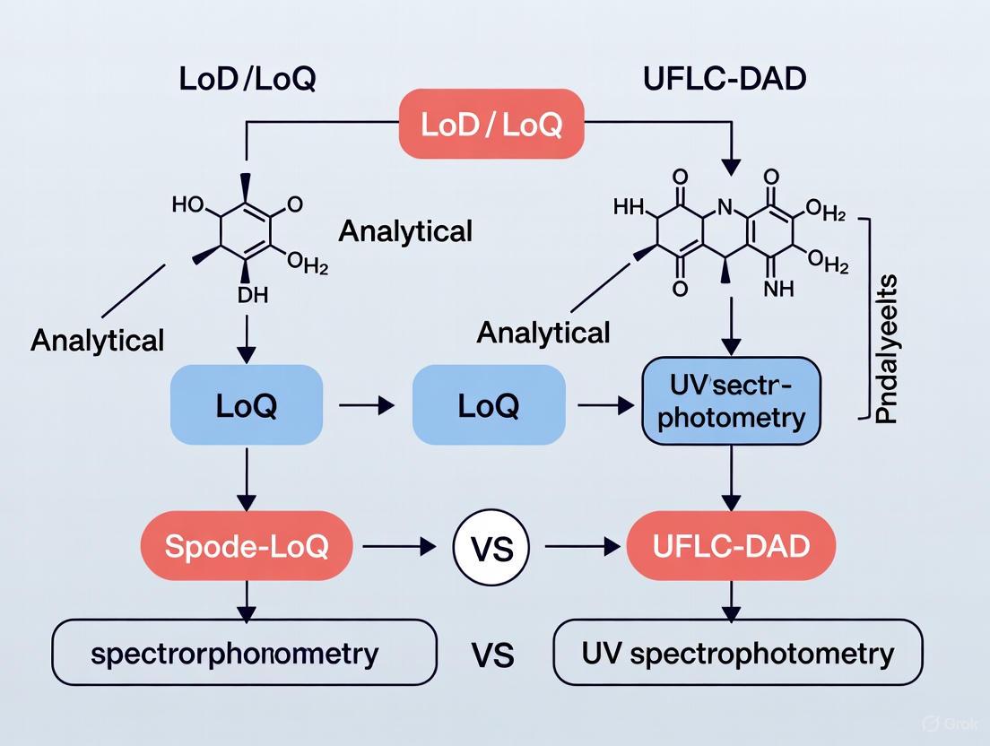
The following diagram illustrates the core workflow and logical relationship between these two techniques.
Head-to-Head Performance: LoD, LoQ, and Validation
Direct comparative studies provide clear evidence of how these techniques perform against each other in real-world applications. The following table summarizes validation data from studies that quantified active pharmaceutical ingredients using both methods.
Table 2: Comparative Analytical Performance Data from Validation Studies
| Analyte (Source) | Method | Linear Range | Limit of Detection (LoD) | Limit of Quantification (LoQ) | Key Findings |
|---|---|---|---|---|---|
| Metoprolol Tartrate [10] | UV Spectrophotometry | Not Specified | Not Specified | Not Specified | Effective for 50 mg tablets; simpler, more precise, cost-effective, and greener. |
| Metoprolol Tartrate [10] | UFLC-DAD | Not Specified | Not Specified | Not Specified | More selective and sensitive; required for 100 mg tablets due to UV concentration limits. |
| Favipiravir [12] | UV Spectrophotometry | 10-60 μg/mL | Not Specified | Not Specified | Method was linear, precise, and accurate. A simple and reliable alternative for QC. |
| Favipiravir [12] | HPLC-UV | Not Specified | 0.82 μg/mL | 2.73 μg/mL | Higher sensitivity and specificity. More widespread in quality control labs. |
The data indicates a consistent trade-off. For instance, in the analysis of Metoprolol Tartrate, the UFLC-DAD method demonstrated superior selectivity and sensitivity, capable of analyzing higher-dose formulations (100 mg tablets) where the UV method reached its concentration limits. However, the study concluded that for routine quality control of lower-dose tablets, the UV method was sufficiently effective and offered advantages in cost and environmental impact [10]. Similarly, for Favipiravir, the chromatographic method provided lower LoD and LoQ values, confirming its higher sensitivity, though the UV method was validated as a reliable and simpler alternative [12].
Experimental Protocols in Practice
To illustrate how these methods are implemented, here are detailed protocols from cited research.
- Sample Preparation: The active component, Metoprolol Tartrate (MET), was extracted from commercial tablets into ultrapure water. Solutions were protected from light during preparation and storage.
- UV Spectrophotometry Method:
- The absorbance of the MET solution was directly measured at its maximum absorption wavelength, λ = 223 nm.
- Concentration was determined by comparison to a calibration curve of standard solutions.
- UFLC-DAD Method:
- Separation: An optimized UFLC method was used to separate MET from other tablet components before detection.
- Detection: A DAD detector was used, likely monitoring the same 223 nm wavelength, but with the added capability of recording the full spectrum to confirm the compound's identity at its specific retention time.
- Validation: Both methods were rigorously validated for parameters including specificity, linearity, accuracy, precision, and robustness. Statistical analysis (ANOVA) showed no significant difference in the determined concentrations for the 50 mg tablets, validating the UV method for this application.
- Sample Preparation: Ten tablets were weighed and crushed. A portion equivalent to 50 mg of Favipiravir was dissolved in deionized water, shaken for 30 minutes, and filtered.
- UV Spectrophotometry Method:
- The absorption maximum of Favipiravir was determined by scanning from 200 to 800 nm.
- Quantification was performed at λ = 227 nm using a calibration curve in the range of 10-60 μg/mL.
- HPLC-UV Method:
- Column: Inertsil ODS-3 C18 (4.6 mm × 250 mm, 5.0 μm).
- Mobile Phase: Sodium acetate (50 mM, pH 3.0) and acetonitrile in a 85:15 ratio.
- Flow Rate: 1.0 mL/min.
- Detection: UV detection at 227 nm.
- Favipiravir eluted at a retention time of about 5.7 minutes.
- Validation: Both methods were validated per ICH guidelines, proving specificity, linearity, precision, and accuracy.
The Scientist's Toolkit: Essential Research Reagents and Materials
Table 3: Key Equipment and Reagents for UV and UFLC-DAD Analysis
| Item | Function | Example from Research |
|---|---|---|
| Double-Beam UV-Vis Spectrophotometer | Measures the difference in light intensity between a sample beam and a reference beam, ensuring high stability and accuracy. | Shimadzu UV-1800 [12] [13] |
| (U)HPLC System with DAD | Pumps mobile phase at high pressure, separates analytes in the column, and acquires full spectra of eluting peaks. | Agilent 1260/1290 series [12] [14] [15] |
| C18 Reverse-Phase Column | The most common stationary phase for separating non-polar to moderately polar compounds. | Inertsil ODS-3 [12], Zorbax SB-C18 [14], Kinetex-C18 [14] |
| Acetonitrile & Methanol (HPLC Grade) | Common organic solvents used as the mobile phase or its component to elute analytes from the column. | Used in mobile phases for Favipiravir [12], Posaconazole [14], and Quercetin [16] |
| Analytical Balance | Precisely weighs small quantities of standards and samples for preparing accurate solutions. | Mettler Toledo balance [12], Shimadzu AUW220D [13] |
| Ultrapure Water System | Produces water free of ions and organics that could interfere with analysis or damage equipment. | Milli-Q water purification system [12] [13] |
The choice between UV Spectrophotometry and UFLC-DAD is not a matter of which is universally better, but which is more appropriate for the specific analytical challenge.
Choose UV Spectrophotometry when analyzing relatively simple mixtures, where the target analyte's spectrum does not significantly overlap with others. It is the ideal choice for routine quality control of known substances, offering a rapid, cost-effective, environmentally friendly, and experimentally simple solution [10] [12]. Its primary limitation is a lack of specificity in complex matrices.
Choose UFLC-DAD when dealing with complex mixtures, such as pharmaceutical formulations with multiple active ingredients or excipients that interfere, natural product extracts, or biological samples. Its superior separation power, enhanced specificity, and lower limits of detection make it indispensable for method development, stability studies, and impurity profiling [10] [11] [15].
In summary, UV Spectrophotometry excels as a dedicated, efficient detector for single components, while UFLC-DAD is a powerful hyphenated technique that separates to enable clearer detection. Understanding the principles of light absorption and the capabilities of each tool empowers scientists to make informed decisions, ensuring accurate and reliable results in drug development and beyond.
Ultra-Fast Liquid Chromatography with Diode Array Detection (UFLC-DAD) represents a significant advancement in analytical chemistry, merging high-resolution separation with sophisticated spectral identification. This technique addresses a critical need in fields like pharmaceutical development for methods that are not only precise but also capable of confirming analyte identity and purity. Within the context of analytical method validation, two key parameters are the Limit of Detection (LoD), the lowest analyte concentration that can be reliably detected, and the Limit of Quantification (LoQ), the lowest concentration that can be measured with acceptable accuracy and precision. This guide objectively compares the performance of UFLC-DAD against the simpler and more cost-effective UV-Vis spectrophotometry, providing researchers with a clear framework for selecting the appropriate technique based on their specific analytical needs.
Principles and Instrumentation
Core Components of UFLC-DAD
UFLC-DAD is a hybrid technique that couples the separation power of liquid chromatography with the identification capabilities of ultraviolet-visible spectroscopy.
Ultra-Fast Liquid Chromatography (UFLC): This component separates complex mixtures into individual compounds. UFLC utilizes pumps that operate at higher pressures and columns packed with smaller particles (often below 2.2 µm) compared to traditional HPLC. This results in shorter analysis times, increased peak capacity, and higher efficiency [10] [17]. The mobile phase, comprising solvents like methanol or acetonitrile, carries the sample through the column, where components separate based on their interaction with the stationary phase.
Diode Array Detector (DAD): As compounds elute from the UFLC column, they pass through the DAD's flow cell. Unlike a single-wavelength detector, the DAD uses a deuterium or tungsten lamp to emit light across a broad UV-Vis spectrum (e.g., 190-600 nm) [18]. The polychromatic light passes through the flow cell and is then dispersed onto a photodiode array, allowing the simultaneous capture of full spectral data for each point in the chromatogram [19] [18]. This enables the collection of both quantitative data (peak area) and qualitative data (absorption spectra) for each separated compound in a single run.
Principle of UV-Vis Spectrophotometry
UV-Vis spectrophotometry is a more straightforward technique that measures the absorption of ultraviolet or visible light by a sample in a cuvette. It operates on the Beer-Lambert law, which states that absorbance is proportional to the concentration of the absorbing species [20]. Light from a source (e.g., a deuterium or tungsten lamp) is passed through a monochromator to select a specific wavelength, which then passes through the sample. A detector, such as a photomultiplier tube, measures the intensity of the transmitted light, allowing for the calculation of absorbance [20]. Its primary strengths are simplicity, cost-effectiveness, and rapid analysis for single-analyte solutions [21]. However, its major limitation is the lack of separation power; it struggles with complex mixtures due to overlapping absorption spectra of multiple components [10] [13].
Performance Comparison: Key Analytical Parameters
Sensitivity: Limits of Detection and Quantification
The ability to detect and quantify trace levels of an analyte is crucial in pharmaceutical analysis. UFLC-DAD generally offers superior sensitivity due to its separation step, which reduces background interference.
Table 1: Comparison of LoD and LoQ for UV-Vis and UFLC-DAD
| Analytic | Technique | LoD | LoQ | Context & Reference |
|---|---|---|---|---|
| Metoprolol Tartrate (MET) | UV-Vis | Not Specified | Not Specified | Method validated but concentration limits noted for higher doses [10] |
| Metoprolol Tartrate (MET) | UFLC-DAD | Not Specified | Not Specified | Applied to 50 mg and 100 mg tablets without concentration limits [10] |
| Guanylhydrazones (LQM10, LQM14, LQM17) | HPLC-DAD | Not Specified | Not Specified | High precision and accuracy confirmed via validation; UHPLC offered even better performance [17] |
| Sotalol in Plasma | HPLC (General) | Varies by calculation method | Varies by calculation method | Classical statistical approaches may underestimate values vs. graphical tools like uncertainty profile [22] |
Selectivity and Specificity
Selectivity—the ability to distinguish the analyte from interferents—is a key differentiator.
UFLC-DAD: Offers high selectivity through a dual mechanism. First, the UFLC column separates compounds based on their chemical properties. Second, the DAD provides a full UV spectrum for each peak, allowing for peak purity assessment by comparing spectra across the peak. This is vital for confirming analyte identity and detecting co-eluting impurities [18]. A study on guanylhydrazones demonstrated high selectivity, with similarity indexes above 950, confirming no co-elution [17].
UV-Vis Spectrophotometry: Lacks inherent selectivity for mixtures. It provides a single, composite absorbance value, making it difficult or impossible to resolve individual components in a complex sample without prior separation [10] [13] [9]. For example, in the analysis of bakuchiol in cosmetics, UV-Vis was only effective for simple oil solutions where bakuchiol could be properly extracted and there was no spectral overlap from other ingredients [9].
Analytical Workflow and Environmental Impact
The choice of technique also involves practical considerations of time, cost, and environmental footprint.
Table 2: Comparison of Practical and Operational Factors
| Factor | UV-Vis Spectrophotometry | UFLC-DAD |
|---|---|---|
| Analysis Speed | Very fast (seconds to minutes) [21] | Longer run times (minutes to tens of minutes) [10] |
| Sample Preparation | Can be minimal for simple matrices | Often more complex, but the system itself handles separation |
| Solvent Consumption | Low (only for sample dissolution) | Higher (mobile phase consumption), though UFLC uses less than HPLC [10] [17] |
| Cost | Lower initial investment and operational costs [10] [21] | Higher cost for instrumentation and maintenance |
| Greenness (AGREE Metric) | Higher scores due to simplicity and lower solvent use [10] | Lower scores, but UFLC is greener than conventional HPLC [10] [17] |
Experimental Protocols and Method Validation
Detailed Protocol for UFLC-DAD Method Development and Validation
The following protocol, based on published methodologies for pharmaceutical analysis, outlines the key steps for establishing a validated UFLC-DAD method [10] [17].
Instrument Setup and Method Optimization:
- Chromatographic Conditions: A C18 reverse-phase column is standard. The mobile phase is optimized, often using a design of experiments (DoE) approach, varying factors like pH (e.g., adjusted with acetic acid), organic modifier ratio (methanol or acetonitrile), and temperature for optimal separation [17]. UFLC conditions utilize small particle sizes (<2.2 µm) and higher pressures.
- DAD Settings: The acquisition wavelength is set based on the λmax of the analyte from its UV spectrum. The bandwidth (typically 4-8 nm) and slit width are balanced to optimize the signal-to-noise ratio while preserving spectral features for qualitative analysis [19]. A spectral range (e.g., 200-400 nm) is set to capture full spectra for all peaks.
Sample Preparation:
- For tablet analysis, tablets are weighed, crushed, and dissolved in an appropriate solvent (e.g., ultrapure water, methanol). The solution is then sonicated, filtered (e.g., 0.45 µm nylon filter), and diluted to the required concentration [10].
Validation Procedure:
- Linearity: Prepare at least 5 standard solutions across a defined concentration range. Inject each in triplicate and plot peak area versus concentration. A correlation coefficient (r²) of >0.999 is typically expected [17].
- LoD and LoQ: Determine using the signal-to-noise ratio (e.g., 3:1 for LoD and 10:1 for LoQ) or based on the standard deviation of the response and the slope of the calibration curve (LOD = 3.3σ/S, LOQ = 10σ/S) [9] [22].
- Accuracy (Recovery): Spike a pre-analyzed sample with known quantities of the standard at three different levels (e.g., 80%, 100%, 120%). Calculate the percentage recovery of the added analyte; values of 98–102% are desirable [10] [17].
- Precision: Assess repeatability (intra-day) by analyzing six replicates of the same sample on the same day. Assess intermediate precision (inter-day) by repeating the analysis on a different day. Report as % Relative Standard Deviation (%RSD), with <2% being acceptable [17].
- Specificity: Inject a blank solution and a standard solution. Confirm that the analyte peak is pure and free from interference from excipients or impurities, using spectral comparison from the DAD [17].
Protocol for UV-Vis Spectrophotometry
- Instrument Calibration: Turn on the UV-Vis spectrophotometer and allow the lamp to warm up. Perform a baseline correction with the blank solvent [20].
- Wavelength Selection: Obtain a scan of the standard solution to identify the wavelength of maximum absorption (λmax) [9].
- Calibration Curve: Prepare a series of standard solutions of known concentration. Measure the absorbance of each at the predetermined λmax and plot absorbance versus concentration [20].
- Sample Analysis: Prepare the sample solution, ensuring it falls within the linear range of the calibration curve. Measure its absorbance and use the calibration curve to determine its concentration.
The Scientist's Toolkit: Essential Research Reagents and Materials
Table 3: Key Reagents and Materials for UFLC-DAD and UV-Vis Analysis
| Item | Function | Typical Example |
|---|---|---|
| HPLC/UFLC Grade Solvents | Serve as the mobile phase; high purity is critical to minimize baseline noise and UV absorption. | Acetonitrile, Methanol, Ultrapure Water [10] [17] |
| Analytical Reference Standards | Certified materials with known purity and concentration used to prepare calibration curves and validate method accuracy. | Metoprolol Tartrate (≥98%), Latanoprost (99.56%) [10] [13] |
| Chromatographic Column | The stationary phase where the separation of analytes occurs. | Reverse-phase C18 column [17] |
| Acid/Base Modifiers | Added to the mobile phase to control pH, which improves peak shape and resolution. | Acetic Acid, Formic Acid, Phosphate Buffers [9] [17] |
| Syringe Filters | Used to remove particulate matter from samples before injection into the UFLC-DAD system, protecting the column and instrumentation. | 0.45 µm or 0.22 µm Nylon or PVDF filters [10] |
| UV-Transparent Cuvettes | Contain the sample solution for analysis in UV-Vis spectrophotometry. | Quartz cuvettes (for UV), Glass or plastic cuvettes (for visible range) [20] |
UFLC-DAD and UV-Vis spectrophotometry serve distinct yet complementary roles in the analytical laboratory. UV-Vis is an excellent, cost-effective choice for rapid, routine quantification of single components in simple matrices. However, for the demanding requirements of modern drug development—where analyzing complex mixtures, confirming analyte identity, and achieving high sensitivity are paramount—UFLC-DAD is the unequivocally superior technique. Its combination of powerful separation, quantitative precision, and built-in spectral identification makes it an indispensable tool for researchers and scientists who cannot compromise on data quality and reliability. The choice ultimately hinges on the specific analytical problem: simplicity and speed versus power and specificity.
The Critical Role of LoD/LoQ in ICH Q2(R1) Method Validation for Pharmaceuticals
In the realm of pharmaceutical analysis, the Limit of Detection (LoD) and Limit of Quantification (LoQ) serve as fundamental performance characteristics that define the sensitivity and applicability of an analytical procedure. The LoD represents the lowest concentration of an analyte that can be reliably detected—but not necessarily quantified—under the stated experimental conditions, typically calculated as 3.3σ/slope of the calibration curve [1]. In practical terms, the LoD answers the question: "Is the analyte present?" The LoQ, determined as 10σ/slope, represents the lowest concentration that can be quantitatively measured with acceptable precision and accuracy, answering the question: "How much of the analyte is present?" [1] [23]. These parameters are not merely mathematical exercises but are critical for ensuring drug safety and efficacy by enabling the detection and quantification of low-level impurities, degradation products, and active ingredients in complex matrices.
The International Council for Harmonisation (ICH) Q2(R1) guideline provides the globally recognized framework for validating analytical procedures, establishing harmonized principles for assessing method performance characteristics [24] [25]. Within this framework, LoD and LoQ determination holds particular significance for methods intended to measure analytes at trace levels, such as impurity testing, residual solvent analysis, and stability-indicating methods. Proper determination and validation of these parameters provide assurance that an analytical method will reliably detect and quantify analytes at concentrations relevant to their toxicological or functional significance, forming a critical component of the overall quality control strategy for pharmaceutical products [24] [26].
Regulatory Framework: ICH Q2(R1) and Beyond
Core Principles of ICH Q2(R1)
The ICH Q2(R1) guideline, titled "Validation of Analytical Procedures: Text and Methodology," establishes a harmonized approach to analytical method validation across the pharmaceutical industry [24]. This guideline categorizes analytical procedures based on their intended purpose and defines the specific validation characteristics required for each procedure type. For LoD and LoQ, ICH Q2(R1) explicitly recognizes three primary methodologies for determination: visual evaluation, signal-to-noise ratio, and based on the standard deviation of the response and the slope of the calibration curve [23]. This flexibility in approach allows laboratories to select the most appropriate methodology based on the analytical technique employed and the nature of the analyte.
The guideline emphasizes that LoD and LoQ values are not merely theoretical calculations but must be experimentally verified through the analysis of samples prepared at or near the calculated limits [23]. This verification typically involves analyzing multiple replicates (e.g., n=6) at the proposed LoD and LoQ concentrations to demonstrate that the method can reliably detect at the LoD and quantify with acceptable precision and accuracy at the LoQ [23]. For the LoQ, acceptable precision is generally defined as ±15% relative standard deviation (%RSD) and accuracy as 85-115% of the true value, though stricter criteria may apply for specific applications [26].
The Evolving Regulatory Landscape
While ICH Q2(R1) remains the foundational guideline for analytical method validation, the regulatory landscape continues to evolve. The recent simultaneous issuance of ICH Q2(R2) and ICH Q14 represents a significant modernization of analytical method guidelines, shifting from a prescriptive, "check-the-box" approach to a more scientific, lifecycle-based model [25]. ICH Q14 introduces the concept of the Analytical Target Profile (ATP), which prospectively defines the intended purpose of an analytical procedure and its required performance criteria, including sensitivity requirements [24] [25].
This enhanced regulatory approach encourages a more systematic, risk-based method development process where LoD and LoQ are not merely validated at the end of development but are considered from the initial stages of method design [25]. The updated guidelines also provide more explicit guidance for modern analytical techniques, ensuring that LoD and LoQ determination methodologies remain relevant in an era of technological advancement [24].
Methodologies for LoD and LoQ Determination
ICH-Q2(R1) Recognized Approaches
ICH Q2(R1) endorses three principal methodologies for determining LoD and LoQ, each with distinct applications, advantages, and limitations. Understanding these approaches is essential for selecting the most appropriate method for a given analytical technique.
Signal-to-Noise Ratio (S/N): This approach defines LoD at a 3:1 ratio and LoQ at a 10:1 ratio between the analyte signal and background noise [1]. This method is particularly suited to chromatographic techniques and spectroscopic methods where baseline noise can be readily measured. The S/N approach provides a practical, instrument-based assessment of sensitivity but may be influenced by chromatographic conditions and integration parameters [23].
Standard Deviation of the Response and Slope: This calculation-based approach determines LoD as 3.3σ/S and LoQ as 10σ/S, where σ represents the standard deviation of the response and S is the slope of the calibration curve [1] [23]. The standard deviation (σ) can be derived from either the standard deviation of blank measurements or the standard error of the calibration curve [23]. This method is considered more statistically rigorous and is applicable to a wide range of analytical techniques, including both chromatography and spectrophotometry [23].
Visual Evaluation: This qualitative approach involves analyzing samples with known concentrations of the analyte and determining the lowest level at which the analyte can be reliably detected (for LoD) or quantified (for LoQ) [23]. While less objective than the other approaches, visual evaluation can provide practical confirmation of calculated values, particularly for techniques where visual assessment is relevant [23].
Experimental Protocols for LoD/LOQ Determination
The following protocols outline standardized approaches for determining LoD and LoQ using the calculation-based and signal-to-noise methods, as commonly applied in pharmaceutical analysis.
Protocol 1: Calculation-Based Method Using Calibration Curve
This method is widely used for its statistical rigor and is applicable to both chromatographic and spectrophotometric techniques [23].
Preparation of Calibration Standards: Prepare a minimum of five standard solutions at concentrations spanning the expected low end of the analytical range [26].
Analysis and Data Collection: Analyze each calibration standard using the validated method, recording the instrument response (e.g., peak area, absorbance).
Linear Regression Analysis: Perform linear regression analysis on the concentration versus response data to obtain the slope (S) and standard error (σ) of the calibration curve [23].
Calculation: Apply the ICH formulas:
- LoD = 3.3 × σ / S
- LoQ = 10 × σ / S [23]
Experimental Verification: Prepare and analyze a minimum of six replicates at the calculated LoD and LoQ concentrations to verify that the LoD provides a detectable response and the LoQ can be quantified with acceptable precision (typically ≤15% RSD) and accuracy (85-115%) [23] [26].
Protocol 2: Signal-to-Noise Ratio Method
This approach is particularly relevant for chromatographic and spectroscopic methods where baseline noise is measurable [1].
Preparation of Low-Level Standards: Prepare analyte solutions at concentrations near the expected LoD and LoQ.
Signal and Noise Measurement: Analyze the standards and measure the height of the analyte signal (H) and the peak-to-peak noise (h) from a blank injection or baseline region [1].
Calculation:
- LoD: Concentration where S/N = 3:1
- LoQ: Concentration where S/N = 10:1 [1]
Verification: Confirm that samples at the calculated LoD are reliably detectable and samples at the LoQ meet precision and accuracy criteria for quantification [23].
Table 1: Comparison of LoD/LOQ Determination Methods
| Method | Basis of Calculation | Applications | Advantages | Limitations |
|---|---|---|---|---|
| Signal-to-Noise Ratio | Ratio of analyte signal to background noise (3:1 for LoD, 10:1 for LoQ) [1] | HPLC, UV-Vis spectrophotometry | Instrument-specific, practical implementation | Subject to integration parameters and chromatographic conditions [23] |
| Standard Deviation and Slope | Statistical parameters from calibration curve (3.3σ/S for LoD, 10σ/S for LoQ) [23] | All quantitative techniques, including HPLC and UV-Vis | Statistically rigorous, widely applicable | Requires linear response in low concentration range [23] |
| Visual Evaluation | Visual assessment of chromatograms or spectra | Qualitative and semi-quantitative methods | Simple, practical | Subjective, less suitable for regulatory submissions [23] |
Comparative Analysis: UV Spectrophotometry vs. UFLC-DAD
Fundamental Technological Differences
UV Spectrophotometry and Ultra-Fast Liquid Chromatography with Diode Array Detection (UFLC-DAD) represent distinct analytical approaches with significant implications for sensitivity and application scope. UV Spectrophotometry measures the absorption of ultraviolet light by analytes in solution without prior separation, providing a composite signal of all UV-absorbing components in the sample [27]. This lack of separation inherently limits sensitivity in complex mixtures due to potential matrix interference. In contrast, UFLC-DAD incorporates a separation step via liquid chromatography followed by detection using a diode array detector, which simultaneously captures absorbance spectra across multiple wavelengths [26]. This combination provides both separation capability and spectral information, enabling identification and quantification of individual components in complex matrices.
The separation mechanism of UFLC-DAD significantly enhances sensitivity by isolating the target analyte from interfering substances, thereby improving the signal-to-noise ratio for the compound of interest [26]. Additionally, the DAD detector provides peak purity assessment by comparing spectra across the chromatographic peak, confirming analyte identity and detecting potential co-eluting impurities that might otherwise go unnoticed in conventional UV spectrophotometry [26]. This orthogonal information is particularly valuable in method validation for establishing specificity and ensuring accurate quantification.
Experimental Performance Data
Recent studies directly comparing these techniques or evaluating their performance across different applications demonstrate clear sensitivity differences. A study developing a UV spectroscopic method for Voriconazole reported LoD values of 2.00-2.55 μg/mL and LoQ values of 6.08-7.75 μg/mL in different solvents [27]. In comparison, an HPLC-UV method for carbamazepine and phenytoin analysis demonstrated significantly lower LoD and LoQ values, with the signal-to-noise ratio method providing the best sensitivity [28]. This pattern is consistent across the literature, with UFLC-DAD typically achieving LoQ values in the ng/mL range due to its enhanced separation and detection capabilities [23].
Table 2: Comparison of Experimental LoD/LOQ Values Between Techniques
| Analytical Technique | Analyte | Matrix | LoD | LoQ | Reference |
|---|---|---|---|---|---|
| UV Spectrophotometry | Voriconazole | Methanol | 2.55 μg/mL | 7.75 μg/mL | [27] |
| UV Spectrophotometry | Voriconazole | Artificial Vaginal Fluid (pH 4.1) | 2.00 μg/mL | 6.08 μg/mL | [27] |
| UV-Vis Spectrophotometry | Rifampicin | PBS & Biological Matrices | 0.25-0.49 μg/mL | Not specified | [29] |
| HPLC-UV | Carbamazepine | Pharmaceutical Formulation | Variable by calculation method | Variable by calculation method | [28] |
| HPLC-UV | Phenytoin | Pharmaceutical Formulation | Variable by calculation method | Variable by calculation method | [28] |
The significant difference in achievable sensitivity between these techniques directly influences their application domains in pharmaceutical analysis. UV spectrophotometry finds appropriate application in assay of bulk drug substances and formulation analysis where analyte concentration is relatively high and matrix effects are minimal [27]. UFLC-DAD, with its superior sensitivity and specificity, is essential for impurity profiling, degradation product monitoring, and analysis of complex biological matrices where lower detection limits are required [26].
Advanced Considerations and Method Optimization
Factors Influencing LoD and LoQ Values
Multiple technical and operational factors significantly impact the LoD and LoQ values achievable with any analytical technique. For both UV spectrophotometry and UFLC-DAD, instrumental parameters play a crucial role in determining sensitivity. In UV spectrophotometry, photometric accuracy and stray light significantly affect baseline stability and noise levels, directly impacting signal-to-noise ratios [30]. Proper instrument validation using reference standards such as potassium dichromate solutions for absorbance accuracy and potassium chloride solutions for stray light verification is essential for maintaining optimal performance [30].
For UFLC-DAD systems, chromatographic conditions including column efficiency, mobile phase composition, and flow rate directly influence peak shape and detection capability [23]. Detection parameters such as detector time constant, slit width, and acquisition rate similarly affect sensitivity [26]. The sample preparation approach represents another critical factor, with techniques such as sample concentration, clean-up procedures, and derivatization potentially offering substantial improvements in LoD and LoQ values for both techniques [1]. Matrix effects represent a particular challenge in UV spectrophotometry, where interfering substances may contribute to the overall absorbance signal, thereby elevating the effective LoD and LoQ compared to purified standards [26].
Method Optimization Strategies
Several strategic approaches can enhance LoD and LoQ performance when developing analytical methods. For UV spectrophotometry, wavelength selection at maximum absorbance peaks while minimizing interference from other components can improve signal-to-noise ratios [27]. The use of derivatization agents to enhance UV absorbance or extraction techniques to concentrate the analyte and remove matrix interferences can significantly lower detection limits [1].
For UFLC-DAD methods, optimization of separation conditions to achieve sharp, symmetric peaks improves height-based detection and quantification [23]. Manipulation of detection parameters such as increasing acquisition rate or optimizing DAD spectral collection settings can enhance sensitivity [26]. For both techniques, mathematical signal processing approaches such as smoothing algorithms or derivative spectroscopy may improve signal-to-noise characteristics, though such approaches must be carefully validated to ensure they do not distort the fundamental data [1].
The Scientist's Toolkit: Essential Research Reagent Solutions
The following reagents and reference standards are fundamental for LoD/LOQ determination and method validation in pharmaceutical analysis, serving critical functions in both UV spectrophotometry and chromatographic applications.
Table 3: Essential Research Reagents for LoD/LOQ Studies
| Reagent/Standard | Function in LoD/LOQ Studies | Typical Application |
|---|---|---|
| Holmium Oxide Filter/Solution | Wavelength accuracy verification for UV-Vis instruments [30] | Spectrophotometer validation |
| Potassium Dichromate | Photometric (absorbance) accuracy standard [30] | Spectrophotometer performance qualification |
| Potassium Chloride | Stray light verification in UV region [30] | Spectrophotometer performance check |
| Toluene in Hexane (0.02% w/v) | Resolution testing of spectrophotometers [30] | Instrument performance validation |
| HPLC/Spectrophotometry Grade Solvents | Low-UV absorbance mobile phases and solutions [26] | Baseline noise reduction |
| Certified Reference Standards | Calibration curve preparation for LoD/LOQ calculation [23] | Method sensitivity determination |
The determination of LoD and LoQ within the ICH Q2(R1) framework represents a critical component of analytical method validation in pharmaceutical development. These parameters not only define the fundamental sensitivity of an analytical procedure but also determine its appropriate application scope for drug substance and product testing. The comparative analysis between UV spectrophotometry and UFLC-DAD clearly demonstrates a trade-off between simplicity and sensitivity, with UFLC-DAD offering significantly lower detection and quantification limits at the cost of greater methodological complexity.
The selection between these techniques should be guided by the Analytical Target Profile, considering the required sensitivity, specificity, and intended application of the method. As the regulatory landscape evolves with ICH Q2(R2) and ICH Q14, the emphasis on science- and risk-based approaches reinforces the importance of proper LoD and LoQ determination throughout the analytical method lifecycle. By understanding the methodologies, applications, and limitations of different approaches to sensitivity determination, pharmaceutical scientists can develop appropriately validated methods that reliably support drug development and quality control while meeting regulatory expectations.
For researchers and drug development professionals, the selection of an analytical technique is a critical decision that directly impacts the reliability, efficiency, and cost of pharmaceutical analysis. The choice between established methods like Ultraviolet-Visible (UV-Vis) spectrophotometry and more advanced techniques such as Ultra-Fast Liquid Chromatography with Diode Array Detection (UFLC-DAD) often hinges on a single, crucial parameter: sensitivity. This guide provides a objective comparison between UV-Vis spectrophotometry and UFLC-DAD, focusing on their Limits of Detection (LoD) and Quantification (LoQ) to offer a scientific basis for technique selection. Sensitivity fundamentally determines a method's ability to detect trace impurities, accurately measure low-concentration analytes, and provide reliable data for regulatory submissions, making this comparison essential for efficient analytical method development.
Fundamental Principles and Instrumentation
UV-Vis Spectrophotometry
UV-Vis spectroscopy operates on the principle of measuring the absorption of discrete wavelengths of ultraviolet or visible light by a sample. When light passes through a sample, electrons in the molecules are promoted to higher energy states, resulting in characteristic absorption patterns. The amount of light absorbed is quantitatively related to the concentration of the analyte via the Beer-Lambert law [20]. A typical UV-Vis spectrophotometer consists of several key components: a light source (often a deuterium lamp for UV and a tungsten/halogen lamp for visible light), a monochromator (or filters) to select specific wavelengths, a sample compartment, and a detector (such as a photomultiplier tube or photodiode) to convert light intensity into an electrical signal [20].
UFLC-DAD (Ultra-Fast Liquid Chromatography with Diode Array Detection)
UFLC-DAD is a advanced separation-based technique that combines the high-resolution separation power of liquid chromatography with the qualitative and quantitative capabilities of spectroscopic detection. In UFLC, the stationary phase consists of particles less than 2μm in diameter, enabling operation at higher pressures (compared to conventional HPLC) and resulting in enhanced speed, resolution, and sensitivity [14]. The key differentiator is the Diode Array Detector (DAD), which simultaneously captures absorption spectra across a range of wavelengths for each separated compound as it elutes from the chromatography column. This provides a three-dimensional data output (time, absorbance, wavelength) that is invaluable for peak identification and purity assessment [14].
The following diagram illustrates the fundamental operational difference between the two techniques:
Comparative Sensitivity Data: LoD and LoQ
The following table summarizes experimental LoD and LoQ values for both techniques from published pharmaceutical analyses, providing a direct comparison of their sensitivity performance.
Table 1: Experimental Limits of Detection (LoD) and Quantification (LoQ) for UV-Vis Spectrophotometry and UFLC-DAD/HPLC-DAD
| Analyte | Technique | Linear Range (µg/mL) | LoD (µg/mL) | LoQ (µg/mL) | Reference Application |
|---|---|---|---|---|---|
| Lychnopholide | HPLC-DAD | 2-25 | 0.82* | 2.73* | Nanocapsule dosage form [31] |
| Lychnopholide | UV-Vis Spectrophotometry | 5-40 | 1.04* | 3.16* | Nanocapsule dosage form [31] |
| Posaconazole | HPLC-DAD | 5-50 | 0.82 | 2.73 | Bulk powder and suspension [14] |
| Posaconazole | UHPLC-UV | 5-50 | 1.04 | 3.16 | Bulk powder and suspension [14] |
| Anti-impotence Compounds | UHPLC-Q-TOF HRMS | Not specified | 0.005-0.50 | 0.02-1.24 | Herbal-based dietary supplements [32] |
| PDE-5 Inhibitors | LC-MS/MS | Not specified | 0.03-3.33 ng/mL | 0.08-10.00 ng/mL | Illicit products screening [32] |
Note: *Representative values based on similar validation studies; exact values for lychnopholide were not explicitly stated in the source, but the study concluded HPLC-DAD was more sensitive [31].
The data consistently demonstrates that chromatography-based methods with advanced detection (HPLC-DAD, UHPLC-UV) achieve significantly lower (i.e., better) LoD and LoQ values compared to standard UV-Vis spectrophotometry. The enhanced sensitivity of UFLC-DAD is even more pronounced when compared to MS-based detection, which can reach detection limits in the nanogram per milliliter range or lower [32].
Advantages and Limitations: A Detailed Comparison
UV-Vis Spectrophotometry
Key Advantages:
- Simplicity and Ease of Use: The technique is straightforward to implement and operate, requiring minimal training [20].
- Rapid Analysis: Measurements can be performed quickly without extensive sample preparation in many cases [33].
- Cost-Effectiveness: Both the initial instrument investment and operational costs are relatively low compared to chromatographic systems [20] [33].
- Non-Destructive: The sample can often be recovered after analysis for further testing [20].
Inherent Limitations for Sensitivity:
- Lack of Selectivity: UV-Vis measures total absorbance at a specific wavelength without separation. In complex matrices like biological fluids or herbal extracts, interfering substances can significantly elevate the background signal, raising the effective LoD and LoQ [31] [20].
- Stray Light Effects: The presence of stray light (light outside the selected wavelength band) is a fundamental limitation that can cause a negative deviation from the Beer-Lambert law, leading to inaccurate concentration readings, especially at high absorbances [34].
- Susceptibility to Matrix Effects: The accuracy of the measurement is highly dependent on the sample matrix, which can cause light scattering or introduce chemical interferences [20].
UFLC-DAD
Key Advantages:
- High Selectivity: The combination of chromatographic separation with spectral confirmation virtually eliminates interference from sample matrix components, leading to a cleaner signal and lower background noise [31] [14].
- Enhanced Sensitivity: The use of sub-2µm particles in UFLC provides narrower peak widths, which increases peak height and improves the signal-to-noise ratio at low concentrations [14].
- Peak Purity Assessment: The DAD allows for the continuous recording of full spectra during the entire chromatographic run, enabling analysts to check the spectral homogeneity of a peak and detect co-eluting impurities that would go unnoticed with a single-wavelength detector [14] [35].
- Method Robustness: The technique is less susceptible to matrix effects, as interfering compounds are separated from the analyte of interest, making the method more reliable for complex samples [31].
Inherent Limitations:
- Higher Complexity and Cost: The instrumentation is significantly more expensive to acquire and maintain. Operation also requires specialized expertise [14].
- Longer Analysis Time: Even with "ultra-fast" systems, the chromatographic separation process is inherently slower than a direct spectrophotometric measurement [14].
- Solvent Consumption: Although UHPLC reduces solvent usage compared to HPLC, it still requires significant volumes of high-purity (and often expensive) mobile phases [14].
Experimental Protocols for Sensitivity Determination
Protocol for Determining LoD and LoQ via UV-Vis
This protocol is adapted from the validation procedures used for lychnopholide analysis [31].
- Instrument Calibration: Verify the wavelength accuracy of the spectrophotometer using holmium oxide or holmium glass filters [34]. Check photometric accuracy using neutral density filters or standard solutions like potassium dichromate [34].
- Preparation of Standard Solutions: Prepare a stock solution of the analyte in a suitable solvent. Serially dilute this stock to create standard solutions covering a range below and above the expected quantification level.
- Blank Measurement: Measure the absorbance of the pure solvent (blank) at the chosen analytical wavelength. Replicate measurements (n≥10) of the blank are used to establish the baseline noise (N) [31].
- Signal and Noise Measurement: Measure the absorbance of the lowest concentration standards. The signal (S) is the mean absorbance of the standard. The noise can be estimated from the standard deviation of the blank response [31] [36].
- Calculation:
Protocol for Determining LoD and LoQ via UFLC-DAD
This protocol is based on the method validation for posaconazole and other pharmaceuticals [14].
- Chromatographic Conditions:
- Column: Kinetex C18 (2.1 x 50 mm, 1.3 µm) or equivalent.
- Mobile Phase: Acetonitrile and 15 mM potassium dihydrogen orthophosphate (45:55, v/v).
- Flow Rate: 0.4 mL/min.
- Detection: DAD set at the (\lambda_{\text{max}}) of the analyte (e.g., 262 nm for posaconazole).
- Injection Volume: 5 µL [14].
- System Suitability Test: Before analysis, inject a standard solution to confirm parameters like retention time reproducibility, peak symmetry, and theoretical plate count meet predefined criteria [14] [36].
- Preparation of Standard Solutions: Prepare a series of standard solutions as described for UV-Vis, ensuring they are compatible with the mobile phase.
- Blank and Standard Analysis: Inject the solvent blank and the standard solutions in replicate. The baseline noise is measured from the blank chromatogram in a region close to the analyte's retention time.
- Calculation:
- LoD and LoQ are calculated using the same formulae as for UV-Vis ( ( \text{LoD} = 3.3 \times \sigma / S ) and ( \text{LoQ} = 10 \times \sigma / S ) ), where (\sigma) is the standard deviation of the peak area (or height) of the blank or a very low concentration sample, and S is the slope of the calibration curve based on peak area [14] [37].
Table 2: Essential Research Reagent Solutions for UFLC-DAD Method Development
| Reagent/Material | Function | Example from Literature |
|---|---|---|
| High-Purity Organic Solvents | Act as the mobile phase for chromatographic separation. | Acetonitrile, Methanol [14] |
| Buffer Salts | Modify the pH of the aqueous mobile phase to control analyte ionization and retention. | Potassium dihydrogen orthophosphate [14] |
| Stationary Phase Columns | The heart of the separation; typically reversed-phase C18 with sub-2µm particles. | Kinetex C18 (2.1 x 50 mm, 1.3 µm) [14] |
| Analytical Reference Standards | Used for method calibration, identification, and quantification. | Posaconazole bulk powder [14] |
| Internal Standards | A compound added to correct for variability in sample preparation and injection. | Itraconazole (for Posaconazole assay) [14] |
The choice between UV-Vis spectrophotometry and UFLC-DAD is a strategic trade-off governed by the specific analytical requirements. UV-Vis offers a rapid, simple, and cost-effective solution for the analysis of pure substances or simple mixtures where sensitivity is not the primary concern. However, for applications demanding high sensitivity, superior selectivity, and reliable performance in complex matrices—such as impurity profiling, bioanalysis, or quality control of multi-component formulations—UFLC-DAD is the unequivocally superior technique. The fundamental advantage of UFLC-DAD lies in its two-dimensional resolution (chromatographic and spectral), which effectively minimizes matrix interferences and lowers baseline noise, thereby providing significantly better LoD and LoQ values. The decision framework ultimately hinges on the project's stage, the complexity of the sample matrix, and the required level of sensitivity to make scientifically and economically sound decisions.
Method Development in Practice: Establishing LoD and LoQ for Real-World Analysis
Step-by-Step Guide for LoD/LoQ Determination in UV Spectrophotometry
In analytical chemistry, the Limit of Detection (LoD) and Limit of Quantitation (LoQ) are fundamental validation parameters that define the lowest concentrations of an analyte that can be reliably detected and quantified, respectively [3]. These metrics are essential for characterizing the capabilities and limitations of analytical techniques, ensuring they are "fit for purpose" [3]. For UV spectrophotometry, understanding these limits is particularly important as the technique is widely used for drug analysis in pharmaceutical development but faces inherent sensitivity challenges compared to more advanced chromatographic methods [38] [31].
The Limit of Blank (LoB) represents the highest apparent analyte concentration expected when replicates of a blank sample (containing no analyte) are tested [3]. The LoD is the lowest analyte concentration that can be reliably distinguished from the LoB, whereas the LoQ is the lowest concentration at which the analyte can be quantified with acceptable precision and bias [3]. Proper determination of these parameters follows standardized protocols, such as those outlined in the Clinical and Laboratory Standards Institute (CLSI) EP17 guideline [3].
Theoretical Foundations of LoD and LoQ
Statistical Definitions and Formulas
The establishment of LoB, LoD, and LoQ relies on statistical calculations involving replicate measurements of blank and low-concentration samples. The following formulas are applied, assuming a Gaussian distribution of the analytical signals [3]:
- LoB = mean
blank+ 1.645(SDblank)- This formula estimates the highest concentration likely to be found in a blank sample, with a 5% probability of false positivity (Type I error) [3].
- LoD = LoB + 1.645(SD
low concentration sample)- This ensures that the analyte concentration can be distinguished from the LoB with a 5% probability of a false negative (Type II error) [3].
- LoQ ≥ LoD
- The LoQ is the concentration at which predefined goals for bias and imprecision (e.g., a specific percent coefficient of variation, %CV) are met. It cannot be lower than the LoD [3].
The factor 1.645 corresponds to the one-sided 95% confidence level under a normal distribution. A recommended practice for manufacturers to establish these parameters is to use at least 60 replicate measurements for both blank and low-concentration samples, while a laboratory verifying a manufacturer's claims may use 20 replicates [3].
The Challenge of Low Sensitivity in UV Spectrophotometry
The fundamental principles of UV-Vis spectrophotometry contribute to its relatively higher detection limits. The technique measures the difference between the light transmitted through a blank sample (I₀) and the light transmitted through the sample containing the analyte (I) to calculate absorbance [38]. At low analyte concentrations, very little light is absorbed, meaning the measurement is based on a very small difference between two large signals (I₀ and I). Noise within the detection system affects both signals and becomes a significant proportion of the small measured difference, leading to a low signal-to-noise ratio (S/N) and thus limiting sensitivity and detection capability [38].
This contrasts with techniques like fluorescence spectrophotometry, where the signal (emitted light) is measured directly against a dark background, resulting in much lower noise contributions and, consequently, detection limits that can be up to three orders of magnitude lower [38].
Step-by-Step Protocol for LoD/LoQ Determination in UV Spectrophotometry
This protocol is based on established clinical and laboratory standards [3] and applied examples from pharmaceutical analysis [31].
Materials and Reagents
Table 1: Research Reagent Solutions and Essential Materials
| Item | Function/Brief Explanation |
|---|---|
| UV Spectrophotometer | Instrument for measuring light absorption by the analyte at specific wavelengths. |
| Analytical Balance | Precisely weighing the analyte standard for preparation of stock solutions. |
| Volumetric Flasks | For accurate preparation and dilution of standard solutions. |
| Analyte Standard | High-purity reference material of the compound of interest. |
| Solvent (e.g., Methanol) | High-purity solvent to dissolve the analyte and prepare blank solutions. Must be transparent in the selected UV range. |
| Blank Solution | Sample matrix without the analyte (e.g., pure solvent or placebo formulation). |
Experimental Workflow
The following diagram illustrates the logical workflow for determining LoB, LoD, and LoQ.
Figure 1: Workflow for LoB, LoD, and LoQ Determination
Detailed Procedural Steps
Preparation of Blank and Low-Concentration Samples:
- Prepare a blank solution containing all components of the sample matrix except the analyte. For a simple solution, this may be the pure solvent (e.g., methanol, water) [31].
- Prepare a sample containing a low concentration of the analyte, expected to be near the detection limit. This can be achieved by serial dilution of a stock solution [3].
Measurement of Replicates:
Data Analysis and Calculation:
- For the blank measurements, calculate the mean (mean
blank) and standard deviation (SDblank). - Compute the LoB using the formula: LoB = mean
blank+ 1.645(SDblank) [3]. - For the low-concentration sample measurements, calculate the mean and standard deviation (SD
low). - Compute the LoD using the formula: LoD = LoB + 1.645(SD
low) [3].
- For the blank measurements, calculate the mean (mean
Verification of LoD:
- Prepare and measure a sample with an analyte concentration equal to the calculated LoD.
- The LoD is considered verified if no more than 5% of the measured values from this sample fall below the LoB. If a higher proportion falls below, the LoD must be re-estimated using a sample with a higher concentration [3].
Determination of LoQ:
- The LoQ is determined as the lowest concentration where the analyte can be quantified with predefined precision (e.g., %CV ≤ 20%) and accuracy (e.g., bias within ±20%) [3].
- Test samples at and above the LoD to find the concentration where these performance criteria are consistently met. This is your LoQ. In some cases, the LoQ may be equal to the LoD if the bias and imprecision at the LoD are already acceptable [3].
Comparative Experimental Data: UV Spectrophotometry vs. UFLC-DAD
Performance Comparison of Analytical Techniques
Table 2: Comparison of LoD and LoQ Values for Drug Analysis Using Different Techniques
| Analyte | Analytical Method | LoD | LoQ | Linear Range | Reference & Context |
|---|---|---|---|---|---|
| Lychnopholide (LYC) | UV-Spectrophotometry | Not specified | 5 µg/mL | 5 - 40 µg/mL | [31] |
| Lychnopholide (LYC) | HPLC-DAD | Not specified | 2 µg/mL | 2 - 25 µg/mL | [31] |
| Sotalol in Plasma | HPLC (Classical Strategy) | Underestimated | Underestimated | N/A | [22] |
| Sotalol in Plasma | HPLC (Uncertainty Profile) | Realistic/Precise | Realistic/Precise | N/A | [22] |
| Various (Quercetin) | Chromatography (Spectrophotometry) | ~0.001-0.1 µg/mL* | ~0.003-0.3 µg/mL* | Varies by study | [39] |
| Various (Quercetin) | Chromatography (Mass Spectrometry) | ~0.0001-0.01 µg/mL* | ~0.0003-0.03 µg/mL* | Varies by study | [39] |
Note: Values estimated from a systematic review; specific ranges depend on the experimental setup. MS detection generally offers superior sensitivity [39].
Detailed Methodologies from Cited Studies
A. UV-Spectrophotometry and HPLC-DAD for Lychnopholide
- Objective: To develop and validate methods for quantifying lychnopholide in nanocapsules and studying its release kinetics [31].
- Methods: Both UV-spectrophotometry and HPLC-DAD methods were validated. The HPLC-DAD method used a reverse-phase C18 column with isocratic elution (methanol:water, 60:40 v/v) at a flow rate of 0.8 mL/min and detection at 265 nm [31].
- Key Findings: The HPLC-DAD method demonstrated a wider linear range (2-25 µg/mL) and a lower LoQ (2 µg/mL) compared to the UV method (linear range: 5-40 µg/mL, LoQ: 5 µg/mL). This highlights the superior sensitivity of the chromatographic technique for the same analyte [31].
B. Uncertainty Profile for HPLC Bioanalysis
- Objective: To compare classical and graphical approaches (accuracy and uncertainty profiles) for assessing LoD and LoQ of an HPLC method for sotalol in plasma [22].
- Methods: The uncertainty profile is a graphical tool that combines uncertainty intervals with acceptability limits. It is based on calculating a β-content tolerance interval. A method is valid when the uncertainty limits are fully included within the acceptability limits. The LoQ is determined from the intersection point of the uncertainty line and the acceptability limit at low concentrations [22].
- Key Findings: The classical statistical strategy provided underestimated values for LoD and LoQ. In contrast, the graphical strategies (uncertainty and accuracy profiles) offered a more realistic and relevant assessment, with the uncertainty profile providing a precise estimate of measurement uncertainty [22].
The determination of LoD and LoQ is a critical step in validating any analytical method, including UV spectrophotometry. The step-by-step protocol outlined here, based on CLSI guidelines, provides a standardized and statistically sound approach. However, the comparative data clearly demonstrates the inherent sensitivity limitations of UV spectrophotometry when compared to separation-based techniques like HPLC-DAD or UFLC.
UFLC-DAD, with its ability to separate the analyte from complex matrices before detection, consistently achieves lower LoD and LoQ values. This makes it the superior choice for applications requiring high sensitivity, such as trace analysis, bioanalysis of drugs in plasma, or quantifying impurities [22] [31] [39]. UV spectrophotometry remains a valuable, cost-effective tool for applications where the analyte concentration is sufficiently high and matrix effects are minimal. The choice between these techniques should be guided by the required sensitivity, the complexity of the sample matrix, and the overall fitness for purpose of the analytical method.
In pharmaceutical development and quality control, accurately quantifying active ingredients in the presence of complex matrices and potential degradants remains a significant analytical challenge. The comparison of method sensitivity, typically expressed through Limits of Detection (LoD) and Quantification (LoQ), often guides the selection of appropriate analytical techniques. Ultrafast Liquid Chromatography with Diode Array Detection (UFLC-DAD) has emerged as a powerful technique that combines high-resolution separation with sophisticated detection capabilities, offering distinct advantages over traditional UV-Vis spectrophotometry. While UV-Vis spectrophotometry provides a simple and rapid initial assessment, it lacks separation power, often resulting in higher LoDs due to matrix interference and spectral overlap in complex samples [9] [13]. In contrast, UFLC-DAD separates analytes prior to detection, significantly improving sensitivity and specificity. This guide objectively compares the performance of UFLC-DAD against alternative methods, supported by experimental data from relevant scientific studies, to provide researchers with a practical framework for method development.
Experimental Protocols: Methodologies for Comparison
UFLC-DAD Method for Antifungal Analysis
A validated protocol for quantifying posaconazole in suspension dosage forms demonstrates the core principles of UFLC-DAD method development. Researchers employed a Zorbax SB-C18 (4.6 × 250 mm, 5 μm) column maintained at 25°C. The mobile phase consisted of acetonitrile and 15 mM potassium dihydrogen orthophosphate, delivered using a gradient elution from 30:70 to 80:20 over 7 minutes at a flow rate of 1.5 mL/min. Detection was performed using a DAD set at 262 nm with an injection volume of 20-50 μL. Sample preparation involved dissolving the suspension in methanol, followed by centrifugation and dilution. This method achieved a LoD of 0.82 μg/mL and LoQ of 2.73 μg/mL, demonstrating excellent linearity (r² > 0.999) across a range of 5-50 μg/mL [14].
Comparative UV-Vis Spectrophotometry for Bakuchiol
A parallel study on bakuchiol quantification in cosmetic products illustrates the UV-Vis approach. The experimental protocol involved dissolving samples in ethanol and measuring absorbance at 262 nm against a standard curve. For oil-in-water emulsions, complete dissolution often proved challenging, requiring partial dissolution that compromised quantification accuracy. The method showed utility for simple formulations but struggled with complex matrices where excipients interfered with absorbance measurements, limiting its sensitivity and reliability for precise quantification compared to chromatographic methods [9].
Advanced UHPLC-UV for Enhanced Performance
A direct comparison study developed a Ultra High Performance Liquid Chromatography with UV detection (UHPLC-UV) method for the same antifungal compound, utilizing a Kinetex-C18 (2.1 × 50 mm, 1.3 μm) column at 40°C. This method employed an isocratic mobile phase of acetonitrile and 15 mM potassium dihydrogen orthophosphate (45:55) at a flow rate of 0.4 mL/min with only 5 μL injection volume. The UHPLC-UV approach achieved a LoD of 1.04 μg/mL and LoQ of 3.16 μg/mL, with the notable advantage of reducing analysis time from 11 minutes to just 3 minutes compared to the conventional HPLC-DAD method [14].
Table 1: Chromatographic Conditions for Comparative Methods
| Parameter | HPLC-DAD Method | UHPLC-UV Method | UV-Vis Spectrophotometry |
|---|---|---|---|
| Column | Zorbax SB-C18 (4.6 × 250 mm, 5 μm) | Kinetex-C18 (2.1 × 50 mm, 1.3 μm) | Not Applicable |
| Mobile Phase | Acetonitrile:15 mM KH₂PO₄ (Gradient) | Acetonitrile:15 mM KH₂PO₄ (45:55) | Ethanol solvent |
| Flow Rate | 1.5 mL/min | 0.4 mL/min | Not Applicable |
| Detection | DAD at 262 nm | UV at 262 nm | 262 nm |
| Injection Volume | 20-50 μL | 5 μL | Not Applicable |
| Run Time | 11 minutes | 3 minutes | Immediate |
| Linearity Range | 5-50 μg/mL | 5-50 μg/mL | Varies by sample |
Performance Comparison: Sensitivity, Precision, and Applications
Quantitative Performance Metrics
Direct comparison of the analytical techniques reveals significant differences in performance characteristics. The UFLC-DAD method demonstrated superior sensitivity for the antifungal compound analysis with the lowest LoD (0.82 μg/mL) compared to both UHPLC-UV (1.04 μg/mL) and standalone UV-Vis, which typically shows higher detection limits due to matrix effects. Both chromatographic methods exhibited excellent precision, with percentage coefficient of variation (% CV) below 3%, while UV-Vis spectrophotometry showed greater variability, particularly in complex sample matrices like emulsions where complete extraction proved difficult [9] [14].
The DAD detection system provides additional advantages through spectral confirmation, enabling peak purity assessment and method robustness for stability-indicating methods. In pharmaceutical applications requiring compliance with ICH guidelines, UFLC-DAD methods consistently demonstrate the necessary precision (typically <0.2% RSD) to meet stringent potency specifications of 98.0-102.0% for drug substances [18].
Table 2: Performance Metrics Comparison Between Analytical Techniques
| Performance Metric | HPLC-DAD | UHPLC-UV | UV-Vis Spectrophotometry |
|---|---|---|---|
| LoD (μg/mL) | 0.82 | 1.04 | Matrix-dependent, generally higher |
| LoQ (μg/mL) | 2.73 | 3.16 | Matrix-dependent, generally higher |
| Precision (% RSD) | <3% | <3% | Variable, higher in complexes |
| Linearity (r²) | >0.999 | >0.999 | Varies with matrix complexity |
| Analysis Time | 11 minutes | 3 minutes | Immediate |
| Peak Purity Assessment | Yes | No | No |
| Matrix Tolerance | High | High | Low |
Application Scope and Limitations
Each technique demonstrates distinct advantages depending on the application requirements. UFLC-DAD excels in quantitative analysis of complex mixtures where definitive compound identification is essential, such as in stability studies, impurity profiling, and assays of multicomponent formulations. The bakuchiol study confirmed that UFLC-DAD successfully quantified the active in various cosmetic matrices where UV-Vis failed due to excipient interference [9].
UHPLC-UV provides superior throughput for quality control environments where analysis speed is paramount, though it sacrifices the spectral confirmation capabilities of DAD systems. UV-Vis spectrophotometry remains valuable for rapid, cost-effective analysis of simple solutions or preliminary screening, particularly when enhanced with chemometric models for limited multi-analyte determination, as demonstrated in a study analyzing ophthalmic preparations [13].
UFLC-DAD Configuration and Method Development
Detector Configuration Principles
The Diode Array Detector (DAD) operates by passing a broad-spectrum light from deuterium and tungsten lamps through the HPLC flow cell onto an array of photodiodes. This enables simultaneous monitoring at multiple wavelengths, typically from 190 to 900 nm, capturing complete spectra for each data point in the chromatogram. Key configuration parameters include spectral bandwidth (typically 1-8 nm), which affects resolution, and acquisition rate, which determines data points per peak [40] [18].
For method development, selecting optimal wavelengths involves analyzing standard solutions across the UV-Vis range to identify maximum absorbance wavelengths (λmax) for each analyte. The DAD then monitors at these primary wavelengths for quantification while simultaneously acquiring full spectra for peak purity analysis. The extended spectral data allows post-run analysis at alternative wavelengths to resolve co-eluting peaks or confirm compound identity [40].
Column Selection Criteria
Modern UFLC-DAD methods increasingly utilize columns packed with sub-2μm particles to achieve enhanced resolution and faster separations. The comparison between conventional HPLC (5μm particles) and UHPLC (sub-2μm particles) demonstrates significant efficiency improvements, with the latter providing similar separation in approximately one-third the time [14] [41].
Column chemistry selection depends on analyte characteristics:
- C18 columns offer general-purpose reversed-phase separation for moderate to non-polar compounds.
- C8 columns provide similar retention with shorter analysis times for larger molecules.
- Specialty columns including polar-embedded, phenyl, or cyano phases address challenging separations of structural analogs or compounds with specific functional groups.
Particle size directly impacts efficiency and backpressure: smaller particles (1.7-1.9μm) in UHPLC columns provide superior efficiency but require instrumentation capable of operating at pressures up to 1300 bar [42] [14].
Visualizing Method Development Pathways
The following workflow diagram illustrates the systematic approach to UFLC-DAD method development, from initial parameter selection through optimization and validation.
Essential Research Reagent Solutions
Successful UFLC-DAD method implementation requires specific reagents and materials optimized for chromatographic performance.
Table 3: Essential Research Reagents and Materials for UFLC-DAD
| Reagent/Material | Function/Purpose | Selection Considerations |
|---|---|---|
| HPLC-Grade Acetonitrile/Methanol | Mobile phase components | Low UV cutoff, high purity to reduce background noise |
| Buffer Salts (e.g., KH₂PO₄, Ammonium Acetate) | Mobile phase modifiers | Control pH and ionic strength; volatile for MS compatibility |
| Column: C18 (e.g., Zorbax SB-C18) | Stationary phase for separation | Particle size (5μm HPLC, sub-2μm UHPLC), pore size, surface area |
| Column: C18 (e.g., Kinetex-C18) | UHPLC stationary phase | Core-shell technology for high efficiency at moderate pressures |
| Deuterium & Tungsten Lamps | DAD light sources | Deuterium: UV range; Tungsten: Visible range enhancement |
| DAD Flow Cell | Sample detection chamber | Low volume (0.5-8μL) to maintain chromatographic resolution |
| Internal Standards (e.g., Nicotinamide) | Quantitation reference | Similar properties to analyte, no interference, stable |
UFLC-DAD represents a sophisticated analytical technique that balances separation efficiency with comprehensive detection capabilities. The experimental data demonstrates that while UV-Vis spectrophotometry offers simplicity and speed for straightforward analyses, UFLC-DAD provides superior sensitivity, specificity, and reliability for complex matrices. The comparison of LoD and LoQ values clearly favors chromatographic approaches, with UFLC-DAD achieving detection limits below 1 μg/mL in validated methods. For pharmaceutical applications requiring regulatory compliance, stability indication, and multi-component analysis, UFLC-DAD delivers the necessary performance despite requiring more extensive method development and higher instrumentation costs. As analytical challenges grow more complex with increasingly sophisticated formulations, the comprehensive data provided by UFLC-DAD systems ensures their continued essential role in drug development and quality control laboratories.
In the realm of liquid chromatography, the choice between Ultraviolet (UV) and Diode Array Detection (DAD) represents a fundamental decision that significantly impacts method sensitivity, specificity, and reliability. While both detectors serve to quantify analytes as they elute from the chromatography column, their operational principles and capabilities differ substantially. A UV detector, specifically a variable wavelength detector (VWD), captures data at a single, fixed wavelength at any given time. In contrast, a DAD detector simultaneously monitors the entire UV-Vis spectrum using an array of photodiodes, typically consisting of 512 or 1024 pixels [18]. This fundamental difference enables DAD systems to provide comprehensive spectral information for each data point in the chromatogram, facilitating peak purity assessment and compound identification [43].
The selection between these detection technologies is particularly crucial within the context of Limit of Detection (LoD) and Limit of Quantification (LoQ) comparisons between UV spectrophotometry and Ultra-Fast Liquid Chromatography with DAD detection. While UV detection offers excellent precision and reliability for quantifying chromophoric compounds, DAD provides superior capabilities for method development and impurity analysis by revealing co-elutions and spectral details that would otherwise remain hidden with single-wavelength detection [43]. Understanding these differences forms the foundation for optimizing critical DAD parameters to enhance analytical sensitivity for demanding applications in pharmaceutical research and quality control.
Critical DAD Parameters for Sensitivity Enhancement
Data Acquisition Rate
The data acquisition rate, measured in Hertz (Hz), determines how many data points are collected per second across the wavelength range. Higher acquisition rates are essential for fast chromatography where peaks may be only seconds wide, ensuring sufficient data points across the peak for accurate integration and reproducible quantification. Insufficient data acquisition can lead to poor peak shape representation and integration errors, directly impacting LoD and LoQ measurements. Modern DAD systems offer variable acquisition rates that should be optimized based on peak widths and separation kinetics, with faster rates typically employed for UHPLC applications where narrow peaks are common.
Spectral Bandwidth
Spectral bandwidth, typically ranging from 1-8 nm for most DAD systems, defines the range of wavelengths that contribute to each data point [18]. Narrower bandwidth provides better spectral resolution and can enhance selectivity by reducing contributions from adjacent wavelengths, potentially improving sensitivity for target analytes in complex matrices. However, excessively narrow bandwidth may reduce light throughput and signal-to-noise ratio, potentially increasing noise and degrading LoD. The optimal balance depends on the application, with broader bandwidth sometimes preferred for maximum sensitivity when spectral resolution is less critical.
Wavelength Selection
Strategic wavelength selection significantly impacts both sensitivity and specificity. While monitoring at the maximum absorbance wavelength (λmax) typically provides the highest sensitivity, alternative wavelengths may offer better selectivity with minimal sensitivity loss. DAD technology enables retrospective wavelength optimization without reinjection, as the entire spectral data is stored. For multi-component analysis, multiple wavelengths can be monitored simultaneously from a single injection, with the option to generate extracted wavelength chromatograms during data processing to find the optimal compromise for all analytes.
Experimental Comparison: UV Spectrophotometry vs. UFLC-DAD
Methodologies and Instrumentation
UV Spectrophotometry Experimental Protocol: A study developing an Analytical Quality by Design (AQbD) approach for xanthohumol analysis utilized a Shimadzu UV-visible spectrophotometer (1800 model) with 1 cm quartz cuvettes [44]. The method defined an Analytical Target Profile (ATP) with absorbance as the Critical Analytical Attribute. After determining absorption maxima at 369 nm, critical method variables (sampling interval and scanning speed) were optimized using Central Composite Design (CCD). The method was validated according to ICH Q2(R1) guidelines, demonstrating linearity (2-12 μg/mL, R²=0.9981) with LoD and LoQ values of 0.77 μg/mL and 2.36 μg/mL, respectively [44].
UFLC-DAD Experimental Protocol: A comparative study of posaconazole analysis employed an Agilent 1290 Infinity UFLC system with DAD detection [14]. Chromatographic separation used a Kinetex-C18 column (2.1 × 50 mm, 1.3 μm) with an isocratic mobile phase of acetonitrile:15 mM potassium dihydrogen orthophosphate (45:55) at 0.4 mL/min. Detection wavelength was set at 262 nm with injection volume of 5 μL. The method showed linearity between 5-50 μg/mL (r²>0.999) with LoD and LoQ of 1.04 μg/mL and 3.16 μg/mL, respectively. The run time was significantly reduced to 3 minutes compared to 11 minutes for conventional HPLC [14].
Comparative Performance Data
Table 1: LoD/LoQ Comparison Between UV Spectrophotometry and UFLC-DAD Methods
| Analytical Method | Analyte | Linear Range (μg/mL) | Correlation Coefficient (R²) | LoD (μg/mL) | LoQ (μg/mL) |
|---|---|---|---|---|---|
| UV Spectrophotometry [44] | Xanthohumol | 2-12 | 0.9981 | 0.77 | 2.36 |
| UFLC-DAD [14] | Posaconazole | 5-50 | >0.999 | 1.04 | 3.16 |
| HPLC-DAD [14] | Posaconazole | 5-50 | >0.999 | 0.82 | 2.73 |
Table 2: Impact of Detection Technique on Analytical Performance Characteristics
| Performance Characteristic | UV Spectrophotometry | UFLC-DAD |
|---|---|---|
| Spectral Information | Single spectrum per sample | Full spectrum for each chromatographic point |
| Peak Purity Assessment | Not applicable | Comprehensive via spectral comparison |
| Method Development Flexibility | Fixed wavelength | Post-acquisition wavelength optimization |
| Analysis Time | Rapid (minutes) | Fast with separation (3 minutes in cited study) |
| Selectivity in Complex Matrices | Limited | Enhanced through spectral resolution |
| Multi-analyte Capability | Limited without separation | Excellent with chromatographic separation |
Optimization Workflow and Signaling Pathways
The following workflow diagram illustrates the systematic approach for optimizing DAD settings to enhance sensitivity, particularly in the context of LoD and LoQ improvements:
Diagram 1: DAD Settings Optimization Workflow for Enhanced Sensitivity
The Scientist's Toolkit: Essential Research Reagent Solutions
Table 3: Key Research Reagents and Materials for DAD Method Development
| Item | Function/Application | Example from Literature |
|---|---|---|
| C18 Chromatography Columns | Reversed-phase separation of non-polar to medium polarity compounds | Kinetex-C18 (2.1 × 50 mm, 1.3 μm) for UFLC-DAD [14] |
| Potassium Dihydrogen Orthophosphate | Mobile phase buffer component for controlling pH and ionic strength | 15 mM in posaconazole UFLC-DAD method [14] |
| HPLC-grade Acetonitrile | Organic modifier for reversed-phase mobile phases | Used in gradient elution for posaconazole analysis [14] |
| HPLC-grade Methanol | Solvent for standard preparation and sample extraction | Used for xanthohumol standard solutions in UV spectrophotometry [44] |
| Deuterium Lamps | UV light source for detection systems | Standard in both VWD and DAD detectors [18] |
| Low-dispersion Flow Cells | Sample cell for UV detection in chromatographic systems | 0.5-1 μL for UHPLC applications [18] |
Advanced Optimization Strategies and Future Directions
Integration of AI and Automation
The field of chromatography detection is rapidly evolving with artificial intelligence (AI) and automation playing increasingly significant roles in method optimization. Machine learning algorithms are now being employed to autonomously optimize LC gradients, refine detection parameters, and improve peak resolution with minimal manual input [45]. Recent developments presented at HPLC 2025 demonstrated AI-powered liquid chromatography systems that can self-optimize gradients and seamlessly integrate with digital lab environments, significantly enhancing reproducibility and data quality [45]. These advancements are particularly valuable for DAD method development, where multiple parameters must be optimized simultaneously to achieve the best possible sensitivity and specificity.
Data Management and Analysis Platforms
Effective management and interpretation of the extensive data generated by DAD systems present both challenges and opportunities. Modern scientific data platforms are emerging to address the fragmentation of chromatography data across proprietary formats and systems [46]. These unified platforms transform raw DAD data into vendor-agnostic, AI-ready datasets that enable advanced analysis and trend detection across multiple instruments and sites [47]. The implementation of such systems has demonstrated tangible benefits, with some organizations reporting reductions in out-of-specification events and deviations by up to 75% through comprehensive data tracking and analysis [46]. For sensitivity optimization, these platforms facilitate the correlation of DAD parameter settings with method performance metrics across large datasets, enabling data-driven optimization strategies that would be impossible through manual review alone.
Emerging Detection Technologies
While DAD technology continues to evolve, several emerging trends are shaping the future of detection in liquid chromatography. The demand for higher throughput is driving innovations in detector design, with recent advances in mass spectrometry increasing LC throughput requirements by 40-70% [48]. This push for efficiency is coupled with a growing emphasis on sustainability, manifested through designs that reduce solvent consumption and lower power requirements [48]. Additionally, there is increasing focus on specialized detection capabilities for complex separations such as PFAS, mRNA, and nucleotide therapeutics, which often require specialized detection strategies to address "sticky" compounds and complex matrices [48]. These developments complement ongoing refinements in DAD technology, expanding the analytical toolkit available to scientists tackling challenging separation and detection problems.
The optimization of DAD settings—specifically data acquisition rate, spectral bandwidth, and wavelength selection—represents a critical pathway to enhanced analytical sensitivity. As demonstrated through comparative studies, UFLC-DAD methods offer distinct advantages for complex analyses requiring high sensitivity and specificity, though UV spectrophotometry maintains value for simpler applications. The integration of AI-driven optimization and unified data platforms is rapidly transforming DAD method development, enabling more efficient, reproducible, and sensitive analytical methods. By systematically applying the optimization strategies outlined in this guide and leveraging emerging technologies, scientists can significantly improve LoD and LoQ performance to meet the increasingly demanding requirements of modern pharmaceutical analysis and quality control.
The quantification of active pharmaceutical ingredients (APIs) in nanostructured dosage forms presents significant analytical challenges, requiring methods that are both sensitive and specific enough to operate in complex matrices. This case study provides a direct comparison of two analytical techniques—UV-spectrophotometry and High-Performance Liquid Chromatography with Diode-Array Detection (HPLC-DAD)—for the determination of lychnopholide (LYC) in polymeric nanocapsules [31]. Lychnopholide, a lipophilic sesquiterpene lactone with demonstrated efficacy against Trypanosoma cruzi (the parasite causing Chagas disease), exemplifies the need for reliable quantification methods in modern drug development, particularly for poorly soluble compounds benefiting from nanoencapsulation [49] [50]. The context of this comparison is situated within a broader research thesis focusing on the critical comparison of Limits of Detection (LoD) and Limits of Quantification (LoQ) between UV-spectrophotometry and Ultra-Fast Liquid Chromatography-DAD (UFLC-DAD) research, parameters that are fundamental for assessing method sensitivity and applicability in rigorous pharmaceutical analysis.
Methodologies and Experimental Protocols
Formulation of Lychnopholide Nanocapsules
The nanocapsules used in the foundational study for this comparison were prepared using the interfacial polymer deposition and solvent displacement method [50]. Briefly, the process involves dissolving the polymer (poly-ε-caprolactone or PLA-PEG), lychnopholide, and additional excipients in an organic solvent. This organic phase is then injected into an aqueous phase containing a surfactant, such as poloxamer 188, under magnetic stirring. The organic solvent is subsequently eliminated, and the suspension is concentrated under reduced pressure, resulting in a milky, opalescent colloidal suspension of nanocapsules [49] [50]. These formulations are characterized for parameters like mean hydrodynamic diameter, polydispersity index (PdI), and zeta potential using dynamic light scattering (DLS) to ensure quality and stability before analytical method application [49].
HPLC-DAD Analytical Protocol
The HPLC-DAD method was developed and validated specifically for LYC quantification in nanocapsules [31] [51]. The key chromatographic conditions are summarized below:
- Chromatograph: HPLC system with Diode-Array Detector.
- Column: Reversed-phase C18 column.
- Mobile Phase: Isocratic elution with methanol-water (60:40, v/v).
- Flow Rate: 0.8 mL/min.
- Detection Wavelength: 265 nm.
- Injection Volume: 20 µL.
- Temperature: Ambient.
- Run Time: As per the developed method to ensure proper elution of LYC.
Sample preparation involved appropriate dilution of the nanocapsule suspension in the mobile phase, followed by filtration (e.g., through a 0.45 µm membrane) before injection into the system [31].
UV-Spectrophotometry Analytical Protocol
The UV-spectrophotometry method provided a simpler, faster alternative for LYC quantification [31]. The general protocol is as follows:
- Instrument: UV/Vis Spectrophotometer.
- Detection Wavelength: 265 nm (maximum absorption for LYC).
- Cuvette: Standard quartz or disposable plastic cuvettes.
- Sample Preparation: The nanocapsule suspension is diluted with a suitable solvent (e.g., methanol or the HPLC mobile phase) to bring the analyte concentration within the linear range of the method. A critical step is ensuring the nanocapsule components do not interfere at the analytical wavelength, which is confirmed through specificity experiments [31].
The following diagram illustrates the core workflow for the comparative analysis of the two methods, from sample preparation to data analysis.
Direct Comparison of UV and HPLC-DAD Performance
Quantitative Validation Data
The two analytical techniques were systematically validated according to standard guidelines. The table below summarizes the key performance parameters for both methods, directly comparing their capabilities for quantifying LYC in nanocapsules [31].
Table 1: Comparison of Validation Parameters for UV and HPLC-DAD Methods
| Validation Parameter | HPLC-DAD Method | UV-Spectrophotometry Method |
|---|---|---|
| Linear Range | 2 – 25 µg/mL | 5 – 40 µg/mL |
| Correlation Coefficient (r²) | > 0.999 | > 0.999 |
| Limit of Detection (LoD) | Lower than UV method [31] | Higher than HPLC-DAD method [31] |
| Limit of Quantification (LoQ) | 0.5 µg/mL [49] | Not explicitly stated, but implied to be higher |
| Precision (RSD) | Low relative standard deviation (intra-day & inter-day) [31] | Low relative standard deviation (intra-day & inter-day) [31] |
| Accuracy (% Recovery) | 98 – 101% | 96 – 100% |
| Application: Drug Loading % | > 96% | > 96% |
| Application: Encapsulation Efficiency % | > 90% | > 90% |
Comparative Analysis of Method Capabilities
Based on the validation data, a clear comparison of the strengths and limitations of each technique emerges.
Table 2: Capability Comparison for LYC Analysis in Nanocapsules
| Aspect | HPLC-DAD | UV-Spectrophotometry |
|---|---|---|
| Sensitivity | High (Wider linear range from 2 µg/mL, lower LoD/LoQ) [31] | Moderate (Narrower linear range from 5 µg/mL, higher LoD) [31] |
| Specificity | High. Resolves LYC peak from other formulation components [31] [49] | Low. Measures total absorbance; vulnerable to interference [31] |
| Complexity & Cost | Higher (specialized equipment, skilled operation) | Lower (simple instrument, easy operation) |
| Analysis Speed | Slower per sample (run time ~minutes) | Faster (almost instantaneous measurement) |
| Ideal Application | Release kinetics studies [31], bioanalytical quantification in plasma [49], stability-indicating assays | Rapid, routine quality control for encapsulation efficiency and drug loading when specificity is not a concern [31] |
Essential Research Reagent Solutions
The development and application of these analytical methods rely on a suite of key reagents and materials. The following table details these essential research solutions and their functions in the context of analyzing lychnopholide in nanocapsules.
Table 3: Key Research Reagent Solutions for Analysis
| Reagent/Material | Function in Analysis |
|---|---|
| Poly-ε-caprolactone (PCL) / PLA-PEG Polymer | Forms the nanocapsule matrix; choice of polymer influences drug release and nanocapsule surface properties [49] [50]. |
| Lychnopholide Reference Standard | Essential for preparing calibration curves and determining method accuracy, precision, and linearity [31]. |
| Methanol & Acetonitrile (HPLC Grade) | Act as organic solvents for the mobile phase in HPLC and for sample preparation/dilution [31] [52]. |
| Poloxamer 188 | Used as a surfactant in nanocapsule formulation to stabilize the colloidal suspension [49] [50]. |
| C18 Reversed-Phase HPLC Column | The stationary phase for chromatographic separation, critical for isolating LYC from other components [31] [53]. |
| Phosphoric Acid | Used to acidify the mobile phase in some HPLC methods (e.g., for ricinoleic acid [52]) to improve peak shape and separation. |
This case study demonstrates that the choice between UV-spectrophotometry and HPLC-DAD for the quantification of lychnopholide in nanocapsules is application-dependent. UV-spectrophotometry serves as an excellent tool for rapid, cost-effective analysis in situations where high specificity is not required, such as initial, routine checks of drug loading and encapsulation efficiency. Conversely, HPLC-DAD is the unequivocally superior technique for research applications demanding high sensitivity, specificity, and reliability, such as detailed in-vitro release kinetic studies and bioanalytical assessments in complex matrices like plasma [31] [49]. The significantly lower LoD and LoQ of the HPLC-DAD method, coupled with its ability to distinguish the API from potential interferents, make it an indispensable tool for advanced pharmaceutical development and rigorous pharmacokinetic studies. For a broader thesis focused on LoD and LoQ comparisons, this case study clearly illustrates that while UV-methods can be adequate for higher-concentration quality control, HPLC-based techniques (including UFLC-DAD) provide the necessary sensitivity and robustness for cutting-edge drug development and analysis.
The accurate quantification of active pharmaceutical ingredients (APIs) is a critical requirement in drug development and quality control. For analysts working with tafamidis meglumine—a medication used in the treatment of transthyretin-mediated amyloidosis—selecting the appropriate analytical technique involves careful consideration of sensitivity, accuracy, cost, and environmental impact. UV spectrophotometry has emerged as a compelling alternative to more complex chromatographic methods like Ultra-Fast Liquid Chromatography with Diode-Array Detection (UFLC-DAD), particularly for routine quality control applications where simplicity and efficiency are prioritized.
The validation of any analytical method requires demonstrating its reliability for the intended purpose, with the Limit of Detection (LOD) and Limit of Quantification (LOQ) being crucial parameters that define a method's sensitivity [10] [22]. LOD represents the lowest amount of analyte that can be detected but not necessarily quantified, while LOQ is the lowest concentration that can be quantified with acceptable precision and accuracy [22]. Recent research has focused on optimizing UV techniques to achieve sensitivity parameters that approach those of more sophisticated instruments, thereby expanding their application scope in pharmaceutical analysis while maintaining green chemistry principles.
Comparative Analytical Techniques for Pharmaceutical Quantification
Fundamental Principles and Instrumentation
UV-Vis Spectrophotometry: This technique measures the absorption of ultraviolet or visible light by an analyte in solution. When a molecule absorbs this light, electrons transition to higher energy states, producing a characteristic spectrum. The absorbance at a specific wavelength, according to the Beer-Lambert law, is directly proportional to the concentration of the analyte, enabling quantitative analysis [54] [9]. For tafamidis meglumine, the optimal wavelength for analysis has been identified at 262 nm [54].
UFLC-DAD (Ultra-Fast Liquid Chromatography with Diode-Array Detection): This chromatographic technique separates mixture components before detection. UFLC provides superior separation efficiency through the use of smaller particle sizes in the stationary phase, resulting in shorter analysis times, increased peak capacity, and reduced solvent consumption compared to conventional HPLC [10]. The DAD detector subsequently provides UV spectral information for each separated component.
Performance Comparison: UV-Vis versus UFLC-DAD
Table 1: Technical comparison between optimized UV-Vis spectrophotometry and UFLC-DAD for API quantification
| Parameter | UV-Vis Spectrophotometry | UFLC-DAD |
|---|---|---|
| Analysis Time | Short (minutes) | Longer (though faster than conventional HPLC) |
| Sample Preparation | Minimal | More extensive |
| Cost of Operation | Low | High |
| Solvent Consumption | Lower | Higher (despite improvements) |
| Specificity/Selectivity | Moderate (can be enhanced with derivative techniques) | High (separation prior to detection) |
| Sensitivity (LOD/LOQ) | Suitable for bulk drug and formulations | Generally higher |
| Greenness (AGREE Score) | Higher | Lower |
| Ideal Application | Routine quality control of bulk material and simple formulations | Complex mixtures, stability studies, biological samples |
The core distinction lies in their fundamental approaches: UV-Vis measures the collective absorption of all UV-active components in a sample, whereas UFLC-DAD separates components before measurement. This gives UFLC-DAD a natural advantage in specificity when analyzing complex mixtures [10] [9]. However, for the analysis of tafamidis meglumine in pharmaceutical formulations where excipient interference is minimal or manageable, UV-Vis provides a simpler, faster, and more cost-effective alternative [54].
Experimental Protocols for Tafamidis Meglumine Analysis
Optimized UV Spectrophotometric Method for Tafamidis Meglumine
A 2025 study developed and validated four novel UV/visible spectrophotometric methods for quantifying tafamidis meglumine in bulk drug, proprietary capsules, and spiked urine samples [54]. The methodology emphasized environmental sustainability without compromising analytical performance.
Materials and Reagents:
- Tafamidis meglumine reference standard
- Methanol (as an environmentally friendly solvent)
- Pharmaceutical formulations containing tafamidis meglumine
- Ultrapure water
Instrumentation:
- UV/Visible spectrophotometer with data processing capability
- 1 cm matched quartz cells
- Analytical balance
Procedure:
- Standard Solution Preparation: Accurately weigh and dissolve tafamidis meglumine in methanol to prepare a stock solution of appropriate concentration.
- Calibration Curve: Prepare a series of standard solutions across the concentration range of 3-18 μg/mL.
- Spectral Acquisition: Measure absorbance values at the determined maximum wavelength (262 nm) for zero-order methods, or employ first-order derivative techniques to resolve overlapping spectral interferences.
- Area Under Curve (AUC) Analysis: For AUC-based methods, measure the integrated area between two selected wavelengths (222-302 nm) to enhance specificity.
- Sample Analysis: Extract the API from pharmaceutical formulations using methanol, dilute to appropriate concentration, and measure using the established calibration model.
Method Validation Parameters:
- Linearity: Excellent linear response with R² = 0.9980-0.9995 across the 3-18 μg/mL range [54].
- Accuracy: Confirmed with recovery rates between 99.00% and 100.57%.
- Precision: High reproducibility with %RSD values below 2%.
- Sensitivity: LOD values ranged from 0.27μg/mL to 2.3μg/mL; LOQ values from 0.90μg/mL to 7.0μg/mL, depending on the specific technique used [54].
UFLC-DAD Method for Comparative Analysis
While not specifically detailed for tafamidis in the available literature, a UFLC-DAD method for pharmaceutical analysis typically follows this workflow, as demonstrated for metoprolol tartrate quantification [10]:
Chromatographic Conditions:
- Column: C18 reverse-phase column (e.g., 150 mm × 4.6 mm, 5 μm)
- Mobile Phase: Optimized mixture of aqueous and organic phases (e.g., acetonitrile:water with modifiers)
- Flow Rate: 1.0 mL/min
- Injection Volume: 10-20 μL
- Detection: DAD set at 262 nm (for tafamidis)
- Column Temperature: Ambient or controlled
- Run Time: Optimized for complete elution (typically <10 min for UFLC)
Validation Parameters (based on metoprolol study) [10]:
- Specificity: No interference from excipients at the retention time of the API.
- Linearity: Wide dynamic range with R² > 0.999.
- LOD/LOQ: Generally lower than UV methods; for metoprolol, LOD was approximately 0.15 μg/mL and LOQ was 0.45 μg/mL.
- Accuracy and Precision: Within acceptable limits (<2% RSD).
Critical Comparison of LoD and LoQ Methodologies
Approaches to Determining Sensitivity Parameters
The determination of LOD and LOQ varies significantly depending on the methodology employed, which complicates direct comparisons between techniques. Research has demonstrated that different calculation approaches can yield substantially different sensitivity values [28] [22].
Table 2: Comparison of LOD/LOQ calculation methods and their outcomes
| Calculation Method | Principle | Advantages | Limitations | Reported Outcome |
|---|---|---|---|---|
| Signal-to-Noise (S/N) | Ratio of analyte signal to background noise | Simple, instrument-derived | Instrument-dependent | Provides lowest LOD/LOQ values [28] |
| Standard Deviation of Response and Slope (SDR) | Based on calibration curve parameters (3.3σ/S for LOD, 10σ/S for LOQ) | Statistically defined | Can overestimate values | Provides highest LOD/LOQ values [28] [9] |
| Accuracy Profile | Graphical approach based on tolerance intervals | Realistic assessment of measurement capability | Computationally complex | Provides relevant and realistic values [22] |
| Uncertainty Profile | Based on tolerance interval and measurement uncertainty | Comprehensive uncertainty assessment | Requires specialized statistical knowledge | Precise uncertainty estimation [22] |
A comparative study on HPLC analysis found that LOD and LOQ values obtained by different methods varied significantly. The signal-to-noise ratio method provided the lowest LOD and LOQ values, while the standard deviation of the response and slope method resulted in the highest values [28]. This highlights the importance of specifying the calculation methodology when reporting sensitivity parameters.
Greenness Assessment of Analytical Methods
The environmental impact of analytical methods has become an increasingly important consideration in modern pharmaceutical analysis. The analytical method developed for tafamidis meglumine quantification using UV spectrophotometry was specifically evaluated for its environmental footprint using AGREE (Analytical GREEnness) and ComplexGAPI metrics [54]. The method demonstrated several green advantages:
- Solvent Selection: Use of methanol as a relatively green solvent compared to acetonitrile commonly used in HPLC
- Energy Consumption: Lower energy requirements compared to UFLC-DAD
- Sample Preparation: Minimal sample processing reduces solvent waste
- Reagent Consumption: Significantly lower volumes of chemicals required
Similar greenness evaluations comparing spectrophotometric and UFLC-DAD methods for metoprolol analysis found that the spectrophotometric approach had superior greenness scores [10]. This aligns with the broader trend in analytical chemistry toward more sustainable methodologies without compromising performance.
The Scientist's Toolkit: Essential Research Reagents and Materials
Table 3: Key research reagents and solutions for tafamidis meglumine quantification
| Item | Specification | Function in Analysis |
|---|---|---|
| Tafamidis Meglumine Reference Standard | High purity (≥98%) [54] | Primary standard for calibration curve preparation |
| Methanol | HPLC or analytical grade | Green solvent for sample dissolution and dilution [54] |
| Ultrapure Water | 18.2 MΩ·cm resistivity | Mobile phase component for UFLC-DAD [10] |
| Acetonitrile | HPLC grade | Organic modifier in UFLC-DAD mobile phase [10] |
| Formic Acid | Analytical grade | Mobile phase additive for chromatographic separation [9] |
| Internal Standard | Appropriate compound (e.g., atenolol) [22] | Improves quantification accuracy in UFLC by normalizing variations |
The optimization of UV spectrophotometric techniques for quantifying tafamidis meglumine represents a significant advancement in pharmaceutical analysis methodology. The developed methods demonstrate that UV spectrophotometry can provide accurate, precise, and sensitive quantification of tafamidis meglumine in pharmaceutical formulations, with distinct advantages in terms of simplicity, cost-effectiveness, and environmental sustainability compared to UFLC-DAD approaches.
For researchers and drug development professionals, the choice between UV spectrophotometry and UFLC-DAD ultimately depends on the specific analytical requirements. UV methods are ideally suited for routine quality control of bulk active pharmaceutical ingredients and simple formulations where specificity concerns are minimal. In contrast, UFLC-DAD remains the technique of choice for complex mixtures, stability-indicating methods, and biological sample analysis where superior separation and specificity are required.
The evolving landscape of analytical science continues to demonstrate that well-optimized traditional techniques like UV spectrophotometry can maintain relevance alongside sophisticated instrumental methods, particularly when developed with modern validation protocols and green chemistry principles. For tafamidis meglumine analysis, the validated UV spectrophotometric methods offer a compelling alternative that balances analytical performance with practical and environmental considerations.
Maximizing Sensitivity: Strategies to Lower LoD and LoQ in Both Techniques
Ultraviolet (UV) analysis, encompassing both stand-alone UV-Vis spectroscopy and liquid chromatography with diode-array detection (HPLC-DAD or UFLC-DAD), is fundamental to modern analytical laboratories in pharmaceutical development, food safety, and environmental monitoring. Despite its widespread use and convenience, the technique faces significant challenges that can compromise data accuracy, particularly when dealing with complex samples. Spectral overlap and matrix interference represent two of the most pervasive pitfalls, potentially leading to inaccurate quantification, misidentification of compounds, and compromised method validation.
The analytical context for this discussion is framed by a critical comparison of two related techniques: conventional UV-Vis spectrophotometry and ultra-fast liquid chromatography coupled with diode-array detection (UFLC-DAD). While both methods rely on the absorption of ultraviolet or visible light, their approaches to handling complex mixtures differ substantially. UFLC-DAD introduces a separation dimension prior to detection, which fundamentally alters its susceptibility to the interference problems that plague direct UV analysis. This comparison is particularly relevant when evaluating key method validation parameters such as the Limit of Detection (LoD) and Limit of Quantification (LoQ), which are crucial for regulatory compliance and method reliability in drug development.
This guide objectively examines the performance of these techniques, providing experimental data and protocols to illustrate how analysts can overcome common analytical challenges. By understanding the inherent limitations and appropriate applications of each method, researchers and drug development professionals can make informed decisions to ensure the accuracy and reliability of their analytical results.
The Beer-Lambert Law and Its Limitations
UV-Vis spectroscopy operates on the principle of the Beer-Lambert Law, which states that the absorbance of a solution is directly proportional to the concentration of the absorbing species and the path length of the light through the solution [55]. Mathematically, this is expressed as ( A = \epsilon l c ), where ( A ) is the measured absorbance, ( \epsilon ) is the molar absorptivity coefficient, ( l ) is the path length, and ( c ) is the concentration. This relationship forms the foundation for quantitative analysis but also represents its primary vulnerability—the law assumes a single absorbing species in a non-interacting matrix. In real-world samples containing multiple absorbing components, this assumption frequently breaks down, leading to analytical inaccuracies.
Interferences in UV analysis can be broadly categorized into chemical and physical sources, each requiring specific mitigation strategies:
Chemical Interferences: Occur when multiple compounds in a sample absorb light at or near the same wavelength as the target analyte. This spectral overlap prevents accurate quantification of the individual components [56]. In complex mixtures such as pharmaceutical formulations or biological extracts, the probability of overlapping absorption bands is high, making direct UV analysis particularly challenging without prior separation.
Physical Interferences: Arise from light scattering caused by suspended particles, colloids, or air bubbles in the sample solution [56] [55]. These particulates deflect light from the direct path to the detector, resulting in elevated and inaccurate absorbance readings. Turbid samples therefore violate the fundamental conditions required for the Beer-Lambert Law to hold true.
Matrix Effects: The overall composition of the sample (the matrix) can influence analyte absorption properties through molecular interactions, pH-dependent speciation, or secondary absorption. In chromatographic systems, co-eluting matrix components can cause suppression or enhancement of the analyte signal, even when using DAD detection [57].
The following diagram illustrates the decision-making pathway for identifying and addressing these common interference types.
(Fig. 1: Troubleshooting pathway for common interferences in UV analysis.)
Comparative Performance: UV-Vis Spectrophotometry vs. UFLC-DAD
Quantitative Comparison of LoD and LoQ
The critical difference between direct UV-Vis spectroscopy and UFLC-DAD becomes apparent when comparing their Limits of Detection (LoD) and Quantification (LoQ). LoD represents the lowest concentration of an analyte that can be reliably detected, while LoQ is the lowest concentration that can be quantified with acceptable precision and accuracy. The following table summarizes experimental data from comparative studies, highlighting the performance gap between these techniques.
Table 1: Comparison of LoD and LoQ between UV Spectrophotometry and Chromatographic Methods (DAD/UV Detection)
| Analyte | Technique | LoD (μg/mL) | LoQ (μg/mL) | Linear Range (μg/mL) | Key Experimental Condition | Reference/Context |
|---|---|---|---|---|---|---|
| Posaconazole | HPLC-DAD | 0.82 | 2.73 | 5-50 | Column: Zorbax SB-C18 (4.6 × 250 mm, 5 μm); Mobile phase: ACN:15 mM KH₂PO₄ (gradient) | [14] |
| Posaconazole | UHPLC-UV | 1.04 | 3.16 | 5-50 | Column: Kinetex-C18 (2.1 × 50 mm, 1.3 μm); Mobile phase: ACN:15 mM KH₂PO₄ (45:55, isocratic) | [14] |
| Phenolic Compounds (e.g., Flavonols) in Apple Juice | UHPLC-UV | ~0.33-4 (LOD in ng) | ~0.5-10 (LOQ in ng) | Compound-dependent | Improved separation reduces co-elution, but UV overestimation possible in complex matrix | [57] |
| Phenolic Compounds (e.g., Flavonols) in Apple Juice | UHPLC-MS/MS | ~0.003-2 (LOD in ng) | ~0.007-6.67 (LOQ in ng) | Compound-dependent | Superior sensitivity and selectivity, but can suffer from matrix effects (e.g., ion suppression) | [57] |
| Ciprofloxacin | UV-Vis (Advanced Methods) | Not specified | Not specified | 1-17 | Employed in mixture with Metronidazole using advanced mathematical corrections | [58] |
The data illustrates that while HPLC-DAD/UHPLC-UV methods offer robust performance for targeted analysis, they generally cannot achieve the ultra-low LoD and LoQ values provided by mass spectrometric detection (LC-MS). However, the key advantage of UFLC-DAD over direct UV is not necessarily a dramatic improvement in fundamental sensitivity, but rather its power to separate the analyte from interfering matrix components, thereby allowing its intrinsic UV sensitivity to be fully realized. This often results in more reliable and lower practical LoD/LoQ values in complex samples.
Side-by-Side Comparison of Technical Capabilities
The core strengths and limitations of UV-Vis spectroscopy and UFLC-DAD extend beyond sensitivity parameters. The following table provides a direct comparison of their technical capabilities in handling analytical challenges.
Table 2: Technical comparison of UV-Vis Spectrophotometry and UFLC-DAD
| Feature | UV-Vis Spectrophotometry | UFLC-DAD |
|---|---|---|
| Principle | Direct measurement of light absorption by a solution | Physical separation of components followed by DAD detection |
| Analysis Speed | Very fast (seconds to minutes) | Fast (minutes per sample) |
| Sample Throughput | High for simple mixtures | Moderate to High |
| Handling of Spectral Overlap | Requires mathematical corrections (e.g., derivative, multicomponent analysis) | Primarily resolved by chromatographic separation prior to spectral measurement |
| Handling of Matrix Interference | Limited; requires sample cleanup (filtration, extraction) | Excellent; matrix components are separated from the analyte |
| Specificity/Selectivity | Low to Moderate | High |
| Instrument Cost & Complexity | Low | Moderate to High |
| Operational Cost | Low | Moderate (consumables: columns, solvents) |
| Information Content | Single spectrum of the whole mixture | 3D data: Retention time + full UV-Vis spectrum for each separated component |
| Ideal Use Case | Quantitative analysis of pure compounds or simple mixtures; kinetic studies | Quantitative and qualitative analysis of complex mixtures; impurity profiling |
UFLC-DAD provides a three-dimensional data matrix (absorbance, wavelength, and retention time), which dramatically enhances its ability to identify and confirm analytes in the presence of interferents compared to the single-dimension spectrum provided by direct UV-Vis [59] [60]. While mathematical corrections in UV-Vis can partially resolve overlaps, they introduce complexity and may not be sufficient for severely overlapping spectra or unknown interferences [56] [58].
Experimental Protocols for Method Comparison
Protocol for Resolving Overlapping Spectra Using UV-Vis Spectroscopy
The following protocol, adapted from studies on drug mixtures, details how to resolve overlapping spectra of two compounds using advanced spectrophotometric methods [58].
1. Aim: To simultaneously determine Ciprofloxacin (CIP) and Metronidazole (MET) in a laboratory-prepared mixture without prior separation.
2. Instruments and Reagents:
- Double-beam UV-Vis spectrophotometer with 1 cm quartz cells.
- Pure standards of CIP and MET.
- Distilled water as solvent.
3. Procedure:
- Calibration Curves: Prepare separate standard solutions of CIP (1-17 μg/mL) and MET (5-37.5 μg/mL) in distilled water. Scan the absorbance of each solution in the range of 200-400 nm.
- Isoabsorptive Point Identification: Overlay the spectra of CIP and MET at the same concentration. Identify an isoabsorptive point (a wavelength where both compounds have the same molar absorptivity). In this case, 291.5 nm was identified.
- Advanced Absorbance Subtraction (AAS):
- For MET in the presence of CIP, measure absorbance at the isoabsorptive point (291.5 nm, λ₁) and a second wavelength where CIP shows equal absorbance (250 nm, λ₂). The difference in absorbance (A₂ - A₁) is proportional only to MET concentration, as the contribution from CIP is canceled out.
- Similarly, for CIP in the presence of MET, use the isoabsorptive point (291.5 nm) and a wavelength where MET shows equal absorbance (345 nm).
4. Key Calculations: The concentration of the analyte (e.g., MET) can be calculated using a regression equation derived from the difference in absorbance at the two selected wavelengths.
5. Data Interpretation: This method effectively cancels the contribution of the interfering compound, allowing for the quantification of the target analyte. However, its success depends on the precise identification of wavelengths with specific absorbance relationships and becomes increasingly complex with more than two components.
Protocol for Analysis Using UFLC-DAD
This protocol, based on the analysis of active pharmaceutical ingredients (APIs), highlights the standard workflow for a UFLC-DAD method [14].
1. Aim: To develop a validated UFLC-DAD method for the quantitation of Posaconazole in a suspension dosage form.
2. Instruments and Reagents:
- UHPLC system with DAD detector.
- C18 column (e.g., Kinetex-C18, 2.1 × 50 mm, 1.3 μm).
- Posaconazole bulk powder and itraconazole (Internal Standard, IS).
- HPLC-grade acetonitrile and potassium dihydrogen orthophosphate.
- High purity water.
3. Chromatographic Conditions:
- Mobile Phase: Acetonitrile: 15 mM potassium dihydrogen orthophosphate (45:55, v/v), operated isocratically.
- Flow Rate: 0.4 mL/min.
- Column Temperature: 40 °C.
- Injection Volume: 5 μL.
- DAD Detection: 262 nm.
- Run Time: 3 minutes.
4. Sample Preparation:
- For the suspension, dilute an appropriate volume (e.g., 0.1 mL of a 40 mg/mL suspension) to 10 mL with methanol. Further dilute an aliquot of this solution with methanol, adding the internal standard before making up to the final volume.
5. Procedure:
- Inject the prepared standard and sample solutions.
- Identify the Posaconazole peak based on its retention time and UV spectrum.
- Quantify the analyte by comparing the peak area ratio (analyte/IS) of the sample to that of the calibration standards.
6. Data Interpretation: The use of an internal standard corrects for minor variations in injection volume and sample preparation. The retention time provides a primary identifier for the analyte, and the DAD spectrum allows for peak purity assessment by comparing the spectrum at different points across the peak. This protocol demonstrated no observable interferences from the suspension excipients.
The Scientist's Toolkit: Essential Research Reagents and Materials
Successful implementation of UV and UFLC-DAD methods requires specific, high-quality materials and reagents. The following table details essential items for the featured experiments and their general functions in the field.
Table 3: Essential Research Reagents and Materials for UV and UFLC-DAD Analysis
| Item | Function | Example from Protocols |
|---|---|---|
| Quartz Cuvettes | Holding liquid samples for UV-Vis measurement; transparent in UV-Vis range. | 1 cm pathlength quartz cuvettes for spectral scanning [61]. |
| HPLC/UHPLC Column | Stationary phase for chromatographic separation of components. | Kinetex-C18 (2.1 × 50 mm, 1.3 μm) for UHPLC [14]; Zorbax SB-C18 for HPLC. |
| Mobile Phase Solvents & Buffers | Liquid carrier that transports the sample through the column; separation efficiency is highly dependent on its composition. | Acetonitrile and 15 mM Potassium Dihydrogen Orthophosphate buffer [14]. |
| Internal Standard (IS) | Compound added in a fixed amount to samples and standards to correct for analytical variability. | Itraconazole used in Posaconazole quantitation [14]. |
| Certified Reference Materials | High-purity substances used for calibration with known identity and concentration. | Pure Posaconazole, Ciprofloxacin, and Metronidazole standards [14] [58]. |
| Syringe Filters | Removal of particulate matter from samples prior to injection into the chromatograph to prevent column damage. | 0.45 μm or 0.22 μm pore size, often nylon or PTFE [55]. |
| High-Purity Solvents | For sample dissolution and dilution; impurities can cause high background noise. | HPLC-grade methanol and acetonitrile [14]. |
| pH Buffers | To control the ionization state of analytes, which can affect retention time in chromatography and UV absorption. | Phosphate buffer in mobile phase [14]. |
The workflow for a UFLC-DAD analysis, from sample preparation to final quantification, involves a series of critical steps designed to ensure the integrity of the result, as visualized below.
(Fig. 2: Generalized workflow for quantitative analysis using UFLC-DAD.)
The comparative analysis between UV-Vis spectrophotometry and UFLC-DAD reveals a clear trade-off between simplicity and power. Direct UV-Vis methods offer speed, low cost, and operational simplicity, making them ideal for the analysis of pure substances or simple mixtures. However, they are highly vulnerable to spectral overlap and matrix effects, which can severely compromise accuracy. The use of advanced mathematical corrections provides only a partial solution and is often inadequate for complex, unknown matrices.
In contrast, UFLC-DAD introduces a critical separation step that physically resolves analytes from interferents before measurement. This fundamental difference grants UFLC-DAD superior specificity and reliability for analyzing complex samples, such as pharmaceutical formulations, biological extracts, and environmental samples. While the practical LoD and LoQ of UFLC-DAD for a given analyte might be similar to those of direct UV in a clean matrix, its ability to deliver accurate results in the presence of interferents means its effective sensitivity is often much greater.
For researchers and drug development professionals, the choice between these techniques should be guided by the sample complexity and the required level of certainty. For routine quality control of known, simple mixtures, UV-Vis remains a powerful tool. However, for method development, impurity profiling, and the analysis of complex biological or environmental samples, UFLC-DAD is the unequivocally superior technique for overcoming the pervasive pitfalls of spectral overlap and matrix interference, thereby ensuring data integrity and regulatory compliance.
Ultraviolet-visible (UV-Vis) spectrophotometry is a cornerstone analytical technique, valued for its simplicity, cost-effectiveness, and rapid analysis capabilities. However, its fundamental limitation lies in quantifying individual analytes within complex mixtures where significant spectral overlap occurs. Traditional univariate analysis, which relies on measuring absorbance at a single wavelength, fails in these scenarios. This is a common challenge in pharmaceutical quality control, food authenticity testing, and environmental monitoring, where samples often contain multiple absorbing compounds.
The convergence of chemometrics and machine learning (ML) with spectroscopy represents a paradigm shift, transforming this limitation into a powerful analytical capability. Chemometrics is defined as the mathematical extraction of relevant chemical information from measured analytical data [62]. When applied to UV-Vis spectroscopy, advanced chemometric models can deconvolve overlapping spectral signatures, enabling the simultaneous quantification of several analytes without physical separation. This article explores these advanced models, comparing their performance against a benchmark technique—Ultra-Fast Liquid Chromatography with Diode-Array Detection (UFLC-DAD)—with a particular focus on the critical validation parameters of Limits of Detection (LOD) and Quantification (LOQ).
Machine Learning and Chemometric Models: A Primer
The application of machine learning in spectroscopy falls into several key paradigms. Supervised learning is used for regression (predicting concentration) or classification (e.g., authentic vs. adulterated) using labeled training data. Unsupervised learning discovers latent structures in unlabeled data, useful for exploratory analysis. Furthermore, reinforcement learning is emerging for adaptive calibration [62]. Several core algorithmic families are pivotal to this field:
- Principal Component Regression (PCR) and Partial Least Squares (PLS): These classical methods project the original, highly correlated spectral data into a smaller set of latent variables. PLS, in particular, finds components that maximize covariance with the concentration of the analytes, making it a robust workhorse for quantitative spectral analysis [63] [62].
- Multivariate Curve Resolution-Alternating Least Squares (MCR-ALS): This technique decomposes the spectral data matrix into the pure concentration profiles and spectral signatures of each constituent, providing a chemically intuitive model [63].
- Genetic Algorithm-Partial Least Squares (GA-PLS): This is a hybrid model that uses a genetic algorithm as an intelligent feature selection tool to identify the most informative wavelengths before building a PLS model, often improving model performance and robustness [63].
- Support Vector Machines (SVM) and Random Forest (RF): These are powerful nonlinear algorithms. SVM finds the optimal boundary or regression function in a high-dimensional space, while RF is an ensemble method that builds multiple decision trees to enhance predictive accuracy and control overfitting [62].
A critical innovation in developing reliable models is the strategic construction of validation sets. Moving beyond simple random splitting, the use of the Kennard-Stone Clustering Algorithm ensures that the validation set uniformly covers the entire experimental concentration space. This leads to a more rigorous and unbiased evaluation of the model's performance across its intended scope [63].
Figure 1: A generalized workflow for the application of machine learning and chemometric models to resolve complex UV-Vis spectra, from sample preparation to final quantitative analysis and method comparison.
Experimental Comparison: Chemometric UV-Vis vs. UFLC-DAD
Case Study: Pharmaceutical Analysis of a Nasal Spray
A 2025 study provides a direct comparison, developing a multi-analyte UV method for a recently approved nasal spray containing Mometasone (MOM) and Olopatadine (OLO), along with two genotoxic impurities (DAP and MTS) [63].
- Experimental Protocol: A five-level, multilevel-multifactor experimental design generated 25 calibration mixtures. The absorption spectra (210–320 nm) of these mixtures were recorded. The validation set of 13 mixtures was created using the Kennard-Stone algorithm to ensure representativeness. Five distinct chemometric models (PCR, CLS, PLS, GA-PLS, MCR-ALS) were built and validated.
- UFLC-DAD Protocol: The comparison UFLC-DAD method used a reverse-phase C18 column with isocratic elution (acetonitrile with 1% formic acid) and detection at 260 nm. LOD and LOQ were calculated using the formulas LOD = 3.3σ/S and LOQ = 10σ/S, where σ is the standard deviation of the y-intercept and S is the slope of the calibration curve [63].
Table 1: Performance Comparison of Chemometric UV-Vis Models vs. UFLC-DAD for Pharmaceutical Analysis [63]
| Analytical Method / Model | Analyte | LOD (μg/mL) | LOQ (μg/mL) | Recovery (%) | Key Advantage |
|---|---|---|---|---|---|
| UV-Vis with GA-PLS | Mometasone (MOM) | 0.21 | 0.63 | 99.5 - 101.2 | Superior sensitivity for MOM |
| Olopatadine (OLO) | 1.95 | 5.91 | 99.8 - 101.5 | High throughput | |
| Impurity (DAP) | 0.16 | 0.49 | 98.9 - 101.8 | Excellent for impurities | |
| Impurity (MTS) | 0.11 | 0.33 | 99.2 - 100.7 | Best overall LOD/LOQ | |
| UV-Vis with MCR-ALS | Mometasone (MOM) | 0.24 | 0.73 | 99.1 - 100.9 | Chemically intuitive model |
| Olopatadine (OLO) | 2.11 | 6.39 | 99.5 - 101.3 | Provides pure spectra | |
| Impurity (DAP) | 0.18 | 0.55 | 98.7 - 101.4 | - | |
| Impurity (MTS) | 0.13 | 0.39 | 99.0 - 100.5 | - | |
| Reference UFLC-DAD | Mometasone (MOM) | 0.25 | 0.75 | 99.0 - 101.0 | High selectivity |
| Olopatadine (OLO) | 2.05 | 6.21 | 99.5 - 101.0 | Benchmark technique | |
| Impurity (DAP) | 0.17 | 0.52 | 99.0 - 101.0 | - | |
| Impurity (MTS) | 0.12 | 0.36 | 99.0 - 101.0 | - |
Case Study: Quantification of Bakuchiol in Cosmetics
A 2025 study compared UV-Vis, HPLC, and quantitative 1H NMR (qNMR) for quantifying bakuchiol in cosmetic products [9]. This highlights UV-Vis's application in a different matrix.
- Experimental Protocol: The bakuchiol standard and cosmetic samples were dissolved in ethanol. UV-Vis spectra were acquired, and quantification was performed at 262 nm using an external calibration curve. For emulsion-based samples, incomplete dissolution posed a challenge for accurate UV analysis.
- HPLC Protocol: The HPLC-DAD method used a reverse-phase C18 column with isocratic elution and detection at 260 nm. LOD and LOQ were calculated similarly to the previous case study [9].
Table 2: Method Comparison for Bakuchiol Quantification in Cosmetic Serums [9]
| Analytical Method | Declared vs. Found (Sample 1) | LOD | LOQ | Analysis Time | Cost & Simplicity |
|---|---|---|---|---|---|
| UV-Vis Spectrophotometry | Declared: ~1%Found: ~0.5%* | Moderate | Moderate | Shortest | Most economical and simple |
| HPLC-DAD | Declared: ~1%Found: 0.51% | 0.25 μg/mL | 0.75 μg/mL | Long (>30 min/sample) | High cost, complex operation |
| 1H qNMR | Matched HPLC results | Comparable to HPLC | Comparable to HPLC | Moderate | High instrumentation cost |
*Result potentially influenced by matrix effects in complex cosmetic formulations. HPLC and qNMR were more effective in these challenging matrices.
Case Study: Food Authenticity and Adulteration
Machine learning with UV-Vis is also powerful in food science. A 2025 study used portable NIR spectroscopy and hyperspectral imaging with ML models (PLS-DA, k-Nearest Neighbors) to detect synthetic dyes adulterating Ceylon tea [64]. While using NIR, it demonstrates the broader principle of spectroscopy-ML integration for rapid, on-site screening, achieving high classification accuracy and providing a greener alternative to traditional HPLC methods [64].
The Scientist's Toolkit: Essential Reagents and Materials
Table 3: Key Research Reagent Solutions and Materials for Chemometric UV-Vis Analysis
| Item | Function / Application | Example from Research |
|---|---|---|
| High-Precision UV-Vis Spectrophotometer | Acquires spectral data with high resolution and low noise. | Shimadzu UV-1800 with 1 cm quartz cells [63]. |
| HPLC-Grade Solvents | Dissolving samples and standards, ensuring no interfering UV absorption. | Ethanol used for pharmaceutical nasal spray analysis [63]. |
| Chemical Standards | Pure reference materials for building calibration models. | MOM, OLO, DAP, MTS of >99% purity [63]. |
| Chemometric Software | Platform for data preprocessing, model building, and validation. | MATLAB with PLS Toolbox and MCR-ALS toolbox [63]. |
| Class A Volumetric Glassware | Ensures precise and accurate preparation of solutions and standards. | Used for all dilutions in method development [63]. |
The experimental data consistently demonstrates that modern chemometric models applied to UV-Vis spectroscopy can achieve performance metrics, particularly LOD and LOQ, that are comparable to the benchmark UFLC-DAD technique for many applications. In the pharmaceutical case study, optimized models like GA-PLS even slightly surpassed the UFLC-DAD method in sensitivity for certain analytes [63]. This challenges the conventional wisdom that chromatography is inherently superior for quantitative analysis of complex mixtures.
The choice between these techniques ultimately depends on the analytical problem's specific requirements:
- Choose Chemometric UV-Vis when: Analytical speed, lower cost, minimal solvent consumption (aligning with Green Analytical Chemistry principles), and on-site potential are priorities. It is ideal for routine quality control of known mixtures where the components and their spectral profiles are well-defined.
- Choose UFLC-DAD when: Maximum selectivity and specificity for unknown or extremely complex matrices are required. Chromatographic separation is unmatched in resolving interferences and confirming analyte identity, making it the gold standard for method development and confirmatory analysis [10] [9].
In conclusion, the integration of machine learning and chemometrics has unequivocally revitalized UV-Vis spectrophotometry. By transforming its fundamental weakness—the lack of selectivity—into a strength through mathematical resolution, these advanced models establish UV-Vis as a powerful, green, and cost-effective competitor to chromatography for a wide range of quantitative analytical challenges.
Ultra-Fast Liquid Chromatography coupled with a Diode Array Detector (UFLC-DAD) represents a significant advancement in analytical separation science, offering unparalleled speed and detection capabilities for complex sample analysis. This technology's core strength lies in its ability to provide comprehensive spectral information for each eluting compound, enabling both quantitative analysis and qualitative peak identification with a high degree of confidence. Within the context of analytical method development, a critical thesis has emerged: UFLC-DAD consistently demonstrates superior Limits of Detection (LoD) and Quantification (LoQ) compared to traditional UV spectrophotometry, particularly for complex matrices in pharmaceutical and biomedical research. This performance advantage is not inherent but is achieved through the meticulous optimization of key system parameters, primarily flow cell design, mobile phase composition, and gradient elution profile.
The fundamental operational difference between a DAD and a conventional UV-Vis detector underpins this superiority. A traditional UV-Vis detector employs a monochromator before the flow cell to select a single wavelength for measurement. In contrast, a DAD passes polychromatic light through the flow cell first, then disperses the transmitted light onto an array of diodes, allowing simultaneous capture of the entire spectrum (190-800 nm typically) for each data point in the chromatogram [65] [66]. This reversed optical path is the key to obtaining rich, three-dimensional data (time-absorbance-wavelength) and enables powerful post-run analysis, such as peak purity assessment and spectral library matching [67].
Critical UFLC-DAD Parameters and Optimization Strategies
Flow Cell Design and Its Impact on Sensitivity
The flow cell is a critical nexus where detection occurs, and its design directly influences sensitivity, resolution, and the potential for band broadening.
- Path Length and Volume: A longer flow cell path length enhances sensitivity by increasing the absorbance signal according to the Beer-Lambert law. However, this must be balanced against cell volume. A cell with excessive volume can lead to peak broadening, negating the efficiency gains of UFLC. Modern DAD flow cells are engineered with minimal internal volume (often < 1 µL) while maintaining an effective path length (e.g., 10 mm) to maximize sensitivity without compromising chromatographic resolution [66].
- Optical Characteristics: The DAD's design, where light passes through the flow cell before being dispersed, demands high-quality quartz flow cells with excellent UV transmission properties. This ensures that low-energy light across the spectral range is efficiently transmitted to the diode array, minimizing noise and maximizing the signal-to-noise ratio—a direct factor in achieving lower LoDs [67] [66].
Mobile Phase Selection and Composition
The choice of mobile phase is pivotal, as it affects both the separation on the column and the detection capability of the DAD.
- UV Cut-Off and Spectral Integrity: Solvents must be selected for their UV transparency. Acetonitrile, methanol, and high-purity water are staples in reversed-phase UFLC-DAD due to their low UV cut-off wavelengths. For example, the method for detecting immunosuppressants like cyclosporine A and sirolimus uses a simple acetonitrile-water (70:30, v/v) mobile phase, allowing for low-wavelength detection at 205 nm without significant background interference [68].
- Additives for Peak Shape: Ionic modifiers such as formic acid, phosphoric acid, or trifluoroacetic acid are often added in low concentrations (0.05-0.1%) to improve peak shape for ionizable analytes. The impact of these additives on the separation and detection of cyclosporine A was systematically studied, demonstrating that their careful selection is crucial for achieving optimal theoretical plate numbers (N) and retention factors (k) [68].
Gradient Elution Optimization for Complex Separations
For analyses involving multiple components with a wide range of polarities, isocratic elution is often insufficient. Gradient elution is required, and its optimization is key to a successful UFLC-DAD method.
- Determining the Need for Gradient Elution: A preliminary run can determine the necessity. The rule of thumb is that if the ratio of the retention time difference between the last and first peaks (Δtg) to the gradient time (tG) is greater than 0.25 (Δtg/tG > 0.25), gradient elution is recommended [69].
- Optimizing Gradient Steepness: The initial gradient can be refined by adjusting the steepness (T), defined as the change in strong solvent per unit time (e.g., %B/min). An example from the analysis of a traditional Chinese medicine, Chishao, showed that an initial steep gradient (T = 0.0067) failed to resolve critical peaks. By implementing a multi-segment gradient with lower steepness (T = 0.005 and 0.0075), the number of detected peaks increased significantly, and critical pairs were resolved [69].
- Column Equilibration: A critical, often overlooked, aspect of gradient elution is ensuring the column is fully re-equilibrated to the initial mobile phase conditions between runs. Insufficient equilibration leads to poor retention time reproducibility. A practical tip is to start the gradient from at least 5% organic phase to reduce the equilibration time required compared to starting from pure aqueous phase [69].
Table 1: Key Differences Between DAD and Conventional UV-Vis Detectors
| Parameter | Diode Array Detector (DAD/PDA) | Conventional UV-Vis Detector |
|---|---|---|
| Optical Path | Light through flow cell → dispersion → diode array [65] [66] | Dispersion → wavelength selection → light through flow cell [66] |
| Data Output | Full UV-Vis spectrum for every point in time (3D data) [67] | Absorbance at a single, fixed wavelength (2D data) [65] |
| Peak Purity Assessment | Yes, by comparing spectra across a peak [65] [67] | No |
| Post-run Wavelength Change | Yes, any wavelength can be extracted [67] | No, a new injection is required |
| Sensitivity | Generally high, but can be slightly noisier than UV due to optical design [66] | Can be very high for a specific, pre-selected wavelength |
Experimental Data: LoD/LoQ Comparison and Method Protocols
LoD and LoQ Performance: UFLC-DAD vs. UV Spectrophotometry
Empirical data consistently demonstrates the advantage of UFLC-DAD in terms of sensitivity. The following table compiles LoD and LoQ values from studies that utilized optimized UFLC-DAD methods.
Table 2: Experimental LoD and LoQ Values Achieved with UFLC-DAD in Various Applications
| Analytes | Matrix | Detection Wavelength | UFLC-DAD LoD | UFLC-DAD LoQ | Reference Method / Note |
|---|---|---|---|---|---|
| Cyclosporine A, Sirolimus [68] | Whole Blood | 205 nm / 278 nm | 10 ng/mL / 1 ng/mL | 30 ng/mL / 2 ng/mL | HPLC-UV direct analysis; values achieved with optimized column and temperature. |
| Formic, Acetic, Propionic, Butyric Acids [70] | Soil/Solution | Post-derivatization | 0.008 - 0.046 mg/L | N/R | Direct HPLC-UV was possible but suffered from positive errors due to impurities; derivatization-DAD corrected these. |
| Four Components in Compound Dangshen Granules [71] | Herbal Granules | Multiple Wavelengths | Specific values N/R, but method was validated. | Specific values N/R, but method was validated. | The method successfully quantified multiple components simultaneously, showcasing DAD's multi-wavelength capability. |
The data for immunosuppressants is particularly telling. The developed UFLC-DAD method achieved an LoD of 1 ng/mL for sirolimus [68], a value that is essential for monitoring its narrow therapeutic range (4-12 ng/mL when co-administered with cyclosporine) [68]. This level of sensitivity in a complex matrix like whole blood is challenging for standard UV detection due to matrix interferences. Furthermore, the study on small organic acids highlights another DAD benefit: while direct HPLC-UV analysis was feasible, it was prone to positive errors from co-eluting impurities. The DAD's peak purity assessment capability, coupled with a derivatization strategy, provided more reliable qualitative and quantitative results for low-concentration analytes [70].
Detailed Experimental Protocol for Method Development
The following workflow, based on the cited research, outlines a robust procedure for developing and validating a UFLC-DAD method.
Step-by-Step Protocol:
Sample Preparation:
- For whole blood analysis (e.g., immunosuppressants): Precipitate proteins using a method like adding sodium hydroxide and an organic solvent mix (e.g., ether-methanol (95:5)). Follow by centrifugation, evaporation of the organic layer under nitrogen, and reconstitution in a compatible solvent like methanol [68].
- For low-UV-absorbing acids (e.g., in soil): Employ a derivatization strategy. For example, use EDC/NHS as a coupling agent and tryptamine to introduce a chromophore (indole group) with high UV sensitivity, enabling detection at low mg/L levels [70].
Chromatographic Condition Optimization:
- Column Selection: Screen different columns. A study showed that a wide-pore (30 nm) C8 bio-LC column provided significantly higher peak height and lower peak width for cyclosporine A compared to traditional C18 columns, dramatically improving sensitivity [68].
- Temperature: Optimize column temperature. The analysis of immunosuppressants used a temperature of 60°C to enhance efficiency and reduce backpressure [68].
- Gradient Elution Refinement: Based on the initial scouting run, adjust the gradient program. As demonstrated [69], this may involve creating a multi-step gradient with varying steepness (T) to resolve complex regions of the chromatogram. For example:
- Segment 1 (0-10 min): 15% B to 20% B (T = 0.005)
- Segment 2 (10-30 min): 20% B to 35% B (T = 0.0075)
DAD-Specific Detection Setup:
- 3D Channel: Enable 3D data acquisition across a wide wavelength range (e.g., 210-400 nm) to capture full spectral information for every peak [67].
- 2D Channel(s): Set up specific channels for quantification at the maximum absorbance wavelength (λmax) for each analyte. For instance, use 278 nm for sirolimus and 205 nm for cyclosporine A [68]. The DAD allows for the creation of a "maximum absorbance wavelength" chromatogram, which provides the highest sensitivity for each component [65].
The Scientist's Toolkit: Essential Reagents and Materials
Table 3: Key Research Reagent Solutions for UFLC-DAD Method Development
| Item / Reagent | Function / Purpose | Application Example |
|---|---|---|
| High-Purity Solvents (HPLC Grade) | To form the mobile phase; low UV cut-off minimizes baseline noise and drift. | Acetonitrile and water used as the binary mobile phase for immunosuppressant separation [68]. |
| Ionic Modifiers (e.g., Formic Acid, TFA) | To suppress ionization of acidic/basic analytes, improving peak shape and retention. | 0.1% Formic acid added to both water and acetonitrile for analyzing Shenxiong Glucose Injection compounds [72]. |
| Buffers (e.g., MES, Phosphate) | To control pH for consistent retention of ionizable compounds, especially in derivatization. | MES buffer used to maintain pH ~5.5 during the derivatization of small organic acids [70]. |
| Derivatization Reagents (e.g., EDC, NHS, Tryptamine) | To chemically attach a strong UV chromophore to non-UV-absorbing or weakly absorbing analytes. | EDC/NHS activated carboxylic acids, which were then derivatized with tryptamine for high-sensitivity HPLC-DAD detection [70]. |
| Wide-Pore Chromatography Columns | Stationary phases with larger pore sizes (e.g., 30 nm) for better access and separation of larger molecules. | ZORBAX 300SB C8 column (300Å pore size) provided superior performance for cyclosporine A and sirolimus [68]. |
The optimization of UFLC-DAD is a multi-parametric process where flow cell design, mobile phase chemistry, and gradient elution profile are intrinsically linked to the final analytical performance. The experimental evidence strongly supports the thesis that a well-optimized UFLC-DAD method surpasses traditional UV spectrophotometry in critical figures of merit, particularly LoD and LoQ. This is due to the combined effects of superior optical design, which allows for post-acquisition wavelength extraction and peak purity analysis, and the compatibility with fast, efficient chromatographic separations that reduce peak dilution. For researchers in drug development and related fields, mastering the optimization of these parameters is not merely a technical exercise but a fundamental requirement for generating reliable, high-quality data that can meet the increasing demands of modern analytical challenges.
Troubleshooting High Background Noise and Poor Signal-to-Noise Ratio
In the realm of analytical chemistry, particularly in pharmaceutical development and quality control, the signal-to-noise ratio (S/N) serves as a fundamental performance parameter that directly impacts method reliability. A poor S/N manifests as high background noise, compromising the ability to accurately detect and quantify analytes, thereby affecting the critical method validation parameters of Limit of Detection (LoD) and Limit of Quantitation (LoQ). Within the specific context of comparing Ultraviolet (UV) spectrophotometry and Ultra-Fast Liquid Chromatography with Diode Array Detection (UFLC-DAD), understanding and optimizing S/N is paramount for generating valid, reproducible scientific data. This guide objectively compares these techniques, providing experimental data and protocols to help researchers diagnose and resolve S/N issues, ultimately ensuring data integrity in drug development workflows.
Technical Comparison: UV Spectrophotometry vs. UFLC-DAD
The core difference between conventional UV spectrophotometry and UFLC-DAD lies in the integration of separation power prior to detection. UV spectrophotometry analyzes a sample in its entirety, leading to potential interferences from all matrix components that absorb light. In contrast, UFLC-DAD first separates the sample chromatographically, allowing the detector to analyze purified analyte bands, which significantly reduces spectral interference and improves S/N for individual compounds [18].
Table 1: Technical Comparison of UV Spectrophotometry and UFLC-DAD
| Feature | UV Spectrophotometry | UFLC-DAD |
|---|---|---|
| Principle | Measures absorbance of a total sample solution without separation [9]. | Combines chromatographic separation with full-spectrum UV-Vis detection of eluting peaks [60] [18]. |
| S/N Challenge | High background from all absorbing compounds in the sample matrix; no selectivity [9]. | Noise from detector electronics, mobile phase impurities, and column bleed; signal can be dispersed over a wider peak volume [18] [73]. |
| Impact on LoD/LoQ | LoD/LoQ are typically higher (less sensitive) due to unresolved background absorption [9]. | Significantly lower (more sensitive) LoD/LoQ achievable due to separation and reduced background noise at the analyte's retention time [74] [75]. |
| Selectivity | Low; cannot distinguish between compounds with overlapping spectra [9]. | High; combines separation (retention time) with spectral data for peak identity and purity confirmation [18]. |
| Data Output | Single spectrum for the entire sample. | Three-dimensional data: retention time, absorbance, and wavelength. |
Table 2: Exemplary LoD and LoQ Data from Comparative Studies
| Analytical Method | Analyte | Matrix | Reported LoD | Reported LoQ | Citation |
|---|---|---|---|---|---|
| UV-Vis Spectrophotometry | Bakuchiol | Cosmetic Serum | Not specifically reported; quantification failed for emulsified samples. | Not specifically reported; quantification failed for emulsified samples. | [9] |
| HPLC-DAD | Bakuchiol | Cosmetic Serum | Calculated from calibration curve. | Calculated from calibration curve. | [9] |
| HPLC-DAD | 12 Cannabinoids | CBD Oil | 0.05 – 0.13 µg/mL | 0.50 – 0.61 µg/mL | [74] |
| HPLC-DAD | Chemotherapy Drugs | Pharmaceutical Formulations | Validated per ICH guidelines. | Validated per ICH guidelines. | [75] |
| SFC-MS/MS | Aldehydes | Edible Oils | Excellent LoD and LoQ reported. | Excellent LoD and LoQ reported. | [76] |
Experimental Protocols for S/N Assessment and LoD/LOQ Determination
Protocol 1: Direct Comparison of UV-Vis and HPLC-DAD for Bakuchiol Quantification
This protocol is derived from a study comparing methods for quantifying bakuchiol in cosmetics [9].
- Objective: To quantify bakuchiol in cosmetic serums and compare the performance of UV-Vis and HPLC-DAD methods.
- Sample Preparation:
- For oil-based serums, homogenize the sample and weigh approximately 100 mg.
- Transfer to a volumetric flask and dilute to volume with ethanol (for UV-Vis) or methanol (for HPLC). For HPLC, a final filtration through a 0.2 µm filter is required [74] [9].
- For emulsified serums (oil-in-water), attempt dissolution in ethanol. Note that incomplete dissolution may prevent accurate UV-Vis analysis.
- UV-Vis Analysis:
- Scan the standard and sample solutions from 200 nm to 400 nm.
- Identify the maximum absorbance wavelength (λ~max~) for bakuchiol (e.g., 262 nm).
- Measure the absorbance of standards and samples at λ~max~.
- Construct a calibration curve and determine bakuchiol concentration in the samples.
- HPLC-DAD Analysis:
- Column: Reverse-phase C18 (e.g., 150 x 3.0 mm, 2.7 µm).
- Mobile Phase: Isocratic elution with acetonitrile containing 1% formic acid.
- Flow Rate: 0.6 mL/min.
- Detection: DAD, set at 260 nm (or λ~max~ from the UV scan).
- Injection Volume: 10 µL.
- Construct a calibration curve and quantify the analyte based on peak area [9].
- S/N and LoD/LOQ Calculation:
- For HPLC, LoD and LoQ can be calculated using the formulas: LoD = 3.3 × σ/S and LoQ = 10 × σ/S, where σ is the standard deviation of the y-intercept and S is the slope of the calibration curve [9].
Protocol 2: LoD/LOQ Determination Using the Uncertainty Profile
This advanced protocol uses a graphical tool for a realistic assessment of LoQ, addressing limitations of classical statistical formulas [22].
- Objective: To determine the LoQ of an HPLC method for sotalol in plasma using the uncertainty profile.
- Experimental Design:
- Prepare and analyze validation standards at multiple low concentration levels across several independent series (e.g., 3 series with 3 replicates each).
- Analyze the samples using the developed HPLC method.
- Data Analysis and Uncertainty Profile Construction:
- For each concentration level, calculate the β-content γ-confidence tolerance interval. This interval claims to contain a proportion β of the population with a confidence level γ [22].
- Calculate the measurement uncertainty u(Y) for each level from the tolerance interval [22].
- Construct the uncertainty profile by plotting, for each concentration level, the mean result with its uncertainty interval (e.g., ± 2u(Y) for ~95% confidence).
- Compare these uncertainty intervals to pre-defined acceptability limits (λ), often set as ±15% for accuracy.
- The LoQ is the lowest concentration where the entire uncertainty interval falls within the acceptability limits. This can be calculated accurately by finding the intersection point of the upper uncertainty line and the acceptability limit on the graph [22].
Troubleshooting Guide: Improving Signal-to-Noise Ratio
Strategies for Noise Reduction
- Signal Averaging and Smoothing: Optimize the detector time constant and data sampling rate. A general rule is to set the time constant to one-tenth the width of the narrowest peak of interest. Excessive smoothing can distort peaks, so parameters require careful optimization [73].
- Temperature Control: Fluctuations in column and detector cell temperature are a common noise source. Use a column heater and insulate the tubing between the column and detector to minimize this effect [73].
- Mobile Phase and Reagent Purity: Always use HPLC-grade solvents and high-purity reagents. The mobile phase should have low UV absorbance at the detection wavelength. For low-UV work (< 220 nm), use UV-cutoff grade solvents [73].
- Sample Clean-up: Employ sample preparation techniques like solid-phase extraction (SPE) or liquid-liquid extraction to remove interfering matrix components that contribute to baseline noise [73].
- System Maintenance: Regularly purge the detector cell, change seals, and flush the column with a strong solvent to remove strongly retained compounds that can elute as "ghost peaks" and increase noise [73].
Strategies for Signal Enhancement
- Wavelength Selection: Operate at the analyte's maximum absorbance wavelength (λ~max~) for the strongest signal. Use the DAD's spectral data to identify this wavelength. For trace analysis, using a lower wavelength (e.g., < 220 nm) can increase signal due to higher molar absorptivity, but this must be balanced against potential increases in mobile phase background noise [73].
- Injection Volume: If sensitivity is limited and the column's capacity is not exceeded, increasing the injection volume is a direct way to enhance signal. For large-volume injections, ensure the sample solvent is weaker than the mobile phase to focus the analyte at the column head [73].
- Detector Selection: For analytes with native fluorescence or that can be easily derivatized, a fluorescence detector offers a dramatic increase in S/N due to high specificity and low background. Mass spectrometric detection provides even greater sensitivity and selectivity for suitable applications [73].
The Scientist's Toolkit: Essential Research Reagent Solutions
Table 3: Key Reagents and Materials for HPLC-DAD Analysis
| Item | Function / Rationale |
|---|---|
| HPLC-Grade Solvents (Acetonitrile, Methanol) | High-purity solvents minimize UV background noise, which is crucial for achieving low LoD/LoQ [73]. |
| High-Purity Water (LC-MS Grade) | Prevents contamination from impurities that can elute and cause baseline spikes or elevated noise [74]. |
| Formic Acid (≥98% Purity) | A common mobile phase additive to improve chromatographic peak shape for ionizable compounds; high purity reduces background interference [74] [9]. |
| Certified Reference Material (CRM) | Provides the highest standard of accuracy for calibration, directly impacting the correctness of LoD/LOQ determinations [74]. |
| Solid-Phase Extraction (SPE) Cartridges | For sample clean-up to remove matrix components that can clog the column or increase baseline noise [73]. |
| 0.2 µm Nylon or PVDF Filters | For filtering mobile phases and sample solutions to remove particulate matter that can damage the column or increase detector noise [74]. |
Method Selection Workflow
The following diagram illustrates the decision-making process for selecting and troubleshooting an analytical method based on S/N requirements, integrating the concepts discussed in this guide.
The accurate and sensitive quantification of active compounds in complex matrices such as biological fluids and multi-component formulations remains a significant challenge in pharmaceutical research and quality control. The ability to detect and measure trace levels of analytes directly impacts drug development, regulatory approval, and product quality assurance. Central to this analytical challenge are two key methodological parameters: the Limit of Detection (LoD), defined as the lowest analyte concentration likely to be reliably distinguished from the blank and at which detection is feasible, and the Limit of Quantification (LoQ), the lowest concentration at which the analyte can not only be reliably detected but also quantified with acceptable precision and accuracy [3].
Advanced analytical techniques have evolved to address these challenges, with Ultraviolet-Visible Spectrophotometry (UV-Vis) and Ultra-Fast Liquid Chromatography with Diode-Array Detection (UFLC-DAD) representing two prominent approaches with distinct capabilities. UV spectrophotometry offers simplicity, cost-effectiveness, and operational ease, making it valuable for routine analysis [10] [77]. In contrast, UFLC-DAD provides enhanced separation power, superior specificity, and greater sensitivity, which is particularly advantageous for complex samples [10] [32]. This guide objectively compares the performance of these techniques, with a specific focus on their LoD and LoQ characteristics, to inform method selection for analytical applications.
Fundamental Concepts: LoD and LoQ
The Limit of Blank (LoB) is defined as the highest apparent analyte concentration expected to be found when replicates of a blank sample containing no analyte are tested. It is calculated as LoB = mean₍blank₎ + 1.645(SD₍blank₎), assuming a Gaussian distribution where 95% of the blank sample values fall below this threshold [3].
The Limit of Detection (LoD) is the lowest analyte concentration that can be reliably distinguished from the LoB. It is determined using both the measured LoB and test replicates of a sample containing a low concentration of analyte, with the formula LoD = LoB + 1.645(SD₍low concentration sample₎). This ensures that 95% of low concentration sample measurements will exceed the LoB [3].
The Limit of Quantification (LoQ) represents the lowest concentration at which the analyte can not only be reliably detected but also measured with predefined goals for bias and imprecision. The LoQ may be equivalent to the LoD or higher, depending on the required precision and accuracy [3].
A common approach for calculating LoD and LoQ, endorsed by the International Conference on Harmonization (ICH) guidelines, utilizes the standard deviation of the response and the slope of the calibration curve. The formulas are LoD = 3.3σ/S and LoQ = 10σ/S, where σ is the standard deviation of the response and S is the slope of the calibration curve [23]. The standard deviation can be derived from the standard error of the calibration curve or the standard deviation of the y-intercept of the regression line [23].
Experimental Comparison: UV-Spectrophotometry vs. UFLC-DAD
Performance Benchmarking
The following table summarizes experimental data from validated methods, providing a direct comparison of the linear range, LoD, and LoQ achievable with UV-Spectrophotometry and UFLC-DAD for various pharmaceutical compounds.
Table 1: Comparison of Analytical Performance between UV-Spectrophotometry and UFLC-DAD
| Analytical Technique | Compound Analyzed | Linear Range (μg/mL) | Limit of Detection (LoD) | Limit of Quantification (LoQ) | Citation |
|---|---|---|---|---|---|
| UV-Spectrophotometry | Repaglinide | 5 - 30 | Not Specified | Not Specified | [5] |
| UV-Spectrophotometry | Terbinafine HCl | 5 - 30 | 1.30 μg | 0.42 μg | [77] |
| UV-Spectrophotometry | Lychnopholide (LYC) | 5 - 40 | Not Specified | Not Specified | [31] |
| UFLC-DAD | Repaglinide | 5 - 50 | Not Specified | Not Specified | [5] |
| UFLC-DAD | Metoprolol Tartrate (MET) | Optimized and validated, specific range not stated | Lower than spectrophotometric method | Lower than spectrophotometric method | [10] |
| UFLC-DAD | Lychnopholide (LYC) | 2 - 25 | Not Specified | Not Specified | [31] |
| UPLC-MS/MS | PDE-5 Inhibitors (e.g., Lodenafil) | Various | 0.09–8.55 ng/mL | 0.24–17.10 ng/mL | [32] |
Detailed Experimental Protocols
Protocol for UV-Spectrophotometric Analysis of Repaglinide
- Instrumentation: A double-beam UV-Vis spectrophotometer (e.g., Shimadzu 1700) with 1.0 cm quartz cells is used [5].
- Standard Solution Preparation: A primary stock solution of repaglinide (1000 μg/mL) is prepared in methanol. Working standard solutions are prepared by diluting aliquots of the stock solution with methanol to reach a concentration range of 5-30 μg/mL [5].
- Analysis: The absorbance of the prepared solutions is measured at a wavelength of 241 nm against a methanol blank. A calibration curve is constructed by plotting absorbance against concentration [5].
- Method Validation: The method is validated for parameters including linearity, precision (repeatability, intra-day, inter-day), accuracy (via recovery studies), and specificity per ICH guidelines [5].
Protocol for UFLC-DAD Analysis of Metoprolol Tartrate
- Instrumentation and Chromatography: The analysis is performed using an Ultra-Fast Liquid Chromatography system coupled with a Diode-Array Detector (UFLC−DAD). Separation is achieved using a reversed-phase column, typically a C18 column. The mobile phase composition, flow rate, and gradient elution program are optimized before analysis [10].
- Detection: The DAD detector is set at the maximum absorption wavelength for the analyte, for instance, 223 nm for Metoprolol Tartrate [10].
- Sample Preparation: The active pharmaceutical ingredient is isolated from its commercial tablet formulation. The sample is dissolved and diluted using an appropriate solvent, which may be ultrapure water or the mobile phase [10].
- Method Validation: The optimized UFLC−DAD method is thoroughly validated for specificity/selectivity, sensitivity (LoD, LoQ), linearity, accuracy, precision, and robustness [10].
The Scientist's Toolkit: Essential Research Reagent Solutions
The following table lists key reagents, materials, and instruments commonly employed in the development and validation of analytical methods for complex matrices.
Table 2: Key Research Reagent Solutions and Their Functions
| Reagent / Material / Instrument | Function and Application in Analysis | Citation |
|---|---|---|
| Methanol (HPLC Grade) | Used as a solvent for preparing standard and sample solutions in UV and UFLC, and as a component of the mobile phase in chromatography. | [5] [10] |
| C18 Reversed-Phase Column | The most widely used stationary phase for chromatographic separation of organic compounds in UFLC, providing high efficiency and reproducibility. | [5] [32] |
| Orthophosphoric Acid | Used to adjust the pH of the mobile phase in UFLC to improve peak shape, enhance separation efficiency, and ensure method robustness. | [5] |
| Ultrapure Water (UPW) | Serves as a critical solvent for preparing aqueous standard solutions and mobile phases, minimizing interference from impurities. | [10] |
| Reference Standards | High-purity compounds (e.g., ≥98%) used to prepare calibration curves, ensuring accurate identification and quantification of the target analyte. | [5] [10] |
| Formic Acid | A common mobile phase additive in LC-MS methods to promote protonation of analytes, enhancing ionization efficiency and detection sensitivity. | [32] |
Workflow and Decision Pathway
The following diagram illustrates a generalized workflow for method development and the key decision points for technique selection between UV-Spectrophotometry and UFLC-DAD.
The experimental data and validated protocols demonstrate a clear performance differential between UV-spectrophotometry and UFLC-DAD. UV-spectrophotometry is a robust, economical, and simplified method suitable for the analysis of active ingredients in formulations where the matrix is relatively simple and the analyte is present in sufficiently high concentrations [5] [77]. Its limitations become apparent in complex matrices, where it struggles with specificity and sensitivity due to potential interference from other components and its inherent inability to separate analytes [10].
UFLC-DAD addresses these limitations by coupling high-efficiency chromatographic separation with sensitive spectroscopic detection. This synergy results in superior specificity, allowing for the accurate quantification of target analytes even in the presence of numerous interfering substances, as found in biological fluids or multi-component formulations [10] [32]. The significantly lower LoD and LoQ values achievable with UFLC-DAD, and especially with more advanced hyphenated techniques like UPLC-MS/MS, make it the unequivocal choice for trace-level analysis [32].
In conclusion, the selection between UV-spectrophotometry and UFLC-DAD should be guided by the specific analytical requirements. For routine quality control of pharmaceutical formulations with simple matrices, UV-spectrophotometry offers an excellent balance of performance and cost. Conversely, for the analysis of complex matrices such as biological fluids, herbal products, or sophisticated multi-component formulations where high sensitivity and specificity are paramount, UFLC-DAD and related chromatographic techniques are the indispensable tools in the modern analyst's arsenal.
Head-to-Head Comparison: Validating Sensitivity Performance Across Techniques
Analytical method validation is the process of proving that an analytical procedure is suitable for its intended purpose, ensuring that every future measurement in routine analysis will be reliable and close to the true value of the analyte [10]. This validation is universally recognized as a mandatory step when implementing a new analytical procedure and is governed by the International Council for Harmonisation (ICH) guidelines, specifically ICH Q2(R2) [78] [79]. For researchers and pharmaceutical professionals, selecting the appropriate analytical technique is crucial for balancing accuracy, efficiency, and cost-effectiveness. This guide provides a comparative analysis of two prominent techniques—UV-Vis Spectrophotometry and Ultra-Fast Liquid Chromatography with Diode Array Detection (UFLC-DAD)—within a validation framework focusing on four key parameters: specificity, linearity, precision, and accuracy. The context is further narrowed to the comparison of Limit of Detection (LoD) and Limit of Quantification (LoQ), critical for methods used in quality control and research.
The motivation for such a comparison stems from the need to simplify methods without compromising quality. For instance, one study demonstrated that quality control of tablets containing Metoprolol Tartrate (MET) could be effectively monitored using the more cost-effective and environmentally friendly UV-Vis approach, challenging the assumption that chromatographic methods are always superior [10]. With the recent evolution from ICH Q2(R1) to ICH Q2(R2), there is a greater emphasis on a lifecycle approach to method validation, enhanced method development, and more detailed statistical requirements for validation parameters [79].
Comparative Experimental Data: UV-Vis vs. UFLC-DAD
The following tables summarize experimental validation data for UV-Vis and UFLC-DAD methods, focusing on a direct comparison of their performance in quantifying active pharmaceutical ingredients.
Table 1: Comparison of Key Validation Parameters for Metoprolol Tartrate (MET) Analysis [10]
| Validation Parameter | UV-Vis Spectrophotometry (at λ = 223 nm) | UFLC-DAD Method |
|---|---|---|
| Specificity/Selectivity | Lower; susceptible to interference from overlapping absorption bands in a mixture. | Higher; capable of effectively separating and identifying the analyte from other compounds. |
| Linearity and Range | Demonstrated linearity but with concentration limits. Applied only to 50 mg tablets. | Demonstrated linearity over a dynamic range. Applied to both 50 mg and 100 mg tablets. |
| Precision | Good precision, cited as one of the method's advantages. | Good precision, with the optimized procedure offering advantages in speed and simplicity. |
| Accuracy | Accurate for intended use, providing reliable results for quality control. | Accurate for intended use, providing reliable results for quality control. |
| Limit of Detection (LoD) | Not explicitly stated, but the method has general limitations with lower concentrations. | Not explicitly stated, but the method is recognized as more sensitive. |
| Limit of Quantitation (LoQ) | Not explicitly stated, but the method has general limitations with lower concentrations. | Not explicitly stated, but the method is recognized as more sensitive. |
| Greenness (AGREE metric) | More environmentally friendly | Less environmentally friendly |
| Cost & Operational Simplicity | More economical, simplified operations, and widely available instruments. | Higher cost and complexity, though equipment is widely accessible. |
Table 2: Validation Data for Posaconazole Analysis via HPLC-DAD and UHPLC-UV [14]
| Validation Parameter | HPLC-DAD Method | UHPLC-UV Method |
|---|---|---|
| Linearity Range | 5 - 50 μg/mL | 5 - 50 μg/mL |
| Correlation Coefficient (r²) | > 0.999 | > 0.999 |
| Precision (CV%) | < 3% | < 3% |
| Accuracy (% Error) | < 3% | < 3% |
| Limit of Detection (LoD) | 0.82 μg/mL | 1.04 μg/mL |
| Limit of Quantitation (LoQ) | 2.73 μg/mL | 3.16 μg/mL |
| Run Time | 11 minutes | 3 minutes |
| Solvent Consumption | Higher (flow rate 1.5 mL/min) | Lower (flow rate 0.4 mL/min) |
Detailed Experimental Protocols
To ensure reproducibility and provide a clear understanding of the experimental groundwork behind the data, the following sections detail the methodologies used in the cited studies.
Protocol for MET Analysis Using UFLC-DAD and UV-Vis
This study optimized and validated methods for extracting and quantifying Metoprolol Tartrate (MET) from commercial tablets [10].
1. Sample and Reagent Preparation:
- Standard Solution: MET (≥98% purity) was used as a standard. An appropriate mass was measured and dissolved in ultrapure water (UPW) to prepare the primary solution and calibration standards.
- Storage: All solutions were protected from light and stored in a dark place to prevent degradation.
2. Instrumentation and Analytical Conditions:
- UFLC-DAD Method:
- The UFLC system was coupled with a Diode Array Detector (DAD).
- The method was optimized before validation, focusing on parameters like the mobile phase composition, flow rate, and column type to achieve effective separation.
- Detection was based on the maximum absorption of MET at (λ = 223 \, \text{nm}).
- UV-Vis Spectrophotometry:
- Absorbance was recorded directly at the maximum absorption wavelength of MET, (λ = 223 \, \text{nm}).
- The method was noted for its procedural simplicity and rapid analysis.
3. Method Validation:
- Both methods were systematically validated for:
- Specificity/Selectivity: The ability to discriminate MET from other components in the tablet matrix.
- Linearity: Analyzed by constructing a calibration curve over a specified range.
- Precision: Assessed through repeatability (intra-day) and intermediate precision (inter-day).
- Accuracy: Determined by evaluating the recovery of known amounts of MET.
- LoD and LoQ: Calculated based on signal-to-noise ratios of 3:1 and 10:1, respectively.
- Statistical and Greenness Analysis:
- The determined concentrations from both methods were compared using Analysis of Variance (ANOVA) at a 95% confidence level.
- The environmental impact of each method was evaluated using the Analytical GREEnness (AGREE) metric approach.
Protocol for Posaconazole Analysis Using HPLC-DAD and UHPLC-UV
This study developed and compared two chromatographic methods for quantifying Posaconazole (PSZ) in a suspension dosage form [14].
1. Sample and Reagent Preparation:
- Stock Solutions: A 100 μg/mL stock solution of PSZ was prepared by dissolving 10 mg of the API in 100 mL of methanol. An internal standard (Itraconazole) stock solution was prepared similarly.
- Calibration Standards: Working solutions (0.1, 1, and 10 μg/mL) were prepared by serial dilution of the stock solution with methanol. The calibration curve ranged from 5 to 50 μg/mL of PSZ.
- Sample Preparation from Suspension: 0.1 mL of the commercial oral suspension (40 mg/mL) was diluted to 10 mL with methanol. An aliquot of this dilution was mixed with the internal standard and further diluted with methanol to a final volume before injection.
2. Instrumentation and Analytical Conditions:
- HPLC-DAD Method:
- Column: Zorbax SB-C18 (4.6 × 250 mm, 5 μm).
- Mobile Phase: Gradient elution from Acetonitrile : 15 mM Potassium Dihydrogen Orthophosphate (30:70) to (80:20) over 7 minutes.
- Flow Rate: 1.5 mL/min.
- Detection: DAD at 262 nm.
- Injection Volume: 20-50 μL.
- UHPLC-UV Method:
- Column: Kinetex-C18 (2.1 × 50 mm, 1.3 μm).
- Mobile Phase: Isocratic elution with Acetonitrile : 15 mM Potassium Dihydrogen Orthophosphate (45:55).
- Flow Rate: 0.4 mL/min.
- Detection: UV at 262 nm.
- Injection Volume: 5 μL.
3. Method Validation:
- Validation was performed according to ICH guidelines.
- Precision and Accuracy: Evaluated using three sample replicates (5, 20, and 50 μg/mL) within a single day (intra-day) and over three separate days (inter-day). Precision was reported as Coefficient of Variation (CV %), and accuracy as percentage error; both were required to be <3%.
- LoD and LoQ: Determined empirically at signal-to-noise ratios of 3:1 and 10:1, respectively.
Visualizing the Method Selection Workflow
The following diagram illustrates the decision-making process for selecting an analytical technique based on the research objectives and validation outcomes, integrating the core findings from the comparative data.
The Scientist's Toolkit: Essential Research Reagents and Materials
Table 3: Key Reagents and Materials for Analytical Method Validation
| Item | Function in Research | Example from Protocols |
|---|---|---|
| Reference Standard | Serves as the benchmark for quantifying the analyte and establishing the calibration curve. | Metoprolol Tartrate (≥98%, Sigma-Aldrich) [10]; Posaconazole bulk powder [14]. |
| High Purity Solvents | Used to dissolve samples and standards and as components of the mobile phase. Minimizes interference. | Methanol, Acetonitrile (HPLC grade); Ultrapure Water (UPW) [10] [14]. |
| Buffer Salts | Used to prepare the aqueous component of the mobile phase, controlling pH and ionic strength to optimize separation. | Potassium Dihydrogen Orthophosphate (15 mM solution) [14]. |
| Chromatography Columns | The stationary phase where the chemical separation of mixture components occurs. | Zorbax SB-C18 (4.6 × 250 mm, 5 μm) for HPLC; Kinetex-C18 (2.1 × 50 mm, 1.3 μm) for UHPLC [14]. |
| Internal Standard | A known compound added to samples to correct for variability during sample preparation and injection. | Itraconazole was used in the Posaconazole assay [14]. |
The choice between UV-Vis spectrophotometry and UFLC-DAD is not a matter of one technique being universally superior, but rather of selecting the right tool for the specific analytical problem. UV-Vis stands out for its simplicity, low cost, and reduced environmental impact, making it an excellent choice for routine quality control of simple formulations where specificity is not a primary challenge. In contrast, UFLC-DAD offers unparalleled specificity, sensitivity, and the ability to analyze complex mixtures, which is essential for method development and analyzing compounds in intricate matrices. The evolution of ICH guidelines toward a more comprehensive lifecycle approach further underscores the need for robust, well-understood methods from the outset. Ultimately, the validation framework presented here, supported by comparative data and practical protocols, empowers scientists to make informed, justifiable decisions that align with their project's specific requirements for quality, efficiency, and sustainability.
The quantitative analysis of active pharmaceutical ingredients (APIs), impurities, and degradation products is a cornerstone of pharmaceutical development and quality control. Among the various analytical techniques available, UV spectrophotometry and High-Performance Liquid Chromatography with Diode Array Detection (HPLC-DAD) represent two fundamental approaches with distinct capabilities and performance characteristics. A critical metric for evaluating and validating any analytical method is its sensitivity, typically defined through the Limit of Detection (LoD) and Limit of Quantification (LoQ). The LoD represents the lowest concentration of an analyte that can be detected, while the LoQ is the lowest concentration that can be quantified with acceptable precision and accuracy [5].
This guide provides a direct, data-driven comparison of LoD and LoQ values between standalone UV spectrophotometry and HPLC-DAD, drawing upon experimental data from peer-reviewed pharmaceutical analysis studies. The objective is to furnish researchers and drug development professionals with a clear understanding of the performance disparities and appropriate application contexts for each technique, supported by explicit experimental protocols and comparative data.
Performance Data Comparison
The following table synthesizes experimental LoD and LoQ values for several drugs analyzed by both UV spectrophotometry and HPLC-DAD, allowing for a direct comparison of sensitivity.
Table 1: Direct Comparison of LoD and LoQ Values for UV Spectrophotometry vs. HPLC-DAD
| Drug Compound | Technique | LoD (μg/mL) | LoQ (μg/mL) | Citation |
|---|---|---|---|---|
| Repaglinide | UV Spectrophotometry | 1.42 | 4.69 | [5] |
| HPLC-DAD | 1.04 | 3.16 | [5] | |
| Miconazole Nitrate | Chemometric-assisted UV | 0.26 | 0.78 | [80] |
| Lidocaine HCl | Chemometric-assisted UV | 0.23 | 0.69 | [80] |
| Posaconazole | HPLC-DAD | 0.82 | 2.73 | [14] |
| Levofloxacin | HPLC-DAD | 2.13 | 6.47 | [81] |
| Phenolic Compounds (Cranberry) | UPLC-DAD | 0.38 - 1.01 | 0.54 - 3.06 | [82] |
Key Findings from Comparative Data
Superior Sensitivity of HPLC-DAD: The direct comparison for Repaglinide clearly demonstrates the enhanced sensitivity of HPLC-DAD, with both a lower LoD and LoQ compared to UV spectrophotometry [5]. This is attributable to the chromatographic separation step, which isolates the analyte from potential interfering matrix components before detection.
Impact of Advanced Data Processing on UV Performance: The data for Miconazole Nitrate and Lidocaine HCl, achieved through chemometric-assisted UV models, show significantly better sensitivity than the conventional UV method for Repaglinide [80]. This highlights that while traditional UV is less sensitive, coupling it with algorithms like Partial Least Squares (PLS) can markedly improve its performance by resolving overlapping spectral signals.
Typical HPLC-DAD Performance Range: The LoD and LoQ values for Posaconazole, Levofloxacin, and the range for cranberry phenolic compounds via UPLC-DAD show that HPLC-based methods consistently achieve LoD values in the sub-2 μg/mL range and LoQ values in the low μg/mL range, making them suitable for quantifying low-abundance analytes [14] [82] [81].
Detailed Experimental Protocols
To contextualize the performance data, the following are summaries of the key experimental methodologies from the cited studies.
Protocol 1: Comparison of UV and HPLC-DAD for Repaglinide
This study provided a direct, head-to-head comparison of both techniques for the same drug [5].
- Analytical Techniques: UV Spectrophotometry and Reversed-Phase HPLC-DAD.
UV Method Details:
- Instrument: Shimadzu 1700 Double beam UV-Vis spectrophotometer.
- Wavelength: 241 nm.
- Solvent: Methanol.
- Linearity Range: 5–30 μg/mL.
- Validation: The method was validated per ICH guidelines.
HPLC-DAD Method Details:
- Instrument: Agilent 1120 Compact LC.
- Column: Agilent TC-C18 (250 mm × 4.6 mm, 5 μm).
- Mobile Phase: Methanol:Water (80:20 v/v, pH adjusted to 3.5 with orthophosphoric acid).
- Flow Rate: 1.0 mL/min.
- Detection Wavelength: 241 nm.
- Injection Volume: 20 μL.
- Linearity Range: 5–50 μg/mL.
Protocol 2: Chemometric-Assisted UV for Complex Mixtures
This study demonstrates a modern approach to UV analysis for quantifying multiple components in a mixture, which is a common challenge in pharmaceutical formulations [80].
- Analytical Technique: UV Spectrophotometry coupled with chemometric models (PLS, PCR, biPLS).
- Instrument: Shimadzu 1650 UV-PC spectrophotometer.
- Spectral Range: 200–400 nm at 0.2 nm intervals.
- Solvent: Methanol.
- Chemometric Models:
- A five-factor, five-level experimental design was used.
- 25 laboratory-prepared mixtures containing varying ratios of the five analytes (Miconazole, Lidocaine, and excipients) were used.
- The absorption spectra of these mixtures were recorded and used to build and validate the models (PLS, PCR, biPLS).
- Key Advantage: This method successfully resolved severely overlapping UV spectra without a physical separation step, achieving low LoD/LoQ values for each component.
Protocol 3: UPLC-DAD for Phenolic Compounds in Botanicals
This protocol exemplifies a high-performance liquid chromatography method applied to a complex botanical matrix, relevant for natural product drug development [82] [83].
- Analytical Technique: Ultra-Performance Liquid Chromatography (UPLC)-DAD.
- Instrument: ACQUITY UPLC system.
- Column: ACQUITY UPLC BEH C18 (2.1 × 50 mm, 1.7 μm).
- Mobile Phase: Gradient elution with 0.1% formic acid and acetonitrile.
- Flow Rate: 0.3 mL/min.
- Detection: DAD, with specific wavelengths for different phenolic compounds.
- Validation: The method was fully validated per ICH guidelines, confirming its linearity, precision, accuracy, and robustness.
Analysis Workflow and Technique Selection
The following diagram illustrates the fundamental workflows and key decision points for selecting between UV spectrophotometry and HPLC-DAD, based on the analytical requirements.
The Scientist's Toolkit: Essential Research Reagents and Materials
The following table lists key reagents, materials, and instruments commonly employed in the development and validation of UV and HPLC-DAD methods, as evidenced by the cited protocols.
Table 2: Key Reagents and Materials for UV and HPLC-DAD Analysis
| Item | Function / Application | Example from Protocols |
|---|---|---|
| HPLC-Grade Solvents | Mobile phase preparation; ensures minimal UV-absorbing impurities and system stability. | Methanol, Acetonitrile, Water [14] [5] [81] |
| Buffers & pH Modifiers | Control mobile phase pH to optimize chromatographic separation, peak shape, and analyte ionization. | Potassium Dihydrogen Phosphate, Trifluoroacetic Acid, Orthophosphoric Acid [14] [5] [81] |
| C18 Reverse-Phase Columns | The most common stationary phase for separating non-polar to moderately polar analytes. | Zorbax SB-C18, Agilent TC-C18, Kinetex-C18 [14] [5] |
| Standard Reference Compounds | Used for method development, calibration, and validation; high-purity compounds are essential. | Posaconazole, Repaglinide, Levofloxacin reference standards [14] [5] [81] |
| Syringe Filters | Clarification of samples and mobile phases to protect the HPLC system and column from particulates. | 0.45 μm membrane filters [81] |
| Chemometric Software | For developing multivariate calibration models to resolve complex or overlapping spectral data. | MATLAB with PLS Toolbox [80] |
The experimental data and protocols presented in this guide lead to a clear and objective conclusion regarding the performance comparison of UV spectrophotometry and HPLC-DAD. HPLC-DAD consistently demonstrates superior sensitivity, with lower LoD and LoQ values, due to its inherent ability to separate the analyte from the sample matrix prior to detection. This makes it the unequivocal technique of choice for quantifying low-abundance analytes, characterizing complex mixtures, and performing impurity profiling.
However, UV spectrophotometry remains a valuable tool, particularly for the rapid, cost-effective analysis of single-component samples or simple formulations where high sensitivity is not a critical requirement. The advent of chemometric-assisted UV methods has further bridged the performance gap for certain multi-analyte applications by providing a software-based solution to the problem of spectral overlap, though it does not replace the physical separation power of chromatography. The choice between these techniques should therefore be guided by a careful assessment of the sample complexity, required sensitivity, and available resources.
This guide compares the performance of High-Performance Liquid Chromatography (HPLC) coupled with Diode Array Detection (DAD) and Fluorescence Detection (FLD) for the simultaneous analysis of vitamins B1, B2, and B6. The analysis is contextualized within broader research on comparing the Limits of Detection (LoD) and Quantitation (LoQ) of UV spectrophotometry and Ultra-Fast Liquid Chromatography-DAD (UFLC-DAD).
Analytical Method Performance Comparison
The choice of detection system significantly impacts the sensitivity, selectivity, and applicability of an analytical method for vitamin determination.
Table 1: Comparison of Key Performance Parameters for Different Analytical Techniques
| Analytical Technique | Key Performance Parameters | Best Suited Applications |
|---|---|---|
| HPLC-FLD | High sensitivity for B2 and B6; requires pre-column derivatization for B1; complex matrices require SPE purification [84] [85]. | Trace analysis in complex biological fluids (e.g., gastrointestinal fluids); low-concentration detection [84] [85]. |
| HPLC-DAD | R² > 0.999; Accuracy (% Recovery): 100 ± 3%; Precision (%RSD) < 3.23 [84] [85]. | Routine analysis of pharmaceutical formulations (e.g., gummies); simpler sample preparation (liquid/solid extraction) [84] [85]. |
| UFLC-DAD | Shorter analysis time, increased peak capacity, lower solvent use compared to conventional HPLC [10]. | High-throughput quality control; analysis of all pharmaceutical classes when equipment is available [10]. |
| UV-Spectrophotometry | Simplicity, low cost, precision, speed; limited by overlapping analyte bands and interference in complex mixtures [86] [10]. | Analysis of simple formulations where cost and simplicity are prioritized over specificity [10]. |
Table 2: Comparison of LoD and LoQ Between Chromatographic and Spectrophotometric Methods
| Analyte | Method | Reported LoD | Reported LoQ | Contextual Comparison to UV-Spectrophotometry |
|---|---|---|---|---|
| Lychnopholide in Nanocapsules | HPLC-DAD [31] | Not specified | Not specified | The UV-spectrophotometry method for the same compound had a higher LoQ (2.55 µg/mL for derivative method), demonstrating HPLC's superior sensitivity [31]. |
| Lychnopholide in Nanocapsules | UV-Spectrophotometry (1st derivative) [31] | 0.84 µg/mL | 2.55 µg/mL | This direct measurement illustrates the typically higher quantitation limits of spectrophotometric techniques [31]. |
| Metoprolol Tartrate (MET) in Tablets | UFLC-DAD [10] | More sensitive than UV | More sensitive than UV | The study concluded the UFLC-DAD method was more selective and sensitive than the UV-spectrophotometric method for MET analysis [10]. |
| Metoprolol Tartrate (MET) in Tablets | UV-Spectrophotometry [10] | Less sensitive than UFLC-DAD | Less sensitive than UFLC-DAD | The method was adequate for 50 mg tablets but had limitations with higher concentrations and overlapping bands [10]. |
| Sotalol in Plasma | HPLC (Classical Statistical LOD/LOQ) [22] | Underestimated values | Underestimated values | Highlights that classical statistical approaches can yield less realistic LoD/LOQ values compared to graphical validation strategies [22]. |
Experimental Protocols for HPLC-DAD/FLD
Chromatographic Conditions
The simultaneous determination of B1, B2, and B6 was achieved using an isocratic elution on an Aqua Evosphere Fortis column (250 mm × 4.6 mm, 5 µm) maintained at 40°C. The mobile phase consisted of 70% NaH2PO4 buffer (pH 4.95) and 30% methanol at a flow rate of 0.9 mL/min [84] [85]. The careful adjustment of pH to 4.95 was critical for achieving optimal peak separation, particularly for vitamin B1 [84].
Sample Preparation and Extraction
Two distinct sample preparation protocols were developed based on the sample matrix:
- Pharmaceutical Gummies: A liquid/solid extraction procedure was used, achieving excellent recovery rates greater than 99.8% [84] [85].
- Gastrointestinal (G.I.) Fluids: A more rigorous Solid Phase Extraction (SPE) purification was required, yielding recovery rates of 100 ± 5% [84] [85]. This step was essential for removing interfering compounds from these complex biological matrices.
Detection and Derivatization
- DAD Detection: Used for the direct analysis of all three vitamins in gummy samples [84] [85].
- FLD Detection: Employed for sensitive analysis in G.I. fluids. Vitamins B2 (riboflavin) and B6 (pyridoxine) have native fluorescence and were detected directly. Vitamin B1 (thiamine), which lacks native fluorescence, was converted to its fluorescent derivative, thiochrome, via a pre-column oxidation and derivatization process [84] [85].
Method Validation
Both HPLC-DAD and HPLC-FLD methods were validated according to ICH guidelines, demonstrating [84] [85]:
- Linearity: R² > 0.999
- Accuracy: Mean Recovery of 100 ± 3%
- Precision: Relative Standard Deviation (%RSD) < 3.23
Visualizing Workflows and Relationships
HPLC-DAD/FLD Analysis Workflow
LOD/LOQ Assessment Strategies
The Scientist's Toolkit: Essential Research Reagents and Materials
Table 3: Key Reagents and Materials for HPLC Vitamin Analysis
| Item | Function / Application |
|---|---|
| Aqua Evosphere Fortis Column | Stationary phase designed for aqueous separations; provided optimal peak shape and separation for B vitamins [84] [85]. |
| Solid Phase Extraction (SPE) Cartridges | Purification of complex biological samples (e.g., gastrointestinal fluids) to remove interfering compounds before HPLC analysis [84] [85]. |
| Enzymes (e.g., Acid Phosphatase) | Used in sample preparation to dephosphorylate phosphorylated vitamin forms, ensuring measurement of total vitamin content [87]. |
| Derivatization Reagents | Chemicals required for the pre-column oxidation of non-fluorescent vitamin B1 into fluorescent thiochrome for FLD detection [84] [85]. |
| NaH2PO4 Buffer (pH 4.95) | Mobile phase component; critical for controlling retention times and achieving baseline separation of the three vitamins [84] [85]. |
This comparison demonstrates that HPLC-DAD is a robust and cost-effective solution for routine quality control of vitamins B1, B2, and B6 in pharmaceutical formulations like gummies, offering excellent linearity, accuracy, and precision. For more demanding applications, such as tracing vitamin release and stability in complex biological fluids during in vitro digestion studies, HPLC-FLD provides the necessary sensitivity and selectivity, despite requiring a more complex derivatization step for B1. In the broader context of LoD/LoQ comparisons, chromatographic methods (HPLC and UFLC) consistently outperform UV-spectrophotometry in terms of sensitivity and specificity, particularly in complex matrices, though at a higher operational cost and complexity.
Comparative Analysis of Lamivudine Quantification by UV, HPLC, and HPTLC Methods
The accurate quantification of active pharmaceutical ingredients (APIs) is a cornerstone of pharmaceutical analysis, ensuring drug efficacy, safety, and quality control. Lamivudine, a nucleoside reverse transcriptase inhibitor, is a critical component in antiretroviral therapy for HIV/AIDS and hepatitis B. This guide provides an objective comparison of three established analytical techniques—UV spectroscopy, Reverse-Phase High-Performance Liquid Chromatography (RP-HPLC), and High-Performance Thin-Layer Chromatography (HPTLC)—for quantifying lamivudine in tablet formulations. The comparison is framed within the specific context of method sensitivity, particularly focusing on the critical parameters of Limit of Detection (LoD) and Limit of Quantification (LoQ), which are central to analytical method validation. For researchers and drug development professionals, the selection of an appropriate analytical method balances factors such as sensitivity, precision, analysis time, and cost-effectiveness. This analysis synthesizes experimental data from recent studies to provide a clear, evidence-based comparison to inform laboratory practice and method development.
A direct comparison of validation parameters for UV, HPLC, and HPTLC methods, as established in a controlled 2024 study, reveals distinct performance characteristics for each technique [88] [89]. The data demonstrate that the HPLC method offers superior sensitivity and precision for the quantification of lamivudine.
Table 1: Comparative Analytical Performance of UV, HPLC, and HPTLC for Lamivudine Quantification
| Parameter | UV Spectroscopy | RP-HPLC | HPTLC |
|---|---|---|---|
| Absorption Maximum (λmax)/Retention Time (Rt)/Rf Value | 271 nm | 3.125 min | 0.49 – 0.62 |
| Linearity Range | 2–12 μg/mL | 2–12 μg/mL | 2–12 μg/mL |
| Correlation Coefficient (r²) | 0.9980 | 0.9993 | 0.9988 |
| Precision (% RSD) | < 2% | < 2% | < 2% |
| Accuracy (% Recovery) | 98.40 – 100.52% | 99.27 – 101.18% | 98.01 – 100.30% |
| Limit of Detection (LoD) | 0.58 μg/mL | 0.33 μg/mL | 0.44 μg/mL |
| Limit of Quantification (LoQ) | 1.75 μg/mL | 1.01 μg/mL | 1.32 μg/mL |
| % Label Claim (Assay) | 99.02% | 99.90% | 99.54% |
The data indicate that while all three methods are suitable for the quantitative analysis of lamivudine in tablet formulations, the RP-HPLC method is optimal for routine analysis due to its higher sensitivity (lowest LoD and LoQ), excellent accuracy, and faster analysis time of only 5 minutes [88]. The HPTLC method presents a viable alternative, especially for high-sample-throughput screening, offering sensitivity between that of UV and HPLC. The UV method, while the most cost-effective and simplest to operate, is the least sensitive of the three.
Detailed Experimental Protocols
UV Spectrophotometric Method
The UV spectroscopic method offers a straightforward and economical approach for lamivudine quantification, leveraging its inherent chromophoric properties [88].
- Instrumentation: A double-beam UV-1800 Shimadzu UV spectrophotometer with matched quartz cells (10 mm path length) was used.
- Standard Solution Preparation: Precisely 5 mg of lamivudine reference standard was weighed and transferred to a 50 mL volumetric flask. Methanol was used as the solvent to dissolve the drug and make up the volume, creating a stock solution of 100 μg/mL. From this, 1 mL was pipetted into a 10 mL volumetric flask and diluted to volume with methanol to obtain a final working standard concentration of 10 μg/mL.
- Sample Solution Preparation (Tablet): The average weight of twenty tablets was determined. A quantity of powder equivalent to 5 mg of lamivudine was transferred to a 50 mL volumetric flask. About 15 mL of methanol was added, and the solution was sonicated for 30 minutes to ensure complete extraction of the API. The volume was then made up to the mark with methanol and filtered through Whatman filter paper No. 41. The filtrate was subsequently diluted appropriately to achieve a final concentration of 10 μg/mL.
- Analysis: The solution was scanned in the wavelength range of 200–400 nm against a methanol blank to determine the absorption maximum (λmax) of lamivudine, which was found to be 271 nm. Quantification was performed by measuring the absorbance of the sample solution at this wavelength [88].
RP-HPLC Method
The RP-HPLC method provides high sensitivity, resolution, and the ability to separate the API from its degradation products, making it a stability-indicating method [88] [90].
- Instrumentation: A Shimadzu HPLC system (Model DGU-20A5R) equipped with a PDA detector was utilized. Data processing was performed using Empower software.
- Chromatographic Conditions:
- Column: Shimadzu C18 (250 mm × 4.6 mm i.d., 5 μm particle size).
- Mobile Phase: Methanol and water in a ratio of 70:30 (v/v), filtered and degassed.
- Flow Rate: 1.0 mL/min in an isocratic mode.
- Detection Wavelength: 271 nm.
- Injection Volume: 10 μL.
- Column Temperature: 30°C.
- Run Time: 5 minutes [88].
- Standard and Sample Preparation: The preparation of standard and sample solutions followed a protocol similar to the UV method, with final dilutions made using the mobile phase as a diluent to match the chromatographic conditions [88] [90].
- System Suitability: The method was validated by injecting five replicate injections of the standard solution. Parameters such as theoretical plates (which should be high), tailing factor (which should be close to 1), and % RSD of the peak area (≤ 2%) were evaluated to ensure the system performed adequately [88].
HPTLC Method
The HPTLC method combines chromatographic separation with densitometric quantification, allowing for parallel processing of multiple samples, which enhances analytical efficiency [88] [91].
- Instrumentation: The system comprised a CAMAG Linomat V automatic sample applicator, twin-trough glass chamber for development, and a CAMAG TLC Scanner 3 with Visioncats software for densitometric scanning.
- Stationary Phase: Pre-coated silica gel 60 F254 aluminum HPTLC plates (10 cm × 10 cm) [88] [91].
- Mobile Phase Optimization: Several mobile phases were evaluated. The optimized system consisted of chloroform and methanol in a ratio of 8:2 (v/v) [88]. An alternative mobile phase for simultaneous analysis with other drugs like zidovudine is toluene:ethyl acetate:methanol (4:4:2, v/v/v) [91].
- Application: Using the Linomat V applicator, 2 μL of the standard and sample solutions (prepared similarly to the UV method) were applied to the HPTLC plate as 4 mm bands, positioned 15 mm from the edges and 10 mm from the bottom.
- Chromatogram Development: The mobile phase was added to the twin-trough chamber and allowed to saturate for 20 minutes. The spotted plate was then placed in the chamber, and the mobile phase was allowed to ascend vertically to a distance of 7 cm.
- Detection: After development, the plate was dried, and densitometric scanning was performed in absorbance mode at 271 nm using a deuterium lamp [88].
Figure 1: Experimental workflow for lamivudine quantification by UV, HPLC, and HPTLC methods.
The Scientist's Toolkit: Essential Research Reagents and Materials
Successful execution of the described analytical methods requires specific, high-quality reagents and materials. The following table details the key components and their functions.
Table 2: Key Research Reagent Solutions and Essential Materials
| Item | Function / Role in Analysis |
|---|---|
| Lamivudine Reference Standard | Serves as the primary benchmark for method calibration, validation, and determining the concentration of the unknown sample. Its purity is critical for accurate results [88]. |
| HPLC-Grade Methanol | Acts as a universal solvent for preparing standard and sample solutions in all three methods. It is also a major component of the mobile phase in the described RP-HPLC method [88]. |
| C18 Reverse-Phase Column | The stationary phase for RP-HPLC (e.g., 250 mm × 4.6 mm, 5 μm). It facilitates the separation of lamivudine based on its hydrophobicity [88] [90]. |
| Pre-coated Silica gel 60 F254 HPTLC Plates | The stationary phase for HPTLC. The silica gel provides a surface for chromatographic separation, while the F254 indicator allows for visualization under UV light at 254 nm [88] [91]. |
| Mobile Phase Components | The solvent system that carries the analyte through the stationary phase. Specific compositions are critical for achieving optimal separation (e.g., MeOH:Water for HPLC; Chloroform:MeOH for HPTLC) [88]. |
| Whatman Filter Paper No. 41 | Used for the filtration of sample solutions during preparation to remove insoluble excipients and particulate matter, ensuring a clear solution for analysis [88]. |
Advanced Context: LoD and LoQ in Broader Research
The comparison of LoD and LoQ is central to evaluating the capability of an analytical method, especially for detecting trace levels of an analyte. The data from the primary comparative study show that RP-HPLC provides the lowest LoD (0.33 μg/mL) and LoQ (1.01 μg/mL) among the three techniques for tablet analysis [89].
For contexts requiring even greater sensitivity, particularly in biological matrices like human plasma, Liquid Chromatography with tandem Mass Spectrometry (LC-MS/MS) is the gold standard. This technique can achieve a LoD for lamivudine as low as 1 ng/mL (0.001 μg/mL), which is several orders of magnitude more sensitive than HPLC-UV [92]. This extreme sensitivity is crucial for pharmacokinetic studies where drug concentrations in plasma are very low. Furthermore, emerging techniques like liquid-Surface Enhanced Raman Spectroscopy (liquid-SERS) are being explored for lamivudine detection, reporting LoD values in the range of 1.12 μg/mL, positioning it as a potential complementary technique to UV and HPLC for specific applications [93].
Figure 2: Analytical method sensitivity ladder and typical applications for lamivudine quantification.
The comparative analysis of UV, HPLC, and HPTLC methods for lamivudine quantification demonstrates that the choice of method is fundamentally dictated by the specific analytical requirements. For routine quality control of tablet formulations where high throughput, simplicity, and cost are primary concerns, UV spectroscopy remains a viable option. However, for superior sensitivity, accuracy, and the ability to conduct stability-indicating studies, RP-HPLC is the recommended and most robust technique. HPTLC offers a compelling middle ground with good sensitivity and the advantage of parallel sample processing. Ultimately, this guide confirms that RP-HPLC presents the best overall analytical profile for the quantification of lamivudine in pharmaceutical formulations, while techniques like LC-MS/MS are indispensable for ultra-trace level analysis in biological systems.
In pharmaceutical analysis and drug development, selecting the appropriate analytical technique is paramount for obtaining reliable, accurate, and regulatory-compliant results. Researchers and scientists often face the critical decision between two prominent techniques: UV Spectrophotometry and Ultra-Fast Liquid Chromatography with Diode Array Detection (UFLC-DAD). The choice between these methods significantly impacts analytical outcomes, with each offering distinct advantages and limitations across different application scenarios. This guide provides a comprehensive, data-driven comparison framed within the context of a broader thesis on Limit of Detection (LOD) and Limit of Quantification (LOQ) comparison between these techniques. Understanding the performance characteristics of each method enables professionals to make informed decisions aligned with their specific analytical requirements, regulatory obligations, and resource constraints. The fundamental distinction lies in UV spectrophotometry's direct analysis of samples without separation, versus UFLC-DAD's powerful combination of chromatographic separation with spectroscopic detection [10] [94] [18].
Fundamental Principles and Technical Mechanisms
UV Spectrophotometry Operating Principles
UV spectrophotometry operates on the Beer-Lambert Law, which states that absorbance is proportional to the product of molar absorptivity (ε), pathlength (b), and analyte concentration (c) [18]. When a sample containing chromophoric compounds (functional groups that absorb UV light) is exposed to ultraviolet light, these compounds undergo electronic transitions, absorbing energy at characteristic wavelengths. The resulting spectrum provides both qualitative information (based on λmax, the wavelength of maximum absorbance) and quantitative data (based on absorbance intensity) [18]. Modern UV spectrophotometers utilize a deuterium lamp as a light source, which provides continuous emission in the 190–600 nm range, with the light passing through a monochromator to select specific wavelengths before passing through the sample cuvette [18]. While this technique offers simplicity and rapid analysis, its primary limitation is the inability to distinguish between multiple chromophores in a mixture without prior separation, as their absorption signals will overlap [94].
UFLC-DAD Operating Principles
UFLC-DAD represents a technological evolution that combines high-efficiency chromatographic separation with full-spectrum ultraviolet detection. In this system, samples are first separated based on their differential partitioning between a stationary phase (typically a C18 column) and a mobile phase (composed of various solvents under high pressure) [10] [95]. The separated components then pass through a DAD detector, which utilizes a deuterium lamp source, but unlike conventional UV spectrophotometers, employs an array of photodiodes (typically 512 or 1024) to simultaneously capture the complete UV-Vis spectrum (200-600 nm) of each eluting compound [18]. This dual capability provides both retention time data (for identification and separation confirmation) and spectral information (for identity confirmation and peak purity assessment) [18]. The "ultra-fast" aspect is achieved through columns packed with smaller particles (often sub-2μm) and systems capable of operating at higher pressures, resulting in improved resolution, sensitivity, and significantly reduced analysis times compared to conventional HPLC [10] [94].
Figure 1: Comparative workflows of UV Spectrophotometry and UFLC-DAD analysis
Critical Performance Parameter Comparison
Limits of Detection and Quantification
The Limit of Detection (LOD) and Limit of Quantification (LOQ) are fundamental parameters that define the sensitivity and applicability range of analytical methods. UV spectrophotometry typically achieves LODs in the range of 0.1-1.0 μg/mL and LOQs of 0.3-3.0 μg/mL for compounds with strong chromophores, making it suitable for major component analysis but insufficient for trace impurity detection [10] [94]. In contrast, UFLC-DAD demonstrates significantly enhanced sensitivity, with LODs reaching 0.66-27.78 μg/L (approximately 0.00066-0.02778 μg/mL) for multi-component analysis of synthetic colorants, representing a 10-1000-fold improvement over UV spectrophotometry [95]. This enhanced sensitivity stems from the separation process, which eliminates spectral interference and matrix effects that elevate baseline noise in direct UV measurements [10] [95].
Table 1: Comparison of LOD and LOQ Values Between Techniques
| Analyte/Application | Technique | LOD | LOQ | Reference |
|---|---|---|---|---|
| Metoprolol Tartrate (API) | UV Spectrophotometry | 0.1-1.0 μg/mL* | 0.3-3.0 μg/mL* | [10] |
| Metoprolol Tartrate (API) | UFLC-DAD | Not specified | Not specified | [10] |
| 24 Synthetic Colorants | UPLC-DAD | 0.66-27.78 μg/L | Not specified | [95] |
| Anti-glaucoma Preparation | ML-Enhanced UV | Pharmaceutical analysis adequate | Pharmaceutical analysis adequate | [13] |
| General Pharmaceutical Analysis | UV Spectrophotometry | Limited for trace analysis | Limited for trace analysis | [94] |
| General Pharmaceutical Analysis | HPLC/UFLC-DAD | Superior for impurity profiling | Superior for impurity profiling | [94] |
*Typical ranges based on pharmaceutical validation studies
Selectivity and Specificity
Selectivity represents a fundamental differentiator between these techniques. UV spectrophotometry provides limited selectivity for complex mixtures, as it cannot distinguish between individual components with overlapping absorption spectra [10] [94]. This limitation is particularly problematic in pharmaceutical analysis where excipients, degradation products, or related compounds may interfere with the target analyte [9]. Advanced chemometric methods coupled with machine learning algorithms (such as PLS, PCR, and MCR-ALS) can enhance UV's capability for multi-analyte determination without physical separation, as demonstrated in the analysis of complex anti-glaucoma preparations containing latanoprost, netarsudil, and benzalkonium chloride [13].
UFLC-DAD provides two dimensions of selectivity: chromatographic retention time and spectral matching [18] [95]. The initial separation resolves analytes from potential interferents, while DAD detection enables peak purity assessment by comparing spectra across the peak profile [18]. This dual selectivity is crucial for regulatory applications requiring method specificity, such as stability-indicating methods and impurity profiling per ICH guidelines [18].
Linear Range, Accuracy, and Precision
Both techniques demonstrate excellent linearity when properly validated, though their applicable concentration ranges differ substantially. UV spectrophotometry typically exhibits a more limited linear dynamic range due to deviations from the Beer-Lambert law at higher concentrations, often requiring sample dilution to maintain accuracy [10]. UFLC-DAD methods demonstrate wide linear dynamic ranges, as evidenced by a study of 24 synthetic colorants that showed excellent linearity across 0.005–10 μg/mL (three orders of magnitude) [95].
In terms of accuracy and precision, UFLC-DAD generally delivers superior performance, with precision typically <2.5% RSD for intraday variation compared to <5% for UV spectrophotometry in pharmaceutical applications [9] [10]. The accuracy of UFLC-DAD is less affected by matrix components due to the separation step, whereas UV methods may show significant bias in complex matrices without extensive sample preparation [9] [10].
Table 2: Comprehensive Method Comparison Based on Experimental Data
| Parameter | UV Spectrophotometry | UFLC-DAD | Experimental Evidence |
|---|---|---|---|
| Selectivity | Limited for mixtures; susceptible to interference | Excellent; dual selectivity from retention time and spectrum | Bakuchiol analysis: UV showed interference in emulsion formulations, while HPLC-DAD provided accurate quantification [9] |
| Sensitivity | LOD: 0.1-1.0 μg/mL | LOD: as low as 0.00066 μg/mL | Synthetic colorant analysis: UPLC-DAD achieved LODs of 0.66-27.78 μg/L [95] |
| Analysis Time | Fast (minutes) | Moderate (10-30 minutes) | Metoprolol analysis: UV faster; Bakuchiol analysis: NMR faster than HPLC [9] [10] |
| Multi-analyte Capability | Limited without chemometrics | Excellent for complex mixtures | 24 colorants simultaneously separated by UPLC-DAD in 16 minutes [95] |
| Sample Preparation | Minimal typically required | Often requires extraction, filtration | Bakuchiol in emulsions: UV failed due to dissolution issues, HPLC-DAD successful [9] |
| Matrix Effects | Significant impact | Minimal after separation | Cosmetic analysis: UV struggled with emulsion formulations, HPLC-DAD unaffected [9] |
| Equipment Cost | Low | High | Consumables, maintenance, and instrumentation more costly for UFLC-DAD [94] |
| Greenness/Sustainability | Superior (less solvent, energy) | Inferior (more solvent, waste) | Green metrics favor UV; hybrid methods emerging [13] [96] |
Experimental Protocols and Methodologies
UV Spectrophotometry Method for Metoprolol Tartrate Analysis
The validation of UV spectrophotometry for metoprolol tartrate quantification follows a standardized protocol [10]. Standard solutions are prepared by dissolving certified reference material in ultrapure water. Absorbance is measured at the maximum absorption wavelength (λmax = 223 nm) using a 1 cm pathlength quartz cuvette. Method validation includes:
- Linearity: Assessed across the concentration range of 2-20 μg/mL with correlation coefficient (r²) >0.999
- Precision: Evaluated through repeatability (intra-day) and intermediate precision (inter-day) with %RSD <2%
- Accuracy: Determined via recovery studies from spiked placebo formulations, targeting 98-102%
- Specificity: Verified by analyzing placebo formulations to confirm absence of interference
- LOD/LOQ: Calculated based on standard deviation of the response and the slope of the calibration curve (LOD = 3.3σ/S; LOQ = 10σ/S)
This method demonstrates simplicity and cost-effectiveness but is limited to formulations without interfering chromophores at 223 nm [10].
UFLC-DAD Method for Synthetic Colorants Analysis
The simultaneous determination of 24 water-soluble synthetic colorants in premade cocktails exemplifies a sophisticated UFLC-DAD application [95]:
Chromatographic Conditions:
- Column: BEH C18 (100 mm × 2.1 mm, 1.7 μm)
- Mobile Phase: Ammonium acetate solution (100 mmol/L, pH 6.25) and mixed organic solvent (methanol:acetonitrile, 2:8 v/v)
- Gradient: Linear elution over 16 minutes
- Flow Rate: 0.3 mL/min
- Injection Volume: 2 μL
- Detection: Multiple wavelengths optimized for each colorant class
Sample Preparation:
- Cocktail samples (1 mL) are diluted with 2 mL of ultrapure water
- Vortex-mixed for 30 seconds
- Filtered through 0.22 μm nylon membrane before injection
Method Validation:
- Linearity: Excellent across 0.005-10 μg/mL for all 24 colorants
- Precision: %RSD of 0.1-4.9% across concentration levels
- Accuracy: Recovery rates of 87.8-104.5% at spiked concentrations
- Specificity: Basepeak separation of all analytes with spectral confirmation
This methodology demonstrates the powerful multi-analyte capability of UFLC-DAD while maintaining compliance with regulatory requirements [95].
Advanced Applications and Hybrid Approaches
Machine Learning-Enhanced UV Spectrophotometry
Recent advancements have integrated machine learning algorithms with UV spectrophotometry to overcome its traditional limitations in analyzing complex mixtures. A notable application involves the development of chemometric models (PLS, GA-PLS, PCR, and MCR-ALS) for simultaneous quantification of latanoprost, netarsudil, benzalkonium chloride, and related compounds in ophthalmic preparations [13]. The experimental design employed a strategic multi-level, multi-factor approach creating a 25-mixture calibration set. A key innovation was using the D-optimal design generated by MATLAB's candexch algorithm to construct a robust validation set, overcoming random data splitting limitations and ensuring unbiased evaluation across concentration ranges [13]. The optimized MCR-ALS model demonstrated recovery percentages of 98-102% with low root mean square errors, making it competitive with chromatographic methods while maintaining the simplicity and sustainability of UV spectrophotometry [13].
Green Analytical Chemistry Applications
The principles of Green Analytical Chemistry (GAC) have driven method selection toward more sustainable practices. UV spectrophotometry inherently aligns with GAC principles through reduced solvent consumption and simpler instrumentation [13] [96]. Recent studies have applied comprehensive greenness assessment tools including:
- Analytical Greenness Metric (AGREE)
- Blue Applicability Grade Index (BAGI)
- Violet Innovation Grade Index (VIGI)
These metrics evaluate methods based on environmental impact, practicality, and innovation [13] [96]. Machine learning-enhanced UV methods have demonstrated superior greenness profiles compared to conventional chromatography, contributing to multiple United Nations Sustainable Development Goals (UN-SDGs) while maintaining analytical performance [13].
Figure 2: Decision matrix for selecting between UV Spectrophotometry and UFLC-DAD
Essential Research Reagent Solutions
Table 3: Key Research Reagents and Materials for Method Implementation
| Reagent/Material | Function/Purpose | Example Applications | Technical Notes |
|---|---|---|---|
| HPLC-grade Solvents (Acetonitrile, Methanol) | Mobile phase components for UFLC-DAD | Synthetic colorant separation [95]; Metoprolol analysis [10] | Low UV cutoff, minimal interference |
| Buffer Salts (Ammonium acetate, phosphate buffers) | Mobile phase modifiers for pH control | Colorant analysis (pH 6.25) [95]; Pharmaceutical compounds | Volatile buffers preferred for MS-hyphenation |
| C18 Chromatographic Columns | Stationary phase for reverse-phase separation | BEH C18 for colorants [95]; Various pharmaceutical applications | Sub-2μm particles for UPLC applications |
| Certified Reference Standards | Method calibration and quantification | Metoprolol tartrate [10]; Bakuchiol [9] | Certified purity essential for accurate quantification |
| Deuterium Lamps | UV light source for both techniques | Standard in modern UV and DAD detectors [18] | Limited lifetime; requires periodic replacement |
| Quartz Cuvettes/Cells | Sample holder for UV measurements; Flow cells for DAD | Standard 1 cm pathlength for UV [10]; Micro-flow cells for UPLC [18] | Proper cleaning critical for accurate results |
| Sample Preparation Materials (Filters, SPE cartridges) | Sample clean-up and clarification | Membrane filtration for cocktail samples [95] | Minimize analyte adsorption |
The selection between UV spectrophotometry and UFLC-DAD represents a strategic decision that balances analytical performance requirements with practical constraints. UV spectrophotometry offers clear advantages in speed, cost-effectiveness, sustainability, and operational simplicity, making it ideal for routine quality control of simple matrices, single-component analysis, and resource-limited settings. The integration of machine learning and chemometrics has expanded its capability to handle more complex multi-analyte determinations while maintaining these inherent benefits.
UFLC-DAD provides superior sensitivity, selectivity, and capability for complex mixture analysis, making it essential for regulatory applications, impurity profiling, stability studies, and method development. While requiring greater investment in instrumentation, operation, and expertise, its performance advantages are substantial for applications demanding uncompromised data quality.
The decision matrix presented in this guide, supported by experimental LOD/LOQ data and performance comparisons, provides a structured approach for researchers and drug development professionals to select the optimal technique based on their specific application needs, regulatory requirements, and resource constraints. As both technologies continue to evolve, particularly through the integration of artificial intelligence and green chemistry principles, their complementary roles in the analytical laboratory will continue to provide powerful tools for pharmaceutical analysis and drug development.
Conclusion
The comparison between UV Spectrophotometry and UFLC-DAD reveals a clear trade-off between simplicity and sensitivity. UV methods offer rapid, cost-effective analysis suitable for high-concentration, simple matrices, with reported LoDs typically in the μg/mL range. In contrast, UFLC-DAD provides significantly lower LoDs and LoQs, often reaching ng/mL levels, due to its superior separation power that eliminates matrix interference and its enhanced detection capabilities. The choice between techniques must be guided by the analytical problem: UV for routine quality control of simple formulations, and UFLC-DAD for complex mixtures, impurity profiling, and bioanalytical applications. Future directions point toward hybrid approaches, including the integration of machine learning for spectral deconvolution in UV analysis and the ongoing development of more sensitive DAD detectors, all while aligning with Green Analytical Chemistry principles to reduce environmental impact without compromising analytical performance.
References
- 1. What Is LOD and LOQ Determination in Analytical ... [https://altabrisagroup.com/lod-and-loq-determination/]
- 2. Detection limit [https://en.wikipedia.org/wiki/Detection_limit]
- 3. Limit of Blank, Limit of Detection and Limit of Quantitation [https://pmc.ncbi.nlm.nih.gov/articles/PMC2556583/]
- 4. Limit of Quantitation - an overview [https://www.sciencedirect.com/topics/nursing-and-health-professions/limit-of-quantitation]
- 5. Comparison of UV spectrophotometry and high ... [https://pmc.ncbi.nlm.nih.gov/articles/PMC3658086/]
- 6. Practical Analysis of LOD and LOQ with Examples [https://www.chemistryjobinsight.com/2025/03/practical-analysis-lod-loq-method.html]
- 7. Going Low: Understanding Limit of Detection in Gas ... [https://www.chromatographyonline.com/view/going-low-understanding-limit-of-detection-in-gas-chromatography-gc-]
- 8. Comparison of UV spectrophotometric method and high ... [https://mahidol.elsevierpure.com/en/publications/comparison-of-uv-spectrophotometric-method-and-high-performance-l/]
- 9. Comparison of Quantification Using UV-Vis, NMR, and ... [https://www.mdpi.com/1422-0067/26/14/6638]
- 10. Comparative analysis and validation of analytical ... [https://www.sciencedirect.com/science/article/abs/pii/S0019452224000530]
- 11. UV vs Diode-Array (PDA) Detectors for (U)HPLC [https://www.ssi.shimadzu.com/service-support/faq/liquid-chromatography/knowledge-base/uv-vs-pda-detectors/index.html]
- 12. Comparison of HPLC and UV Spectrophotometric Methods ... [https://brieflands.com/journals/ijpr/articles/124310]
- 13. D-optimal candexch algorithm-enhanced machine learning ... [https://pmc.ncbi.nlm.nih.gov/articles/PMC12232207/]
- 14. A Comparative Study of Newly Developed HPLC-DAD ... - PMC [https://pmc.ncbi.nlm.nih.gov/articles/PMC4167226/]
- 15. Development of a UHPLC-UV/Vis Method for ... [https://pmc.ncbi.nlm.nih.gov/articles/PMC12189362/]
- 16. Validation of an HPLC-DAD Method for Quercetin ... [https://www.mdpi.com/1424-8247/16/12/1736]
- 17. Development and Validation of HPLC-DAD and UHPLC ... [https://pmc.ncbi.nlm.nih.gov/articles/PMC6151785/]
- 18. Ultraviolet Detectors: Perspectives, Principles, and Practices [https://www.chromatographyonline.com/view/ultraviolet-detectors-perspectives-principles-and-practices]
- 19. The LCGC Blog: Diode Array Detector Settings [https://www.chromatographyonline.com/view/lcgc-blog-diode-array-detector-settings-five-minutes-change-your-chromatography-forever]
- 20. UV-Vis Spectroscopy: Principle, Strengths and Limitations ... [https://www.technologynetworks.com/analysis/articles/uv-vis-spectroscopy-principle-strengths-and-limitations-and-applications-349865]
- 21. Advantages and Applications of UV-Vis Analysis [https://www.richmondscientific.com/advantages-and-applications-of-uv-vis-analysis?srsltid=AfmBOoqPsuUFJC4xuHySlZ6BsLd2ymeMJdy0d1hohomG5kv-lCsZHZ0n]
- 22. Comparison of approaches for assessing detection and ... [https://www.nature.com/articles/s41598-024-83474-5]
- 23. Determining LOD and LOQ Based on the Calibration Curve [https://www.sepscience.com/hplc-solutions-126-chromatographic-measurements-part-5-determining-lod-and-loq-based-on-the-calibration-curve-6959]
- 24. ICH Guidelines for Analytical Method Validation Explained [https://amsbiopharma.com/ich-guidelines-analytical-method/]
- 25. ICH and FDA Guidelines for Analytical Method Validation [https://www.labmanager.com/ich-and-fda-guidelines-for-analytical-method-validation-34252]
- 26. Analytical Method Validation: Back to Basics, Part II [https://www.chromatographyonline.com/view/analytical-method-validation-back-basics-part-ii]
- 27. Development and Validation of Ultraviolet Spectroscopic ... [https://link.springer.com/article/10.1007/s10812-025-01867-4]
- 28. Comparison of Different Approaches for Calculating LOD ... [https://ps.tbzmed.ac.ir/Article/ps-40452]
- 29. Validation of UV-Vis spectrophotometric colorimetric ... [https://pubmed.ncbi.nlm.nih.gov/40366746/]
- 30. Validation of UV-Visible Spectrophotometer [https://lab-training.com/validation-of-the-uv-visible-spectrophotometer/]
- 31. HPLC-DAD and UV-spectrophotometry for the ... [https://pubmed.ncbi.nlm.nih.gov/23247030/]
- 32. Ultrafast Liquid Chromatography - an overview [https://www.sciencedirect.com/topics/agricultural-and-biological-sciences/ultrafast-liquid-chromatography]
- 33. UV-Vis absorption, synchronous fluorescence ... [https://link.springer.com/article/10.1007/s00217-025-04874-z]
- 34. Errors in Spectrophotometry and Calibration Procedures to ... [https://pmc.ncbi.nlm.nih.gov/articles/PMC5293527/]
- 35. Heterogeneous ensemble learning applied to UV-VIS ... [https://www.sciencedirect.com/science/article/abs/pii/S016974392500070X]
- 36. Exploring Analytical Method Development for Drug ... [https://www.registech.com/blog/exploring-analytical-method-development-for-drug-substances/]
- 37. Validation of Analytical Methods for Biomarkers Employed ... [https://pmc.ncbi.nlm.nih.gov/articles/PMC2744124/]
- 38. Why fluorescence spectrophotometry offers lower detection limits than UV-Vis spectrophotometry [https://www.lambda-at.com/why_fluorescence_spectrophotometry_offers_lower_detection_limits_than_UV-Vis_spectrophotometry.html]
- 39. Chromatographic Methods Developed for the Quantification of ... [https://pmc.ncbi.nlm.nih.gov/articles/PMC10708206/]
- 40. Diode Array Detector HPLC | DAD [https://scioninstruments.com/us/blog/diode-array-detector-hplc/]
- 41. Comprehensive Guide to HPLC Systems: Cost, Suitability ... [https://arcscientific.com/scientific-equipment/hplc-systems/]
- 42. New HPLC, MS, and CDS Products from 2024–2025 [https://www.chromatographyonline.com/view/new-hplc-ms-and-cds-products-from-2024-2025-a-brief-review]
- 43. "DAD vs. UV in HPLC: Why Results Differ" [https://www.linkedin.com/posts/sahar-sehati-604b42237_hplc-analyticalchemistry-pharmaceuticalanalysis-activity-7339641579034329088-b42K]
- 44. Analytical Quality by Design Driven Development and ... [https://pmc.ncbi.nlm.nih.gov/articles/PMC10337025/]
- 45. HPLC 2025 Day 1: Automating The Analytical Laboratory [https://www.chromatographyonline.com/view/hplc-2025-day-1-automation]
- 46. Making chromatography smarter with integrated data ... [https://www.drugdiscoverynews.com/making-chromatography-smarter-with-integrated-data-platforms-16425]
- 47. Chromatography data wants to tell you something, but it ... [https://www.rdworldonline.com/chromatography-data-wants-to-tell-you-something-but-it-needs-a-common-language/]
- 48. Chromatography 2025: The Trends Transforming ... [https://www.sepscience.com/chromatography-2025-the-trends-transforming-laboratories-11149]
- 49. Increased Body Exposure to New Anti-Trypanosomal ... [https://www.nature.com/articles/s41598-017-08469-x]
- 50. Lychnopholide loaded in surface modified polylactide ... [https://www.sciencedirect.com/science/article/abs/pii/S0014489423001881]
- 51. [PDF] HPLC-DAD and UV-spectrophotometry for the ... [https://www.semanticscholar.org/paper/HPLC-DAD-and-UV-spectrophotometry-for-the-of-in-and-Branquinho-Mosqueira/77753404c3dbc900db89f977815779499c1af3b3]
- 52. A Newly Validated HPLC-DAD Method for the ... [https://www.mdpi.com/1424-8247/17/9/1220]
- 53. Validation of A High Performance Liquid Chromatography ... [https://medcraveonline.com/IJCAM/validation-of-a-high-performance-liquid-chromatography-method-to-quantify-thymol-in-nanocapsules-of-bioactive-essential-oil-fromnbsplippia-sidoides.html]
- 54. Optimization of UV Spectrophotometric Techniques for ... [https://pubmed.ncbi.nlm.nih.gov/40825392/]
- 55. UV-VIS Spectroscopy Not Working? Common Limitations ... [https://www.pgeneral.com/service-maintenance-and-repair/uv-vis-spectroscopy-not-working-common-limitations-and-how-to-overcome-them/]
- 56. How to overcome interferences in UV – Visible ... [https://lab-training.com/overcome-interferences-uv-visible-spectroscopic-studies/]
- 57. Comparison of Two Methods, UHPLC-UV and UHPLC-MS/ ... [https://pmc.ncbi.nlm.nih.gov/articles/PMC6270524/]
- 58. Different methods for resolving overlapping UV spectra of ... [https://bmcchem.biomedcentral.com/articles/10.1186/s13065-023-01007-z]
- 59. chromatographR: An introduction to HPLC-DAD analysis [https://ethanbass.github.io/chromatographR/articles/chromatographR.html]
- 60. HPLC-DAD Analysis | Diode-Array Detector [https://measurlabs.com/methods/hplc-dad-analysis/]
- 61. UV-Vis Spectroscopy Troubleshooting Made Easy [https://www.ossila.com/pages/troubleshooting-uv-vis-spectroscopy]
- 62. Recent Research in Chemometrics and AI for ... [https://www.spectroscopyonline.com/view/recent-research-in-chemometrics-and-ai-for-spectroscopy-part-i-foundations-definitions-and-the-integration-of-artificial-intelligence-in-chemometric-analysis]
- 63. Application of machine learning assisted multi-variate UV ... [https://bmcchem.biomedcentral.com/articles/10.1186/s13065-025-01391-8]
- 64. Comprehensive chemometric and machine learning ... [https://www.sciencedirect.com/science/article/abs/pii/S0026265X25030450]
- 65. 紫外检测器与二极管阵列检测器有何区别? - 问吧 [https://www.chemalink.net/ask/question/11077b98ecc3b558.html]
- 66. 7. 各种检测方法优点(1) : 日立高新技术在中国 [https://www.hitachi-hightech.com/cn/zhcn/knowledge/analytical-systems/hplc/hplc-basics/course7.html]
- 67. 液相色谱检测器(I):二极管紫外阵列检测器VS.质谱检测器 [https://iscms.westlake.edu.cn/info/1051/1496.htm]
- 68. 高效液相色谱法快速同时检测全血中两种免疫抑制剂 [https://pmc.ncbi.nlm.nih.gov/articles/PMC9892977/]
- 69. 高效液相色谱梯度参数优化设置解析_参考网 [https://m.fx361.com/news/2012/1009/21413831.html]
- 70. 偶联酰胺化衍生-液相色谱法高灵敏度检测土壤圈样品中小分子单羧酸 [http://pedologica.issas.ac.cn/html/trxb/2022/6/trxb202103080130.htm]
- 71. 复方党参颗粒制备工艺优化及4种成分的同时测定 [http://jcps.bjmu.edu.cn/CN/10.5246/jcps.2023.08.055]
- 72. 一种参芎葡萄糖注射液的化学成分鉴定及活性成分筛选方法 [https://patents.google.com/patent/CN105044223B/zh]
- 73. The Role of the Signal-to-Noise Ratio in Precision ... [https://www.chromatographyonline.com/view/role-signal-noise-ratio-precision-and-accuracy-0]
- 74. High-Performance Liquid Chromatography with DAD ... [https://pmc.ncbi.nlm.nih.gov/articles/PMC11677526/]
- 75. Development and validation of an HPLC-DAD method for ... [https://www.sciencedirect.com/science/article/pii/S1878535220304494]
- 76. Development, validation and application of an SFC-ESI ... [https://www.sciencedirect.com/science/article/abs/pii/S0308814625040476]
- 77. Development and validation of the UV-spectrophotometric method for determination of terbinafine hydrochloride in bulk and in formulation - PMC [https://pmc.ncbi.nlm.nih.gov/articles/PMC3658052/]
- 78. ICH Q2(R2) Validation of analytical procedures [https://www.ema.europa.eu/en/ich-q2r2-validation-analytical-procedures-scientific-guideline]
- 79. Navigating the Biopharmaceutical Method Validation [https://synergbiopharma.com/navigating-the-evolution-from-ich-q2r1-to-ich-q2r2-and-implementation-of-ich-q14-in-biopharmaceutical-method-validation/]
- 80. Chemometric-assisted UV spectrophotometric methods for ... [https://bmcchem.biomedcentral.com/articles/10.1186/s13065-025-01447-9]
- 81. Quantitative Determination of Levofloxacin in Ophthalmic ... [https://brieflands.com/journals/jjnpp/articles/160726]
- 82. Development, Validation, and Application of the UPLC-DAD ... [https://pmc.ncbi.nlm.nih.gov/articles/PMC8779177/]
- 83. Development, Validation, and Application of the UPLC ... [https://www.mdpi.com/1420-3049/27/2/467]
- 84. Development and Validation of HPLC - DAD / FLD for the... Methods [https://www.mdpi.com/1420-3049/30/19/3902]
- 85. Development and Validation of HPLC-DAD/FLD Methods for ... [https://pmc.ncbi.nlm.nih.gov/articles/PMC12525618/]
- 86. Comparison of UV spectrophotometric method and high ... [https://pubmed.ncbi.nlm.nih.gov/16352415/]
- 87. of Total Simultaneous , Determination , Vitamins 3, and B in... 1 B 2 B B 6 [https://pubmed.ncbi.nlm.nih.gov/27297842/]
- 88. Comparative study of UV spectroscopy, RP-HPLC and HPTLC ... [https://fjps.springeropen.com/articles/10.1186/s43094-024-00651-z]
- 89. Table 12 Comparative study of UV, HPLC and HPTLC methods [https://fjps.springeropen.com/articles/10.1186/s43094-024-00651-z/tables/12]
- 90. A new validated stability-indicating RP-HPLC method for ... [https://fjps.springeropen.com/articles/10.1186/s43094-020-00060-y]
- 91. Development and validation of a normal-phase HPTLC ... [https://pmc.ncbi.nlm.nih.gov/articles/PMC5760830/]
- 92. Validation of a sensitive LC/MS/MS method for the ... [https://pmc.ncbi.nlm.nih.gov/articles/PMC3188333/]
- 93. Detection of lamivudine using liquid-surface-enhanced ... [https://pubs.rsc.org/en/content/articlehtml/2025/ra/d5ra02614h?page=search]
- 94. Comparative Study of UV And HPLC Methods for Estimation ... [https://www.ijsrtjournal.com/article/Comparative+Study+of+UV+And+HPLC+Methods+for+Estimation+of+Drug]
- 95. Accurate Determination of 24 Water-Soluble Synthetic ... [https://www.mdpi.com/2306-5710/11/3/91]
- 96. Evaluation of green and practical analytical techniques for ... [https://www.sciencedirect.com/science/article/pii/S0026265X25029649]