UV-Vis vs. UFLC-DAD: A Strategic Guide for Analytical Method Selection in Pharmaceutical Analysis
This article provides a comprehensive framework for researchers, scientists, and drug development professionals to strategically select between UV-Vis spectrophotometry and UFLC-DAD (Ultra-Fast Liquid Chromatography with Diode Array Detection) for their...
UV-Vis vs. UFLC-DAD: A Strategic Guide for Analytical Method Selection in Pharmaceutical Analysis
Abstract
This article provides a comprehensive framework for researchers, scientists, and drug development professionals to strategically select between UV-Vis spectrophotometry and UFLC-DAD (Ultra-Fast Liquid Chromatography with Diode Array Detection) for their analytical needs. It covers the foundational principles of both techniques, explores their specific methodological applications in pharmaceutical analysis, offers troubleshooting and optimization strategies, and details validation protocols. By synthesizing key performance indicators and practical considerations, this guide empowers professionals to make informed decisions that enhance accuracy, efficiency, and regulatory compliance in method development and quality control.
Core Principles: Demystifying How UV-Vis Spectrophotometry and UFLC-DAD Work
Ultraviolet-Visible (UV-Vis) spectroscopy is an analytical technique that measures the amount of discrete wavelengths of UV or visible light that are absorbed by or transmitted through a sample in comparison to a reference or blank sample [1]. This property is influenced by the sample composition, providing information on what is in the sample and at what concentration [1]. The technique covers light wavelengths in the ultraviolet (190-400 nm) and visible (400-800 nm) regions of the electromagnetic spectrum [2]. When light at these wavelengths interacts with molecules, electrons in the molecules become excited and move to higher energy states, resulting in characteristic absorption patterns that serve as fingerprints for identifying substances and determining their concentrations [2].
The fundamental principle underlying UV-Vis spectroscopy is the Beer-Lambert Law, which relates the attenuation of light to the properties of the material through which light is traveling [3]. This law enables the transition of UV-Vis spectroscopy from a qualitative identification technique to a powerful quantitative analytical method applicable across chemistry, pharmacy, environmental science, and food and beverage quality control [2]. This technical guide explores the core principles of UV-Vis spectroscopy and provides a comparative framework for researchers deciding between UV-Vis and Ultra-Fast Liquid Chromatography with Diode Array Detection (UFLC-DAD) for analytical applications.
Fundamental Principles of Light-Matter Interaction
Electronic Transitions
The core mechanism of UV-Vis spectroscopy involves electronic transitions within molecules. When photons of UV or visible light encounter a molecule, they can be absorbed if their energy precisely matches the energy difference between a ground state and an excited state orbital of electrons in that molecule [2]. Molecules that absorb light in the UV-Vis range are called chromophores and typically contain π-electrons, unsaturated bonds, or atoms with non-bonding orbitals [2].
The specific amount of energy required for electronic transitions depends on the molecular structure and bonding environment [1]. Different molecules therefore absorb different wavelengths, creating unique absorption spectra that can be used for identification [2]. The energy of light is inversely proportional to its wavelength, meaning shorter wavelengths carry more energy and are more effective at promoting electrons to higher energy states [1].
Transmittance and Absorbance
When light passes through a sample, its intensity decreases due to absorption. This attenuation is quantified through two fundamental parameters:
Transmittance (T) is defined as the ratio of the transmitted intensity (I) over the incident intensity (I₀) [4]:
- ( T = \frac{I}{I_0} )
- Commonly expressed as percentage transmittance: ( \%T = \frac{I}{I_0} \times 100 )
Absorbance (A) has a logarithmic relationship to transmittance [4]:
- ( A = \log{10} \left( \frac{I0}{I} \right) = -\log_{10} T )
The relationship between absorbance and transmittance means that an absorbance of 0 corresponds to 100% transmittance (no absorption), while an absorbance of 1 corresponds to 10% transmittance (90% absorption) [4]. This logarithmic scale makes absorbance more practical for quantitative measurements across a wide range of concentrations.
Table 1: Relationship Between Absorbance and Transmittance
| Absorbance | Transmittance | Light Absorbed | Light Transmitted |
|---|---|---|---|
| 0 | 100% | 0% | 100% |
| 0.3 | 50% | 50% | 50% |
| 1 | 10% | 90% | 10% |
| 2 | 1% | 99% | 1% |
| 3 | 0.1% | 99.9% | 0.1% |
The Beer-Lambert Law
Mathematical Formulation
The Beer-Lambert Law (also known as Beer's Law) establishes a quantitative relationship between absorbance and the properties of an absorbing solution [3]. The law states that absorbance is directly proportional to both the concentration of the absorbing species and the path length of light through the solution [3]. This relationship is mathematically expressed as:
A = εlc
Where:
- A is the absorbance (unitless)
- ε is the molar absorptivity or molar extinction coefficient (L·mol⁻¹·cm⁻¹)
- l is the path length of light through the solution (cm)
- c is the concentration of the absorbing species (mol·L⁻¹)
The molar absorptivity (ε) is a measure of how strongly a chemical species absorbs light at a particular wavelength [3]. A larger molar absorptivity indicates a greater probability of the electronic transition occurring, making the substance a more effective light absorber at that wavelength [3].
Historical Context and Theoretical Foundation
The Beer-Lambert Law represents a combination of two historical discoveries. Lambert's Law, formulated by Johann Heinrich Lambert, states that absorbance and path length are directly proportional [5]. Beer's Law, developed by August Beer, establishes that concentration and absorbance are directly proportional [5]. The modern formulation merges these two relationships into a single comprehensive law [6].
The theoretical foundation begins with the observation that the rate of decrease in light intensity with material thickness is proportional to the incident light intensity [5]. This leads to a differential equation that, when solved, yields the logarithmic relationship formalized in the Beer-Lambert Law [6] [5]. Although the law is widely applied, it's important to recognize that it represents an approximation that neglects certain electromagnetic effects and light scattering phenomena [6].
Practical Applications and Examples
The primary utility of the Beer-Lambert Law in analytical chemistry is the determination of unknown concentrations. By measuring the absorbance of a solution at a specific wavelength and knowing the molar absorptivity and path length, the concentration can be calculated as:
c = A / (εl)
For example, if a sample has an absorbance of 1 at a specific wavelength, this means that 90% of the light at that wavelength has been absorbed, and only 10% is transmitted [3]. This quantitative relationship enables researchers to create calibration curves using standard solutions of known concentrations, then use these curves to determine concentrations of unknown samples [4].
Table 2: Beer-Lambert Law Components and Their Significance
| Component | Symbol | Units | Physical Significance |
|---|---|---|---|
| Absorbance | A | Unitless | Measure of light absorbed by sample |
| Molar Absorptivity | ε | L·mol⁻¹·cm⁻¹ | Measure of how strongly a species absorbs light at specific wavelength |
| Path Length | l | cm | Distance light travels through sample |
| Concentration | c | mol·L⁻¹ | Amount of absorbing species in solution |
Instrumentation and Measurement
Components of a UV-Vis Spectrophotometer
A UV-Vis spectrophotometer consists of four essential components that work together to measure light absorption [1] [2]:
Light Source: Provides broad-spectrum UV and visible light. Common configurations include:
- Single xenon lamp for both UV and visible ranges
- Two-lamp system: deuterium lamp for UV (190-400 nm) and tungsten or halogen lamp for visible light (400-800 nm) [1]
Wavelength Selector: Monochromators containing diffraction gratings are most common, separating light into narrow wavelength bands [1]. Typical systems have 1200-2000 grooves per mm, providing a balance between resolution and usable wavelength range [1].
Sample Container: Holds the sample in a cuvette with standard path length of 1 cm. Quartz cuvettes are essential for UV measurements as glass and plastic absorb UV light [1].
Detector: Converts transmitted light intensity into an electrical signal. Photomultiplier tubes (PMTs) are common for their sensitivity to low light levels, while photodiodes and charge-coupled devices (CCDs) are also used [1].
Measurement Process
The measurement process involves comparing light intensity through a sample to that through a reference solution (blank) [3] [1]. In double-beam instruments, the light beam is split, passing simultaneously through sample and reference cuvettes for immediate comparison [2]. The instrument electronics then calculate absorbance using the relationship ( A = \log{10} \left( \frac{I0}{I} \right) ), where I₀ is the reference intensity and I is the sample intensity [3].
Diagram 1: UV-Vis spectrophotometer workflow in double-beam configuration
Experimental Protocols and Method Validation
Standard Quantitative Analysis Protocol
Materials and Reagents:
- UV-Vis spectrophotometer with deuterium and tungsten lamps
- Quartz cuvettes (1 cm path length)
- Analytical balance (±0.0001 g accuracy)
- Volumetric flasks (appropriate volumes)
- High-purity solvents
- Standard reference material of analyte
Procedure:
- Preparation of Standard Solutions: Create a series of standard solutions with known concentrations covering the expected sample concentration range.
Blank Measurement: Fill a cuvette with solvent alone and measure baseline absorbance across desired wavelength range.
Spectrum Acquisition: Scan each standard solution to identify wavelength of maximum absorption (λ_max).
Calibration Curve: Measure absorbance of standard solutions at λ_max and plot absorbance versus concentration.
Sample Measurement: Measure absorbance of unknown samples at the same λ_max.
Quantification: Calculate sample concentration using the linear regression equation from the calibration curve.
Validation Parameters:
- Linearity: Coefficient of determination (R²) should exceed 0.995
- Precision: Relative standard deviation (RSD) of replicate measurements <2%
- Accuracy: Recovery of spiked standards should be 95-105%
- Limit of Detection (LOD): Typically 3×signal-to-noise ratio
- Limit of Quantification (LOQ): Typically 10×signal-to-noise ratio
Research Reagent Solutions
Table 3: Essential Research Reagents and Materials for UV-Vis Spectroscopy
| Reagent/Material | Specification | Function | Application Notes |
|---|---|---|---|
| Quartz Cuvettes | 1 cm path length, UV-transparent | Sample container | Essential for UV measurements (<350 nm) |
| Deuterium Lamp | 190-400 nm spectral range | UV light source | Typical lifetime 1000 hours |
| Tungsten/Halogen Lamp | 350-800 nm spectral range | Visible light source | More stable than xenon lamps |
| Methanol/Acetonitrile | HPLC grade | Solvent | Low UV cutoff, minimal interference |
| Buffer Salts | Analytical grade | pH control | Phosphate buffer common for biomolecules |
| Standard Reference Materials | Certified purity | Calibration | NIST-traceable standards recommended |
Comparative Analysis: UV-Vis Spectroscopy vs. UFLC-DAD
Technical Comparison
The choice between UV-Vis spectroscopy and UFLC-DAD depends on analytical requirements, sample complexity, and available resources [7]. Both techniques utilize the Beer-Lambert Law and light absorption principles but differ significantly in implementation and application.
Table 4: Technical Comparison Between UV-Vis Spectroscopy and UFLC-DAD
| Parameter | UV-Vis Spectroscopy | UFLC-DAD |
|---|---|---|
| Principle | Absorption of light by chromophores | Separation followed by absorption detection |
| Analysis Time | Minutes | 10-30 minutes |
| Sample Volume | 0.5-3 mL (cuvette); µL-range for microvolume systems | 1-100 µL injection |
| Concentration Range | Limited by Beer-Lambert linearity (typically A<2) | Wider dynamic range through dilution |
| Sample Complexity | Limited resolution for mixtures | High resolution for complex mixtures |
| Specificity | Lower, unless unique chromophores present | High, due to retention time + spectrum |
| Equipment Cost | $3,000-$60,000 [2] | Significantly higher |
| Operational Cost | Low (minimal solvents) | Higher (solvent consumption, column replacement) |
| Method Development | Simple and rapid | Complex and time-consuming |
Pharmaceutical Application Case Study
A recent comparative study of metoprolol tartrate (MET) quantification in pharmaceuticals demonstrated the practical implications of technique selection [7]. The UV-Vis method recorded absorbance at λ_max = 223 nm, while UFLC-DAD employed chromatographic separation before detection [7].
Key Findings:
- Both methods were validated for specificity, sensitivity, linearity, accuracy, precision, and robustness
- UFLC-DAD offered advantages in speed and simplicity for complex formulations
- UV-Vis provided simplicity, precision, and lower cost but had limitations regarding sample volume and higher concentration detection
- Statistical analysis (ANOVA) showed no significant difference in determined MET concentrations between techniques at 95% confidence level
- Greenness assessment using Analytical GREEnness (AGREE) metric favored UV-Vis spectroscopy
Diagram 2: Method selection guide for UV-Vis versus UFLC-DAD
Application-Specific Considerations
Choose UV-Vis Spectroscopy when:
- Analyzing pure compounds or simple mixtures
- High-throughput analysis is priority
- Resources are limited (budget, training)
- Sample amounts are not constrained
- Method development time must be minimized
- Green chemistry principles are important [7]
Choose UFLC-DAD when:
- Analyzing complex mixtures without unique chromophores
- Specificity is critical (e.g., isomers, co-eluting compounds)
- Sample is limited or precious
- Wider dynamic range is required
- Confirmation of compound identity is essential through retention time and spectrum
The complementary nature of these techniques is evident in comprehensive studies, such as wine aging research, where UV-Vis absorption spectroscopy provides rapid screening while HPLC-DAD delivers specific compound identification and quantification [8].
UV-Vis spectroscopy, grounded in the Beer-Lambert Law, remains a cornerstone analytical technique due to its simplicity, cost-effectiveness, and broad applicability. The principle of electronic transitions induced by UV-Vis light provides both qualitative identification through spectral fingerprints and quantitative analysis through absorbance-concentration relationships.
For researchers and drug development professionals, technique selection between UV-Vis and UFLC-DAD represents a strategic decision balancing analytical needs with practical constraints. UV-Vis excels in simplicity, speed, and cost-efficiency for appropriate applications, while UFLC-DAD provides superior resolution and specificity for complex samples. Understanding the fundamental principles outlined in this guide enables informed methodological choices that optimize analytical outcomes within project constraints.
The continued relevance of UV-Vis spectroscopy in modern laboratories is evidenced by its widespread application across quality control, pharmaceutical analysis, environmental monitoring, and research, where its operational principles continue to make it an indispensable tool in the analytical sciences.
Ultra-Fast Liquid Chromatography (UFLC) represents a significant evolution in separation science, enabling dramatically reduced analysis times and enhanced resolution compared to conventional High-Performance Liquid Chromatography (HPLC). When coupled with a Diode-Array Detector (DAD), also known as a Photodiode-Array Detector (PDA), this integrated system provides not only superior separation power but also comprehensive spectral data for each analyte. This architecture is particularly valuable in pharmaceutical analysis, food safety testing, and environmental monitoring where both the identity and quantity of compounds must be determined with high confidence [9] [10].
The core innovation of UFLC-DAD technology lies in its ability to simultaneously deliver high-speed separations through columns packed with sub-2-micron particles operating at elevated pressures, while collecting full ultraviolet-visible (UV-Vis) spectra for each eluting peak. This dual capability addresses a critical need in modern analytical laboratories: the requirement for methods that are both fast and information-rich. Unlike single-wavelength UV detectors that merely confirm presence based on retention time, DAD detection adds a second dimension of spectral confirmation, enabling researchers to perform peak purity assessment and identify unknown compounds within a single chromatographic run [11] [12].
Core Components and Operating Principles
UFLC Separation Module
The UFLC subsystem is engineered for high-pressure operation, typically up to 1000 bar or more, which enables the use of columns packed with stationary phases of 1.7-1.9 μm particle size. These smaller particles provide enhanced efficiency according to the Van Deemter equation, maintaining optimal performance at higher linear velocities and thereby allowing faster separations without sacrificing resolution [10]. The pumping system must deliver precise, pulse-free flow rates even at elevated pressures, with typical analytical-scale flow rates ranging from 0.1-2.0 mL/min. The autosampler must introduce samples with minimal dispersion while maintaining precision at low injection volumes (often 1-10 μL), and the column oven provides precise temperature control to ensure retention time stability [13].
Compared to conventional HPLC systems limited to approximately 400 bar, UFLC systems can reduce analysis times by 3-5 times while improving resolution by approximately 70% [10]. This separation power is particularly valuable for complex samples such as herbal supplements, pharmaceutical formulations, and biological matrices where multiple analytes with similar structural characteristics must be resolved [9]. The ability to rapidly separate complex mixtures makes UFLC the foundation upon which the comprehensive detection capabilities of the DAD can be fully utilized.
Diode-Array Detection Technology
The DAD represents a significant advancement over conventional UV-Vis detectors through its ability to monitor the entire UV-Vis spectrum simultaneously. While a variable wavelength detector (VWD) uses a monochromator to select a single wavelength before the light passes through the flow cell, the DAD employs a fundamentally different optical design: light from the source passes through the flow cell first, then is dispersed by a diffraction grating onto an array of photodiodes [12]. This reversed optical path enables the detector to capture the full absorption spectrum (typically 190-800 nm) for each data point throughout the chromatographic run.
The key components of a modern DAD system include:
- Light Source: A deuterium lamp for the UV region (190-400 nm), often supplemented with a tungsten lamp for the visible range (400-800 nm) [12] [1].
- Flow Cell: Typically with a pathlength of 10 mm but significantly reduced volume (0.5-1 μL for UHPLC applications) to minimize post-column band broadening [12].
- Diffraction Grating: Usually a blazed holographic grating with 300-2000 grooves per mm that disperses the light after it passes through the flow cell [1].
- Photodiode Array: Consisting of 512-1024 individual diodes, each measuring the intensity at a specific wavelength (approximately 1 nm resolution) [12].
This configuration enables the detector to collect up to 80 spectra per second, providing comprehensive spectral information for even the narrowest peaks generated by UFLC systems, which can have peak widths of just 1-2 seconds [13].
Figure 1: Optical pathway in a Diode-Array Detector (DAD)
Comparative Technical Specifications
Performance Comparison: UFLC-DAD vs. HPLC-UV
The combination of UFLC with DAD detection provides significant advantages over conventional HPLC with single-wavelength UV detection across multiple performance parameters. The following table summarizes these key differences based on data from validation studies and technical specifications:
Table 1: Performance comparison between UFLC-DAD and conventional HPLC-UV systems
| Parameter | HPLC-UV | UFLC-DAD | Improvement |
|---|---|---|---|
| Operating Pressure | 200-400 bar [10] | 600-1000 bar [10] | 2.5x increase |
| Analysis Time | 10-30 min [10] | 1-5 min [10] | 5-10x reduction |
| Detection | Single wavelength [12] | Full spectrum (190-800 nm) [11] | Spectral confirmation |
| Peak Capacity | 100-200 [14] | 200-500 [14] | 2x increase |
| Solvent Consumption | 5-10 mL/run [14] | 1-2 mL/run [14] | 5x reduction |
| Data Collection Rate | 5-20 Hz [13] | 20-80 Hz [13] | 4x increase |
The enhanced performance of UFLC-DAD systems is particularly evident in method validation parameters. In a study comparing methods for anticancer guanylhydrazones, UFLC-DAD demonstrated superior precision with RSD values ≤1.27% compared to ≤2.81% for HPLC-UV, while maintaining excellent linearity (r²>0.999) for all compounds [14]. Similar results were reported in a study of posaconazole analysis, where UHPLC achieved a run time of just 3 minutes compared to 11 minutes for HPLC, with equivalent sensitivity and linearity [10].
Research Reagent Solutions for UFLC-DAD Analysis
Successful implementation of UFLC-DAD methods requires careful selection of reagents and consumables compatible with the high-pressure environment and detection requirements. The following table outlines essential materials and their functions:
Table 2: Essential research reagents and materials for UFLC-DAD applications
| Item | Function/Application | Technical Specifications |
|---|---|---|
| C18 Reverse Phase Column | Stationary phase for separation [9] | Sub-2μm particles, 50-100mm length, 2.1-3.0mm ID [10] |
| Acetonitrile (HPLC Grade) | Organic mobile phase component [9] | Low UV cutoff (<200nm), high purity for sensitivity [9] |
| Formic Acid/Acetic Acid | Mobile phase modifier [9] | 0.1% concentration for peak symmetry [14] |
| Methanol (HPLC Grade) | Alternative organic modifier [14] | For less hydrophobic compounds [14] |
| Ammonium Acetate | Buffer for mass spectrometry compatibility [9] | 1-10mM concentration [9] |
| Quartz Flow Cells | DAD detection path [12] | 10mm pathlength, <1μL volume [12] |
The selection of appropriate mobile phase components is particularly critical for DAD detection, as these must provide both satisfactory chromatographic separation and UV transparency at the wavelengths of interest. Acetonitrile with 0.1% formic acid is a frequently employed mobile phase in UFLC-DAD methods for pharmaceutical analysis, as evidenced by multiple methods for PDE-5 inhibitors and related compounds [9]. The acidic modifier improves peak shape while maintaining compatibility with both the separation and detection systems.
Operational Methodologies and Experimental Protocols
Standard UFLC-DAD Method Development Workflow
Developing a robust UFLC-DAD method requires systematic optimization of both separation and detection parameters. The following workflow represents a generalized approach applicable to most small molecule analyses:
Sample Preparation: Implement appropriate extraction and clean-up procedures. For complex matrices such as dietary supplements or biological fluids, liquid-liquid extraction (LLE) or solid-phase extraction (SPE) may be necessary. In the analysis of PDE-5 inhibitors in illicit products, simple extraction with water/acetonitrile containing 1% formic acid followed by sonication and centrifugation provided satisfactory results [9].
Column Selection: Choose a appropriate UHPLC column based on analyte characteristics. Reverse-phase C18 columns (50-100 mm × 2.1 mm, 1.7-1.9 μm) are most commonly employed for small molecules. The column temperature is typically maintained at 40°C to ensure retention time stability [9].
Mobile Phase Optimization: Develop gradient elution conditions. A typical initial approach employs a binary gradient with 0.1% formic acid in water (Mobile Phase A) and 0.1% formic acid in acetonitrile (Mobile Phase B). The gradient might begin at 5-10% B, increasing to 90-95% B over 3-10 minutes depending on analyte hydrophobicity [9].
DAD Spectral Collection: Configure spectral acquisition parameters. Set the wavelength range to 190-400 nm for most UV-absorbing compounds, with a spectrum acquisition rate of 10-20 Hz (higher for very fast separations). Select appropriate monitoring wavelengths for quantification based on analyte spectra [11].
System Suitability Testing: Establish criteria for retention time stability, peak symmetry, resolution, and signal-to-noise ratio before implementing the method for routine analysis [14].
Figure 2: UFLC-DAD method development workflow
Peak Purity Assessment Protocol
One of the most powerful applications of DAD detection is peak purity assessment, which helps determine if a chromatographic peak represents a single compound or multiple co-eluting substances. The standard protocol involves:
Spectral Acquisition: Collect full UV-Vis spectra (typically 190-400 nm) throughout the elution of the chromatographic peak at multiple points: upslope, apex, and downslope [11].
Spectral Comparison: Normalize the spectra and compare them using the instrument software. Modern DAD systems automatically calculate a peak purity index or purity angle based on the spectral similarity [12].
Threshold Determination: Establish acceptance criteria for peak purity. A purity angle less than the purity threshold (as determined by analysis of a standard) indicates a pure peak, while a larger value suggests potential co-elution [11].
Advanced Deconvolution: For partially resolved peaks, utilize advanced software features such as Shimadzu's i-PDeA function, which employs spectral data to mathematically resolve overlapping peaks without physical separation [11].
This peak purity assessment capability is particularly valuable in pharmaceutical analysis where it is necessary to demonstrate method specificity and the absence of interfering peaks near the analyte of interest, as required by ICH guidelines [12].
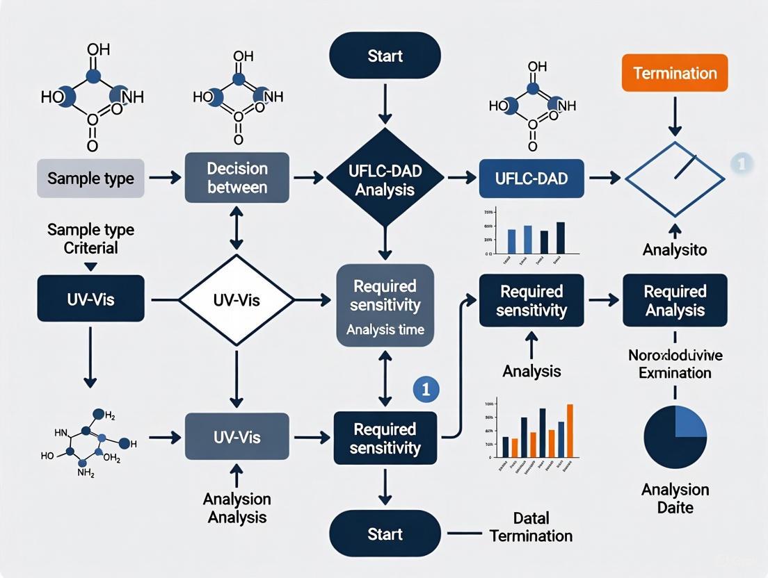
Decision Framework: When to Select UV-Vis vs. UFLC-DAD
Application-Based Selection Criteria
The choice between a conventional UV-Vis detector and a UFLC-DAD system depends on multiple factors related to the analytical requirements, sample characteristics, and operational constraints. The following decision framework provides guidance for selecting the appropriate technology:
Figure 3: Detector selection decision framework
Specific Application Scenarios
Choose UFLC-DAD when:
- Method Development and Unknown Screening: The full spectral capability enables compound identification through library matching and peak purity assessment [11].
- Regulated Pharmaceutical Analysis: ICH Q3A guidelines require demonstration of specificity and peak purity for stability-indicating methods [12].
- Complex Natural Product Analysis: Herbal supplements and botanical extracts often contain multiple compounds with similar retention times but different spectra [9].
- Impurity Profiling: The combination of high separation efficiency and spectral data facilitates identification and quantification of low-level impurities [9].
Choose HPLC-UV when:
- Routine Quality Control: For well-characterized compounds where only quantification is needed, single-wavelength detection suffices [15].
- Budget Constraints: UV detectors are more cost-effective than DAD systems, both in initial investment and maintenance [16].
- Limited Technical Expertise: UV detectors are simpler to operate and maintain [12].
- Methods with Established Specificity: When interference has been ruled out through previous validation studies [14].
A hybrid approach employing HPLC with DAD detection may be appropriate when separation requirements are modest but spectral confirmation is desirable. Similarly, UFLC with UV detection may be optimal when speed is essential but the analytes are well-characterized [13].
UFLC-DAD architecture represents the convergence of high-efficiency separation technology with comprehensive spectroscopic detection, providing an powerful analytical tool for modern laboratories. The integration of sub-2-micron particle chromatography with full-spectrum UV-Vis detection enables researchers to not only separate complex mixtures rapidly but also obtain critical spectral data for compound identification, confirmation, and purity assessment within a single analysis.
The decision to implement UFLC-DAD versus conventional HPLC-UV involves careful consideration of analytical requirements, sample complexity, and operational constraints. For applications demanding the highest level of confidence in compound identity or dealing with unknown components, the additional information provided by DAD detection is invaluable. For routine analysis of well-characterized compounds where throughput and cost-efficiency are primary concerns, UV detection may remain sufficient. As analytical challenges continue to evolve toward more complex samples and shorter turnaround times, UFLC-DAD technology offers a robust solution that balances separation power with comprehensive detection capabilities.
Selecting the appropriate analytical technique is a critical decision in research and drug development. The choice between Ultraviolet-Visible (UV-Vis) spectroscopy and Ultra-Fast Liquid Chromatography with a Diode Array Detector (UFLC-DAD) hinges on a fundamental understanding of their core components and how these components dictate the capabilities and limitations of each system. This guide provides an in-depth technical comparison of the light sources, wavelength selectors, and detectors that form the foundation of both instruments. By framing this comparison within the context of specific analytical needs—from rapid, pure compound analysis to the separation and identification of complex mixtures—this guide empowers scientists to make informed, strategic decisions for their research.
Core Component Comparison
The analytical performance of UV-Vis and UFLC-DAD systems is directly determined by their core optical components. The table below provides a detailed, quantitative comparison of these subsystems.
Table 1: Technical Comparison of Core Components in UV-Vis Spectrophotometers and UFLC-DAD Systems
| Component | UV-Vis Spectrophotometer | UFLC-DAD System |
|---|---|---|
| Light Sources | • Deuterium (D₂) lamp: Covers UV range (∼190–400 nm) [1].• Tungsten-Halogen (W) lamp: Covers visible range (∼350–1000 nm) [1].• Xenon lamp: Single, high-intensity source for both UV and visible ranges; more expensive and less stable [1]. | • Typically uses a Deuterium (D₂) lamp and a Tungsten (W) lamp to cover the UV-Vis spectrum [17].• Lamps are integrated into a single detector module. |
| Wavelength Selectors | • Monochromator: Most common; uses a diffraction grating (often ≥1200 grooves/mm) for wavelength selection [1].• Filters: Absorption, interference, cutoff, and bandpass filters are used to narrow wavelengths or for specific applications [1]. | • Incorporates a monochromator (e.g., with a diffraction grating) after the sample flow cell [17].• This allows polychromatic light to pass through the sample first. |
| Detectors | • Photomultiplier Tube (PMT): Common; high sensitivity for low-light detection [1].• Photodiodes & CCDs: Semiconductor-based detectors [1]. | • Utilizes a Photo Diode Array (PDA) consisting of multiple diodes (e.g., 512 or 1024), each measuring a specific wavelength simultaneously [17]. |
| Key Operational Characteristics | • Sequential wavelength measurement.• Requires a reference (blank) measurement for baseline correction [1]. | • Simultaneous, full-spectrum acquisition at every data point during the chromatographic run [18].• Provides a three-dimensional data output: retention time, absorbance, and wavelength [17]. |
Experimental Protocols
The distinct configurations of these components lead to different standard operating procedures. The following workflows, derived from recent research, illustrate the application-specific protocols for each technique.
Protocol 1: Rapid Contamination Detection in Microalgae Cultures using UV-Vis and Machine Learning
This protocol highlights the use of UV-Vis for rapid, in-vivo analysis of complex biological samples without the need for separation [19].
- Sample Preparation: Grow cultures of the microalgae of interest (e.g., Chlorella vulgaris). Introduce potential biological contaminants, such as the flagellate Poterioochromonas malhamensis or the rotifer Brachionus plicatilis. For robustness testing, culture samples under different stress conditions, such as salt-stressed media [19].
- Instrument Setup: Configure a UV-Vis spectrophotometer with a light source covering 200–1000 nm and a 10-mm path length cuvette holder. A handheld spectrometer can be used for portability [19].
- Data Acquisition: Collect UV-Vis absorption spectra of both uncontaminated and contaminated cultures. The distinct spectral fingerprints of chlorophylls, carotenoids, and other pigments serve as the analytical signal [19].
- Data Analysis: Employ machine learning for data analysis. Use Principal Component Analysis (PCA) to classify the spectral data and distinguish between uncontaminated and contaminated cultures, even under challenging salt-stress conditions that alter pigment profiles [19].
Protocol 2: Comprehensive Analysis of Wine Aging Markers using UFLC-DAD
This protocol demonstrates the use of UFLC-DAD for separating, identifying, and quantifying specific compounds within a complex mixture [8].
- Sample Collection: Obtain a set of sweet botrytized wine samples with a known and wide age range (e.g., 3 to 60 years) [8].
- Chromatographic Separation:
- Column: Use a reversed-phase C18 column.
- Mobile Phase: Employ a binary gradient, typically consisting of water (with acid modifier) and an organic solvent like acetonitrile or methanol.
- Injection: Automatically inject a precise volume of filtered wine sample.
- DAD Detection & Data Acquisition:
- Set the DAD to acquire full spectra (e.g., from 190–400 nm) for every data point during the chromatographic run.
- Set the data acquisition rate to at least 5–20 Hz to ensure sufficient data points across narrow UHPLC peaks [20].
- Use a narrow bandwidth (e.g., 1–4 nm) for high spectral resolution and a small step size (e.g., 1 nm) for smooth spectral curves [20].
- Compound Identification & Quantitation:
- Identify phenolic compounds (e.g., catechin, gallic acid, caftaric acid) by matching their retention times and UV-Vis spectra against those of authentic standards analyzed under identical conditions [8].
- Quantify the compounds using calibration curves built from standards.
- Chemometric Analysis: Perform statistical analysis (e.g., Spearman correlation) to establish a high correlation between the concentration of specific markers (e.g., caftaric acid, catechin, gallic acid) and the chronological age of the wine [8].
System Workflows and Logical Relationships
The fundamental difference in data acquisition between the two techniques—sequential versus simultaneous—dictates their analytical workflows and applications. The following diagram visualizes the operational logic of each system.
The Scientist's Toolkit: Essential Research Reagents and Materials
Successful execution of the protocols depends on the use of specific, high-quality materials. The following table lists key reagents and their functions in the featured experiments.
Table 2: Essential Research Reagents and Materials
| Item Name | Function / Analytical Role |
|---|---|
| Microalgae Cultures (e.g., Chlorella vulgaris) | Serves as the model biological system for developing rapid contamination screening methods using its natural pigment fingerprints [19]. |
| Phenolic Compound Standards (Catechin, Gallic Acid, etc.) | Used for compound identification and creating quantitative calibration curves in HPLC-DAD analysis of complex samples like wine [8]. |
| HPLC/DAD Solvents (Acetonitrile, Methanol, Acidified Water) | Form the mobile phase for chromatographic separation; purity is critical to maintain low background noise and ensure peak resolution [8]. |
| Quartz Cuvettes | Essential for UV range measurements in spectrophotometry, as quartz is transparent down to ~190 nm, unlike plastic or glass [1]. |
| Syringe Filters (0.45 µm or 0.22 µm) | Used to clarify and particulate-free liquid samples (e.g., wine, culture media) prior to injection into the HPLC system to prevent column damage [8]. |
Decision Framework: UV-Vis vs. UFLC-DAD
The choice between UV-Vis and UFLC-DAD is not a matter of which instrument is superior, but which is more appropriate for the specific analytical question. The following framework guides this decision.
Choose UV-Vis Spectroscopy when:
- The analytical goal is rapid quantification of a known compound in a pure or simple mixture [21].
- The sample is a simple solution and does not require separation of components [21].
- You need to monitor reaction kinetics or time-dependent processes in real-time [1].
- Cost-effectiveness, speed, and ease of use are primary concerns, and the necessary information can be derived from a bulk absorption measurement [21].
Choose UFLC-DAD when:
- The sample is a complex mixture requiring high-resolution separation of multiple components before detection [21].
- Peak purity assessment and identification of co-eluting compounds is necessary, as the DAD's full-spectrum capability can reveal impurities invisible to a single-wavelength detector [18].
- Spectral confirmation of identity is required, as the UV-Vis spectrum of a peak provides a additional identification point alongside its retention time [17] [18].
- The analysis involves unknown compounds or impurities, and you need to retrospectively extract chromatograms at different wavelengths without reinjecting the sample [18].
This technical guide provides an in-depth examination of three fundamental parameters in ultraviolet-visible (UV-Vis) spectroscopy and ultra-fast liquid chromatography with diode array detection (UFLC-DAD): spectral bandwidth, pathlength, and molar absorptivity. Understanding the interplay between these parameters is crucial for researchers and drug development professionals when selecting appropriate analytical techniques and optimizing method performance. This review synthesizes current principles and practices to establish a framework for informed decision-making between conventional UV-Vis and UFLC-DAD methodologies, supported by experimental data and technical specifications relevant to pharmaceutical analysis.
The selection of appropriate detection technology is pivotal in analytical method development for pharmaceutical research and quality control. Ultraviolet-visible (UV-Vis) detection remains the workhorse in quality control laboratories due to its reliability, ease of use, and universal response to chromophoric compounds, while diode array detection (DAD) in liquid chromatography systems provides enhanced capabilities for peak identification and purity assessment [12]. The performance of both techniques hinges on three interdependent parameters: spectral bandwidth, which determines wavelength resolution; pathlength, which governs signal intensity according to Beer-Lambert law; and molar absorptivity, an intrinsic molecular property defining absorption strength [12] [22]. This whitepaper examines these critical parameters within the context of selecting between conventional UV-Vis and UFLC-DAD systems, providing technical guidance for method optimization in drug development workflows.
Fundamental Principles and Definitions
Molar Absorptivity (ε)
Molar absorptivity (also known as molar extinction coefficient) is an intrinsic property of a chemical substance that defines how strongly it absorbs light at a specific wavelength [23] [24]. According to the Beer-Lambert law, absorbance (A) is directly proportional to molar absorptivity (ε), pathlength (b), and concentration (c): A = ε × b × c [12] [22]. Compounds with high molar absorptivity (typically >10,000 L·mol⁻¹·cm⁻¹) produce strong detection signals even at low concentrations, making them ideal for trace analysis [23]. For drug substances with low native molar absorptivity, analytical sensitivity can be improved through derivatization strategies that create charge-transfer complexes with enhanced absorption properties [23].
Pathlength (b)
Pathlength is the distance light travels through the sample in a flow cell or cuvette, typically measured in millimeters or centimeters [12] [25]. In accordance with Beer-Lambert law, absorbance increases linearly with pathlength, making it a crucial factor for detection sensitivity [25]. HPLC flow cells typically have pathlengths of 10mm, while UHPLC cells are shorter (0.5-1mm) to maintain low dispersion volumes [12]. As a rule of thumb, the flow cell volume should be approximately one-third of the peak volume at half height to minimize band broadening [25].
Spectral Bandwidth (SBW)
Spectral bandwidth is defined as the width of the wavelength of light at half the maximum intensity (Full Width at Half Maximum or FWHM) [26]. This parameter critically influences measurement accuracy, resolution, and ability to distinguish fine spectral features. There are distinct considerations for different instrument types:
- Monochromator-based instruments: SBW is determined by the physical slit width and optical dispersion, with narrower slits providing higher resolution but reduced light throughput [26].
- Diode-array instruments: SBW is often fixed and determined by the design and spacing of the diodes in the array, allowing simultaneous measurement of all wavelengths [26].
The relationship between spectral bandwidth and the natural bandwidth of the sample's absorption band is crucial; for high measurement accuracy (≥99.5%), the ratio of spectral bandwidth to natural bandwidth should be 0.1 or less [26].
Quantitative Comparison of Technical Parameters
Flow Cell Specifications for UV Detection
Table 1: Comparison of HPLC/UFLC flow cell characteristics based on pathlength
| Path Length | Cell Volume | Typical Applications | Linearity Limit (AU) | Sensitivity Consideration |
|---|---|---|---|---|
| 1.0 cm | 1.0 µL | Standard HPLC analysis | ~200 mAU | Balanced sensitivity and dispersion |
| 6.0 cm | 4.0 µL | Trace analysis | ~333 mAU (55.5 mAU/cm) | Higher signal-to-noise ratio; increased band broadening risk |
| 0.5-1.0 mm | 0.5-1.0 µL | UHPLC applications | Instrument-dependent | Minimal dispersion for sharp peaks |
Data adapted from Agilent flow cell specifications and chromatography literature [12] [25].
Molar Absorptivity Values for Selected Compounds
Table 2: Representative molar absorptivity values for pharmaceutical compounds
| Compound/Core Structure | λmax (nm) | Molar Absorptivity (ε, L·mol⁻¹·cm⁻¹) | Structural Features |
|---|---|---|---|
| Furo[3,2-e]indolizine (R1=R2=H) | 355 | 1.7 × 10⁴ | Base heteroaromatic system |
| Furo[3,2-e]indolizine (R1=R2=4-CNPh) | 434 | 3.2 × 10⁴ | Extended π-system with electron-withdrawing groups |
| Vitamin B1 (after oxidation) | ~375 | Varies with derivatization | Thiochrome formation |
| Vitamin B2 | ~445 | ~1.2 × 10⁴ | Isoalloxazine ring |
| Vitamin B6 | ~325 | ~8.0 × 10³ | Pyridoxine structure |
Data compiled from multiple sources including furoindolizine studies and vitamin分析方法 [27] [24].
Experimental Protocols and Methodologies
Determination of Molar Absorptivity
Protocol for Empirical ε Measurement:
Solution Preparation: Prepare a stock solution of accurately known concentration (typically 1-10 mM) using a high-purity standard and appropriate solvent.
Dilution Series: Create a series of 5-8 dilutions covering the expected linear range of Beer-Lambert law (typically yielding absorbances of 0.1-1.0 AU).
Spectroscopic Measurement: Using a spectrophotometer with appropriate SBW setting (≤1 nm recommended), measure absorbance at λmax for each dilution using a cuvette with precisely known pathlength.
Data Analysis: Plot absorbance versus concentration and perform linear regression. The slope of the line (A/c) equals ε × b, allowing calculation of ε using the known pathlength.
Validation: Verify linearity (R² > 0.999) and repeat with at least three independent stock solutions for precision assessment [23] [22].
Simultaneous Vitamin Analysis Using HPLC-DAD/FLD
Chromatographic Conditions:
- Column: Aqua C18 (250 mm × 4.6 mm, 5 μm)
- Temperature: 40°C
- Mobile Phase: Isocratic elution with 70% NaH₂PO₄ buffer (pH 4.95) and 30% methanol
- Flow Rate: 0.9 mL/min
- Detection: DAD for vitamins B2 and B6; FLD for vitamin B1 after pre-column derivatization
Sample Preparation:
- Liquid/Solid Extraction: For pharmaceutical gummies, homogenize and extract with suitable solvent (e.g., acidified methanol)
- Derivatization for B1: Oxidize thiamine to fluorescent thiochrome using alkaline potassium ferricyanide
- Cleanup: For complex matrices (e.g., gastrointestinal fluids), employ Solid Phase Extraction (SPE) with C18 cartridges
- Validation Parameters: Demonstrate linearity (R² > 0.999), accuracy (mean recovery 100 ± 3%), and precision (RSD < 3.23%) [27]
Instrument Selection Guide: UV-Vis vs. UFLC-DAD
Technical Comparison for Pharmaceutical Applications
Table 3: Decision matrix for detector selection based on analytical requirements
| Analytical Requirement | Recommended Detector | Optimal Parameter Settings | Rationale |
|---|---|---|---|
| Routine quantification of chromophoric compounds | Single wavelength UV-Vis | Pathlength: 10 mm; SBW: 4-8 nm | High precision (<0.2% RSD), reliability, simplicity [12] |
| Peak purity/purity assessment | DAD/PDA | SBW: 1-4 nm; full spectrum collection | Spectral comparison across peak (upslope vs. downslope) [12] |
| Method development/unknown identification | DAD/PDA | Full spectrum (190-800 nm); SBW: ≤4 nm | Library matching and multi-wavelength analysis capability [12] |
| Low abundance analytes | Long pathlength UV cell or pre-column derivatization | Pathlength: 30-60 mm; optimized ε | Enhanced sensitivity via Beer-Lambert law [23] [25] |
| Compounds with low molar absorptivity | Derivatization or universal detection | Chemical modification to enhance ε | Complex formation to improve detectability [23] |
| High-resolution separations (UHPLC) | DAD with low-dispersion flow cell | Pathlength: 0.5-1 mm; cell volume: <1 μL | Maintains separation efficiency with minimal peak broadening [12] |
Research Reagent Solutions for Absorption Spectroscopy
Table 4: Essential materials and reagents for method development
| Reagent/ Material | Function/Application | Technical Considerations |
|---|---|---|
| Deuterium Lamp | UV light source for 190-400 nm | Standard in modern UV-Vis and DAD detectors; requires periodic replacement [12] |
| Charge Transfer Complex Reagents (DDQ, TCNQ) | Electron acceptors for enhancing detectability | Improve molar absorptivity of donor compounds; selectivity depends on donor properties [23] |
| C18 SPE Cartridges | Sample cleanup and preconcentration | Essential for complex matrices (biological fluids); improves method sensitivity and column lifetime [27] |
| Buffer Salts (e.g., NaH₂PO₄) | Mobile phase modification | Controls pH for separation and stability; affects chromophore ionization and λmax [27] |
| Pre-column Derivatization Reagents | Convert non-UV-absorbing compounds to detectable forms | Enables detection of vitamins (B1→thiochrome), amines, thiols via fluorescence or enhanced UV absorption [27] |
Visualizing Parameter Relationships
Parameter Interdependence in UV Detection
The strategic selection between UV-Vis and UFLC-DAD detection methodologies requires thorough understanding of three fundamental parameters—spectral bandwidth, pathlength, and molar absorptivity—and their interplay in governing analytical performance. UV-Vis detection remains optimal for routine quantification where high precision and reliability are paramount, while UFLC-DAD provides superior capabilities for method development, peak purity assessment, and analysis of complex mixtures. By applying the technical principles and experimental frameworks presented in this guide, researchers can make informed decisions that optimize detection strategies for specific pharmaceutical applications, ultimately enhancing method performance and analytical confidence in drug development workflows.
The Historical Evolution and Enduring Role of UV Detection in HPLC
The development of High-Performance Liquid Chromatography (HPLC) represents a cornerstone achievement in analytical chemistry, enabling the separation, identification, and quantification of complex mixtures with remarkable precision. While numerous detection methods have emerged over the decades, ultraviolet (UV) detection remains one of the most widely utilized techniques across pharmaceutical, clinical, and environmental laboratories. Its enduring prominence stems from an exceptional balance of reliability, ease of use, and universal response to chromophoric compounds [12].
The evolution of UV detection technology has progressed from simple fixed-wavelength instruments to sophisticated diode array systems capable of full-spectrum monitoring. This progression has occurred within the broader context of HPLC's own development, which traces its modern beginnings to the mid-1960s [28]. Understanding this historical trajectory provides valuable insights for contemporary researchers faced with methodological choices, particularly when deciding between conventional UV-Vis and more advanced diode array detection (DAD) for analytical applications.
This article examines the technical evolution of UV detection, its current applications in modern analytical science, and practical guidance for detector selection within research and development frameworks.
Historical Development of HPLC and UV Detection
The Birth of Modern Liquid Chromatography
The foundational principles of high-performance liquid chromatography were established through pivotal work in the 1960s that transformed traditional liquid chromatography into a powerful analytical technique. The seminal year of 1966 is often cited as a critical starting point, marked by two significant developments: Piel's work using finely ground silica particles in narrow-bore columns under pressure, and Hamilton's application of smaller ion-exchange resins for amino acid analysis [28]. These innovations demonstrated the profound impact of particle size and pressure on separation efficiency, fulfilling the prediction made by Martin and Synge in 1941 that "the smallest HETP should be obtainable by using very small particles and a high pressure difference across the length of the column" [28].
The true revolution in HPLC column technology began with Professor Csaba Horváth's development of pellicular particles (now called superficially porous particles or SPPs) around 1968 [28]. These particles featured an impenetrable solid core (37-50 µm glass beads) coated with a thin, porous outer layer (1-2 µm) of stationary phase. This design provided an order of magnitude improvement in efficiency compared to the larger, fully porous particles used previously, primarily because the thin porous layer restricted diffusion paths for analyte molecules [28].
Evolution of UV Detection Technology
The development of reliable UV detectors was equally crucial to HPLC's success. Early UV detection systems were fixed-wavelength detectors using low-pressure mercury lamps with a strong 254 nm emission line [12]. Introduced in the late 1960s, these detectors represented a significant advancement, with one 1968 model reporting noise of approximately ±0.2 mAU—about 50 times less sensitive than modern detectors [12]. While limited in flexibility, these robust detectors established UV detection as a fundamental HPLC component.
The next significant advancement came with variable wavelength detectors (VWD), which incorporated deuterium lamps and monochromators to allow user-selectable wavelengths across the UV-Vis spectrum (typically 190-600 nm) [12]. The heart of these systems was a movable diffraction grating that could be rotated to select specific wavelengths through an exit slit, typically with a spectral bandwidth of 5-8 nm [12]. This flexibility enabled method development at the optimal wavelength for specific analytes, greatly enhancing detection selectivity.
The most transformative development in UV detection emerged with the introduction of photodiode array detectors (PDA or DAD) in the 1980s [12]. Unlike variable wavelength detectors that use a monochromator before the flow cell, DADs employ a "reverse optics" design where white light passes through the flow cell first before being dispersed onto an array of photodiodes [29]. This fundamental architectural difference enables simultaneous monitoring of multiple wavelengths and collection of complete spectra for each data point during chromatographic separation [12] [29].
Table 1: Historical Evolution of HPLC UV Detection Technologies
| Time Period | Detector Type | Key Characteristics | Performance Metrics | Applications |
|---|---|---|---|---|
| Late 1960s | Fixed Wavelength | Low-pressure mercury lamps (254 nm); cutoff filters; simple optical design | Noise: ~±0.2 mAU [12] | Basic separations of chromophoric compounds |
| 1980s | Variable Wavelength (VWD) | Deuterium lamps; monochromators; selectable wavelengths (190-600 nm) | Spectral bandwidth: 5-8 nm [12] | Method development; targeted compound analysis |
| 1980s-Present | Diode Array (DAD/PDA) | Reverse optics; full spectrum collection (190-950 nm); photodiode arrays (512-1024 elements) | Simultaneous multi-wavelength monitoring; spectral acquisition [12] [11] | Peak purity; method development; unknown identification |
The timeline of HPLC and UV detection development reveals a pattern of incremental improvements interspersed with revolutionary breakthroughs. The period from 1966 to approximately 1975 established the fundamental principles, while the 1980s brought sophisticated optical designs and electronics that dramatically improved performance. Modern systems continue this evolution with enhanced sensitivity, reduced noise levels, and advanced data processing capabilities.
Fundamental Principles and Technical Evolution
Operating Principles of UV Detection
The fundamental principle underlying all UV detection for HPLC is the Beer-Lambert Law, which mathematically describes the relationship between analyte concentration and light absorption [29]. According to this law, absorbance (A) is proportional to the product of the molar absorptivity (ε), pathlength (b), and analyte concentration (c): A = ε × b × c [12]. This relationship enables quantitative analysis when properly calibrated with standards of known concentration.
UV detectors specifically measure the UV absorbance of HPLC eluent as it passes through a flow cell, providing a continuous signal that correlates with analyte concentration [12]. Key components shared across all UV detector types include:
- Light Source: Typically a deuterium (D₂) lamp providing continuous emission in the 190-600 nm range, sometimes augmented with a tungsten lamp for visible light detection (up to 950 nm) [12]
- Flow Cell: A small, flow-through device with quartz windows defining a fixed pathlength (typically 10 mm), with volumes ranging from 8-18 µL for HPLC to 0.5-1 µL for UHPLC applications [12]
- Photodetector: Converts transmitted light intensity into electrical signals for data processing [29]
The essential difference between detector types lies in how they select and process wavelengths. Variable wavelength detectors use a monochromator before the flow cell to select a specific wavelength, while diode array detectors pass polychromatic light through the flow cell first, then disperse it onto a photodiode array for full spectrum capture [29].
Technical Specifications and Performance Metrics
Several key parameters define UV detector performance and suitability for specific applications:
- Pathlength: Typically 10 mm in standard flow cells, directly affecting sensitivity according to Beer's Law [12]
- Spectral Bandwidth: The range of wavelengths passing through the flow cell, typically 5-8 nm for VWD and adjustable for DAD [12]
- Noise: Modern detectors typically exceed the historical benchmark of ±1×10⁻⁵ AU [12]
- Dispersion: Primarily dependent on flow cell volume and design, contributing to overall system band broadening [12]
Table 2: Comparison of Modern UV Detector Types for HPLC
| Parameter | Variable Wavelength Detector (VWD) | Diode Array Detector (DAD/PDA) |
|---|---|---|
| Optical Design | Pre-cell monochromator with movable diffraction grating [12] | Post-cell fixed diffraction grating with diode array [29] |
| Wavelength Selection | Single or limited wavelengths selected before flow cell [12] | Full spectrum (190-950 nm) captured simultaneously [11] |
| Spectral Data | Limited to preselected wavelengths | Complete UV-Vis spectrum for every time point [12] |
| Primary Applications | Routine quantitative analysis where spectral data is not essential [12] | Peak purity, method development, unknown identification [11] |
| Flow Cell Volume | 8-18 µL (HPLC); 0.5-1 µL (UHPLC) [12] | Similar to VWD, with specialized micro-flow cells available |
| Advantages | Lower cost, sufficient for many routine applications | Spectral information for peak identification and purity assessment [12] [11] |
The evolution of detector technology has progressively addressed limitations while expanding capabilities. Early fixed-wavelength detectors provided limited application range, while variable wavelength detectors introduced method development flexibility. Diode array detectors represented the most significant advancement, enabling retrospective data analysis and peak purity assessment without method redevelopment [11].
UV Detection in Contemporary Analytical Applications
Pharmaceutical Analysis and Therapeutic Drug Monitoring
UV detection remains extensively employed in pharmaceutical analysis and therapeutic drug monitoring (TDM), where its combination of precision, reliability, and cost-effectiveness offers particular advantages. A 2023 study demonstrated the development of a practical HPLC-UV platform for quantifying serum levels of five different drugs (carbamazepine, phenytoin, lamotrigine, vancomycin, and voriconazole) during routine clinical work [30]. The method incorporated solid-phase extraction (SPE) using monolithic C18-silica disk cartridges followed by HPLC-UV analysis on a Chromolith HighResolution RP-18 column [30].
This platform successfully addressed the need for in-hospital TDM without requiring sophisticated MS instrumentation, achieving excellent correlation with both immunoassay (cobas analyzer) and HPLC-MS/MS reference methods [30]. The validation results demonstrated that HPLC-UV methods can deliver the precision required for clinical decision-making, with the authors noting that "the higher precision achievable with UV detection (<0.2% RSD) is pivotal and necessary in regulatory testing because a typical potency specification for drug substances is 98.0 to 102.0%" [12].
Method Comparison and Validation Studies
Comparative studies continue to highlight the appropriate applications and limitations of UV detection. A 2019 study directly compared HPLC-UV with UV-Vis spectrophotometry for quantifying levofloxacin released from mesoporous silica microspheres/nano-hydroxyapatite composite scaffolds [31]. While both methods showed excellent linearity (R²=0.9991 for HPLC-UV vs. R²=0.9999 for UV-Vis), significant differences emerged in recovery rates, particularly at medium concentrations (110.96% for HPLC-UV vs. 99.50% for UV-Vis) [31].
The study concluded that "it is not accurate to measure the concentration of drugs loaded on biodegradable composite composites by UV-Vis" alone, emphasizing that HPLC separation prior to detection is essential for accurate quantification in complex matrices [31]. This distinction highlights the continuing value of HPLC-UV for analyses where sample complexity or interfering substances complicate direct spectrophotometric measurement.
Advanced Applications and Hybrid Techniques
Modern applications increasingly combine HPLC-UV with advanced data processing techniques. A 2025 study on wine aging employed HPLC-DAD alongside chemometric methods to establish correlations between phenolic compound content and wine age [8]. The diode array detection enabled simultaneous quantification of multiple phenolic compounds (catechin, caffeic acid, caftaric acid, gallic acid, protocatechuic acid, and p-coumaric acid) while establishing characteristic spectral fingerprints for quality assessment [8].
Similarly, a 2024 study demonstrated the quantification of active pharmaceutical ingredients (acetylsalicylic acid, caffeine, and paracetamol) in solid formulations using UV-Vis diffuse reflectance spectroscopy with multivariate data processing, validating results against a reference HPLC-DAD method [32]. These applications illustrate how UV detection continues to evolve through integration with advanced data analysis techniques rather than being displaced by more expensive detection methods.
Experimental Protocols and Methodologies
HPLC-UV Method Development for Therapeutic Drug Monitoring
The following protocol, adapted from a 2023 clinical study, outlines a standardized approach for developing HPLC-UV methods for therapeutic drug monitoring [30]:
Sample Preparation:
- Solid-Phase Extraction: Use monolithic C18-silica disk centrifugal spin-cartridges (e.g., MonoSpin C18)
- Cartridge Pretreatment: Sequentially pass 500 µL of acetonitrile and 500 µL of water through the cartridge by centrifugation at 5,000 rpm (2,400×g) for 1 minute
- Sample Loading: Filter patient serum through a 0.45 µm syringe filter, load 150 µL onto the pretreated cartridge, and centrifuge for 3 minutes
- Washing: Pass 500 µL of water through the cartridge by centrifugation for 2 minutes
- Elution: Pass 150 µL of elution solution (aqueous 50% acetonitrile for most drugs; 30% for vancomycin) through the cartridge by centrifugation for 1 minute; collect the eluate for analysis
HPLC-UV Analysis:
- Column: Chromolith HighResolution RP-18 (100 mm × 4.6 mm i.d.)
- Mobile Phase: Varies by drug; typically gradient or isocratic mixtures of aqueous buffer and acetonitrile
- Column Temperature: 40°C
- Detection: Diode array detector with monitoring at drug-specific wavelengths (e.g., 220 nm for carbamazepine, 240 nm for voriconazole)
- Validation: Assess linearity, accuracy, precision, recovery, and limit of quantification according to ICH guidelines
Advanced Method Optimization with Experimental Design
A 2024 study on posaconazole quantification demonstrated the application of experimental design and machine learning for HPLC-UV method optimization [33]. The protocol included:
Chromatographic Optimization:
- Factor Evaluation: Assess five critical parameters (total organic phase %, methanol % in organic phase, mobile phase pH, column temperature, and flow rate) using a fractional 2-level factorial design
- Response Monitoring: Measure retention time, peak resolution, and peak symmetry
- Machine Learning Integration: Apply artificial neural networks (ANN) combined with genetic algorithms (GA) to identify optimal conditions
Extraction Optimization:
- Liquid-Liquid Extraction: Evaluate volume of extraction solvent, salt addition, pH, extraction time, and centrifugation time
- Recovery Assessment: Determine extraction efficiency for both drug and internal standard
The optimized method achieved a retention time of 8.2 minutes with recovery above 98%, demonstrating excellent sensitivity (LOQ: 50 ng/mL) using only 150 µL of plasma sample [33]. This approach illustrates how modern method development extends beyond traditional one-factor-at-a-time optimization to more efficient multivariate strategies.
Diagram 1: HPLC-UV Analytical Workflow. This flowchart illustrates the standardized workflow for HPLC-UV analysis, from sample preparation through data analysis.
The Scientist's Toolkit: Essential Research Reagents and Materials
Table 3: Essential Materials for HPLC-UV Analysis in Pharmaceutical Research
| Category | Specific Examples | Function and Application Notes |
|---|---|---|
| SPE Cartridges | Monolithic C18-silica disk cartridges (e.g., MonoSpin C18) [30] | Rapid sample cleanup and concentration; suitable for small sample volumes (150 µL serum) |
| HPLC Columns | Chromolith HighResolution RP-18 (100 mm × 4.6 mm) [30]; Sepax BR-C18 (250 × 4.6 mm, 5 µm) [31] | Fast separations with low backpressure; suitable for pharmaceutical compounds |
| Mobile Phase Components | Acetonitrile (HPLC-grade), methanol, ammonium acetate, phosphate buffers, tetrabutylammonium bromide [30] [31] | Create optimal separation conditions; ion-pairing agents improve peak shape for ionizable compounds |
| Reference Standards | USP/EP certified reference standards for target analytes (e.g., carbamazepine, phenytoin, voriconazole) [30] | Method calibration and quality control; essential for quantitative accuracy |
| Internal Standards | Compound-specific stable isotopes or structural analogs (e.g., ciprofloxacin for levofloxacin analysis) [31] | Monitor extraction efficiency and account for procedural variations |
Guidance for Detector Selection: UV vs. DAD in Research Applications
Decision Framework for Detector Selection
Choosing between variable wavelength UV detectors and diode array detectors requires careful consideration of analytical requirements, budget constraints, and application scope. The following guidelines support informed decision-making:
Select Variable Wavelength UV Detection when:
- Analyzing known compounds with well-characterized absorbance maxima
- Performing routine quantitative analysis with established methods
- Working within budget constraints that preclude DAD acquisition
- Analyzing samples with minimal spectral interference from matrix components [12]
Opt for Diode Array Detection when:
- Developing new methods for unknown or complex samples
- Requiring peak purity assessment for method validation
- Analyzing compounds with potentially co-eluting interferents
- Needing spectral data for confirmatory identification
- Working with compounds having dissimilar absorbance spectra that can be distinguished spectrally [11]
Advanced DAD Applications and Capabilities
Modern diode array detectors offer capabilities extending beyond basic spectral collection. The i-PDeA (intelligent Peak Deconvolution Analysis) function available in some systems enables mathematical resolution of co-eluting peaks based on their spectral differences [11]. This advanced capability provides a powerful alternative to method redevelopment when complete chromatographic separation proves challenging.
For pharmaceutical applications, peak purity assessment represents a critical DAD application. By comparing spectra across different regions of a chromatographic peak, analysts can detect potential co-elution not apparent from the chromatographic profile alone [11]. This capability is particularly valuable for stability-indicating methods and impurity profiling, where the International Council for Harmonisation (ICH) guidelines require demonstrating specificity and the ability to detect degradants [12].
UV detection has maintained its pivotal role in HPLC analysis through continuous technological evolution that has expanded its capabilities while preserving its fundamental advantages of robustness, reproducibility, and accessibility. From its beginnings with fixed-wavelength detectors to contemporary diode array systems, UV detection has adapted to meet the changing demands of analytical science.
The enduring relevance of UV detection is particularly evident in pharmaceutical analysis and therapeutic drug monitoring, where its combination of precision, reliability, and cost-effectiveness continues to support both research and routine applications. While mass spectrometry offers superior sensitivity and specificity for certain applications, UV detection remains the undisputed workhorse for quality control laboratories, especially in regulatory environments where its proven reliability and quantitative precision are essential [12].
For researchers selecting detection strategies, the choice between conventional UV and DAD detection ultimately depends on specific application requirements, with diode array detectors offering clear advantages for method development and verification, while variable wavelength detectors remain sufficient for many routine quantitative applications. As HPLC continues to evolve, UV detection methodologies will likely maintain their central position in the analytical landscape, particularly through integration with advanced data processing techniques and experimental design approaches that maximize their information potential.
Strategic Applications: When to Deploy UV-Vis or UFLC-DAD for Maximum Impact
Within the analytical toolkit available to researchers and drug development professionals, selecting the appropriate technique is fundamental to efficiency and data quality. The choice often hinges on a balance between analytical needs, resource constraints, and time. This guide focuses on the ideal application of standalone UV-Vis spectroscopy for the quantitative analysis of pure compounds, positioning it within a broader methodology that includes more advanced techniques like Ultra-Fast Liquid Chromatography with Diode-Array Detection (UFLC-DAD). While UFLC-DAD offers superior separation for complex mixtures [7], standalone UV-Vis remains a powerful, cost-effective, and straightforward workhorse for specific, well-defined analytical challenges. Its principle, based on measuring the absorption of ultraviolet or visible light by a sample, provides a direct path to quantification when the analyte is known and free of interferents [1] [2]. Understanding its core strengths and limitations allows scientists to make informed decisions, streamlining workflows without compromising data integrity.
Fundamental Principles and Instrumentation of UV-Vis Spectroscopy
Core Mechanism: Electronic Transitions and the Beer-Lambert Law
The fundamental principle of UV-Vis spectroscopy involves the promotion of electrons in a molecule from a ground state to a higher energy, excited state upon absorbing specific wavelengths of light [2]. The energy required for this transition is unique to the molecular structure of the compound, resulting in a characteristic absorption spectrum [1]. This property is harnessed for quantification through the Beer-Lambert Law, which states that the absorbance (A) of a solution is directly proportional to the concentration (c) of the absorbing species and the path length (L) of the light through the sample [1].
The mathematical relationship is expressed as: A = ε * c * L where:
- A is the measured absorbance (unitless)
- ε is the molar absorptivity (L·mol⁻¹·cm⁻¹)
- c is the concentration (mol·L⁻¹)
- L is the path length (cm) [1]
This linear relationship is the cornerstone of quantitative UV-Vis analysis, enabling the determination of an unknown concentration from a measured absorbance value once the absorptivity is established using standard solutions [1].
Key Instrument Components
A UV-Vis spectrophotometer is composed of four main components that work in sequence:
- Light Source: Provides broad-spectrum UV and visible light. Common sources include deuterium lamps (UV) and tungsten or halogen lamps (visible light) [1] [2].
- Wavelength Selector: Typically a monochromator, which isolates a specific, narrow band of wavelengths from the broad-spectrum output of the lamp to probe the sample [1].
- Sample Container: A cuvette that holds the solution. For UV studies, quartz cuvettes are essential as they are transparent to UV light, whereas plastic and glass absorb strongly in this region [1].
- Detector: Measures the intensity of light passing through the sample and converts it into an electrical signal. Common detectors include photomultiplier tubes (PMTs), photodiodes, and charge-coupled devices (CCDs) [1].
Table 1: Key Components of a UV-Vis Spectrophotometer and Their Functions
| Component | Function | Common Types |
|---|---|---|
| Light Source | Emits continuous spectrum of UV/Vis light | Deuterium lamp, Tungsten/Halogen lamp |
| Wavelength Selector | Selects a specific wavelength | Monochromator, Absorption/Interference filters |
| Sample Container | Holds the sample solution for analysis | Quartz cuvette (UV), Glass/plastic cuvette (Vis) |
| Detector | Converts light intensity into electrical signal | Photomultiplier Tube (PMT), Photodiode |
Ideal Applications and Method Validation for Pure Compound Analysis
Standalone UV-Vis spectroscopy excels in applications where the target analyte is a single, well-defined pure compound dissolved in a solvent that does not absorb in the spectral region of interest. Its simplicity, speed, and low cost make it an ideal first-choice technique for several key quantitative tasks in research and development.
Exemplary Use Cases
- Pharmaceutical Quality Control: UV-Vis is extensively used for the assay of active pharmaceutical ingredients (APIs) in pure form. For instance, it can be deployed to quantify metoprolol tartrate in bulk substance analysis before formulation, providing a rapid check for content uniformity and potency [7].
- Concentration Verification in Research: In chemical and biochemical labs, it is the go-to method for determining the concentration of purified compounds, such as proteins, DNA, and RNA, based on their intrinsic absorbance (e.g., at 280 nm for proteins, 260 nm for nucleic acids) [1] [2].
- Beverage and Food Industry: It is robustly applied for quantifying specific additives or ingredients, such as caffeine content in beverages, where the matrix is relatively simple or can be easily prepared to isolate the analyte [34] [2].
- Environmental Monitoring: Online UV-Vis sensors are used to monitor parameters like nitrate concentration in drinking water, leveraging the fact that the water matrix is consistent and the target analyte has a distinct absorption profile [35].
Detailed Experimental Protocol: Quantifying a Pure Pharmaceutical Compound
The following protocol, inspired by the validation study for metoprolol tartrate, outlines a standard procedure for quantifying a pure API [7].
1. Instrument Calibration and Setup:
- Power on the UV-Vis spectrophotometer and allow the lamp to warm up for the time specified by the manufacturer (typically 15-30 minutes).
- Select the absorption mode and set the wavelength to the maximum absorbance (λmax) of the target compound. For metoprolol, this is 223 nm [7].
- Using a matched pair of quartz cuvettes (e.g., 1 cm path length), fill one with the pure solvent (water, buffer) to be used as the blank or reference.
2. Preparation of Standard Solutions:
- Accurately weigh a known quantity of the high-purity reference standard of the compound.
- Dissolve in the appropriate solvent to prepare a primary stock solution of known concentration (e.g., 100 µg/mL).
- Serially dilute the stock solution with the same solvent to prepare a calibration curve spanning a concentration range where absorbance is linear (e.g., 5-50 µg/mL). Ensure all solutions are homogeneous.
3. Sample Preparation:
- For a pure API, accurately weigh a portion of the sample and dissolve it in the same solvent used for the standard curve. The sample concentration should fall within the linear range of the calibration curve. If necessary, dilute the sample accordingly.
4. Data Acquisition and Analysis:
- Measure the absorbance of the blank and ensure the instrument is zeroed.
- Measure the absorbance of each standard solution and the sample solution, in triplicate, against the blank.
- Plot the average absorbance of the standard solutions against their known concentrations to generate a calibration curve. Perform linear regression to obtain the equation of the line (y = mx + c, where y is absorbance and x is concentration).
- Use the regression equation to calculate the concentration of the unknown sample based on its measured absorbance.
Method Validation Parameters
For the results to be scientifically reliable, the analytical method must be validated. The following table summarizes key validation parameters and typical acceptance criteria for a UV-Vis method, as demonstrated in comparative studies [7].
Table 2: Key Validation Parameters for a Quantitative UV-Vis Method
| Validation Parameter | Description | Typical Target for UV-Vis |
|---|---|---|
| Linearity & Range | The ability to obtain results directly proportional to analyte concentration within a specified range. | R² > 0.999 [7] [36] |
| Precision | The closeness of agreement between a series of measurements. Expressed as %RSD (Relative Standard Deviation). | %RSD < 2% [7] [36] |
| Accuracy | The closeness of the measured value to the true value. Often assessed by recovery studies. | Recovery of 80-110% [7] [36] |
| Limit of Detection (LOD) | The lowest concentration that can be detected. | Compound-dependent [36] |
| Limit of Quantification (LOQ) | The lowest concentration that can be quantified with acceptable precision and accuracy. | Compound-dependent [36] |
| Specificity | The ability to assess the analyte unequivocally in the presence of other components. | Verified by a single peak in the spectrum and no interference from the solvent [7]. |
UV-Vis vs. UFLC-DAD: A Strategic Comparison
The decision to use standalone UV-Vis or hyphenated techniques like UFLC-DAD is critical. The table below provides a direct comparison to guide this decision, highlighting that UV-Vis is optimal for simplicity and cost, while UFLC-DAD is necessary for complex mixtures.
Table 3: Strategic Comparison: Standalone UV-Vis vs. UFLC-DAD
| Aspect | Standalone UV-Vis Spectrophotometry | UFLC-DAD |
|---|---|---|
| Best For | Quantitative analysis of a single, pure compound in solution [7] [34]. | Complex mixtures, fingerprinting, and peak purity analysis [7] [11]. |
| Key Advantage | Simplicity, speed, and low cost [7] [34]. Instrument is easy to use and maintain. | High selectivity and specificity; can separate and quantify multiple analytes simultaneously [7] [11]. |
| Throughput | Very high; results in seconds to minutes. | Lower; analysis time per sample is longer (minutes to tens of minutes). |
| Cost | Low initial investment and operational costs [7]. | High initial investment, higher operational costs (columns, solvents). |
| Sensitivity | Good for typical concentrations. | Generally higher sensitivity and lower limits of detection [7]. |
| Environmental Impact | Lower; typically uses small volumes of solvents. | Higher; consumes significant amounts of organic solvents [7]. |
| Data Output | Absorption spectrum; concentration of the analyte. | Chromatogram with retention times; spectra for each peak; concentration of multiple analytes [11]. |
The following decision pathway can help researchers select the appropriate technique:
The Scientist's Toolkit: Essential Reagents and Materials
The following table lists key materials and reagents required for a typical quantitative UV-Vis experiment, as referenced in the protocols above.
Table 4: Essential Research Reagent Solutions and Materials
| Item | Function / Explanation |
|---|---|
| High-Purity Reference Standard | A substance with a known and certified concentration of the target analyte, used to create the calibration curve [7]. |
| Spectrophotometric Grade Solvent | A solvent (e.g., water, methanol, hexane) that is free of UV-absorbing impurities to ensure a low background signal [7]. |
| Quartz Cuvettes (1 cm path length) | Standard sample holders for UV analysis. Quartz is transparent to UV light, unlike plastic or glass [1]. |
| Volumetric Flasks and Precision Pipettes | For accurate preparation and dilution of standard and sample solutions to ensure precise concentrations [7]. |
| UV-Vis Spectrophotometer | The core instrument, which should be properly calibrated and maintained according to the manufacturer's instructions [1] [2]. |
Standalone UV-Vis spectroscopy remains an indispensable technique in the analytical scientist's arsenal. Its simplicity, cost-effectiveness, and rapid quantitative capabilities make it the ideal choice for the analysis of pure compounds in contexts ranging from pharmaceutical quality control to academic research. However, its utility is bounded by its lack of inherent separation power. For pure substances, it offers an unrivalled balance of performance and efficiency. For complex mixtures, the enhanced selectivity of UFLC-DAD is irreplaceable [7]. Therefore, a deep understanding of both techniques empowers researchers and drug development professionals to deploy the right tool for the right job, optimizing resource allocation and ensuring the generation of high-quality, reliable data.
Chromatographic analysis of complex mixtures, particularly in pharmaceutical and natural product research, consistently faces the challenge of co-eluting peaks—a critical obstacle that can compromise accuracy in both qualitative identification and quantitative measurement. The selection of an appropriate detection system is pivotal in addressing this challenge. While single-wavelength UV-Vis detectors have been widely used for their simplicity and cost-effectiveness, they offer limited information when peaks overlap. Ultra-Fast Liquid Chromatography coupled with Diode Array Detection (UFLC-DAD) emerges as a powerful solution, providing a comprehensive spectral dataset that enables researchers to deconvolute co-eluting peaks and verify peak purity with high confidence. This technical guide explores the superior capabilities of DAD technology within the context of modern analytical chemistry, providing researchers and drug development professionals with a clear framework for selecting the optimal detection methodology based on their specific analytical needs.
The fundamental distinction between these detection approaches lies in their data capture methodology. Conventional UV-Vis detectors monitor absorption at one or several predefined wavelengths, generating a chromatogram where co-elution is often indistinguishable from a pure compound peak [11]. In contrast, DAD detectors simultaneously capture the full ultraviolet-visible spectrum (typically 190-900 nm) for each data point across the entire chromatogram, creating a three-dimensional data matrix (time, absorbance, wavelength) that provides a unique spectral fingerprint for each analyte [11]. This rich dataset enables powerful post-acquisition processing, including spectral comparison and peak purity assessment, which are impossible with single-wavelength detection.
Fundamental Principles: UV-Vis Detection Versus DAD Technology
Technical Operating Principles
Understanding the core technological differences between these detection systems is essential for making an informed selection:
UV-Vis Detection: Traditional UV-Vis detectors employ monochromators containing diffraction gratings to isolate specific wavelengths from a broad-spectrum light source before the light passes through the sample flow cell [1]. This configuration only measures absorption at the preselected wavelength(s), typically generating a two-dimensional chromatogram (absorbance vs. time). While this approach can be highly sensitive for target compounds with known absorption characteristics, it provides no spectral information for unexpected or co-eluting compounds.
Diode Array Detection (DAD): DAD technology fundamentally reverses this optical path. In a DAD system, white light passes through the sample flow cell first, after which the transmitted light is dispersed across an array of hundreds of individual diode detectors, each measuring a specific narrow wavelength band simultaneously [11]. This parallel detection scheme captures the complete UV-Vis spectrum approximately every 10-80 milliseconds throughout the chromatographic run, creating a comprehensive three-dimensional dataset that can be sliced to view chromatograms at any wavelength or spectra at any retention time.
Comparative Advantages and Limitations
The table below summarizes the key operational differences and their practical implications for analytical method development:
Table 1: Technical comparison between UV-Vis and DAD detection systems
| Feature | UV-Vis Detector | Diode Array Detector (DAD) |
|---|---|---|
| Spectral Data | Single or few predefined wavelengths | Full spectrum (190-900 nm) continuously |
| Peak Purity Assessment | Not possible without prior separation | Enabled via spectral comparison across peak |
| Unknown Identification | Limited, based on retention time only | Enhanced, via library spectral matching |
| Method Development | Requires preliminary knowledge of λ_max | Optimal wavelength selection post-acquisition |
| Sensitivity | Potentially higher at specific wavelength | Slightly reduced due to narrower bandwidths |
| Cost | Lower initial investment | Higher initial and maintenance costs |
| Data File Size | Relatively small | Significantly larger due to spectral data |
The critical advantage of DAD technology becomes evident when addressing co-elution: the peak purity assessment function allows analysts to compare spectra from different time points across a chromatographic peak (up-slope, apex, down-slope) [11]. Significant spectral differences indicate potential co-elution, even for perfectly symmetrical peaks that would appear pure with single-wavelength detection. This capability is particularly valuable in pharmaceutical analysis, where regulatory requirements demand demonstration of method specificity and peak purity [37].
UFLC-DAD in Practice: Resolving Co-eluting Peaks
Advanced Deconvolution Capabilities
The fundamental power of DAD detection lies in its ability to mathematically resolve co-eluting compounds based on their spectral differences, even when they exhibit nearly identical retention times. Modern DAD systems incorporate sophisticated algorithm-based deconvolution that leverages the full spectral dataset to discriminate between analytes.
Diagram 1: Spectral deconvolution workflow for co-eluting peaks
As illustrated in Diagram 1, the deconvolution process begins with continuous spectral acquisition throughout the chromatographic run. When co-elution is suspected, the system extracts spectra from multiple points across the chromatographic peak and applies mathematical algorithms to identify spectral dissimilarities. Advanced systems like Shimadzu's i-PDeA (Intelligent Peak Deconvolution Analysis) function can then virtually separate the overlapping peaks by leveraging their distinct spectral characteristics, generating both purified spectra and quantified peak areas for each individual component [11].
Experimental Evidence and Validation Studies
Recent pharmaceutical studies demonstrate the practical application of UFLC-DAD for resolving complex mixtures. A 2023 study developed a stability-indicating UPLC-DAD method for simultaneous determination of ivermectin and praziquantel in pharmaceutical tablets, specifically validating the method's specificity in resolving both active pharmaceutical ingredients from interfering excipients, impurities, and degradation products [37]. The researchers achieved baseline separation with resolution values exceeding 1.5 for all critical peak pairs, demonstrating the method's capacity to discriminate between structurally similar compounds.
Another investigation focusing on COVID-19 therapeutics faced significant challenges with peak overlapping between oseltamivir phosphate and dexamethasone, as well as between daclatasivir dihydrochloride and remdesivir, due to their similar hydrophobicity [38]. Through systematic method optimization employing factorial design, the team developed a UPLC-PDA method that successfully resolved all four compounds in under 9.5 minutes while demonstrating linearity (R² > 0.9999) and precision (%RSD < 1.2%) for each analyte [38].
Table 2: Quantitative performance of published UFLC-DAD methods for complex mixtures
| Application | Analytes | Linearity (R²) | Precision (%RSD) | Analysis Time | Reference |
|---|---|---|---|---|---|
| Anthelmintic Tablets | Ivermectin, Praziquantel | >0.9997 | <2.0% | <10 min | [37] |
| COVID-19 Therapeutics | Oseltamivir, Dexamethasone, Daclatasivir, Remdesivir | >0.9999 | <1.2% | 9.5 min | [38] |
| Cranberry Phenolics | Chlorogenic acid, 8 flavonols | >0.999 | <2.0% | <15 min | [36] [39] |
| Traditional Medicine Alkaloids | 9 alkaloids | ≥0.9991 | ≤3.32% | <12 min | [40] |
Comprehensive Method Development Protocol
Instrumentation and Column Selection
Establishing a robust UFLC-DAD method begins with appropriate hardware selection and configuration:
- Chromatography System: Ultra-Fast Liquid Chromatography system capable of maintaining pressures up to 1000 bar (15,000 psi) with minimal extra-column volume to preserve separation efficiency [13].
- Detection System: Diode Array Detector with spectral range of 190-900 nm, spectral resolution of 1-4 nm, and acquisition rate of 10-80 Hz (points per second) to adequately capture narrow UPLC peaks [13].
- Analytical Column: ACQUITY UPLC BEH C18 column (1.7 µm, 2.1 × 50 mm or 2.1 × 100 mm) or equivalent, maintained at 40°C for optimal reproducibility [37] [36].
- Mobile Phase: Gradient elution using high-purity water, acetonitrile, and methanol. For improved peak shape with ionizable compounds, include additives such as 0.1% formic acid or 5 mM ammonium acetate [40] [38].
- Flow Rate: 0.3-0.7 mL/min, optimized based on column dimensions and separation requirements [37] [40].
- Injection Volume: 1-10 µL, using partial loop or flow-through needle injection techniques to minimize carryover [38].
- Spectral Acquisition: Continuous collection of full spectra (190-900 nm) at 2-4 nm resolution throughout the chromatographic run, with simultaneous monitoring of multiple specific wavelengths for quantification (e.g., 239 nm, 245 nm) [37] [38].
Method Optimization and Validation
Following initial setup, systematic optimization ensures robust method performance:
- Gradient Optimization: Employ design of experiments (DOE) approaches to efficiently optimize organic modifier composition and gradient profile. A 23 full factorial design examining factors such as initial organic percentage, gradient time, and temperature can identify optimal conditions while establishing method robustness [38].
- Peak Purity Assessment: Configure DAD software to automatically compare spectra from the up-slope, apex, and down-slope of each chromatographic peak. A peak purity index exceeding 990 (on a 0-1000 scale) typically indicates a spectrally homogeneous peak [11].
- Forced Degradation Studies: To establish the stability-indicating capability of the method, subject representative samples to stress conditions (acid, base, oxidation, heat, and light) according to ICH guidelines. Verify that degradation products are chromatographically resolved from main peaks and from each other, with no significant peak co-elution as confirmed by spectral analysis [37].
- Validation Parameters: Establish method validity per ICH Q2(R1) guidelines, specifically evaluating specificity (via DAD peak purity), linearity (R² > 0.999), accuracy (98-102%), precision (%RSD < 2%), LOD, LOQ, and robustness [37] [36].
Diagram 2: Complete UFLC-DAD analytical workflow
The Scientist's Toolkit: Essential Research Reagents and Materials
Successful implementation of UFLC-DAD methodology requires specific high-quality materials and reagents. The following table details essential components for establishing a robust analytical method:
Table 3: Essential research reagents and materials for UFLC-DAD analysis
| Component | Specification | Function/Purpose | Example Applications |
|---|---|---|---|
| UPLC Column | C18, 1.7 µm, 2.1 × 50-100 mm | High-efficiency separation | Pharmaceutical compounds, natural products [37] [36] |
| Mobile Phase A | High-purity water with 0.1% formic acid | Aqueous component for reversed-phase | Improves ionization for better peak shape [40] |
| Mobile Phase B | Acetonitrile or methanol (HPLC grade) | Organic modifier for gradient elution | Compound elution and separation [37] |
| Additives | Ammonium acetate (5-10 mM) | Buffer capacity for ionizable compounds | COVID-19 drug analysis [38] |
| Reference Standards | Certified reference materials | Method validation and quantification | Ivermectin, praziquantel USP standards [37] |
| Extraction Solvents | Methanol, ethanol, acetonitrile | Sample preparation and extraction | Cranberry phenolic compounds [36] |
| Syringe Filters | 0.22 µm PVDF or nylon | Sample clarification pre-injection | Removal of particulate matter [40] |
| Quality Controls | Independent reference materials | System suitability testing | Verification of method performance [37] |
Strategic Selection Framework: UV-Vis Detection Versus UFLC-DAD
The decision between conventional UV-Vis detection and UFLC-DAD technology should be guided by specific analytical requirements and project objectives. The following framework supports informed methodology selection:
Application-Specific Recommendations
Choose UV-Vis Detection When:
- Analyzing simple mixtures with previously established chromatographic separation
- Target compounds are well-characterized with known spectral properties
- The analytical method is strictly for quantitative analysis of known compounds
- Budget constraints preclude investment in DAD technology
- High sensitivity at specific wavelengths is the primary requirement [1] [11]
Opt for UFLC-DAD Technology When:
- Characterizing complex unknown mixtures or natural products
- Method development for new chemical entities
- Assessing peak purity in stability-indicating methods
- Identifying or confirming suspected co-elution
- Regulatory requirements demand comprehensive specificity data
- Future-proofing methods for potential additional applications [37] [36] [11]
Integration with Complementary Techniques
For complete structural characterization, UFLC-DAD often serves as a complementary technique alongside mass spectrometric detection. While DAD provides valuable spectral information for chromophores, LC-MS/MS delivers molecular weight and fragmentation data [40]. The combination of UFLC-DAD-MS creates a particularly powerful analytical platform for comprehensive mixture analysis, combining retention time, UV-Vis spectrum, and mass spectral data for confident compound identification [40].
UFLC-DAD technology represents a significant advancement over conventional UV-Vis detection for the analysis of complex mixtures, particularly when confronted with the challenge of co-eluting peaks. By providing comprehensive spectral information for every time point in the chromatogram, DAD detection enables researchers to deconvolute overlapping peaks, verify peak purity, and identify unknown components with a level of confidence unattainable with single-wavelength detection. While the technology requires greater initial investment and generates more complex datasets, its value in method development, stability studies, and quality control applications is undeniable. As analytical challenges continue to grow in complexity, particularly in pharmaceutical and natural product research, UFLC-DAD stands as an essential tool for researchers demanding uncompromising accuracy in their separation science.
In pharmaceutical analysis, ensuring the purity of a drug substance is paramount to guaranteeing its safety and efficacy. Impurity and degradation profiling are critical processes that identify and quantify impurities arising from synthesis or storage. A core challenge is ensuring chromatographic methods can distinguish the active pharmaceutical ingredient (API) from its impurities, many of which are structurally similar and may co-elute [41] [42].
This guide focuses on using the Diode Array Detector (DAD), a ubiquitous tool in liquid chromatography, for peak purity assessment. We frame this discussion within a broader analytical strategy: when to use simpler, cost-effective UV-Vis spectrophotometry versus the more sophisticated Ultra-Fast Liquid Chromatography with DAD (UFLC-DAD). The choice hinges on the analysis's goals—initial, rapid screening or definitive, regulatory-grade identification and quantification [7].
Theoretical Foundations of DAD-Based Peak Purity
The Principle of Spectral Comparison
A fundamental assumption in DAD-based peak purity is that each unique chemical compound has a unique UV-Vis spectrum. During chromatographic elution, the DAD continuously captures full spectra across the peak at different time points (e.g., at the upslope, apex, and downslope) [42].
The core concept of spectral peak purity is to determine if all spectra across a chromatographic peak are spectrally identical, suggesting a single component. If the spectra are not identical, it indicates the presence of one or more co-eluting compounds [42]. Commercial software tools answer the question: "Is this chromatographic peak composed of compounds having a single spectroscopic signature?" [42].
Mathematical Basis for Spectral Similarity
Spectral similarity is quantified using vector mathematics. Each spectrum is treated as a vector in n-dimensional space, where 'n' is the number of wavelength data points [42].
- Spectral Contrast Angle (θ): The angle between two spectral vectors determines their similarity. A angle of 0° indicates identical spectral shapes.
- Correlation Coefficient (r): An equivalent measure involves calculating the correlation coefficient between two mean-centered spectra. The relationship is given by ( r = \cos(θ) ) [42].
For example, two similar but distinct isomeric compounds, angelicin and psoralen, have a spectral contrast angle of 11.4° (r = 0.980), visually similar but mathematically distinct [42].
Experimental Protocols for Peak Purity Assessment
Forced Degradation Studies
Forced degradation (stress testing) is vital for demonstrating a method's stability-indicating capability. It involves intentionally degrading the API under various conditions to generate potential impurities [41] [42].
Detailed Protocol:
- Preparation of Stress Solutions:
- Acid Hydrolysis: Treat the API solution with 0.1-1 M HCl (e.g., at 60°C for 1-24 hours).
- Base Hydrolysis: Treat the API solution with 0.1-1 M NaOH (e.g., at 60°C for 1-24 hours).
- Oxidative Degradation: Treat the API solution with 1-3% w/v hydrogen peroxide (e.g., at room temperature for 1-24 hours).
- Photodegradation: Expose the solid API or solution to UV light (e.g., 1.2 million lux hours) [41].
- Thermal Degradation: Heat the solid API at elevated temperatures (e.g., 70-105°C) for days to weeks [41].
- Analysis: Inject the stressed samples into the LC-DAD system. The chromatographic method should be optimized to separate the API from its degradation products.
- Purity Assessment: Perform peak purity analysis on the main API peak in each stress condition. A pure peak confirms the method's stability-indicating property for that condition [42].
Data Acquisition and Processing Workflow
Proper data acquisition and processing are crucial for reliable results. The following diagram illustrates the workflow for assessing peak purity from sample preparation to final interpretation.
Key Steps in the Workflow:
- Baseline Correction: The baseline of the chromatographic peak must be properly defined from the peak start to the peak end limits before analysis [42].
- Spectral Normalization: Spectra are normalized to eliminate the concentration effect and compare only spectral shapes. This makes the purity assessment independent of the component's concentration [42].
- Purity Angle vs. Purity Threshold: The software calculates a Purity Angle (a measure of spectral variance) and compares it to a Purity Threshold (derived from the spectral noise). If the Purity Angle is less than the Purity Threshold, the peak is considered spectrally pure [42].
Comparison of UV-Vis and UFLC-DAD for Analysis
The choice between a standalone UV-Vis spectrophotometer and a full UFLC-DAD system depends on the analysis's complexity and required specificity.
Table 1: Comparison of UV-Vis Spectrophotometry and UFLC-DAD for Pharmaceutical Analysis
| Parameter | UV-Vis Spectrophotometry | UFLC-DAD |
|---|---|---|
| Principle | Measures total absorbance of a sample without separation [7]. | Separates components before individual detection and spectral collection [7]. |
| Specificity/Selectivity | Low. Cannot resolve mixtures with overlapping spectra; best for pure substances or simple formulations [7] [43]. | High. Chromatographic separation combined with spectral confirmation provides high specificity [7] [42]. |
| Sensitivity | Moderate. Can be limited by sample matrix and interfering absorbances [7]. | High. Lower limits of detection and quantification, suitable for trace impurity analysis [7] [44]. |
| Analysis of Complex Mixtures | Not suitable without prior separation. Overlapping bands make quantification complex [7]. | Excellent. Designed for complex mixtures like drug products and environmental samples [7] [44]. |
| Sample Throughput | Very high for single-analyte tests; rapid and simple procedures [7]. | Lower than UV-Vis due to longer run times, but UFLC offers faster analysis than conventional HPLC [7]. |
| Operational Cost & Solvent Consumption | Low cost, minimal solvent use [7]. | Higher cost, significant solvent consumption, though UFLC uses less than HPLC [7]. |
| Primary Application in Purity Assessment | Rapid assay and content uniformity for raw materials or single-component products [7] [32]. | Forced degradation studies, impurity profiling, and stability-indicating method development [41] [42]. |
Greenness and Cost Considerations: Studies validating the analysis of metoprolol tartrate in tablets found that the UV-Vis method was not only substantially more cost-effective but also more environmentally friendly than the UFLC-based method, making it attractive for routine quality control where applicable [7].
Limitations and Advanced Techniques
Inherent Limitations of DAD-Based Purity Assessment
Despite its utility, the DAD-based peak purity approach has critical limitations:
- Similar UV Spectra: Chemically related impurities (especially isomers) often have nearly identical UV spectra. If the spectra are the same, the co-elution will be undetectable by DAD [45]. For example, enantiomers cannot be distinguished by DAD as their UV spectra are identical [45].
- Sensitivity to Minor Components: The technique suffers from inherent limitations in sensitivity for detecting low-level impurities (e.g., <1-5%) that are masked by the spectral signal of the major component [46].
- Concentration Dependence: The ability to detect a co-eluting impurity depends on its relative concentration and how different its spectrum is from the main component.
Orthogonal Techniques and Data Fusion
When DAD results are inconclusive or higher confidence is required, orthogonal techniques are necessary.
- Liquid Chromatography-Mass Spectrometry (LC-MS): MS detection is a powerful orthogonal technique because separation is based on mass-to-charge ratio (m/z) rather than UV spectral characteristics. It can easily distinguish compounds with identical UV spectra but different molecular weights [46] [44]. Multivariate analysis of LC-MS data can generate characteristic ion clusters, providing a visual criterion for peak purity [46].
- Two-Dimensional Liquid Chromatography (2D-LC): 2D-LC provides a robust solution by separating the sample on two different chromatographic columns. A fraction from the first dimension is transferred to a second column with orthogonal separation mechanics, dramatically increasing the probability of resolving co-eluting peaks [42].
- Data Fusion and Chemometrics: Advanced chemometric methods, such as Multivariate Curve Resolution – Alternating Least Squares (MCR-ALS), can be applied to fused data from multiple detectors (e.g., DAD and MS) to resolve complex co-elution problems [47].
The Scientist's Toolkit: Essential Reagents and Materials
Successful impurity profiling and peak purity analysis require specific reagents, materials, and software.
Table 2: Key Research Reagent Solutions for Forced Degradation and Peak Purity Studies
| Item | Function/Application |
|---|---|
| Acids (HCl) & Bases (NaOH) | Used in forced degradation studies for acid and base hydrolysis to simulate potential degradation pathways [41]. |
| Hydrogen Peroxide (H₂O₂) | Used for oxidative forced degradation studies to assess the API's susceptibility to oxidation [41]. |
| High-Purity Solvents (ACN, MeOH) | Essential for preparing mobile phases and sample solutions to avoid interference from solvent impurities [7] [41]. |
| Reference Standards (API, Impurities) | Crucial for method development and validation. Used to confirm retention times, identify impurities, and establish spectral libraries [7] [43]. |
| Chromatographic Columns (C18, Polar, Chiral) | Columns of different selectivities are screened during method development to achieve optimal separation of the API from its impurities [42] [45]. |
| Chemometric Software | Software equipped with peak purity algorithms and multivariate analysis tools (e.g., PCA, MCR-ALS) is critical for processing DAD data and assessing spectral homogeneity [47] [42] [46]. |
The DAD is an indispensable tool for peak purity assessment within impurity and degradation profiling. Its strength lies in its ability to provide a spectral fingerprint for each point in a chromatogram, enabling scientists to detect co-eluting impurities with dissimilar UV spectra. However, its limitations with structurally similar compounds necessitate a clear understanding of its scope.
Framed within the choice of analytical techniques, UV-Vis spectrophotometry serves as an excellent, economical choice for simple, well-defined analyses where specificity is not a primary concern. In contrast, UFLC-DAD is the definitive technique for developing stability-indicating methods, profiling impurities in complex matrices, and conducting forced degradation studies. For the most challenging separations, particularly those involving isomers or very low-level impurities, DAD should be viewed as a first line of defense, with orthogonal techniques like LC-MS and 2D-LC providing the conclusive evidence required to ensure drug safety and efficacy.
Posaconazole is a broad-spectrum triazole antifungal agent critical for treating invasive fungal infections in immunocompromised patients [10] [48]. It inhibits ergosterol synthesis by targeting lanosterol 14-α-demethylase, thereby disrupting fungal cell membrane integrity and causing cell death [49]. Accurate quantification of posaconazole in pharmaceutical formulations and biological matrices is essential for ensuring proper dosing, therapeutic efficacy, and quality control [48]. The analytical landscape for compounds like posaconazole presents significant challenges that require sophisticated method development approaches. Researchers must navigate variables including formulation complexity, concentration ranges, matrix effects, and regulatory requirements when selecting and optimizing analytical techniques.
The fundamental question in modern pharmaceutical analysis revolves around technique selection: when to employ simpler, cost-effective methods like UV-Vis spectroscopy versus more sophisticated separation-based techniques like Ultra-Fast Liquid Chromatography with Diode-Array Detection (UFLC-DAD). This technical guide examines method development for posaconazole as a case study to establish a framework for this decision-making process, providing detailed protocols, comparative data, and structured workflows to inform analytical strategy for pharmaceutical compounds.
Analytical Technique Selection: UV-Vis vs. UFLC-DAD
Fundamental Principles and Instrumentation
UV-Vis Spectroscopy operates on the principle that pharmaceutical compounds containing chromophores absorb light in the ultraviolet or visible regions. The absorbance measured at a specific wavelength (λmax) follows the Beer-Lambert law, enabling quantification [7] [50]. For posaconazole, the absorbance maximum is typically 262 nm [10]. The instrumentation consists of a light source, monochromator, sample holder, and detector, offering relatively simple operation and minimal method development requirements.
UFLC-DAD represents an advanced separation technique that combines ultra-fast liquid chromatography with diode-array detection. UFLC utilizes sub-2μm particles at high pressures (≥800 bar) to achieve superior resolution and faster analysis times compared to conventional HPLC [10] [13]. The DAD detector simultaneously captures absorbance across a spectrum of wavelengths (typically 190-900 nm), providing both quantitative data and spectral information for peak purity assessment and identity confirmation [11]. This dual capability makes UFLC-DAD particularly valuable for method development and troubleshooting.
Comparative Technical Specifications
Table 1: Direct Comparison of UV-Vis and UFLC-DAD Techniques
| Parameter | UV-Vis Spectroscopy | UFLC-DAD |
|---|---|---|
| Selectivity | Limited; susceptible to interference from excipients or impurities [50] | Excellent; resolves analytes from complex matrices [7] [50] |
| Sensitivity | Good for simple assays; LOD in μg/mL range [50] | Superior; can detect ng/mL levels with appropriate sample preparation [49] |
| Analysis Time | Fast (minutes) [50] | Moderate (typically 3-20 minutes) [10] [49] |
| Sample Preparation | Minimal; often just dilution [50] | Extensive; may require extraction, filtration, or preconcentration [49] |
| Information Content | Single-point absorbance measurement [11] | Retention time + full spectral data + quantitative measurement [11] |
| Equipment Cost | Low [50] | High [50] |
| Operational Expertise | Basic technical training | Advanced chromatographic knowledge |
| Environmental Impact | Lower solvent consumption [7] | Higher solvent usage; waste generation [7] |
Method Development Workflow for Pharmaceutical Compounds
The following diagram illustrates the comprehensive decision-making workflow for selecting and developing analytical methods for pharmaceutical compounds like posaconazole:
Analytical Method Selection Workflow
Defining Analytical Objectives and Requirements
The initial stage of method development requires clear definition of analytical objectives, including target analyte, concentration range, sample matrix, required specificity, accuracy, precision, and regulatory constraints [51]. For posaconazole, this includes defining whether the method will be applied to bulk powder, pharmaceutical formulations (suspensions, tablets), or biological samples (plasma, blood), as each presents distinct challenges [10] [48] [49]. Regulatory considerations must be addressed early, with ICH Q2(R2) and Q14 guidelines providing frameworks for method validation and development [52] [51].
UFLC-DAD Method Development Protocol for Posaconazole
Stationary Phase Selection: Begin with C18 columns (e.g., 50-150 mm × 2.1-4.6 mm, 1.3-5 μm particles) [10] [48]. For posaconazole, a Zorbax SB-C18 (4.6 × 250 mm, 5 μm) or Kinetex-C18 (2.1 × 50 mm, 1.3 μm) column has demonstrated excellent separation [10]. The smaller particle sizes (<2 μm) in UHPLC columns provide enhanced resolution and shorter run times [10].
Mobile Phase Optimization: Develop mobile phase using acetonitrile or methanol combined with aqueous buffer. For posaconazole, successful separations have been achieved with acetonitrile:15 mM potassium dihydrogen orthophosphate (45:55, isocratic) or gradient elution from 30:70 to 80:20 [10]. pH adjustment to 6.8-7.0 with phosphate buffer often improves peak shape [48] [49].
Detection Parameters: Set DAD detection at 262 nm for posaconazole based on its absorbance maximum [10] [48]. Collect full spectra (200-400 nm) for peak purity assessment. The spectral data allows confirmation of analyte identity through spectrum matching and detection of potential co-eluting impurities [11].
Chromatographic Conditions Optimization: Utilize experimental design (e.g., fractional factorial designs) to efficiently optimize multiple parameters simultaneously. Key factors include organic phase percentage (40-70%), methanol ratio in organic phase (0-20%), mobile phase pH (5-7), column temperature (25-45°C), and flow rate (0.8-1.5 mL/min) [49]. For posaconazole, optimized conditions have been reported as organic phase (58%), methanol (6%), mobile pH = 7, column temperature (39°C), and flow rate (1.2 mL/min), yielding retention time of 8.2 minutes [49].
UV-Vis Method Development Protocol for Posaconazole
Wavelength Selection: Prepare standard solutions of posaconazole (5-30 μg/mL) in methanol and scan from 200-400 nm to identify absorbance maximum [48]. For posaconazole, λmax = 262 nm [10].
Linearity and Range Establishment: Prepare calibration standards across concentration range (e.g., 2-20 μg/mL) [48]. Measure absorbance and construct calibration curve. The method should demonstrate linearity with r² > 0.999 [48].
Specificity Assessment: Test placebo formulations containing all excipients except active ingredient to confirm absence of interference at analytical wavelength [50]. For simple formulations, this can often be achieved through appropriate dilution and excipient compatibility.
Experimental Protocols and Validation
Detailed UFLC-DAD Protocol for Posaconazole Suspension
Materials and Reagents:
- Posaconazole reference standard (≥98% purity)
- Acetonitrile (HPLC grade)
- Methanol (HPLC grade)
- Potassium dihydrogen orthophosphate (analytical grade)
- Ultrapure water (Milli-Q or equivalent)
- Nylon membrane filter (0.45 μm)
Mobile Phase Preparation: Prepare 15 mM potassium dihydrogen orthophosphate solution in ultrapure water. Adjust pH to 6.8 with dilute orthophosphoric acid. Mix with acetonitrile in ratio 55:45 (aqueous:organic). Filter through 0.45 μm membrane and degas by sonication for 10 minutes [10] [48].
Standard Solution Preparation: Accurately weigh 10 mg posaconazole reference standard into 10 mL volumetric flask. Dissolve and dilute to volume with methanol to obtain 1 mg/mL stock solution. Prepare working standards by appropriate dilution with mobile phase [48].
Chromatographic Conditions:
- Column: Kinetex-C18 (2.1 × 50 mm, 1.3 μm)
- Mobile Phase: Acetonitrile:15 mM potassium dihydrogen phosphate (45:55, isocratic)
- Flow Rate: 0.4 mL/min
- Injection Volume: 5 μL
- Column Temperature: 40°C
- Detection: 262 nm
- Run Time: 3 minutes [10]
Sample Preparation: Transfer 0.1 mL of posaconazole oral suspension (40 mg/mL) to 10 mL volumetric flask. Dilute to volume with methanol and mix thoroughly (Solution A). Transfer 0.1 mL of Solution A to volumetric flask, add 10 μg/mL internal standard (itraconazole), and dilute to final volume with methanol. Filter through 0.45 μm syringe filter before injection [10].
Method Validation Parameters and Acceptance Criteria
Table 2: Method Validation Results for Posaconazole Analytical Methods
| Validation Parameter | UV-Vis Method Performance | UFLC-DAD Method Performance | ICH Acceptance Criteria |
|---|---|---|---|
| Linearity Range | 5-50 μg/mL [10] | 2-20 μg/mL [48] | Specific to analyte range |
| Correlation Coefficient (r²) | >0.999 [10] | >0.999 [10] [48] | ≥0.999 |
| Precision (% RSD) | <2% [50] | <1% [48] | ≤2% |
| Accuracy (% Recovery) | 98-102% [50] | 99.01-99.05% [48] | 98-102% |
| LOD | ~1 μg/mL [50] | 0.82-1.04 μg/mL [10] | Signal-to-noise ~3:1 |
| LOQ | ~3 μg/mL [50] | 2.73-3.16 μg/mL [10] | Signal-to-noise ~10:1 |
| Specificity | Limited for complex matrices [50] | High; resolves from excipients and impurities [10] | No interference |
| Robustness | Sensitive to matrix effects [7] | Maintains performance with minor parameter variations [48] | System suitability within limits |
The Scientist's Toolkit: Essential Research Reagents and Materials
Table 3: Essential Research Reagents and Materials for Posaconazole Analysis
| Item | Specification | Function | Example Sources |
|---|---|---|---|
| Posaconazole Reference Standard | Pharmaceutical secondary standard, ≥98% | Qualification and quantification benchmark | Sigma-Aldrich, BrightGene Co., Ltd. [48] [49] |
| Chromatography Column | C18, 50-150 × 2.1-4.6 mm, 1.3-5 μm particles | Analytical separation | Phenomenex Kinetex, Zorbax SB-C18 [10] |
| HPLC Grade Acetonitrile | HPLC grade, ≥99.9% purity | Mobile phase component | Fisher Scientific, Merck [48] [49] |
| HPLC Grade Methanol | HPLC grade, ≥99.8% purity | Solvent for standard and sample preparation | Fisher Scientific, Merck [48] |
| Potassium Dihydrogen Orthophosphate | Analytical grade | Buffer component for mobile phase | Merck, Riedel-de-Haën [10] |
| Orthophosphoric Acid | Analytical grade, 85% | Mobile phase pH adjustment | Merck [48] [49] |
| Syringe Filters | Nylon membrane, 0.45 μm pore size | Sample filtration before injection | HiMedia, Millipore [48] |
| Itraconazole Internal Standard | Pharmaceutical secondary standard | Internal standard for UFLC-DAD | Commercially available [10] |
Advanced Applications and Recent Innovations
Green Analytical Chemistry Considerations
The environmental impact of analytical methods has become increasingly important in pharmaceutical analysis. Recent studies have compared the greenness of UV-Vis and UFLC-DAD methods using metrics like the Analytical GREEnness (AGREE) approach [7]. UV-Vis spectroscopy generally demonstrates superior environmental performance due to lower solvent consumption and minimal waste generation [7]. UFLC-DAD methods can be made greener through solvent reduction strategies, including method miniaturization, reduced flow rates (0.4 mL/min vs. conventional 1.0 mL/min), and alternative solvent selection [7] [10].
Chemometrics and Machine Learning in Method Optimization
Advanced optimization approaches utilizing experimental design and machine learning represent the cutting edge of analytical method development. For posaconazole analysis, researchers have successfully applied fractional 2-level factorial designs combined with artificial neural networks (ANN) and genetic algorithms (GA) to optimize both chromatography and extraction parameters [49]. This approach efficiently evaluates multiple variables (organic phase percentage, methanol ratio, mobile phase pH, column temperature, flow rate) with minimal experimental runs, identifying optimal conditions that may not be evident through one-factor-at-a-time experimentation [49].
Analysis of Low-Volume Biological Samples
For preclinical pharmacokinetic studies, researchers have developed sensitive UFLC-DAD methods capable of quantifying posaconazole in low-volume plasma samples (100 μL) with an LOQ of 50 ng/mL [49]. This advancement is particularly important for studies in small animals where blood volume is limited. The method combines efficient liquid-liquid extraction with optimized chromatographic conditions, demonstrating the expanding capabilities of UFLC-DAD in challenging analytical scenarios [49].
The selection between UV-Vis and UFLC-DAD methodologies for pharmaceutical analysis of compounds like posaconazole should be guided by specific analytical requirements. UV-Vis spectroscopy offers a rapid, cost-effective solution for simple quality control applications where specificity is not a primary concern, such as raw material identification and single-component formulation analysis [50]. Its simplicity, low cost, and minimal solvent consumption make it ideal for routine analysis in resource-limited settings [7] [50].
UFLC-DAD provides superior separation capability, specificity, and sensitivity for complex matrices, stability-indicating methods, and impurity profiling [7] [10] [50]. The diode-array detection offers the significant advantage of peak purity assessment and spectral confirmation, which is invaluable for method development and troubleshooting [11]. While requiring greater investment in instrumentation and expertise, UFLC-DAD delivers comprehensive analytical information that often justifies its implementation for regulatory submissions and complex analytical challenges.
The evolving landscape of pharmaceutical analysis continues to leverage advancements in both approaches, with recent innovations focusing on green chemistry principles, miniaturization, and computational optimization to enhance efficiency, sustainability, and analytical performance [7] [49].
In the demanding environment of a quality control (QC) laboratory, the choice of analytical technique is a critical decision that balances the need for speed with the depth of information required. This guide provides a detailed comparison between two cornerstone techniques: Ultraviolet-Visible (UV-Vis) Spectrophotometry and Ultra-Fast Liquid Chromatography with Diode Array Detection (UFLC-DAD). Within the context of a broader thesis on analytical method selection, this document argues that UV-Vis is the superior choice for rapid, targeted quantification of known chromophores, whereas UFLC-DAD is indispensable for methods requiring separation, peak identification, and purity assessment of complex mixtures. The following sections will dissect the fundamental principles, operational parameters, and specific application protocols for each technique to guide researchers, scientists, and drug development professionals in making informed, context-appropriate decisions.
Fundamental Principles and Technical Specifications
Understanding the core technology behind each instrument is essential for appreciating their respective strengths and limitations in a QC setting.
UV-Vis Spectrophotometry measures the absorption of light in the ultraviolet (190–400 nm) and visible (400–700 nm) regions of the electromagnetic spectrum by a sample [53] [54]. When a molecule contains a chromophore (a functional group that absorbs UV or visible light), electrons are promoted from a ground state to an excited state. The amount of light absorbed follows the Beer-Lambert Law, which states that absorbance (A) is proportional to the concentration (c) of the analyte, the path length (b) of the cell, and its molar absorptivity (ε): A = εbc [54]. This relationship is the foundation for quantitative analysis. A key limitation is that it provides a composite measurement; if multiple absorbing compounds are present in the sample, their individual contributions to the total absorbance cannot be distinguished without prior separation [54].
UFLC-DAD represents a significant evolution of High-Performance Liquid Chromatography (HPLC). UFLC systems operate at significantly higher pressures (often exceeding 600 bar [55]) compared to conventional HPLC, utilizing sub-2-micron particle columns to achieve superior separation efficiency and drastically reduced run times. The critical differentiator is the Diode Array Detector (DAD), which overcomes a key limitation of single-wavelength UV detectors. While a single-wavelength detector measures absorbance at one predetermined wavelength, a DAD simultaneously captures the entire UV-Vis spectrum (e.g., 190–700 nm) for each point in time as analytes elute from the column [12]. This yields a three-dimensional data matrix (absorbance, wavelength, and time), enabling not only quantification but also peak purity assessment and compound identification based on spectral matching [12] [56].
Table 1: Core Technical Comparison of UV-Vis Spectrophotometry and UFLC-DAD
| Feature | UV-Vis Spectrophotometry | UFLC-DAD |
|---|---|---|
| Analytical Principle | Absorption of light by chromophores in a sample | High-pressure chromatographic separation followed by spectral detection |
| Primary Output | Single spectrum; Absorbance at a chosen wavelength | Chromatogram with a full UV-Vis spectrum for each retention time |
| Data Dimensionality | Two-dimensional (Absorbance vs. Wavelength) | Three-dimensional (Absorbance vs. Wavelength vs. Time) |
| Typical Analysis Time | Seconds to minutes | Minutes to tens of minutes |
| Key Quantitative Law | Beer-Lambert Law | Beer-Lambert Law (applied post-separation) |
| Spectral Bandwidth | Fixed or variable, typically 1-5 nm [54] | Defined by the DAD, often 1-8 nm [12] |
| Information Provided | Total concentration of chromophores | Individual concentration, retention time, and spectral identity of separated components |
Strategic Comparison: Speed vs. Information
The choice between UV-Vis and UFLC-DAD is a direct trade-off between analytical throughput and the richness of the data generated.
The Case for Speed: UV-Vis Spectrophotometry
UV-Vis is the undisputed champion of speed and simplicity for specific, well-defined applications. Its overwhelming advantages in QC are reliability, ease of use, high precision, and a wide linear dynamic range, making it ideal for the quantification of any chromophoric compound [12]. A measurement can be performed in seconds after a simple "blank and measure" procedure, allowing for extremely high throughput. This makes it perfect for applications like raw material identity testing, concentration verification of standard solutions, and monitoring reaction kinetics in real-time. However, its significant limitation is the lack of selectivity if the sample matrix is complex; it cannot distinguish between the target analyte and any interfering substance that absorbs at the same wavelength [54].
The Case for Information: UFLC-DAD
UFLC-DAD sacrifices raw speed for a comprehensive information payload. The chromatographic separation step, even when accelerated by UFLC, is the primary time cost. However, this step is what allows the technique to resolve complex mixtures into their individual components. The DAD then adds a powerful layer of confirmation by providing the UV spectrum of each separated peak. This is critical for peak purity analysis, where the spectrum at the upslope, apex, and downslope of a chromatographic peak are compared to detect co-eluting impurities [12]. Furthermore, the spectral similarity can be used to tentatively identify unknown peaks by comparison with a library of standard spectra, a functionality not possible with single-wavelength detection.
Table 2: Operational Comparison in a Routine QC Context
| Aspect | UV-Vis Spectrophotometry | UFLC-DAD |
|---|---|---|
| Sample Preparation | Often minimal; may require dilution | Typically more involved; filtration is almost always required |
| Method Development | Straightforward (wavelength selection) | Complex (column, mobile phase, and gradient optimization) |
| Analysis Speed | Very High (seconds per sample) | Moderate to High (3-20 minutes per sample) |
| Data Richness | Low (Composite concentration) | Very High (Concentration, retention time, spectral data, purity) |
| Selectivity | Low (unless no interferents are present) | High (separation-based) |
| Ideal Use Case | Quantification of a target analyte in a simple, known matrix | Quantification and identity/purity confirmation in complex mixtures |
| Regulatory Support | Excellent for specific, compendial methods | Excellent for impurity profiling and method specificity [12] |
Experimental Protocols for Routine QC Analysis
The following detailed protocols, drawn from research applications, illustrate the practical implementation of each technique.
Protocol 1: UV-Vis Quantification of Levofloxacin in a Simulated Body Fluid
This protocol, adapted from a study comparing HPLC and UV-Vis, is typical for drug release monitoring [31].
1. Research Reagent Solutions:
- Levofloxacin Standard: The target analyte, a broad-spectrum fluoroquinolone antibiotic. Used to create the calibration curve.
- Simulated Body Fluid (SBF): The dissolution medium, mimicking physiological conditions for drug release studies.
- Methanol (HPLC-grade): A solvent potentially used for preparing standard stock solutions.
2. Equipment:
- UV-Vis Spectrophotometer (e.g., Shimadzu UV-2600)
- Analytical balance
- Volumetric flasks, pipettes
3. Method:
- Wavelength Selection: Scan a standard solution of Levofloxacin (e.g., 25 µg/mL) across 200-400 nm. Identify the wavelength of maximum absorption (λmax), which was found to be 290 nm [31].
- Calibration Curve: Prepare standard solutions of Levofloxacin in SBF across a concentration range (e.g., 0.05 - 300 µg/mL). Measure the absorbance of each standard at 290 nm against an SBF blank. Plot absorbance versus concentration to generate a linear regression equation.
- Sample Analysis: Measure the absorbance of the unknown sample(s) under the same conditions. Calculate the concentration using the regression equation from the calibration curve.
4. Data Analysis: The direct Beer-Lambert relationship allows for straightforward quantification. The study reported a linear regression equation of y = 0.065x + 0.017 with a correlation coefficient (R²) of 0.9999, demonstrating excellent linearity [31].
Protocol 2: UFLC-DAD Analysis of Tocopherols and Tocotrienols in Plant Oils
This protocol, based on the optimization of a pre-column treatment and C18-UFLC method, highlights the power of DAD for complex, multi-analyte profiling [57].
1. Research Reagent Solutions:
- Tocopherol (T) & Tocotrienol (T3) Standards (α, β, γ, δ forms): The target analytes, biologically active fat-soluble antioxidants. Used for identification and calibration.
- α-Tocopheryl Acetate (α-TAc): A common internal standard or fortified antioxidant.
- Methanol, Acetonitrile (HPLC-grade): Used for the mobile phase and sample dilution.
- Trifluoroacetic Anhydride (Derivatization Agent): Used in the pre-column procedure to esterify β- and γ-forms of T and T3 to improve their separation on a C18 column [57].
2. Equipment:
- UFLC System (e.g., Shimadzu i-Series or similar, capable of high-pressure operation)
- DAD Detector
- C18 Analytical Column (e.g., 1.6 µm particle size)
- Fluorescence Detector (FLD) - often used in parallel for higher sensitivity for these compounds
3. Method:
- Chromatographic Conditions:
- Column: C18, maintained at a constant temperature (e.g., 40°C)
- Mobile Phase: Binary gradient. Eluent A: Water, Eluent B: Methanol/Acetonitrile mixture.
- Flow Rate: e.g., 0.5 - 1.5 mL/min
- Injection Volume: e.g., 1 - 10 µL
- DAD Detection: Scan from 190 - 500 nm. Specific quantification wavelengths are monitored, such as 290 nm for tocopherols [57].
- Sample Preparation: Oils are weighed and dissolved/saponified as needed. The extract may undergo derivatization with trifluoroacetic anhydride to aid in the separation of the critical β- and γ- isomers [57].
- Analysis: Inject standards and samples. The gradient elution separates the various T and T3 isoforms based on their hydrophobicity.
4. Data Analysis:
- Quantification: Use retention times and peak areas from the chromatogram at a specific wavelength (e.g., 290 nm) compared to calibration standards to quantify each isoform.
- Peak Purity & Identification: Use the DAD to compare the UV spectrum of each peak in the sample to the spectrum of the corresponding standard stored in a library. The peak purity index is calculated to ensure the peak is homogeneous and not co-eluting with an impurity.
Diagram: Analytical Technique Selection Workflow
The decision between UV-Vis spectrophotometry and UFLC-DAD in routine quality control is not a matter of one technique being universally better than the other. It is a strategic choice dictated by the analytical question at hand. UV-Vis spectrophotometry is the optimal tool for unrivalled speed and efficiency when the application involves the quantification of a known chromophore in a simple, well-understood matrix that is free of interferents. Its simplicity, robustness, and low cost per analysis make it a mainstay for high-volume, targeted QC checks.
Conversely, UFLC-DAD is the technique of choice when the sample is complex and the information requirement extends beyond mere quantification to include separation, identity confirmation, and purity assessment. The initial investment in longer run times and more complex method development is repaid with a high level of confidence in the results, which is critical for impurity profiling, method development, and troubleshooting.
Ultimately, a modern QC laboratory benefits from having both technologies available. They serve complementary roles, and the ability to select the right tool—whether for rapid, routine assessment or for in-depth investigative analysis—is fundamental to maintaining both efficiency and scientific rigor in pharmaceutical development and quality assurance.
Optimizing Performance and Solving Common Problems in Both Techniques
Ultraviolet-Visible (UV-Vis) spectroscopy serves as a fundamental pillar in analytical chemistry, offering a rapid, cost-effective, and straightforward method for quantifying chromophoric compounds. The technique's reliability, ease of use, and high precision make it an indispensable tool, particularly in quality control laboratories across pharmaceuticals, biotechnology, and environmental sectors [12] [15]. However, the simplicity of UV-Vis can be deceptive, as the accuracy and reproducibility of its results are profoundly influenced by sample preparation, contamination control, and a thorough understanding of solvent effects. This guide provides an in-depth examination of these critical troubleshooting areas, framed within a broader context of method selection. By understanding its limitations and strengths relative to more sophisticated techniques like Ultra-Fast Liquid Chromatography with Diode-Array Detection (UFLC-DAD), researchers can make informed decisions on when UV-Vis is sufficient and when a chromatographic approach is necessary for definitive analysis [7].
Sample Preparation Fundamentals
Proper sample preparation is the first and most crucial step in ensuring data integrity. The fundamental goal is to present a homogeneous, contaminant-free sample at an appropriate concentration within a compatible solvent.
Solution-Based Measurements
For solution-based analysis, the sample is dissolved in a solvent and contained in a cuvette with a defined path length. The following protocols, summarized in the table below, are critical for success.
Table: Essential Protocols for Solution-Based UV-Vis Sample Preparation
| Protocol Step | Detailed Procedure | Rationale & Purpose |
|---|---|---|
| Cuvette Cleaning | Execute a standard glass-washing procedure; rinse with last-used solvent followed by a rinsing agent like acetone, isopropyl alcohol (IPA), or deionized water [58]. | Removes residual contaminants from previous experiments that could cause spectral interference or cross-contamination. |
| Solvent Compatibility Rinse | Before loading the sample, rinse the clean cuvette with the same solvent used to dissolve the sample [58]. | Eliminates trace cleaning solvents and ensures the sample environment is chemically consistent, preventing unexpected solvent interactions. |
| Reference Measurement | Fill an identical, clean cuvette with the pure solvent used for sample dissolution and use it to establish the baseline [58]. | Accounts for absorbance from the cuvette walls and the solvent itself, ensuring the final spectrum reflects only the analyte. |
| Concentration Optimization | Prepare samples at concentrations that yield an absorbance within the instrument's linear range (typically 0.1–1.0 AU). Use dilution or a cuvette with a shorter path length for highly concentrated samples [58]. | Prevents signal saturation (too concentrated) or a signal too weak to distinguish from noise (too dilute), ensuring accurate quantification via Beer's Law. |
| Dissolution & Filtration | Ensure complete dissolution of the analyte. Filter solutions using a syringe filter (e.g., 0.45 µm or 0.2 µm pore size) before transferring to the cuvette [58]. | Removes undissolved particles or micro-colloids that cause light scattering, leading to erroneously high absorbance readings. |
Thin Film Preparation
For materials like polymers or coatings, analysis as a solid thin film is more representative of the application. Key considerations include:
- Substrate Selection: Use quartz glass substrates for measurements requiring UV light transmission, as quartz has high transmission across UV and visible wavelengths [58].
- Film Quality: Create smooth, uniform, and pinhole-free films. Substrate cleaning and solution filtration before deposition are essential to minimize defects that scatter light and distort measurements [58].
- Thickness Control: Optimize film thickness via spin speed or precursor concentration. Excessively thick films can completely attenuate the light signal, while overly thin films may provide a weak analyte signal [58].
Contamination and Interference
Contamination is a primary source of error, manifesting as unexpected peaks, elevated baselines, or non-reproducible results.
- Carryover Contamination: Inadequate cleaning of cuvettes is a common pitfall. Residual material from a previous high-concentration sample can contaminate a subsequent low-concentration sample, leading to inaccurate quantification [58].
- Particulate Interference: Undissolved particles or dust in the solution cause light scattering (turbidity), which is measured as absorbance. This is a critical issue when analyzing samples near or below their solubility limit [59].
- Chemical Contaminants: Impurities in solvents or introduced during sample handling can introduce chromophores that absorb in the spectral region of interest. For instance, plasticizers leaching from plastic labware can contaminate samples.
Solvent Effects and Spectral Integrity
The choice of solvent is not merely a convenience; it directly impacts the electronic environment of the chromophore, which can alter the UV-Vis spectrum.
Solvent Polarity and Band Shifts
The polarity of the solvent can cause shifts in the absorption spectrum. Polar solvents can stabilize the excited state of a polar chromophore more effectively than the ground state, resulting in a red shift (bathochromic shift) of the absorption maximum. Conversely, a change to a less polar solvent may lead to a blue shift (hypsochromic shift).
Deviations from Beer's Law
Beer's Law, which states that absorbance is proportional to concentration, assumes a dilute solution of non-interacting molecules. However, in real-world scenarios, particularly with polymers or at high concentrations, this law can break down.
- Molecular Interactions: At high concentrations, solute molecules can interact, forming aggregates (e.g., dimers) that have electronic properties and absorption spectra distinct from the monomer [58] [59].
- Scattering Effects: In polymeric solutions, the large molecules scatter a significant amount of light. This scattered light does not reach the detector and is interpreted as absorbance, leading to a positive deviation from Beer's Law, where the measured absorbance is higher than predicted [59].
Table: Solvent Effects on Polystyrene and Ethylbenzene [59]
| Analyte | Solvent | Specific Extinction Coefficient (at ~260 nm) | Observation |
|---|---|---|---|
| Polystyrene 705 | Cyclohexane | 20.89 dL/g·cm | Exhibits hypochromism (lower absorption) compared to the model compound. |
| Polystyrene 705 | Carbon Tetrachloride | 27.38 dL/g·cm | Higher extinction coefficient shows solvent's influence on absorption intensity. |
| Ethylbenzene | Cyclohexane | 21.78 dL/g·cm | Serves as a low molecular weight model for the polystyrene chromophore. |
| Ethylbenzene | Carbon Tetrachloride | 28.00 dL/g·cm | Confirms the solvent effect is not exclusive to polymers. |
This data highlights that the extinction coefficient—a fundamental parameter for quantification—is not an absolute constant for a substance but is dependent on the solvent system. Using an extinction coefficient determined in one solvent for quantification in another can lead to significant errors.
UV-Vis vs. UFLC-DAD: A Strategic Comparison
Understanding when to use UV-Vis and when to escalate to a chromatographic technique like UFLC-DAD is critical for efficient and accurate research. The following diagram illustrates the decision-making workflow for method selection.
Key Differentiating Factors
- Simplicity vs. Complexity: UV-Vis is ideal for analyzing pure compounds or simple mixtures where the analyte's spectrum is unobstructed [7]. However, it struggles with overlapping absorption bands in complex mixtures, making quantitative analysis of individual components difficult or impossible without prior separation [7]. UFLC-DAD, by coupling physical separation with spectroscopic detection, elegantly solves this problem.
- Specificity and Peak Purity: A defining advantage of UFLC-DAD is its ability to perform peak purity assessment. By comparing UV spectra across a chromatographic peak, it can determine if a peak represents a single, pure compound or is a co-elution of multiple substances [12]. UV-Vis lacks this capability for mixtures.
- Sensitivity and Dynamic Range: UFLC methods are generally more sensitive and can handle a wider dynamic range of concentrations. The spectrophotometric method has limitations in detecting higher concentrations and requires larger sample volumes in some cases [7].
- Cost, Speed, and Environmental Impact: UV-Vis holds a significant advantage in terms of cost-effectiveness, operational simplicity, and analysis speed [7]. It also tends to have a superior greenness score (as measured by metrics like AGREE) due to lower solvent consumption and energy use compared to UFLC [7].
Table: Comparative Analysis of UV-Vis and UFLC-DAD [7]
| Parameter | UV-Vis Spectroscopy | UFLC-DAD |
|---|---|---|
| Principle | Measures light absorption by chromophores in a sample. | Separates components via chromatography, then identifies and quantifies via DAD. |
| Best For | Pure compounds, simple mixtures, quality control of known substances. | Complex mixtures, impurity profiling, peak purity assessment, unknown identification. |
| Selectivity | Low for mixtures; cannot resolve overlapping spectra. | High; physical separation precedes detection. |
| Sensitivity | Good, but can be limited by sample matrix. | Higher; more selective and sensitive for isolated substances. |
| Sample Volume | Can require larger amounts for analysis. | Minimal; advent of UFLC reduces sample and solvent use. |
| Analysis Speed | Very fast (seconds to minutes). | Fast, but longer than UV-Vis due to separation step. |
| Cost | Low (instrument and operation). | High (instrument, maintenance, and solvents). |
| Environmental Impact | Lower solvent consumption; greener. | Higher solvent consumption. |
The Scientist's Toolkit: Essential Research Reagent Solutions
The following table details key materials and their functions for successful UV-Vis experimentation.
Table: Essential Reagents and Materials for UV-Vis Analysis
| Item | Function & Importance |
|---|---|
| High-Purity Solvents (HPLC or Spectrophotometric grade) | Minimize baseline absorbance and chemical interference, especially in the low UV range (<250 nm). |
| Quartz Cuvettes | Essential for UV range analysis due to high UV-visible light transmission; various path lengths allow for concentration optimization. |
| Syringe Filters (0.45 µm, 0.2 µm) | Remove particulate matter from samples to prevent light scattering, a critical step for reliable absorbance measurement. |
| Standard Cuvette Cleaning Solvents (Acetone, IPA) | Effectively remove a wide range of organic residues from cuvettes to prevent carryover contamination. |
| Certified Reference Materials (e.g., NBS Standard Samples) | Provide a traceable standard for instrument qualification, method validation, and verifying the accuracy of measurements [59]. |
Effective troubleshooting in UV-Vis spectroscopy demands a meticulous and scientifically-grounded approach to sample preparation, contamination control, and solvent selection. Recognizing that the measured signal is a product of the entire sample-solvent system, and not just the analyte, is fundamental. Furthermore, a robust analytical strategy involves knowing the limits of UV-Vis. For pure compounds and simple mixtures, it remains an unparalleled tool for fast, cost-effective, and quantitative analysis. However, when faced with complex matrices or the need for definitive identification and peak purity, coupling the power of UV detection with the separation prowess of UFLC-DAD is not just an option—it is a necessity. By applying the protocols and principles outlined in this guide, researchers can confidently produce reliable UV-Vis data and make strategic decisions on analytical method selection.
Ultrafast Liquid Chromatography coupled with a Diode Array Detector (UFLC-DAD) represents a significant advancement in analytical separation sciences, offering enhanced speed, resolution, and detection capabilities compared to conventional High-Performance Liquid Chromatography (HPLC). The core strength of this technique lies in its ability to provide both quantitative data and spectral confirmation for each analyte in a complex mixture. This dual capability is particularly valuable in pharmaceutical development, food safety, and quality control environments, where definitive compound identification is as crucial as accurate quantification. Within a broader research context, understanding when to employ a sophisticated DAD versus a standard UV-Vis detector is fundamental to designing efficient, reliable, and information-rich analytical methods. This guide provides an in-depth technical examination of the three pillars of robust UFLC-DAD method development: column selection, mobile phase optimization, and spectral parameter configuration.
The DAD's ability to scan the entire UV-Vis spectrum simultaneously sets it apart from a single-wavelength UV detector. While a UV detector captures data at a single, fixed wavelength, a DAD records the full spectral profile of each eluting peak [18]. This fundamental difference means that a sample analyzed on both systems may yield chromatograms that look similar at first glance, but the DAD data will contain additional spectral information that can reveal critical details like co-eluting impurities or peak purity issues that are invisible to a standard UV detector [18]. This capability for spectral confirmation makes DAD indispensable for method development and rigorous impurity analysis.
Critical Considerations for Column Selection
The chromatographic column is the heart of the separation process, and its selection directly impacts resolution, efficiency, and analysis time.
Stationary Phase Chemistry: The C18 (Octadecylsilane) phase is the most widely used in reversed-phase UFLC due to its versatile hydrophobicity and broad applicability. For more complex separations, such as distinguishing between β- and γ- forms of tocopherols and tocotrienols, alternative phases like solid-core pentafluorophenyl, C30 silica, or non-silica-based polyvinyl alcohol columns can offer superior selectivity [57]. The selectivity differences arise from the distinct interactions (e.g., hydrophobic, π-π, hydrogen bonding) these phases have with analytes.
Particle Size and Column Dimensions: UFLC's speed and efficiency advantages over traditional HPLC are largely derived from the use of columns packed with smaller particles (typically less than 2 µm) [10]. This allows for operation at higher pressures with the mobile phase running at greater linear velocities, resulting in significantly shorter analysis times and enhanced peak capacity [10]. A study comparing HPLC and UHPLC for posaconazole analysis demonstrated a reduction in run time from 11 minutes to just 3 minutes by switching to a column with 1.3 µm particles [10].
Table 1: Comparison of Column Parameters and Their Effects
| Parameter | Typical Range | Impact on Separation |
|---|---|---|
| Particle Size | 1.3 - 5 µm | Smaller particles increase efficiency and allow faster flow rates, reducing run time. |
| Column Length | 50 - 250 mm | Shorter columns enable faster separations; longer columns generally provide higher resolution. |
| Internal Diameter | 2.1 - 4.6 mm | Narrower diameters enhance MS compatibility and reduce solvent consumption. |
| Pore Size | 80 - 130 Å | Larger pores (e.g., 300 Å) are beneficial for separating large molecules like proteins. |
Mobile Phase Optimization Strategies
The mobile phase is not merely a carrier; it is a critical variable that controls retention, selectivity, and peak shape. Optimization is essential for developing a robust method.
Composition and Elution Modes
The choice of organic solvent (mobile phase B) in reversed-phase chromatography is most frequently between acetonitrile and methanol. Acetonitrile is often preferred for its strong eluting power, low viscosity (leading to lower backpressure), and good UV transparency down to 190 nm [60]. Methanol, while less expensive, has a higher viscosity and greater UV absorbance at low wavelengths, which can limit sensitivity for some applications [60]. Isocratic elution (constant mobile phase composition) is simple and robust, but gradient elution, where the proportion of the organic solvent increases over time, is essential for separating complex mixtures with components of widely varying hydrophobicity. It provides better control over retention times and improves resolution for closely eluting peaks [61].
pH and Additives
For ionizable analytes, the pH of the aqueous mobile phase (mobile phase A) is a powerful tool for manipulating retention and selectivity. A low pH (2-4) is common in pharmaceutical analysis, as it suppresses the ionization of acidic analytes (increasing their retention) and protonates basic analytes and residual silanols on the column, leading to sharper peaks [60]. The use of buffers is necessary to tightly control pH for critical assays. Common volatile, MS-compatible additives include formic acid, acetic acid, and ammonium acetate. Trifluoroacetic acid (TFA) is an effective ion-pairing reagent for proteins and peptides, but can cause ion suppression in MS [60]. Non-volatile phosphate buffers are effective for UV methods but are not suitable for LC-MS.
Table 2: Common Mobile Phase Additives and Their Properties
| Additive | Typical Concentration | pKa | UV Cutoff (nm) | Key Applications & Notes |
|---|---|---|---|---|
| Formic Acid | 0.05 - 0.1% | 3.75 | 210 | Volatile; MS-compatible; good for positive ion mode. |
| Acetic Acid | 0.05 - 0.1% | 4.76 | 210 | Volatile; MS-compatible; milder than formic acid. |
| Ammonium Acetate | 5 - 20 mM | 4.76 (AcOH) | 230 | Volatile buffer; useful for pH ~4-5; MS-compatible. |
| Trifluoroacetic Acid (TFA) | 0.05 - 0.1% | ~0.5 | 210 | Excellent for peptide analysis; can suppress MS signal. |
| Phosphate Salts | 5 - 50 mM | 2.1, 7.2, 12.3 | 200 | Non-volatile; UV-transparent; not MS-compatible. |
Configuring Diode Array Detector Parameters
The DAD is more than a multi-wavelength detector; it is a tool for peak identification and purity assessment.
Wavelength Selection and Spectral Bandwidth
Selecting the appropriate monitoring wavelength is a balance between sensitivity and specificity. The maximum absorbance wavelength (λmax) of the target analyte typically provides the highest sensitivity [12]. However, for complex samples, a wavelength can be chosen to minimize interference from the matrix or co-eluting compounds, even if it is not the λmax. The spectral bandwidth defines the range of wavelengths that are averaged to produce the data point. A narrower bandwidth (e.g., 1-4 nm) can provide better resolution of fine spectral features, while a wider bandwidth (e.g., 5-8 nm) can improve the signal-to-noise ratio [12]. Modern DAD software allows for the extraction of chromatograms at multiple wavelengths from a single injection, facilitating the analysis of compounds with different chromophores.
Peak Purity and Spectral Libraries
A primary advantage of the DAD is its ability to perform peak purity analysis. This is achieved by comparing the UV-Vis spectra taken from different points across a chromatographic peak (e.g., the upslope, apex, and downslope) [12]. A pure peak will have nearly identical spectra across its entire width, while a co-eluting impurity will cause spectral shifts, revealed by a drop in the peak purity index or an increase in the purity angle. Furthermore, the acquired spectra can be searched against commercial or in-house spectral libraries to aid in the tentative identification of unknown peaks, a feature not available with single-wavelength UV detectors.
The Scientist's Toolkit: Essential Research Reagents and Materials
A successful UFLC-DAD analysis relies on high-quality reagents and consumables.
Table 3: Essential Reagents and Materials for UFLC-DAD Method Development
| Item | Function / Purpose | Example Use Case |
|---|---|---|
| HPLC-Grade Solvents | High-purity mobile phase components to minimize baseline noise and contamination. | Acetonitrile, methanol, and water for mobile phase preparation [62]. |
| Buffer Salts & Additives | Control pH and ionic strength to optimize retention and peak shape for ionizable analytes. | Ammonium acetate for a volatile buffer at pH ~6.5 [62]. |
| Column Regeneration Solvents | Clean and store the chromatographic column to extend its lifetime. | High-purity water and acetonitrile or methanol. |
| Syringe Filters | Remove particulate matter from samples to protect the column and system. | 0.22 µm or 0.45 µm nylon or PTFE filters. |
| Vial Inserts | Maximize injection precision, especially for small sample volumes. | Low-volume (e.g., 100-250 µL) inserts for 2 mL vials. |
UV-Vis vs. DAD: A Decision Framework for Researchers
The choice between a standard UV-Vis detector and a DAD should be driven by the analytical goals and method requirements.
Choose a DAD when:
- Peak Purity and Homogeneity must be confirmed, such as in stability-indicating methods for pharmaceuticals [12] [18].
- Spectral Identification of unknown peaks or impurities is necessary, as the DAD allows for library matching.
- Method Development and Optimization is underway, as the ability to retrospectively extract chromatograms at any wavelength saves immense time and resources.
- Analyzing Complex Matrices where co-elution is a potential risk, as the DAD can uncover impurities invisible to a single-wavelength detector [18].
A UV-Vis detector may be sufficient when:
- Routine Quality Control of well-characterized samples is the primary task, where only quantification of known targets is needed.
- Cost and Simplicity are driving factors, as UV detectors are generally less expensive and simpler to operate [7].
- Method Greenness is a priority, as UV methods can sometimes be adapted to use less solvent than more complex DAD methods designed for maximum information [7].
Experimental Protocol: Application in Food Colorant Analysis
A study on the determination of 24 water-soluble synthetic colorants in premade cocktails provides an excellent example of an optimized UFLC-DAD method in practice [62].
Chromatographic Conditions:
- Column: BEH C18.
- Mobile Phase: A) 100 mmol/L ammonium acetate solution (pH 6.25); B) Mixed organic solvent of methanol and acetonitrile (2:8, v/v).
- Elution: Linear gradient elution achieving separation within 16 minutes.
- Detection: DAD with multi-wavelength monitoring.
Performance:
- The method demonstrated excellent linearity (r² > 0.999) across a range of 0.005–10 µg/mL.
- Limits of detection (LODs) were low, ranging from 0.66 to 27.78 µg/L for all 24 colorants.
- The method showed good precision (0.1–4.9%) and recoveries ranging from 87.8% to 104.5%.
This protocol highlights the power of a well-optimized UFLC-DAD method for simultaneous, sensitive, and reliable analysis of a large number of analytes in a complex food matrix.
Mastering the optimization of UFLC-DAD methods requires a systematic and synergistic approach to column selection, mobile phase design, and spectral configuration. The selection of a stationary phase and dimensions dictates the potential efficiency of the separation, while the mobile phase composition, pH, and additives are the primary tools for fine-tuning selectivity and peak shape. Finally, the intelligent configuration of the DAD parameters unlocks the detector's full potential for both quantification and qualification. The strategic decision to use a DAD over a standard UV detector should be grounded in the need for the additional spectral dimension it provides, which is critical for definitive identification, rigorous purity assessment, and confident reporting of results in advanced research and regulated environments.
Managing Detector Saturation and Improving Signal-to-Noise Ratio
In the realm of analytical chemistry, the quality of data is paramount. For researchers and drug development professionals, two persistent challenges threaten data integrity: detector saturation and poor signal-to-noise ratio (SNR). Detector saturation occurs when the intensity of a signal exceeds the detector's maximum measurable limit, causing loss of quantitative data and potentially leading to erroneous conclusions. Simultaneously, a poor SNR can obscure critical information, reducing method sensitivity and reliability. These challenges manifest differently across analytical platforms, particularly when comparing the ubiquitous UV-Vis spectrophotometer with the more specialized Ultra-Fast Liquid Chromatography with Diode Array Detection (UHPLC-DAD).
The choice between these techniques is not merely procedural but strategic, impacting everything from method development timelines to regulatory compliance. Modern UV-Vis systems have evolved significantly, with 2025 models featuring improved optical stability, enhanced detectors, and solid-state light sources that reduce drift and require less frequent calibration [63]. Meanwhile, UHPLC-DAD systems have advanced toward higher pressure capabilities, reduced instrument footprints, and integrated cloud-based data systems that facilitate remote monitoring and data sharing [55] [64]. This technical guide provides an in-depth examination of saturation and SNR challenges within the context of selecting the appropriate analytical technique, complete with practical methodologies for optimization applicable to pharmaceutical research and development.
Theoretical Foundations: Saturation and Signal-to-Noise Principles
Detector Saturation Dynamics
Detector saturation fundamentally stems from the finite dynamic range of any detection system. In UV-Vis spectroscopy, saturation typically occurs when the concentration of an analyte is too high, resulting in absorbance values that exceed the instrument's linear range (generally beyond 2-3 AU according to the Beer-Lambert law). The primary consequences include loss of quantitative data, peak flattening, and the potential for detector damage with prolonged exposure.
In UHPLC-DAD systems, saturation presents additional complexities. The D detector simultaneously captures multiple wavelengths across the UV-Vis spectrum, but each diode has a finite charge-capacity. When analyzing complex samples with components of vastly different concentrations, highly concentrated analytes can saturate the detector at specific wavelengths, compromising data for trace components eluting at similar times. The high sensitivity and narrow peak widths characteristic of UHPLC separations can exacerbate this issue, as peaks are more concentrated in time and space compared to conventional HPLC.
Signal-to-Noise Ratio Fundamentals
Signal-to-noise ratio quantifies the clarity of an analytical signal against background interference. It is formally defined as SNR = S/N, where S is the signal amplitude and N is the standard deviation of the background noise. In practical terms, SNR determines the limit of detection (LOD), limit of quantification (LOQ), and overall method reliability.
Noise sources differ significantly between the two techniques:
- UV-Vis systems contend primarily with photon shot noise, detector thermal noise, and stray light effects.
- UHPLC-DAD systems face these same challenges plus pulsation noise from pumps, injection volume variations, and mobile phase impurities.
Modern instrumentation has made significant strides in noise reduction. Contemporary UV-Vis systems incorporate more robust optical components with fewer moving parts, which improves measurement stability and reduces drift over time [63]. Similarly, latest-generation DAD detectors have addressed historical limitations, as one forum contributor noted: "Modern diode arrays detectors are much quieter than their predecessors" [13].
Table 1: Comparative Analysis of Noise Sources in UV-Vis and UHPLC-DAD Systems
| Noise Category | UV-Vis Spectrophotometry | UHPLC-DAD Systems |
|---|---|---|
| Fundamental Noise | Photon shot noise, Dark current noise | Same as UV-Vis plus pulsation noise from high-pressure pumps |
| Environmental Noise | Stray light, Temperature fluctuations | Mobile phase composition variations, Temperature fluctuations in column compartment |
| Instrument-Specific Noise | Source flicker, Detector thermal noise | Injection volume variability, Flow cell artifacts, Column bleed |
| Sample-Dependent Noise | Light scattering, Bubbles | Matrix effects, Co-eluting peaks |
Technical Comparison: UV-Vis versus UHPLC-DAD for Pharmaceutical Analysis
The decision between UV-Vis and UHPLC-DAD hinges on multiple factors beyond their core detection principles. The following comparison examines their relative advantages and limitations in the context of modern pharmaceutical analysis.
Table 2: Technique Selection Guide: UV-Vis versus UHPLC-DAD for Pharmaceutical Applications
| Parameter | UV-Vis Spectrophotometry | UHPLC-DAD |
|---|---|---|
| Best Application Fit | Single-component analysis, Quality control of raw materials, Fast method development | Complex mixtures, Stability-indicating methods, Impurity profiling |
| Sample Throughput | Very high (seconds per sample) | Moderate (minutes per sample) |
| Information Content | Limited to spectral signature at specified wavelength(s) | Retention time + full spectral data + peak purity assessment |
| Handling of Saturation | Dilution is primary strategy; limited by pathlength | Multi-wavelength monitoring allows data salvage at non-saturated wavelengths |
| SNR Optimization Levers | Pathlength adjustment, Integration time, Signal averaging | Column chemistry, Gradient optimization, Flow cell design |
| Resource Requirements | Lower cost, minimal training, smaller footprint | Higher investment, specialized expertise, larger footprint |
| Regulatory Compliance | Suitable for compendial methods (USP/Ph. Eur.) | Preferred for method specificity requirements |
Strategic Considerations for Technique Selection
The evolving capabilities of both technologies inform modern selection criteria. Recent advancements in UV-Vis focus on user-friendly interfaces, pre-programmed methods, and simplified workflows that enable multidisciplinary teams to generate reliable results without specialized expertise [63]. The latest systems, such as the Techcomp UV2500, are "engineered for high-speed operation, delivering quick, stable readings without compromising precision" [63], making them ideal for high-volume quality control environments where speed and simplicity are paramount.
Conversely, UHPLC-DAD systems have advanced in pressure capabilities (up to 1300 bar in some systems), separation efficiency, and data richness [55]. The trend toward miniaturization addresses laboratory space constraints while reducing solvent consumption aligns with green chemistry initiatives [64]. Furthermore, cloud integration is transforming how chromatographers engage with their instruments, enabling "remote monitoring, seamless data sharing, and consistent workflows across global sites" [64].
For drug development applications, UHPLC-DAD provides critical advantages for method specificity and impurity profiling. As one industry expert noted, "Handling complex separations, such as PFAS, mRNA, and nucleotide therapeutics, demands more specialized techniques," [64] which often necessitates the separation power coupled with spectral verification afforded by DAD detection.
Experimental Protocols for Managing Detector Saturation
Protocol 1: Systematic Identification of Saturation in UV-Vis Analysis
Purpose: To establish a methodology for detecting and correcting saturation in UV-Vis spectrophotometry, particularly for drug substance qualification.
Materials and Equipment:
- Modern UV-Vis spectrophotometer with scanning capability (e.g., Techcomp UV2500) [63]
- Quartz cuvettes of varying pathlengths (1 mm, 10 mm)
- Reference standard of analyte
- Appropriate solvent matched to sample matrix
- Dilution vessels (volumetric flasks, pipettes)
Procedure:
- Initial Spectrum Acquisition: Prepare sample at standard concentration and obtain full UV-Vis spectrum (200-800 nm) using 10 mm pathlength cuvette.
- Saturation Identification: Examine spectrum for absorbance values exceeding 2.0 AU and regions where spectral peaks appear flattened at maximum.
- Pathlength Optimization: For saturated regions, switch to shorter pathlength (1 mm) and re-acquire spectrum.
- Dilution Series: Prepare serial dilutions (1:2, 1:5, 1:10) of original sample and measure absorbance at λmax.
- Linearity Assessment: Plot absorbance versus concentration for each dilution to establish linear range (R² > 0.995).
- Optimal Concentration Determination: Identify concentration yielding absorbance between 0.5-1.5 AU for quantitative work.
Troubleshooting Notes:
- If saturation persists at shortest pathlength and maximum dilution, consider alternative wavelengths with lower molar absorptivity.
- For turbid samples, employ integrating sphere accessories to reduce light scattering effects.
- Verify cuvette cleanliness and absence of air bubbles, which can cause anomalous readings.
Protocol 2: Mitigating Saturation in UHPLC-DAD Methods
Purpose: To develop a UHPLC-DAD method that avoids detector saturation while maintaining sensitivity for trace components in pharmaceutical formulations.
Materials and Equipment:
- UHPLC system with DAD detector (e.g., Agilent Infinity III, Shimadzu i-Series, or Thermo Fisher Vanquish Neo) [55]
- Analytical column appropriate for analytes (e.g., C18, 50-100 mm × 2.1 mm, sub-2μm)
- Mobile phase components (HPLC-grade)
- Reference standards of active pharmaceutical ingredient (API) and known impurities
Procedure:
- Multi-Wavelength Scouting: Inject concentrated API sample and acquire data across 200-400 nm range to identify optimal wavelengths for each component.
- Saturation Check: Review 3D data plot for wavelength channels where signal intensity reaches detector maximum.
- Wavelength Selection: Choose primary quantification wavelength that avoids saturation for major components while maintaining sensitivity for impurities.
- Dynamic Range Expansion: Program detector to automatically switch to less sensitive wavelength (or reduced slit width) during elution of major peaks.
- Injection Volume Optimization: Reduce injection volume from standard 5-10 μL to 1-2 μL for concentrated samples.
- Signal Linearity Verification: Create calibration curves for all analytes across expected concentration range to confirm detector response linearity.
Advanced Saturation Avoidance Strategy: For methods analyzing both API and trace impurities, employ heart-cutting or 2D-LC approaches to physically separate concentrated main peak from impurities before detection. Modern systems like the Thermo Fisher Vanquish Neo UHPLC system offer "tandem direct injection workflow" that can perform "column loading, washing, and equilibration offline and in parallel to the analytical gradient" [55], effectively managing concentration disparities before detection.
Experimental Protocols for Improving Signal-to-Noise Ratio
Protocol 1: SNR Optimization in UV-Vis Spectroscopy
Purpose: To systematically enhance SNR in UV-Vis measurements for low-concentration analytes in biological matrices.
Materials and Equipment:
- UV-Vis spectrophotometer with signal averaging capability
- Temperature-controlled cuvette holder
- High-purity solvents
- Sample clarification kit (syringe filters, 0.22 μm or 0.45 μm)
Procedure:
- Instrument Warm-up: Allow spectrophotometer to warm up for at least 30 minutes to stabilize light source and detector.
- Baseline Optimization: Collect baseline correction with matched solvent in both sample and reference cuvettes.
- Integration Time Adjustment: Increase detector integration time from default (e.g., 0.1 s to 0.5-1.0 s) to improve signal capture.
- Signal Averaging: Employ multiple scan averaging (minimum 8 scans) to reduce random noise.
- Sample Clarification: Centrifuge or filter samples to remove particulate matter that causes light scattering.
- Background Subtraction: For complex matrices, use matrix-matched blank for background subtraction.
- SNR Quantification: Measure signal height at λmax and noise in flat region of spectrum, then calculate SNR = S/N.
Advanced SNR Enhancement: Leverage modern UV-Vis capabilities such as real-time visual feedback and guided workflows available in contemporary systems to minimize user-induced variability [63]. For extremely low concentrations, utilize longer pathlength cuvettes (e.g., 50 mm) when available, applying the Beer-Lambert relationship to enhance detection sensitivity.
Protocol 2: SNR Enhancement for UHPLC-DAD Applications
Purpose: To maximize SNR for trace impurity detection in pharmaceutical products using UHPLC-DAD.
Materials and Equipment:
- UHPLC system capable of high-pressure operation (≥ 1000 bar)
- High-efficiency analytical column (sub-2μm particles)
- Guard column of matching chemistry
- HPLC-grade mobile phase with vacuum degassing
Procedure:
- Column Selection: Choose column with highest efficiency (theoretical plates) for target separations.
- Mobile Phase Optimization: Employ high-purity solvents with 0.2 μm filtration and thorough degassing to reduce pump and detector noise.
- Flow Cell Selection: Utilize minimized volume flow cells compatible with narrow UHPLC peaks (e.g., ≤ 1 μL volume).
- Data Collection Rate: Set DAD acquisition rate sufficiently high (≥ 40 Hz) to adequately define narrow UHPLC peaks [13].
- Spectral Smoothing: Apply post-acquisition mathematical smoothing (e.g., Savitzky-Golay) without compromising peak integrity.
- Selective Wavelength Monitoring: Use extracted chromatograms at wavelengths with maximal analyte absorbance but minimal interference.
Advanced Chromatographic SNR Optimization: Recent innovations in column technology offer additional SNR improvements. Emerging technologies include micropillar array columns featuring "lithographically engineered columns featuring rod-like elements that ensure a uniform flow path" [64], which significantly enhance separation efficiency and peak shape. Additionally, consider stationary phase selectivity to improve resolution between analyte and matrix interferences, fundamentally improving SNR through physical separation rather than electronic processing alone.
The Scientist's Toolkit: Essential Research Reagents and Materials
Table 3: Research Reagent Solutions for Saturation and SNR Challenges
| Item | Function | Application Notes |
|---|---|---|
| Variable Pathlength Cuvettes (e.g., 1 mm, 10 mm) | Enables concentration adjustment without dilution | Critical for preventing saturation in UV-Vis; allows optimal absorbance range (0.1-1.5 AU) |
| On-Line Dilution Systems | Automates sample dilution before injection | Reduces manual handling errors; integrated in advanced UHPLC systems like Thermo Fisher Vanquish Neo [55] |
| High-Purity Solvents & Additives | Minimizes baseline noise and ghost peaks | Essential for low-wavelength UV detection (< 220 nm) in both techniques |
| Sub-2μm UHPLC Columns | Provides superior separation efficiency | Enhances SNR by resolving analytes from matrix interferences; requires high-pressure compatibility |
| Guard Columns/Cartridges | Protects analytical column from matrix components | Extends column life and maintains optimal SNR by preventing contamination |
| Signal Processing Software | Applies mathematical smoothing and filtering | Reduces high-frequency noise without additional experiments; available in most modern CDS |
| Vacuum Degassing Systems | Removes dissolved gases from mobile phase | Reduces pump noise and baseline fluctuations in UHPLC |
| Calibration Standards | Verifies detector linearity and identifies saturation | Regular analysis checks for deviation from established linear range |
Integrated Workflow and Decision Framework
The following diagrams illustrate systematic approaches to managing detector saturation and SNR optimization, integrating both UV-Vis and UHPLC-DAD considerations.
Diagram 1: Detector Saturation Resolution Workflow
Diagram 2: Signal-to-Noise Ratio Optimization Workflow
Managing detector saturation and optimizing signal-to-noise ratio represent fundamental technical challenges that directly impact analytical data quality in pharmaceutical research and development. The strategic selection between UV-Vis spectrophotometry and UHPLC-DAD must consider the specific analytical requirements, with UV-Vis offering simplicity and speed for single-component analysis, while UHPLC-DAD provides superior separation power and specificity for complex mixtures.
Contemporary advancements in both technologies continue to expand their capabilities. UV-Vis systems are evolving toward greater user-friendliness and connectivity [63], while UHPLC-DAD systems are advancing in pressure limits, separation efficiency, and data handling through cloud-based CDS [55] [64]. Regardless of the technique selected, the systematic approaches outlined in this guide—from fundamental principles to practical protocols—provide researchers with a comprehensive framework for overcoming saturation and SNR challenges, ultimately leading to more reliable and defensible analytical results in drug development.
In the pursuit of efficient and accurate analytical methods, high-performance liquid chromatography (HPLC) coupled with diode array detection (DAD) has emerged as a powerful technique for the identification and quantification of chemical compounds. The photodiode array detector (PDA or DAD) represents a significant advancement over conventional ultraviolet-visible (UV-Vis) detectors because it monitors the entire UV-Vis spectrum of material passing through the flow cell in real time using a photodiode imaging sensor, typically consisting of 512 or 1024 pixels or elements [12]. This capability to capture both chromatographic and spectral data simultaneously enables researchers to conduct sophisticated data analysis, including peak purity assessments and spectral deconvolution of co-eluting compounds.
The fundamental challenge in chromatographic separation is the inevitable occurrence of partially or completely co-eluting peaks, especially when analyzing complex mixtures such as pharmaceutical formulations, natural products, or biological samples [65]. Traditional single-wavelength UV detectors struggle to resolve these overlapping peaks, potentially leading to inaccurate quantification and missed identifications. Within this context, spectral deconvolution technologies such as Shimadzu's i-PDeA have emerged as powerful solutions that leverage the full spectral data captured by DAD detectors to mathematically resolve co-eluting compounds without requiring complete physical separation on the column [11]. This technical guide explores the principles, methodologies, and applications of advanced DAD data analysis, with particular emphasis on spectral deconvolution for unresolved peaks, while framing this discussion within the broader decision-making process for selecting appropriate detection techniques in analytical research.
Fundamental Principles of UV-Vis and DAD Detection
Optical Design and Operational Mechanisms
Understanding the fundamental differences between UV-Vis and DAD detectors is crucial for selecting the appropriate detection strategy for specific analytical challenges. Variable wavelength detectors (VWDs) employ a deuterium source and a monochromator system containing a movable diffraction grating that can be rotated to select a specific wavelength through an exit slit [12]. This design allows for measurement at user-selectable specific wavelengths, typically monitoring only one or two wavelengths simultaneously [11]. The monochromatic light passes through the flow cell and impinges on a single photodiode that transforms the light energy into electrical signals.
In contrast, diode array detectors utilize a fundamentally different optical arrangement where polychromatic light from the source passes through the flow cell first, after which the transmitted light is dispersed onto a photodiode array [12]. This reversed optical configuration enables simultaneous capture of the entire spectrum, typically between 190-640 nm, with each diode in the array measuring a specific narrow wavelength band [66]. This fundamental design difference empowers DAD systems with capabilities beyond the reach of conventional UV-Vis detectors, including continuous spectral capture, post-run data reanalysis at different wavelengths, and advanced applications such as peak purity assessment and spectral deconvolution.
Signal Acquisition and Data Structure
The data structure generated by DAD detectors is multidimensional, consisting of absorbance measurements across wavelength and time dimensions. This creates a three-dimensional data matrix (time-wavelength-absorbance) that contains comprehensive information about each analyte emerging from the chromatographic column [11]. The high data-sampling rate of modern DAD detectors, up to 240 Hz, makes them particularly suitable for capturing narrow chromatographic peaks, including those encountered in fast LC and UHPLC applications [66]. Each analyte produces a characteristic absorption spectrum that serves as a chemical fingerprint, dependent on its chromophoric groups and molecular structure [12]. This spectral signature remains consistent for a compound regardless of chromatographic co-elution, forming the theoretical foundation for spectral deconvolution algorithms.
Table 1: Comparison of UV-Vis and DAD Detector Characteristics
| Characteristic | UV-Vis Detector | Diode Array Detector (DAD) |
|---|---|---|
| Optical Design | Monochromator before flow cell | Polychromatic light through flow cell, then dispersion onto diode array |
| Spectral Acquisition | Selected wavelengths sequentially | Full spectrum simultaneously |
| Data Dimensionality | Two-dimensional (time-absorbance) | Three-dimensional (time-wavelength-absorbance) |
| Primary Applications | Target compound quantification when standards are available | Identification, purity assessment, method development |
| Peak Purity Assessment | Limited or impossible | Built-in capability via spectral comparison |
| Spectral Deconvolution | Not available | Possible with appropriate algorithms |
| Typical Cost | Lower | Higher |
Spectral Deconvolution: Theoretical Foundations
Mathematical Principles of Deconvolution
Spectral deconvolution operates on the principle that the absorbance measured at any point in a chromatographic peak representing co-eluting compounds equals the sum of the individual absorbances of each component at their respective concentrations. This relationship follows the Beer-Lambert law, which states that absorbance (A) is proportional to the product of the molar absorptivity (ε), pathlength (b), and concentration (c) of the analyte [12]. For co-eluting compounds, the total absorbance at any wavelength (λ) and time (t) can be expressed as:
A(λ,t) = ε₁(λ)c₁(t)b + ε₂(λ)c₂(t)b + ... + εₙ(λ)cₙ(t)b
Where εᵢ(λ) represents the molar absorptivity of component i at wavelength λ, cᵢ(t) represents the concentration of component i at time t, and b is the pathlength. The deconvolution algorithm employs this relationship to extract individual component spectra and concentration profiles from the mixed signal. The i-PDeA technology implements a robust algorithm that relies on "sound scientific principles, not estimation based on gaussian modeling that has been used in the past" [11]. This approach provides more reliable quantitative results from virtually separated peaks compared to earlier methods that primarily focused on peak shape deconvolution without spectral differentiation.
Requirements for Successful Deconvolution
Successful spectral deconvolution depends on several critical factors. First, the component spectra must be sufficiently distinct, as compounds with nearly identical spectra cannot be reliably separated mathematically [11]. Second, adequate data point density across chromatographic peaks is essential, with recommendations of 20-25 data points across a peak for reliable deconvolution, especially as co-eluting components move closer together [65]. Third, the signal-to-noise ratio must be sufficient to distinguish true spectral features from noise, which may require adjusting instrument parameters or sample concentration. Modern DAD detectors typically exceed historical noise benchmarks of ±1×10⁻⁵ absorbance units, providing the sensitivity needed for effective deconvolution [12].
i-PDeA Deconvolution Technology: Implementation and Workflow
System Configuration and Requirements
The i-PDeA (intelligent Peak Deconvolution Analysis) function represents an advanced implementation of spectral deconvolution technology within Shimadzu's DAD systems. The hardware requirements include a DAD detector with sufficient spectral resolution and data acquisition speed, with Shimadzu systems offering sampling rates up to 240 Hz to accommodate narrow chromatographic peaks [66]. The software component processes the three-dimensional data set, performing the mathematical decomposition of overlapping signals. Proper system configuration must include wavelength calibration and validation of photometric accuracy to ensure the reliability of deconvolution results. The flow cell design also impacts performance, with modern systems employing optofluidic waveguides that improve light transmission for enhanced sensitivity [66].
Step-by-Step Analytical Workflow
The implementation of i-PDeA deconvolution follows a systematic workflow that transforms raw chromatographic data into resolved component information. The process begins with comprehensive data acquisition, where the DAD collects full spectral information throughout the chromatographic run, generating the three-dimensional data matrix that forms the foundation for all subsequent processing [11]. The next stage involves peak detection and region selection, where areas of interest with suspected co-elution are identified based on chromatographic profile abnormalities or prior knowledge.
The core deconvolution algorithm then analyzes the spectral data across the selected region, mathematically resolving the mixed signals into pure component spectra and their individual concentration profiles [11]. This process employs the transformed data in logarithmic space to turn the deconvolution problem into a search for constant patterns, significantly accelerating the computational process compared to conventional approaches. The final validation and interpretation stage assesses the quality of deconvolution through statistical measures and comparison with reference standards when available.
The following diagram illustrates this workflow:
Figure 1: i-PDeA Spectral Deconvolution Workflow
Experimental Protocols for Method Development and Validation
Method Development for Deconvolution Applications
Developing robust chromatographic methods that incorporate spectral deconvolution requires systematic optimization of multiple parameters. First, chromatographic conditions should be optimized to achieve the best possible physical separation before resorting to deconvolution. This includes column selection (C8 or C18 columns with 3-5 μm particle size for HPLC or sub-2 μm for UHPLC), mobile phase composition (acetonitrile or methanol with aqueous buffers), and gradient profile [10] [67]. Temperature optimization (typically 25-40°C) can significantly impact selectivity and should be explored [67]. The use of mobile phase additives such as formic acid or TFA (0.1% concentration) can improve peak shape, though TFA may suppress ionization in LC-MS applications [67].
Second, DAD acquisition parameters must be configured for optimal spectral capture. Wavelength range should encompass the characteristic absorption maxima of target analytes, typically 190-400 nm for most pharmaceuticals [68]. Spectral bandwidth of 1-4 nm provides adequate resolution while maintaining sufficient signal-to-noise ratio. Data collection rate should be sufficiently high to capture 20-25 data points across the narrowest peak of interest [65]. For UHPLC applications with very narrow peaks, higher acquisition rates up to 240 Hz may be necessary [66].
Third, deconvolution parameters require optimization for specific applications. Selection of appropriate spectral regions for deconvolution should focus on wavelengths with significant differences between target analytes. Background correction methods must be validated to minimize interference from mobile phase or matrix components. Threshold settings for peak purity algorithms should be established using control samples to balance sensitivity and specificity.
Validation Procedures for Deconvolution Methods
Validation of analytical methods employing spectral deconvolution should follow ICH guidelines while addressing additional parameters specific to the deconvolution process [68] [10]. The following table outlines key validation parameters and acceptance criteria:
Table 2: Validation Parameters for Spectral Deconvolution Methods
| Validation Parameter | Experimental Approach | Acceptance Criteria |
|---|---|---|
| Specificity | Compare deconvoluted spectra with reference standards | Match factor >990 for pure spectra |
| Linearity | Analyze series of standard mixtures with varying ratios | R² ≥ 0.999 for each component [68] |
| Accuracy | Spiked recovery studies with known mixtures | Recovery 95-105% for each component [68] |
| Precision | Repeat analysis of homogeneous sample (n=6) | RSD < 2% for retention time, < 3% for area [10] |
| Limit of Detection | Signal-to-noise approach for deconvoluted peaks | S/N ≥ 3 for each component |
| Robustness | Deliberate variations in column temp, mobile phase pH | Consistent deconvolution results (purity angle < threshold) |
| Range | Establish minimum and maximum component ratios | Accurate quantification across 50-150% of target ratio |
Practical Applications in Pharmaceutical Analysis
Case Study: Chemotherapy Drug Analysis
The application of HPLC-DAD with spectral deconvolution was demonstrated in the development and validation of a method for determining the chemical stability of five chemotherapy drugs: doxorubicin, epirubicin, etoposide, irinotecan, and topotecan [68]. The researchers developed a single chromatographic method according to ICH, USP, and European Pharmacopoeia guidelines that could separate and quantify these compounds while also determining drug decomposition products. The method demonstrated excellent linearity (R² ≥ 0.999), precision (RSD < 3%), and accuracy (recovery values between 95% and 105%), making it suitable for quality control applications in hospital settings where ensuring drug stability is critical for patient safety [68].
Case Study: Posaconazole Suspension Analysis
A comparative study of newly developed HPLC-DAD and UHPLC-UV assays for the quantitation of posaconazole in bulk powder and suspension dosage form demonstrated the value of DAD detection in pharmaceutical quality control [10]. While the UHPLC-UV assay offered economic and chromatographic separation superiority with a shorter run time (3 minutes versus 11 minutes), the HPLC-DAD method provided the advantage of spectral confirmation of peak identity and purity [10]. Both methods were validated according to ICH guidelines and shown to be linear (r² > 0.999), precise (CV% < 3%), and capable of quantitating posaconazole in suspension dosage form without observable interferences.
Strategic Selection: When to Use UV-Vis vs. DAD Detection
Decision Framework for Detection Selection
The choice between conventional UV-Vis detection and DAD with advanced deconvolution capabilities should be guided by analytical requirements, sample complexity, and operational constraints. Conventional UV-Vis detectors are suitable for applications with well-characterized samples where target compounds are known to be well-separated chromatographically and reference standards are available for confirmation [15]. Their lower cost, simplicity of operation, and adequate performance for many routine applications make them ideal for high-volume quality control laboratories where methods are firmly established and regulatory compliance requires minimal method changes.
DAD detection with deconvolution capability is indicated when method development for complex mixtures, analysis of unknown samples, or confirmation of peak purity is required [11]. The additional spectral dimension provides a powerful tool for identifying unexpected components, detecting co-eluting impurities, and verifying compound identity without requiring complete chromatographic resolution. In research environments where sample composition may vary or method robustness is being established, the information richness of DAD data justifies the additional instrument cost and computational requirements.
Integration with Complementary Detection Techniques
For the most challenging analytical problems, DAD detection often serves as part of a comprehensive detection strategy that may include mass spectrometry (MS) or other specialized detectors. The nondestructive nature of DAD detection makes it ideal for serial connection with MS detectors, providing both spectral and structural information from a single injection [66]. In natural product discovery, for example, aggressive dereplication using UHPLC-DAD-QTOF combines chromatographic separation, UV-Vis spectra, and accurate mass measurements to rapidly identify known compounds and highlight potentially novel metabolites [69]. This integrated approach maximizes information yield while conserving valuable samples.
Successful implementation of spectral deconvolution methods requires both specialized instrumentation and analytical resources. The following table details key components of the research toolkit for scientists working in this field:
Table 3: Essential Research Toolkit for DAD Deconvolution Applications
| Tool/Resource | Specification/Function | Application Notes |
|---|---|---|
| DAD Detector | High-speed acquisition (≥80 Hz), spectral resolution 1-4 nm, noise < ±2.0×10⁻⁵ AU | Ensure compatibility with deconvolution software |
| Chromatography Data System | Advanced software with deconvolution algorithms (e.g., i-PDeA) | Verify processing capabilities for 3D data |
| Reference Standards | High-purity authenticated compounds | Essential for method development and validation |
| Spectral Libraries | Curated UV-Vis spectral databases | Enable compound identification through spectral matching |
| UHPLC/HPLC System | Compatible pumping system, low-dispersion autosampler | Maintain chromatographic integrity for narrow peaks |
| Analytical Columns | C8 or C18, various particle sizes (1.3-5 μm) | Select based on required resolution and pressure limits |
| Mobile Phase Reagents | HPLC-grade solvents, volatile buffers (formate, acetate) | Minimize UV absorption background interference |
Spectral deconvolution technologies such as i-PDeA represent significant advancements in extracting meaningful information from complex chromatographic data. By leveraging the full spectral capabilities of diode array detectors, these mathematical approaches enable researchers to resolve challenging co-elutions that would otherwise require lengthy method redevelopment or result in compromised data quality. When framed within the broader context of detection selection strategy, DAD with deconvolution capabilities offers a powerful solution for method development, impurity profiling, and analysis of complex mixtures where conventional UV detection reaches its limitations. As pharmaceutical and biological analyses continue to push the boundaries of complexity, these advanced data analysis techniques will play an increasingly important role in delivering accurate, reliable results across research and quality control environments.
Within the context of selecting an analytical technique, the choice between a traditional UV-Vis spectrophotometer and an Ultra-Fast Liquid Chromatography (UFLC) system with a Diode Array Detector (DAD) is foundational. UV-Vis is a versatile, cost-effective workhorse for quantitative analysis of high-concentration, pure samples, while LC-DAD provides superior separation and identification capabilities for complex mixtures [70]. However, the analytical integrity of any method is contingent upon two often-overlooked practical aspects: the correct selection of the flow cell and consistent instrument warm-up protocols. Proper flow cell selection is critical for maintaining separation efficiency, especially in UHPLC, while adequate warm-up ensures baseline stability, low noise, and reproducible results. This guide provides an in-depth technical overview of these practices, framed within the broader decision-making process for analytical research.
Part 1: Core Concepts - Detector Types and Their Operational Principles
The decision to use a stand-alone UV-Vis spectrophotometer or a liquid chromatography system with a DAD hinges on the sample complexity and analytical goals. UV-Vis spectrophotometry measures the attenuation of light as it passes through a sample, based on the Beer-Lambert law [70]. It is ideal for direct analysis of samples with known composition and sufficient analyte concentration. In contrast, Liquid Chromatography with Diode Array Detection (LC-DAD) first separates the components of a mixture chromatographically before passing them through a flow cell for UV-Vis spectral analysis [71]. This makes it indispensable for method development, impurity profiling, and analyzing complex samples where multiple compounds may co-elute [18].
The table below summarizes the key characteristics of these two systems:
| Feature | Stand-alone UV-Vis Spectrophotometer | UFLC with Diode Array Detector (DAD) |
|---|---|---|
| Primary Function | Direct concentration measurement of chromophores in solution [70] | Separation and subsequent identification/quantification of mixture components [71] |
| Sample Throughput | Typically higher for simple, pre-cleaned samples | Slower due to chromatographic run times |
| Data Dimensionality | Absorbance vs. Wavelength (2D) | Absorbance vs. Wavelength vs. Time (3D), including full spectra [71] |
| Information Obtained | Quantitative data, reaction monitoring | Retention time, quantitative data, spectral data for peak identity and purity [72] [18] |
| Ideal Application Context | Quality control of pure substances, enzyme kinetics, concentration verification | Method development, complex mixture analysis, impurity/degradant profiling, confirming peak purity [12] [72] |
Deconstructing Detector Optics: VWD vs. DAD
In LC systems, the type of UV-Vis detector has significant implications for the data generated. The two main types are the Variable Wavelength Detector (VWD) and the Diode Array Detector (DAD).
Variable Wavelength Detector (VWD): A VWD uses a monochromator to select a single, specific wavelength from the light source before it passes through the flow cell. The transmitted light at this one wavelength is then measured by a single photodiode [71]. This sequential design allows for high sensitivity at the chosen wavelength and is often robust and cost-effective for routine quantitative analysis where the optimal wavelength is known beforehand [16].
Diode Array Detector (DAD): A DAD employs a fundamentally different optical design. In a DAD, polychromatic light passes through the flow cell first. The transmitted light is then dispersed by a grating onto an array of hundreds of photodiodes, allowing for the simultaneous detection of all wavelengths [12] [71]. This enables the collection of full UV-Vis spectra for every point in the chromatogram, which is crucial for peak purity assessment and method development [72] [18].
The following diagram illustrates the logical workflow for selecting the appropriate analytical technique and detector based on the sample and analytical requirements:
Part 2: The Flow Cell - A Critical System Component
Function and Key Parameters
The flow cell is the interface where the analytical measurement occurs. It is a small, flow-through device with transparent windows (typically quartz) at each end that define the optical path. Its primary function is to contain the sample or HPLC eluent for absorbance measurement while minimizing the contribution to band broadening [12].
Key parameters of a flow cell are:
- Pathlength (b): The distance light travels through the sample inside the cell. According to Beer's Law (A = εbc), absorbance is directly proportional to pathlength, making it a key factor for sensitivity [12].
- Volume: The internal volume of the flow cell. A smaller volume is essential for UHPLC to preserve the high efficiency of separations achieved with sub-2µm particles [12].
- Dispersion: The band-broadening effect of the flow cell, which is largely dependent on its volume and internal flow characteristics [12].
Selection Guide: HPLC vs. UHPLC Flow Cells
Choosing the wrong flow cell can degrade chromatographic resolution, particularly when transferring methods from HPLC to UHPLC or when using narrower-bore columns. The following table provides standard flow cell specifications to guide selection:
| Parameter | Standard HPLC Flow Cell | UHPLC Flow Cell |
|---|---|---|
| Typical Volume | 8 - 18 µL [12] | 0.5 - 1 µL [12] |
| Standard Pathlength | 10 mm [12] | 10 mm (or shorter for high-pressure compatibility) |
| Primary Application | Conventional HPLC with 4.6 mm i.d. columns, 3-5 µm particles | UHPLC with 2.1 mm i.d. or smaller columns, sub-2µm particles |
| Impact of Mismatch | Using an HPLC cell in UHPLC: severe band broadening and loss of resolution. | Using a UHPLC cell in HPLC: potential sensitivity loss due to lower volume, but often acceptable. |
Best Practice: Always match the flow cell volume to the chromatographic system. For UHPLC systems designed for high resolution and speed, a low-dispersion, low-volume flow cell (<1 µL) is mandatory [12].
Part 3: Instrument Warm-Up and Qualification
The Critical Need for Warm-Up
A UV-Vis or DAD detector requires a sufficient warm-up period to achieve stable output from its light source, typically a Deuterium (D₂) lamp. A stable light source is the foundation for a stable baseline, low noise, and accurate quantitative results. Insufficient warm-up can lead to:
- Baseline Drift: A continuous upward or downward movement of the signal over time, making integration inaccurate.
- High Noise: Random fluctuations in the signal, which raise the detection limit and impair the ability to detect and quantify low-level analytes.
Modern instruments may have short ready-state times, but achieving true thermal and electronic stability for high-precision work (like pharmaceutical QC per ICH guidelines) often requires 30 minutes or more of warm-up [12].
Experimental Protocol: System Suitability and Performance Verification
After the instrument has warmed up, verifying its performance against key specifications is a critical step. This is often done through a system suitability test.
Objective: To verify that the detector meets manufacturer specifications for noise, drift, and sensitivity, ensuring it is fit for its intended analytical use.
Materials & Reagents:
- HPLC Grade Water: A transparent, UV-cutoff solvent to create a clean baseline.
- Caffeine Standard: A compound with a known molar absorptivity, used for sensitivity testing (e.g., at ~270 nm) [73].
- Calibrated Attenuator or Certified Neutral Density Filter: For accurate noise measurement.
- Data System: To record and process baseline signals.
Procedure:
- Baseline Stability & Noise:
- Set the detector wavelength to 254 nm or another relevant UV wavelength with the flow cell filled with HPLC grade water.
- With no flow, block the light path to the flow cell (or use an optical filter) to simulate 2-3 AU for 15-20 minutes.
- Record the signal. The peak-to-peak noise should be less than ±1.0 × 10⁻⁵ AU, a benchmark for modern detectors [12].
- Alternatively, monitor the baseline under mobile phase flow conditions for an extended period (e.g., 1 hour) to check for drift.
- Sensitivity (LOD) Check:
- Prepare a caffeine standard at a low concentration (e.g., in the nanogram range) [71].
- Inject the standard and measure the signal-to-noise (S/N) ratio. The S/N should be greater than 3 for a detection limit and greater than 10 for a quantification limit.
The Scientist's Toolkit: Essential Reagents and Materials
The table below lists key materials required for the experiments and procedures described in this guide.
| Item | Function / Explanation |
|---|---|
| HPLC Grade Water | A solvent with low UV absorbance, used for mobile phase preparation and baseline stability tests to minimize background noise. |
| Caffeine Standard | A well-characterized compound with a known chromophore (λ_max ~270 nm), used as a model analyte for detector sensitivity and performance verification [73]. |
| Certified Neutral Density Filter | A calibrated optical filter used during noise tests to accurately simulate high absorbance and verify detector specifications [12]. |
| Quartz Cuvette / Flow Cell | The sample container for UV-Vis or the detection cell for LC. Quartz is essential for UV transparency below ~300 nm. |
| Deuterium (D₂) Lamp | The light source for UV detection, providing a continuous spectrum in the 190-600 nm range. Its stability is crucial for quantitative accuracy [12]. |
In the rigorous field of analytical research and drug development, the reliability of data is non-negotiable. While the strategic choice between UV-Vis and LC-DAD is guided by the scientific question, the analytical outcome is equally dependent on meticulous operational practices. Properly selecting a flow cell that is matched to the chromatographic system's dimensions is fundamental to achieving the promised separation efficiency. Furthermore, respecting the instrument's need for a thorough warm-up period and routinely verifying its performance through system suitability tests are simple yet powerful habits that prevent analytical artifacts and instill confidence in the generated data. By integrating these core technical practices, scientists can ensure their sophisticated instrumentation performs optimally, delivering dependable results that support robust research and regulatory compliance.
Method Validation, Regulatory Compliance, and Data-Driven Comparison
Analytical method validation is the formal process of proving that a method is fit for its intended purpose, ensuring that every future measurement in routine analysis will be sufficiently reliable and accurate [7]. For laboratories in the pharmaceutical sector and related fields, using fully validated analytical methods is now universally accepted and often mandatory [7]. The International Council for Harmonisation (ICH) provides the foundational framework for validation, with the Q2(R2) guideline detailing the consideration of validation elements for registration applications [74]. This process is critical for various applications, including product development, quality control, and research, as it ensures the consistency and accuracy of data supporting product quality and patient safety [7].
This guide focuses on the core validation parameters—Linearity, Precision, Accuracy, Limit of Detection (LOD), and Limit of Quantitation (LOQ)—within the context of choosing between two common analytical techniques: UV-Vis spectrophotometry and Ultra-Fast Liquid Chromatography with Diode-Array Detection (UFLC-DAD). The choice between these techniques involves a careful balance of factors. UV-Vis is popular for its simplicity, low cost, and rapid procedures [7] [75]. In contrast, UFLC-DAD offers superior selectivity and sensitivity, making it suitable for analyzing complex mixtures [7] [10]. Understanding the validation performance of each method is key to making an informed, scientifically sound decision for your analytical research.
Core Validation Parameters: Definitions and Methodologies
Linearity and Range
Linearity evaluates the ability of an analytical procedure to obtain test results that are directly proportional to the concentration of the analyte in a sample. The range is the interval between the upper and lower concentrations for which demonstrated linearity, accuracy, and precision are achieved [74].
Experimental Protocol:
- Prepare a series of standard solutions at a minimum of five concentration levels across the expected working range [76] [75].
- Analyze each solution in triplicate using the optimized method (e.g., measure absorbance in UV-Vis or peak area in UFLC-DAD).
- Plot the instrumental response (y-axis) against the analyte concentration (x-axis).
- Perform linear regression analysis to calculate the correlation coefficient (r), slope, and y-intercept of the calibration curve. A correlation coefficient of r > 0.999 is typically expected for HPLC methods, while UV-Vis methods also strive for values as close to 1.000 as possible [27] [76] [75].
Precision
Precision expresses the closeness of agreement between a series of measurements obtained from multiple sampling of the same homogeneous sample under prescribed conditions. It is typically investigated at three levels: repeatability (intra-day precision), intermediate precision (inter-day precision), and reproducibility [74].
Experimental Protocol:
- Repeatability: Analyze six independent preparations of a single homogenous sample at 100% of the test concentration on the same day by the same analyst [76] [75].
- Intermediate Precision: Determine the same sample on different days, by different analysts, or using different instruments [76]. The results are evaluated using the % Relative Standard Deviation (% RSD). For drug quantification in formulations, an RSD of less than 2% is generally considered acceptable [76] [75].
Accuracy
Accuracy expresses the closeness of agreement between the value found and the value accepted as a true or reference value. It is often determined as the percentage of recovery of the known amount of analyte spiked into the sample [74] [75].
Experimental Protocol (Recovery Study):
- Prepare a pre-analyzed sample to know the initial drug content.
- Spike the sample with known quantities of the standard analyte at three different levels (e.g., 80%, 100%, and 120% of the target concentration) [75].
- Analyze the spiked samples using the proposed method.
- Calculate the percentage recovery using the formula:
Limit of Detection (LOD) and Limit of Quantification (LOQ)
The LOD is the lowest amount of analyte in a sample that can be detected, but not necessarily quantified. The LOQ is the lowest amount of analyte that can be quantitatively determined with suitable precision and accuracy [74].
Experimental Protocol (Based on Standard Deviation and Slope):
- Determine the standard deviation (σ) of the response (y-intercepts) from the linear regression analysis.
- Determine the slope (S) of the corresponding calibration curve.
- Calculate LOD and LOQ using the formulae:
Table 1: Comparison of Typical Validation Performance for UV-Vis and UFLC-DAD
| Parameter | Typical Performance: UV-Vis | Typical Performance: UFLC-DAD |
|---|---|---|
| Linearity (R²) | ≥ 0.999 [75] | ≥ 0.999 [27] [76] |
| Precision (% RSD) | < 2% [75] | < 2% [27] [10] |
| Accuracy (% Recovery) | 98.5% - 100.0% [75] | 100 ± 3% [27] |
| LOD | ~1.30 μg for Terbinafine HCl [75] | 0.82 μg/mL for Posaconazole [10] |
| LOQ | ~0.42 μg for Terbinafine HCl [75] | 2.73 μg/mL for Posaconazole [10] |
Experimental Protocols in Practice
Example Protocol: Validating a UV-Vis Method for Terbinafine HCl
A validated UV-Vis method for terbinafine hydrochloride in bulk and formulations illustrates the practical application of these parameters [75].
- Instrumentation and Reagents: Standard UV-Vis spectrophotometer, terbinafine HCl reference standard, and distilled water.
- Sample Preparation:
- A standard stock solution (100 μg/mL) is prepared in distilled water.
- Working standard solutions (5–30 μg/mL) are prepared by serial dilution.
- Analysis: The absorbance of the solutions is measured at the λmax of 283 nm.
- Validation Data:
- Linearity: The method was linear in the 5–30 μg/mL range with a regression equation of Y = 0.0343X + 0.0294 (r² = 0.999).
- Accuracy: Recovery studies at 80%, 100%, and 120% levels showed results between 98.54% and 99.98%.
- Precision: Intra-day and inter-day precision showed % RSD values below 2%.
- LOD & LOQ: The LOD and LOQ were found to be 1.30 μg and 0.42 μg, respectively [75].
Example Protocol: Validating an RP-HPLC-DAD Method for Tapentadol HCl
A reversed-phase HPLC method with DAD detection for tapentadol HCl in tablets demonstrates validation for a chromatographic technique [76].
- Instrumentation and Chromatography:
- Column: ZORBAX Eclipse Plus C18 (250 mm × 4.6 mm, 5 μm).
- Mobile Phase: 0.1% formic acid in water and acetonitrile (75:25).
- Flow Rate: 1 mL/min.
- Detection: DAD at 272 nm.
- Retention Time: ~5.8 minutes.
- Sample Preparation: Tablets are powdered, dissolved, and diluted with water. Further serial dilutions are made to prepare solutions in the range of 0.2–2.0 μg/mL.
- Validation Data:
- Linearity: Excellent linearity (r² > 0.999) was observed over the range of 0.2–2.0 μg/mL.
- Specificity: The method was able to separate tapentadol from excipients and degradation products formed under alkaline stress, confirming specificity [76].
The Scientist's Toolkit: Essential Research Reagent Solutions
Table 2: Key Reagents and Materials for Analytical Method Development and Validation
| Item | Function / Purpose | Example from Literature |
|---|---|---|
| Reference Standard | Provides the known, high-purity analyte for preparing calibration standards and determining accuracy. | Metoprolol tartrate (≥98%, Sigma-Aldrich) [7]; Tapentadol HCl working standard [76]. |
| Chromatographic Column | Stationary phase for separating mixture components in HPLC/UFLC. | ZORBAX Eclipse Plus C18 column for tapentadol analysis [76]; Kinetex-C18 for UHPLC [10]. |
| HPLC-Grade Solvents | Used to prepare mobile phases and samples; high purity is critical to avoid background noise and interference. | Acetonitrile and methanol (HPLC grade) [10] [76]; Formic acid for mobile phase modification [76]. |
| Buffer Salts | Used to control the pH of the mobile phase, which is critical for achieving good peak shape and separation. | Potassium dihydrogen orthophosphate [10]; Sodium dihydrogen phosphate [27]. |
| Filters | Remove particulate matter from samples and mobile phases to protect instrumentation and columns. | 0.2-μm nylon filter [76]. |
Decision Framework: UV-Vis vs. UFLC-DAD in Analytical Research
The choice between UV-Vis and UFLC-DAD is not a matter of which technique is universally better, but which is more appropriate for the specific analytical problem. The following diagram outlines the key considerations in this decision-making process.
Key Differentiating Factors
- Specificity and Selectivity: This is the most significant differentiator. UV-Vis provides a single absorbance value for the entire sample at a chosen wavelength, making it susceptible to interference from other absorbing compounds [7] [43]. In contrast, UFLC-DAD first separates the mixture, then uses the DAD to obtain full spectral information for each peak. This allows for peak purity assessment by comparing spectra across the peak, helping to confirm the identity of the analyte and detect co-eluting impurities [11] [76].
- Sensitivity (LOD/LOQ): UFLC-DAD generally offers superior sensitivity, with LOD and LOQ values often an order of magnitude lower than those achievable by UV-Vis, as seen in the comparison of methods for different compounds [10] [75]. The separation step concentrates the analyte into a narrow band, resulting in a stronger signal for the detector.
- Analysis Time and Cost: UV-Vis is notably faster for a single measurement and has lower operational costs due to simpler instrumentation and no consumption of expensive chromatographic solvents and columns [7]. UFLC-DAD, while slower per run, can be more efficient in the long run for complex samples as it may reduce or eliminate the need for extensive sample cleanup.
- Sample Complexity: UV-Vis is best suited for the analysis of active ingredients in simple, well-defined formulations where excipients do not interfere [75]. For complex mixtures, such as plant extracts or multi-component pharmaceuticals, the separation power of UFLC-DAD is indispensable [7] [43].
Robust validation according to ICH guidelines is non-negotiable for generating reliable analytical data in drug development and quality control. The parameters of linearity, precision, accuracy, LOD, and LOQ provide the essential metrics for proving a method's fitness for purpose.
The choice between UV-Vis and UFLC-DAD is a strategic decision. UV-Vis spectrophotometry offers a powerful, cost-effective, and rapid solution for the analysis of simple, well-characterized samples, making it ideal for routine quality control in environments with high sample throughput and limited budgets. UFLC-DAD, with its superior separation power, specificity, and sensitivity, is the technique of choice for complex mixtures, method development, and situations requiring unambiguous identification and peak purity confirmation, such as stability-indicating methods.
Researchers and scientists are encouraged to use the decision framework and validation data presented here as a guide for selecting the most appropriate, efficient, and reliable technique for their specific analytical challenges.
The selection of an appropriate analytical technique is a critical decision in pharmaceutical analysis, balancing factors such as sensitivity, specificity, throughput, and cost. This technical guide provides a comprehensive comparison of Ultraviolet-Visible (UV-Vis) spectrophotometry and Ultra-Fast Liquid Chromatography with Diode Array Detection (UFLC-DAD) for drug assay, using the antidiabetic drug repaglinide as a model compound. Through systematic evaluation of validation parameters and experimental data, we demonstrate that while UV-Vis spectrophotometry offers simplicity and cost-effectiveness for routine quality control, UFLC-DAD provides superior separation capability, specificity, and peak purity assessment for complex matrices. The findings presented herein offer researchers and pharmaceutical scientists a structured framework for selecting the optimal analytical approach based on specific application requirements within drug development and quality control workflows.
In pharmaceutical analysis, the choice between UV-Vis spectrophotometry and liquid chromatography-based techniques represents a fundamental decision point in method development. UV-Vis spectrophotometry has long been valued for its simplicity, cost-effectiveness, and rapid analysis capabilities, making it particularly suitable for routine quality control of active pharmaceutical ingredients (APIs) in simple formulations [77]. In contrast, chromatographic techniques coupled with advanced detection systems have emerged as more powerful tools for complex analyses requiring separation, specificity, and comprehensive compound characterization.
Repaglinide, a meglitinide class antidiabetic drug used for treating type 2 diabetes mellitus, serves as an excellent model compound for this comparative analysis due to its well-characterized chromophoric properties and clinical importance [78]. The drug's structure contains aromatic rings that provide strong UV absorption, making it amenable to both analytical techniques. As pharmaceutical formulations become more complex and regulatory requirements more stringent, understanding the performance characteristics of these analytical workhorses becomes increasingly crucial for method selection in research and quality control environments.
This whitepaper systematically evaluates both techniques through the lens of repaglinide analysis, providing experimental protocols, validation data comparisons, and practical guidance for implementation within a pharmaceutical development context.
Theoretical Foundations and Instrumentation
UV-Vis Spectrophotometry Principles
UV-Vis spectrophotometry operates on the Beer-Lambert Law, which states that the absorbance (A) of a solution is directly proportional to the concentration (c) of the absorbing species, the pathlength (b) of the measurement cell, and the molar absorptivity (ε) of the compound [12]. The fundamental equation A = εbc provides the theoretical basis for quantitative analysis, with absorbance measurements typically performed at the wavelength of maximum absorption (λmax) to achieve optimal sensitivity.
For repaglinide, the UV spectrum in methanol exhibits characteristic absorption maxima at approximately 241 nm, with additional peaks reported at 208 nm and 285 nm in some studies [78] [79]. This absorption profile arises from the conjugated aromatic system within the repaglinide molecule, which contains chromophoric groups that absorb electromagnetic radiation in the ultraviolet region. The selection of 241 nm as the primary analytical wavelength for repaglinide quantification represents a balance between strong absorptivity and minimal interference from common excipients and solvent effects.
UFLC-DAD Instrumentation and Detection Principles
UFLC-DAD represents a significant advancement over conventional HPLC systems, employing sub-2μm particle columns and operating at significantly higher pressures (typically up to 1000 bar or greater) to achieve enhanced separation efficiency, resolution, and speed [14]. The key differentiator of DAD detection compared to conventional UV-Vis detection is its ability to simultaneously capture the complete UV-Vis spectrum across a defined wavelength range for each eluting compound throughout the chromatographic run.
The DAD optical system differs fundamentally from conventional UV detectors by reversing the traditional optical path: instead of passing monochromatic light through the flow cell, polychromatic light passes through the flow cell and is then dispersed onto a diode array, allowing simultaneous multi-wavelength detection [12] [11]. This configuration enables continuous spectral acquisition, which facilitates peak purity assessment through spectral comparison across the peak and provides complementary identification power through library spectral matching.
Figure 1: Optical path configuration in Diode Array Detection (DAD)
Experimental Protocols
UV-Vis Spectrophotometric Method for Repaglinide
Reagent Preparation
- Methanol solvent: High-purity HPLC or AR grade methanol serves as the dissolution solvent and blank [78]
- Repaglinide standard stock solution (1000 μg/mL): Accurately weigh 100 mg of repaglinide reference standard and transfer to a 100 mL volumetric flask. Dissolve in and make up to volume with methanol [78]
- Working standard solutions: Prepare serial dilutions from stock solution in methanol to cover the concentration range of 5-30 μg/mL [78]
Sample Preparation (Tablet Formulation)
- Accurately weigh and finely powder 20 tablets [78]
- Transfer an accurately weighed portion of powder equivalent to 10 mg repaglinide to a 100 mL volumetric flask [78]
- Add approximately 30 mL methanol and sonicate for 15 minutes to ensure complete dissolution [78]
- Dilute to volume with methanol and filter to remove insoluble excipients [78]
- Further dilute filtrate with methanol to obtain final concentration within the 5-30 μg/mL linear range [78]
Instrumentation and Analysis Parameters
- Instrument: Double-beam UV-Vis spectrophotometer [78]
- Scanning range: 200-400 nm [78]
- Analytical wavelength: 241 nm [78]
- Cell pathlength: 1 cm quartz cuvette [78]
- Blank: Methanol [78]
UFLC-DAD Method for Repaglinide
Mobile Phase Preparation
- Methanol:water (80:20 v/v): Measure 800 mL HPLC-grade methanol and 200 mL HPLC-grade water [78]
- pH adjustment: Adjust to pH 3.5 using orthophosphoric acid [78]
- Filtration: Filter through 0.45 μm membrane filter and degas by sonication for 10 minutes [78]
Standard and Sample Preparation
- Standard stock solution (1000 μg/mL): Prepare as described in section 3.1.1 [78]
- Working standard solutions: Prepare serial dilutions in mobile phase to cover concentration range of 5-50 μg/mL [78]
- Sample preparation: Follow procedure in section 3.1.2, but use mobile phase instead of methanol for final dilution [78]
Chromatographic Conditions
- Column: Agilent TC-C18 (2) column, 250 mm × 4.6 mm i.d., 5 μm particle size or equivalent C18 column [78]
- Mobile phase: Methanol:water (80:20 v/v, pH 3.5) [78]
- Flow rate: 1.0 mL/min [78]
- Detection: DAD at 241 nm [78]
- Injection volume: 20 μL [78]
- Temperature: Ambient [78]
UHPLC Adaptation with DoE Optimization
For enhanced performance, the method can be adapted to UHPLC conditions:
- Column: C18 column with sub-2μm particles (e.g., 1.8-2.2 μm) [14]
- Flow rate: 0.4-0.6 mL/min (optimize via DoE) [14]
- Injection volume: 1-5 μL [14]
- Temperature: 35-45°C (optimize via DoE) [14]
- Gradient optimization: Employ factorial design to optimize separation conditions [14]
Figure 2: Comparative analytical workflows for UV-Vis and UFLC-DAD methods
Results and Comparative Performance
Method Validation Parameters
Table 1: Comparative method validation parameters for UV-Vis and UFLC-DAD assays of repaglinide
| Validation Parameter | UV-Vis Spectrophotometry | UFLC-DAD | Interpretation |
|---|---|---|---|
| Linearity range | 5-30 μg/mL [78] | 5-50 μg/mL [78] | UFLC-DAD offers wider linear dynamic range |
| Correlation coefficient (r²) | >0.999 [78] | >0.999 [78] | Both demonstrate excellent linearity |
| Precision (% RSD) | <1.50% [78] | <1.50% [78] | Comparable precision for both methods |
| Accuracy (% Recovery) | 99.63-100.45% [78] | 99.71-100.25% [78] | Both show excellent accuracy |
| LOD/LOQ | Not specified in study | Not specified in study | Typically lower for chromatographic methods |
| Specificity | Limited in complex matrices [78] | High - resolves analytes from impurities [78] | UFLC-DAD superior for complex samples |
| Analysis time | ~1-2 minutes per sample | ~10-15 minutes including separation [78] | UV-Vis offers higher throughput |
Additional Comparative Metrics
Table 2: Extended technical comparison of UV-Vis and UFLC-DAD techniques
| Characteristic | UV-Vis Spectrophotometry | UFLC-DAD |
|---|---|---|
| Capital cost | Lower | Significantly higher |
| Operational cost | Lower (minimal solvent consumption) | Higher (solvent consumption & column replacement) |
| Sample throughput | High (minimal preparation, rapid analysis) | Moderate (requires separation time) |
| Multi-component analysis | Limited without chemometrics | Excellent (sequential separation) |
| Peak purity assessment | Not available | Comprehensive via spectral comparison [11] |
| Method development complexity | Straightforward | Requires optimization of multiple parameters |
| Matrix effects | Significant interference likely | Minimal due to separation |
| Regulatory acceptance | Limited for complex formulations | Preferred for most regulatory submissions |
Analysis of Experimental Data
The validation data demonstrates that both methods exhibit excellent precision and accuracy for the quantification of repaglinide in tablet formulations, with percent RSD values below 1.50% and mean recoveries close to 100% [78]. This indicates that either technique would be suitable for routine quality control applications where formulation components are well-characterized and potential interferents are limited.
The primary differentiator emerges in specificity assessment, where UFLC-DAD provides distinct advantages for stability-indicating methods and impurity profiling. The chromatographic separation effectively resolves repaglinide from degradation products and formulation excipients, while the DAD capability enables peak purity verification through spectral comparison across the peak profile [11]. This capability is particularly valuable for forced degradation studies and method robustness evaluation.
For solvent consumption and environmental impact, UHPLC methods have shown significant improvements over conventional HPLC, with one study reporting approximately four times less solvent consumption and substantially reduced injection volumes [14]. This aligns with current initiatives toward green chemistry practices in analytical laboratories.
The Scientist's Toolkit: Essential Research Reagents and Materials
Table 3: Essential research reagents and materials for repaglinide analysis
| Item | Specification/Function | Application in Both Methods |
|---|---|---|
| Repaglinide reference standard | High-purity certified material for calibration | Primary standard for quantitative analysis |
| Methanol | HPLC grade for spectroscopy and mobile phase | Solvent for standard/sample preparation [78] |
| Water | HPLC grade with UV cutoff <200 nm | Mobile phase component [78] |
| Orthophosphoric acid | Analytical grade for pH adjustment | Mobile phase modifier (pH 3.5) [78] |
| Volumetric flasks | Class A glassware for precise volume measurements | Preparation of standard and sample solutions |
| Syringe filters | 0.45 μm pore size, compatible with organic solvents | Sample filtration prior to analysis |
| C18 chromatographic column | 5μm particles for HPLC, sub-2μm for UHPLC | Stationary phase for separation (UFLC-DAD only) |
| Quartz cuvette | 1 cm pathlength, UV-transparent | Sample holder for UV-Vis spectroscopy |
Discussion: Strategic Method Selection Framework
Application-Specific Recommendations
The comparative data indicates that UV-Vis spectrophotometry represents the most appropriate choice for:
- Routine quality control of repaglinide in bulk drug substance
- High-throughput analysis environments where rapid results are prioritized
- Resource-limited settings where instrumentation budgets are constrained
- Stability studies where degradation is unlikely to produce interfering chromophores
Conversely, UFLC-DAD emerges as the preferred technique for:
- Stability-indicating methods requiring separation of degradation products [77]
- Complex formulations containing multiple APIs or excipients with potential interference [80]
- Method robustness evaluation and peak purity assessment [11]
- Regulatory submissions requiring comprehensive validation data
- Research applications where unknown impurities or degradation products may be present
Emerging Trends and Future Perspectives
The evolution of analytical instrumentation continues to impact technique selection. Modern DAD detectors have addressed historical limitations in sensitivity and noise performance, with some contemporary systems approaching the sensitivity levels traditionally associated with single-wavelength UV detectors [13]. Additionally, hybrid approaches that combine rapid UV screening with confirmatory chromatographic analysis are gaining traction in quality control laboratories seeking to optimize efficiency and data quality.
The implementation of experimental design (DoE) methodologies for chromatographic development, as demonstrated in UHPLC method optimization [14], represents a significant advancement over traditional univariate approaches. This systematic methodology enables more efficient method development and enhanced robustness while simultaneously evaluating multiple factors and their interactions.
This comprehensive comparison demonstrates that both UV-Vis spectrophotometry and UFLC-DAD offer distinct advantages for repaglinide analysis, with optimal technique selection dependent on specific application requirements. UV-Vis spectrophotometry provides a rapid, cost-effective solution for routine quality control of simple formulations, while UFLC-DAD delivers superior separation power, specificity, and peak purity assessment capabilities for complex matrices and regulatory applications.
The experimental protocols and validation data presented herein provide pharmaceutical scientists with practical guidance for method implementation, highlighting that technique selection should be driven by factors including sample complexity, required specificity, throughput demands, and regulatory considerations. As analytical technologies continue to evolve, the complementary strengths of these techniques will ensure their continued importance within the pharmaceutical analytical workflow.
In the realm of liquid chromatography, the choice of detector profoundly impacts the reliability and informational depth of an analysis. While single-wavelength ultraviolet-visible (UV-Vis) detectors provide robust quantitative data, diode array detectors (DAD) offer transformative capabilities for peak identification and purity assessment critical in research and drug development. This technical guide examines the fundamental operational differences between these detection technologies and demonstrates how the spectroscopic dimension afforded by DAD enables researchers to detect co-elutions, verify compound homogeneity, and ensure analytical method specificity with confidence unmatched by conventional UV-Vis detection.
Liquid chromatography serves as a pivotal separation technique across pharmaceutical, environmental, and food sciences. The detector, as the final measurement point in this system, converts the separation achievement into actionable data. For researchers and drug development professionals, answering the question "Is this peak pure?" carries significant implications for accurate quantification, method validation, and ultimately, product safety and efficacy [42].
The International Council for Harmonisation (ICH) guidelines emphasize the need for specificity in analytical methods, particularly for stability-indicating methods in pharmaceutical analysis [12]. Detecting and identifying impurities, even at levels as low as 0.05-0.10%, is not merely an analytical exercise but a regulatory requirement with direct impact on patient safety [42]. Historical incidents involving harmful enantiomers or isomers of pharmaceutical compounds further underscore the necessity of rigorous peak purity assessment throughout drug development [42].
Within this context, this whitepaper examines the technical foundations of UV-Vis and DAD detection, establishes DAD's superiority for identification and purity applications through comparative data, provides experimental protocols for implementation, and acknowledges practical limitations to guide informed detector selection for research applications.
Fundamental Principles: How DAD and UV-Vis Detectors Work
Optical Design and Data Acquisition
The fundamental difference between conventional UV-Vis detectors and DAD systems lies in their optical configuration and data acquisition strategy, which directly dictates their analytical capabilities.
Variable Wavelength UV-Vis Detectors employ a sequential measurement approach. Their optical system uses a deuterium lamp (often supplemented with a tungsten lamp for visible range) whose polychromatic light is directed through a monochromator. This monochromator, consisting of an entrance slit, diffraction grating, and exit slit, selects a specific wavelength to pass through the flow cell and onto a single photodiode [12] [81]. A beam splitter directs a portion of the source energy to a reference photodiode to compensate for lamp fluctuations [12]. This design permits high sensitivity at a single wavelength but provides no spectral information beyond that selected point.
Diode Array Detectors revolutionize this approach through parallel detection. In a DAD, light from the source passes directly through the flow cell without wavelength preselection. The transmitted light is then dispersed by a diffraction grating onto an array of hundreds of photodiodes (typically 512 or 1024), enabling simultaneous measurement across the entire UV-Vis spectrum [12] [81]. This continuous acquisition of full spectral data at regular intervals (often 1 second or less) throughout the chromatographic run creates a three-dimensional data cube (absorbance × time × wavelength) that forms the foundation for advanced peak assessment capabilities [81].
Mathematical Foundation of Spectral Comparison
Peak purity assessment in DAD relies on mathematical comparison of spectra acquired across different regions of a chromatographic peak. The underlying principle treats each spectrum as a vector in n-dimensional space, where n equals the number of wavelength data points [42] [82].
Spectral similarity is quantified using the cosine of the angle θ between two spectral vectors (a and b):
where the numerator represents the dot product of the two vectors, and the denominator is the product of their norms (lengths) [42]. This calculation produces a value independent of signal amplitude, relying solely on spectral shape [42].
An equivalent approach uses the correlation coefficient between mean-centered spectra [42] [82]:
When the vectors are mean-centered before calculation, both measures of similarity are equivalent [42]. Commercial software typically presents this as a purity angle or match factor, with higher values indicating greater spectral homogeneity [42] [83].
Table 1: Key Technical Parameters Affecting Spectral Data Quality
| Parameter | Impact on Peak Purity Assessment | Optimal Setting Considerations |
|---|---|---|
| Spectral Bandwidth | Determines wavelength resolution; affects ability to distinguish subtle spectral differences [12] | Narrower bandwidth improves selectivity but increases noise; typically 1-4 nm for DAD [12] |
| Slit Width | Controls amount of light reaching the detector; affects spectral resolution [84] | Wider slits increase sensitivity but decrease spectral resolution due to "bleed" between wavelengths [84] |
| Acquisition Rate | Number of spectra collected across a peak; affects reliability of purity assessment [84] | Should provide 20-30 data points across the narrowest peak; too fast reduces signal-to-noise [84] |
| Wavelength Range | Spectral region used for purity calculations [83] | Should encompass characteristic absorption features while excluding high-noise regions (e.g., below 220 nm) [83] |
Comparative Analysis: DAD vs. UV-Vis for Peak Identification and Purity
Direct Capability Comparison
The three-dimensional data structure acquired by DAD systems provides distinct advantages for method development, peak identification, and purity assessment that extend beyond the capabilities of single-wavelength UV-Vis detection.
Table 2: Functional Comparison of DAD vs. UV-Vis Detection Capabilities
| Analytical Function | UV-Vis Detector | Diode Array Detector | Practical Implication for Research |
|---|---|---|---|
| Peak Identification | Retention time only; limited confidence for unknown peaks | Spectrum + retention time; library matching possible | DAD enables confident identification when reference standards are unavailable |
| Peak Purity Assessment | Indirect via peak shape; unreliable for co-elution | Direct via spectral comparison across peak | DAD detects co-elution even with perfect chromatographic peak shape |
| Method Development | Iterative wavelength optimization required | Post-run wavelength selection; optimal λ determined from data | DAD reduces method development time by up to 70% |
| Selectivity | Fixed at chosen wavelength; may miss optimal detection λ | Multi-wavelength monitoring; ratio techniques possible | DAD improves detection in complex matrices with overlapping peaks |
| Data Re-interrogation | Limited to chosen wavelength(s) | Full spectral data permanently available | DAD enables retrospective investigation without re-injection |
Quantitative Advantages in Pharmaceutical Analysis
In pharmaceutical applications, DAD demonstrates measurable benefits for impurity detection and method specificity. The ability to compare spectra across a peak's elution profile allows detection of impurities even when they co-elute with the main component [42]. While structurally related impurities often have similar spectra, careful examination of spectral evolution can reveal differences that would be invisible to single-wavelength detection [42].
The spectral contrast angle provides a quantitative measure of spectral similarity. For example, in a study comparing angelicin and psoralen (isomeric compounds with similar structures), the spectral contrast angle was measured at 11.4° (r = 0.980), indicating detectable differences despite their similarity [42]. This level of discrimination enables researchers to establish method specificity with greater confidence than peak shape analysis alone.
For drug substances with typical potency specifications of 98.0-102.0%, the higher precision achievable with UV detection (<0.2% RSD) is necessary for regulatory testing [12]. DAD maintains this precision while adding the spectral dimension required for comprehensive peak purity assessment in stability-indicating methods [42].
Experimental Protocols for DAD Peak Purity Assessment
Method Configuration and Parameter Optimization
Proper configuration of DAD acquisition parameters is essential for reliable peak purity results. The following protocol outlines critical steps for method setup:
Spectral Acquisition Range Selection: Set the acquisition range to encompass all relevant absorbance features of the analytes. A typical range of 200-400 nm captures most chromophores, though this should be adjusted based on compound-specific properties [83].
Spectral Bandwidth and Slit Width Optimization: Balance sensitivity and spectral resolution by selecting appropriate bandwidth (typically 1-4 nm) and slit width. Narrower settings improve spectral differentiation but reduce signal-to-noise ratio [84].
Acquisition Rate Determination: Calculate the acquisition rate based on the narrowest peak width:
Aim for 20-30 data points across the narrowest peak to adequately characterize spectral changes [84].
Absorbance Threshold Setting: Establish an absorbance threshold to exclude noisy regions of the peak from purity calculations. Typically, thresholds of 5-10% of peak height focus the analysis on regions with adequate signal-to-noise [84].
Peak Purity Analysis Workflow
The following workflow describes the standard procedure for performing peak purity analysis using DAD data:
Background Correction: Select appropriate background correction to remove mobile phase and matrix contributions. Options include:
- No reference: Not recommended except for raw data examination
- Manual reference: User-selected reference points for unresolved peaks
- Automatic background selection: Software selects baseline points before/after peak for subtraction [84]
Spectra Normalization: Normalize acquired spectra to compensate for concentration changes during peak elution. Normalization can be based on maximum absorbance, spectrum area, or best possible match algorithms [84].
Spectral Comparison: The software compares spectra from upslope, apex, and downslope regions using correlation algorithms [83]. The comparison generates a purity value based on spectral homogeneity.
Result Interpretation: Evaluate the purity index or match factor against established thresholds. In OpenLab CDS, for example, sensitivity settings (typically 50% default) can be adjusted to control threshold stringency [83].
DAD Peak Purity Assessment Workflow
Advanced Spectral Data Processing Techniques
Recent research has explored enhanced spectral processing approaches to improve discrimination of closely eluting compounds:
Multivariate Analysis: Principal component analysis (PCA) of spectral data can reveal differences not apparent in direct spectral comparison [85].
Spectral Derivatives: First and second derivative transformations can enhance subtle spectral differences between similar compounds [82].
Ellipsoid Volume Calculation: An alternative approach computes the volume of an ellipsoid in 3D Cartesian space (using slope, intercept, and correlation coefficient from linear regression of spectral pairs), where smaller volumes indicate higher spectral similarity [82].
Complementary Techniques and Limitations
Recognizing DAD Limitations
Despite its significant advantages, DAD technology has inherent limitations that researchers must acknowledge:
Spectral Similarity Challenges: Chemically related compounds, particularly isomers, often have nearly identical UV spectra, making discrimination difficult or impossible [45]. Enantiomers typically cannot be distinguished by DAD as they possess identical chromophores [45].
Concentration Dependence: Impurities present at low concentrations (<1%) may not significantly alter the composite spectrum, especially when the impurity spectrum resembles the main component [84].
Perfect Co-elution: When compounds co-elute perfectly with identical retention times, their spectral contributions remain constant across the peak, defeating spectral comparison approaches [84] [82].
Chromophore Requirements: Compounds lacking chromophores or with uninformative spectra provide limited data for purity assessment [84].
Complementary Analytical Approaches
When DAD reaches its limitations, orthogonal techniques provide complementary information:
Mass Spectrometry: LC-MS detects compounds based on mass-to-charge ratio, providing orthogonal selectivity independent of chromophores [42] [84]. However, isomers with identical mass may still be challenging to distinguish [45].
Two-Dimensional Liquid Chromatography (2D-LC): Comprehensive 2D-LC separates compounds on two different separation mechanisms, dramatically increasing peak capacity to resolve co-elutions [42].
Chiral Chromatography: For enantiomeric separations, specialized chiral columns provide resolution where spectral detection fails [45].
Essential Research Reagent Solutions
Table 3: Key Materials and Reagents for DAD-Based Purity Assessment
| Reagent/ Material | Function in Analysis | Application Notes |
|---|---|---|
| HPLC Grade Solvents (acetonitrile, methanol) | Mobile phase components | Low UV cutoff enables low-wavelength detection; minimal impurities reduce background noise [85] |
| High-Purity Water (HPLC grade) | Aqueous mobile phase component | Low TOC (<10 ppb) and conductivity (<0.055 µS/cm) essential for minimal baseline disruption [82] |
| Volatile Modifiers (formic acid, ammonium formate) | Mobile phase additives | Improve chromatographic performance while maintaining MS compatibility if used; concentration typically 0.1% [85] |
| Reference Standards | Spectral libraries and quantitative calibration | Certified reference materials essential for establishing reference spectra and retention times [85] |
| Column Regeneration Solutions | HPLC column maintenance | Strong solvents (e.g., 100% acetonitrile or methanol) for removing retained impurities that affect separation [83] |
Diode array detection represents a significant advancement over conventional UV-Vis detection for peak identification and purity assessment in liquid chromatography. The capability to acquire full spectral data throughout the chromatographic run provides researchers with a powerful tool for method development, impurity detection, and analytical specificity verification. While DAD cannot overcome all analytical challenges—particularly with perfect co-elution or nearly identical spectra—its integration into the analytical workflow substantially increases confidence in results compared to single-wavelength detection. For drug development professionals and researchers requiring rigorous peak purity assessment, DAD delivers the specificity advantage essential for method validation, regulatory compliance, and ultimately, product quality and safety assurance.
The selection of an appropriate analytical technique is a critical decision in pharmaceutical research and development, with significant implications for project cost, timeline, and data quality. This technical guide provides a comprehensive cost-benefit analysis comparing Ultraviolet-Visible (UV-Vis) spectroscopy and Ultra-Fast Liquid Chromatography with Diode-Array Detection (UFLC-DAD), framing this comparison within the broader thesis of method selection for analytical research. The escalating complexity of pharmaceutical compounds and growing pressure for sustainable practices necessitate rigorous economic evaluation alongside technical performance metrics [7] [86].
UFLC-DAD represents a sophisticated separation technique that offers high selectivity and sensitivity for complex mixtures, while UV-Vis spectroscopy provides a simpler, more economical alternative for specific applications [7]. Research indicates that for certain quality control applications, such as monitoring active ingredient concentration in pharmaceuticals, UV-Vis can provide sufficient reliability at a fraction of UFLC-DAD's cost [7]. This analysis synthesizes current market data, operational parameters, and technical capabilities to establish a structured framework for technique selection, empowering researchers to make strategically and economically sound decisions.
Market Context and Industry Trends
The chromatography instrumentation market, which includes UFLC-DAD systems, is experiencing robust growth driven by pharmaceutical and biotechnology sectors. The market is projected to grow from USD 10.9 billion in 2025 to USD 18.3 billion by 2035, reflecting a compound annual growth rate (CAGR) of 5.3% [87]. Liquid chromatography systems dominate this market, holding an estimated 56.8% share in 2025, underscoring the technique's central role in modern analytical laboratories [88] [87].
Concurrently, there is increasing emphasis on Green Analytical Chemistry (GAC) principles, which advocate for methods that minimize environmental impact through reduced energy consumption and waste generation [7] [89] [90]. This dual pressure of technological advancement and sustainability creates a compelling case for evaluating simpler techniques like UV-Vis spectroscopy where analytically justifiable, particularly for routine analyses where its limitations regarding specificity and matrix complexity are not constraining factors [7].
UV-Vis Spectroscopy: Principles and Applications
UV-Vis spectroscopy measures the absorption of ultraviolet or visible light by a compound, following the Beer-Lambert law. It is characterized by its simplicity, speed, and low operational cost [7] [90]. The technique is particularly valuable for quantitative analysis of chromophores in purified samples, with methods recently enhanced through coupling with multivariate calibration and artificial neural networks (ANN) to resolve spectral overlaps in mixtures [90].
However, UV-Vis faces limitations in specificity and sensitivity when analyzing complex matrices, as it cannot readily distinguish between compounds with similar absorption spectra without prior separation [7]. It also has constraints in detecting higher concentrations and requires larger sample volumes compared to chromatographic techniques in some configurations [7].
UFLC-DAD: Principles and Applications
UFLC-DAD combines high-efficiency chromatographic separation with full-spectrum UV-Vis detection. The UFLC component provides superior separation efficiency, resolution, and sensitivity compared to conventional HPLC, with shorter analysis times and reduced solvent consumption [7]. The DAD detector enables simultaneous multi-wavelength detection and peak purity assessment, making it invaluable for method development and impurity profiling [7] [43].
This technique excels in analyzing complex mixtures, such as pharmaceutical formulations with multiple active ingredients or biological matrices, where selective quantification is essential [7] [91]. The main drawbacks include higher instrumentation and operational costs, increased method complexity, and greater environmental impact due to solvent consumption [7].
Direct Performance Comparison
Recent studies directly comparing these techniques for pharmaceutical analysis demonstrate that UFLC-DAD provides superior selectivity for complex matrices, while UV-Vis can be sufficient for simpler analyses. Research on metoprolol tartrate quantification found that while UFLC-DAD offered advantages in speed and could handle a wider concentration range, the validated UV-Vis method provided comparable precision and accuracy for routine quality control of tablet formulations [7].
For analysis of bakuchiol in cosmetic products, UV-Vis faced challenges with emulsion-based samples where incomplete extraction occurred, whereas HPLC-DAD successfully quantified the analyte despite matrix complexity [43]. Similarly, in the determination of cardiovascular drugs, advanced UV-Vis methods coupled with chemometrics were able to overcome spectral overlap issues, presenting a green alternative to chromatographic methods for certain applications [90].
Comprehensive Cost Analysis
Initial Investment and Instrument Pricing
The capital investment for UV-Vis and UFLC-DAD systems differs substantially, spanning multiple pricing tiers. This analysis categorizes systems into entry-level, mid-range, and high-end configurations to facilitate accurate comparison.
Table 1: Initial Investment Cost Comparison
| System Tier | UV-Vis Spectroscopy | UFLC-DAD Systems | Key Features Included | |
|---|---|---|---|---|
| Entry-Level | $5,000 - $15,000 | $10,000 - $40,000 | Basic instrument, software, single cell holder (UV-Vis); Isocratic pump, basic DAD, manual injector (UFLC-DAD) | |
| Mid-Range | $15,000 - $30,000 | $40,000 - $100,000 | Automated sample changers, temperature control (UV-Vis); Binary pumps, autosampler, column oven (UFLC-DAD) | |
| High-End | $30,000 - $50,000 | $100,000 - $500,000+ | High-throughput systems, specialized software | Advanced UHPLC capabilities, comprehensive software |
Basic UV-Vis systems represent the most economical entry point, with modern spectrophotometers costing between $5,000 and $15,000 for standard configurations [92] [90]. In stark contrast, entry-level UFLC-DAD systems begin at approximately $10,000-$40,000, with prices escalating dramatically for advanced configurations [92]. High-end UFLC systems incorporating ultra-high-performance capabilities, advanced automation, and coupled techniques can reach $500,000 [92].
The significant price differential stems from the complex engineering requirements of UFLC-DAD systems, which incorporate high-pressure pumps, precision autosamplers, temperature-controlled column compartments, and sophisticated detection systems [86] [92]. Leading manufacturers including Agilent Technologies, Waters Corporation, Thermo Fisher Scientific, and Shimadzu dominate this premium market segment [86] [88].
Operational and Maintenance Costs
Beyond initial acquisition, operational and maintenance expenses constitute a substantial portion of the total cost of ownership, particularly for chromatographic systems.
Table 2: Operational and Maintenance Cost Breakdown
| Cost Category | UV-Vis Spectroscopy | UFLC-DAD Systems | Notes and Frequency |
|---|---|---|---|
| Annual Maintenance | $500 - $2,000 | $5,000 - $20,000 | Service contracts; frequency varies by manufacturer |
| Consumables | Cuvettes, lamps | Columns, solvents, seals, tubing | HPLC-grade solvents significant ongoing cost |
| Solvent Consumption | Minimal (mL) | High (Liters per day) | UFLC uses acetonitrile/methanol + aqueous buffers |
| Software Licensing | Often included | $1,000 - $5,000/year | Compliance-ready software (e.g., Waters Empower) |
| Energy Consumption | Low (100-500W) | Moderate (1-2kW) | Includes auxiliary equipment |
| Waste Disposal | Minimal | Significant (hazardous solvent waste) | Costly and regulated |
UFLC-DAD systems incur substantially higher ongoing costs, with annual service contracts typically costing $5,000-$20,000 depending on system complexity and service level [92]. Consumables represent another significant expense category, with chromatography columns ranging from $300-$1,000 each and requiring regular replacement [92]. High-purity solvents for mobile phases constitute a recurring operational expense, with UFLC-DAD methods typically consuming milliliters to liters of organic solvents daily depending on workload [7] [92].
UV-Vis spectroscopy demonstrates superior operational economy, with minimal consumable costs (primarily cuvettes and occasional lamp replacements) and negligible solvent consumption [7] [90]. The technique's simpler engineering also translates to lower service contract costs, typically $500-$2,000 annually for comprehensive coverage [92].
Hidden and Indirect Costs
Less apparent costs significantly impact the total cost of ownership. UFLC-DAD systems require specialized operator training and ongoing expertise development, contributing to indirect personnel costs [86] [87]. The global shortage of skilled chromatographers further exacerbates these challenges, potentially delaying method development and implementation timelines [86].
UV-Vis systems offer significantly lower indirect costs, with most analytical chemists possessing fundamental spectroscopy knowledge, reducing training requirements [7]. Method development timelines are typically shorter, accelerating analytical throughput for appropriate applications [7] [90].
Experimental Protocols and Methodologies
UV-Vis Method for Cardiovascular Drug Analysis
A recent study demonstrated a green analytical method for simultaneous determination of propranolol, rosuvastatin, and valsartan using UV-Vis spectroscopy coupled with artificial neural networks (ANN) [90].
Reagents and Materials: Propranolol hydrochloride, rosuvastatin calcium, and valsartan reference standards (purity >98%), distilled water, pharmaceutical formulations [90].
Instrumentation: Shimadzu UV-1800 spectrophotometer with 1 cm quartz cells, controlled by UV Probe software (version 2.43) [90].
Procedure:
- Stock Solution Preparation: Dissolve 10 mg of each drug reference standard in 100 mL distilled water to obtain 100 µg/mL stock solutions [90].
- Calibration Set Design: Employ a partial factorial design with 3 factors at 5 levels each, generating 25 samples with concentrations ranging from 2-10 µg/mL for each drug [90].
- Spectra Acquisition: Record UV absorption spectra from 200-400 nm at fast scan speed with 1 nm resolution [90].
- Chemometric Modeling: Develop ANN models using MATLAB R2016a, employing the backpropagation algorithm with Firefly Algorithm (FA) for wavelength selection optimization [90].
- Method Validation: Validate using an external set of 20 samples following ICH guidelines, assessing accuracy, precision, and selectivity through standard addition techniques [90].
This approach successfully addressed the challenge of spectral overlap through advanced chemometrics, demonstrating that UV-Vis can provide reliable quantification of multiple components in formulations without physical separation [90].
UFLC-DAD Method for Vitamin Analysis
A validated UFLC-DAD method for simultaneous determination of vitamins B1, B2, and B6 illustrates the technical capabilities of chromatographic approaches for complex analyses [93].
Reagents and Materials: Thiamine hydrochloride (B1), riboflavin (B2), pyridoxine hydrochloride (B6), methanol HPLC grade, sodium dihydrogen phosphate, formic acid, ultrapure water [93].
Instrumentation: UFLC system with DAD detector, Aqua Evosphere Fortis column (250 mm × 4.6 mm, 5 µm), column oven maintained at 40°C [93].
Chromatographic Conditions:
- Mobile Phase: 70% NaH2PO4 buffer (pH 4.95) and 30% methanol
- Flow Rate: 0.9 mL/min
- Detection: DAD with appropriate wavelength monitoring
- Injection Volume: 10-20 µL
- Run Time: 15 minutes [93]
Procedure:
- Mobile Phase Preparation: Prepare 10 mM NaH2PO4 buffer, adjust pH to 4.95 with dilute NaOH or H3PO4, then mix with methanol in 70:30 ratio and degas [93].
- Standard Solution Preparation: Dissolve accurately weighed vitamins in diluent (H2O-MeOH 50:50 v/v) to prepare stock solutions, then serially dilute to working concentrations [93].
- Sample Preparation: For gummies, employ liquid/solid extraction; for biological fluids, use solid-phase extraction (SPE) for cleanup [93].
- System Equilibration: Equilibrate column with mobile phase for 30-60 minutes until stable baseline achieved [93].
- Validation Parameters: Assess linearity, accuracy, precision, specificity, LOD, LOQ according to ICH guidelines [93].
This method demonstrated excellent separation of the three vitamins with resolution factors >3.3, highlighting UFLC-DAD's capability for analyzing complex mixtures even with similar chemical properties [93].
Decision Framework and Selection Guidelines
The choice between UV-Vis and UFLC-DAD involves multidimensional consideration of analytical requirements, economic constraints, and sustainability goals. The following diagram illustrates the key decision-making workflow:
Diagram 1: Technique selection workflow
Technique Selection Criteria
Choose UV-Vis spectroscopy when:
- Analyzing single-component systems or simple mixtures with minimal spectral overlap [7] [90]
- Capital budget is constrained (<$50,000) [92]
- Rapid analysis throughput is prioritized over comprehensive compound characterization [7]
- Operational costs and environmental impact are significant concerns [7] [90]
- Method development timeline is compressed [7]
Choose UFLC-DAD when:
- Analyzing complex matrices with multiple analytes requiring separation [7] [91]
- Regulatory requirements demand comprehensive method validation and impurity profiling [7] [86]
- Sample amounts are limited, requiring high sensitivity [7]
- Analyte identification confirmation through spectral matching is essential [43]
- Research goals include method scalability to LC-MS applications [91]
Cost-Benefit Decision Matrix
The following matrix synthesizes technical and economic considerations to guide selection:
Table 3: Cost-Benefit Decision Matrix
| Factor | UV-Vis Spectroscopy | UFLC-DAD | Advantage |
|---|---|---|---|
| Capital Cost | $5,000 - $50,000 | $40,000 - $500,000+ | UV-Vis |
| Operational Cost | Low ($1,000-$5,000/year) | High ($10,000-$50,000/year) | UV-Vis |
| Analysis Speed | Fast (minutes) | Slower (10-30 minutes) | UV-Vis |
| Specificity | Low (without chemometrics) | High | UFLC-DAD |
| Sensitivity | Moderate (µg/mL) | High (ng/mL) | UFLC-DAD |
| Multi-analyte Capability | Limited | Excellent | UFLC-DAD |
| Method Development | Simple | Complex | UV-Vis |
| Environmental Impact | Low (AGREE: >0.8) | Moderate (AGREE: ~0.5) | UV-Vis |
| Regulatory Acceptance | Moderate | High | UFLC-DAD |
Hybrid Implementation Strategy
Research organizations can optimize resource allocation by implementing both techniques strategically. A hybrid approach employs UV-Vis for high-volume routine quality control of established products where methods are well-characterized, while reserving UFLC-DAD for method development, complex matrices, and research applications [7]. This balanced strategy maximizes laboratory efficiency while containing costs, particularly beneficial for organizations with diverse analytical requirements across different project phases [7].
The Scientist's Toolkit: Essential Research Reagents and Materials
Successful implementation of either analytical technique requires specific reagents and materials optimized for each methodology.
Table 4: Essential Research Reagents and Materials
| Item | Function | UV-Vis Specificity | UFLC-DAD Specificity |
|---|---|---|---|
| Reference Standards | Method calibration and validation | High-purity (>98%) target analyte | High-purity (>98%) all analytes and potential impurities |
| Solvents | Sample preparation and analysis | Spectroscopic-grade solvents | HPLC-grade solvents (ACN, MeOH) |
| Buffers | Mobile phase/background | pH control for ionizable analytes | Buffer salts (phosphate, acetate); pH control critical |
| Columns | NA for UV-Vis; Separation for UFLC | Not applicable | C18, C8, specialized stationary phases |
| Cuvettes/Cells | Sample containment | Quartz (UV), glass (Vis); pathlength critical | Flow cell with specified pathlength and volume |
| Syringe Filters | Sample clarification | 0.45 µm or 0.2 µm membranes | 0.45 µm or 0.2 µm membranes compatible with organic solvents |
| Vials/Containers | Sample storage and injection | Compatible with sample solvent | Low-UV background; certified for HPLC |
The cost-benefit analysis between UV-Vis spectroscopy and UFLC-DAD reveals a nuanced landscape where technical capability, economic reality, and sustainability objectives must be balanced. UV-Vis spectroscopy offers compelling economic advantages for appropriate applications, with significantly lower capital investment (5-10x less), reduced operational costs, faster analysis times, and superior environmental profile [7] [92] [90]. Recent advances in chemometric modeling have expanded its applicability to more complex analytical challenges previously reserved for chromatographic techniques [90].
Conversely, UFLC-DAD remains indispensable for complex separation challenges, regulatory applications requiring comprehensive validation, and trace analysis demanding high sensitivity [7] [91] [93]. The technique's higher costs are justifiable when its superior separation power and detection capabilities are analytically necessary.
The broader thesis for analytical researchers emphasizes that technique selection should be driven by analytical requirements first, with economic considerations determining how those requirements are most efficiently met. For many routine pharmaceutical analyses, particularly in quality control environments, UV-Vis spectroscopy represents a scientifically sound and economically advantageous option that aligns with green chemistry principles [7] [90]. As sustainability pressures intensify across the pharmaceutical industry, strategic deployment of fit-for-purpose methodologies like UV-Vis will become increasingly essential for maintaining both analytical excellence and economic viability.
In the regulatory landscape, data integrity is paramount, and for analytical techniques in pharmaceutical development and quality control, it is non-negotiable. The foundation of reliable data lies not just in the result but in the integrity of the spectral confirmation that supports it. This guide delves into the critical choice between traditional UV-Vis spectroscopy and Ultra-Fast Liquid Chromatography with Diode Array Detection (UFLC-DAD), a decision that directly impacts the ability to meet stringent regulatory standards. We will explore the technical capabilities of each method, provide detailed protocols for impurity and stability testing, and demonstrate how modern approaches combining spectroscopy with chemometrics are setting new benchmarks for data integrity in regulated laboratories.
Regulatory agencies worldwide are increasingly mandating spectral confirmation for peak purity in analytical data submissions [18]. Data integrity lapses can have severe consequences, including the rejection of premarket submissions and regulatory action against testing facilities [94] [95]. The core principle is that data must be attributable, legible, contemporaneous, original, and accurate (ALCOA+) [95]. In practical terms, for spectral data, this means that the technique employed must be capable of providing unambiguous evidence of a compound's identity and purity, and the raw data must be maintained in its original form with a secure audit trail.
The choice between UV-Vis spectroscopy and UFLC-DAD is fundamentally a choice about the depth and reliability of spectral evidence you can collect. A UV-Vis detector captures data at a single, fixed wavelength, providing a limited snapshot. In contrast, a DAD captures the entire UV-Vis spectrum for each data point, creating a three-dimensional data matrix (time, wavelength, absorbance) that enables powerful post-run analysis for peak identification, purity assessment, and method development [18] [12]. This capability to retroactively interrogate data is a significant advantage for both operational efficiency and regulatory defensibility.
Technical Comparison: UV-Vis Spectroscopy vs. UFLC-DAD
Understanding the fundamental operational differences between these techniques is crucial for selecting the right tool for your application and for justifying that choice during regulatory reviews.
Principle of Operation and Data Output
UV-Vis Spectrophotometry: This technique measures the amount of discrete wavelengths of UV or visible light absorbed by or transmitted through a sample in comparison to a reference [1]. It typically provides a single spectrum or a single absorbance value at a chosen wavelength for a sample. When coupled with a separation technique like HPLC, a single-wavelength UV detector produces a two-dimensional chromatogram (absorbance vs. time).
UFLC with Diode Array Detection (DAD): Also known as a Photodiode Array Detector (PDA), a DAD simultaneously captures absorbance data across a broad range of wavelengths [18] [12]. As the HPLC eluent passes through the flow cell, polychromatic light passes through it and is then dispersed onto an array of hundreds of photodiodes. Each diode measures a narrow band of wavelengths, resulting in a three-dimensional output: absorbance as a function of time and wavelength [12].
Table 1: Core Technical Capabilities and Data Integrity Implications
| Feature | Traditional UV-Vis / Single-Wavelength UV | UFLC with Diode Array Detection (DAD) |
|---|---|---|
| Data Dimension | 2D (Absorbance vs. Time or Wavelength) | 3D (Absorbance vs. Time vs. Wavelength) [18] |
| Spectral Acquisition | Fixed or variable wavelength; sequential | Full spectrum simultaneously captured [12] |
| Peak Purity Assessment | Not possible without multiple injections | Directly possible by comparing spectra across a peak [18] [12] |
| Impurity Detection | Limited; co-eluting peaks may be missed | Enhanced; can reveal impurities via spectral differences [18] |
| Regulatory Defensibility | Lower; limited spectral confirmation | Higher; provides robust spectral evidence for identity and purity [18] |
| Method Development | Requires multiple runs to optimize wavelength | Single run provides data for wavelength optimization [12] |
Key Advantages and Limitations
The advantages of DAD are significant in a regulated environment. Its ability to uncover shoulder peaks or co-elutions that appear as a single, clean peak to a UV detector directly addresses data integrity concerns about the completeness of impurity profiles [18]. Furthermore, the higher precision achievable with UV detection (often <0.2% RSD) is pivotal for regulatory testing where drug substance potency specifications are tight, such as 98.0 to 102.0% [12].
The primary limitation of a standard UV-Vis system is its inability to provide peak purity or confirm identity without re-analysis. The limitation of a DAD is primarily cost and data file size, though the regulatory confidence it provides often justifies the investment. For both techniques, the mobile phase must be optically transparent in the selected wavelength range [12].
Experimental Protocols for Spectral Confirmation
The following protocols illustrate how DAD and UV-Vis are applied in critical pharmaceutical testing scenarios to generate integrity-rich data.
Protocol 1: Impurity Profiling and Peak Purity Assessment using UFLC-DAD
This protocol is designed to identify and quantify impurities while providing definitive proof that the main peak is spectrally pure.
1. Research Reagent Solutions: Table 2: Essential Materials for Impurity Profiling
| Item | Function |
|---|---|
| UFLC-DAD System | Provides high-resolution separation and full-spectrum data capture for each eluting peak [12]. |
| Reference Standard | Used to identify the main analyte and establish its canonical UV spectrum. |
| Stressed Sample | Sample exposed to forced degradation (e.g., heat, light, acid/base) to generate impurities [77]. |
| Suitable HPLC Column | For separating the main analyte from its potential degradants. |
| Mobile Phase Solvents | High-purity solvents transparent in the UV range used for the analysis. |
2. Method Parameters:
- DAD Spectral Range: 210 nm to 400 nm (or based on analyte absorbance)
- Spectral Acquisition Rate: 10-20 spectra/second
- Chromatographic Monitoring Wavelength: The λmax of the main analyte (e.g., 254 nm)
- Injection Volume: As per validated method
3. Procedural Workflow:
- Step 1: System Setup and Calibration. Ensure the DAD is warmed up and wavelength accuracy is verified.
- Step 2: Data Acquisition. Inject the reference standard and the stressed sample. The DAD continuously collects full spectra throughout the run.
- Step 3: Peak Purity Analysis. Using the instrument software, select the main peak in the stressed sample chromatogram. The algorithm compares the UV spectrum at the peak's apex to the spectra at the upslope and downslope, calculating a purity angle or match factor [12].
- Step 4: Impurity Identification. Examine the chromatogram at different wavelengths to maximize the detection of impurities. Use spectral libraries to tentatively identify impurities based on their UV spectrum.
Protocol 2: Drug Stability Testing using UV-Vis Spectroscopy
This protocol uses stand-alone UV-Vis spectrophotometry for rapid, economical assessment of key stability indicators like potency and the presence of contaminants [77].
1. Research Reagent Solutions: Table 3: Essential Materials for Drug Stability Testing
| Item | Function |
|---|---|
| UV-Vis Spectrophotometer | Measures absorbance of the drug solution at specific wavelengths to quantify API and detect changes [1] [77]. |
| Quartz Cuvettes | Required for UV analysis as quartz is transparent to most UV light; standard pathlength is 1 cm [1]. |
| Stability Samples | Drug product subjected to forced, accelerated, or real-time aging under ICH guidelines [77]. |
| Reference Standard | For constructing a calibration curve to quantify the Active Pharmaceutical Ingredient (API). |
| Appropriate Solvent | To dissolve the drug product without interfering in the UV absorbance range of interest. |
2. Method Parameters:
- Wavelength: λmax of the API (e.g., 275 nm)
- Path Length: 1 cm
- Slit Width: As per instrument specification to achieve desired bandwidth
3. Procedural Workflow:
- Step 1: Blank Measurement. Measure the absorbance of the pure solvent to be used as the reference [1].
- Step 2: Calibration Curve. Prepare a series of standard solutions of the reference API and measure their absorbance at λmax. Plot absorbance vs. concentration; the slope is related to the molar absorptivity (ε) by Beer-Lambert's Law (A = εbc) [1].
- Step 3: Sample Analysis. Measure the absorbance of the stability samples at the same λmax.
- Step 4: Data Analysis. Use the calibration curve to determine the concentration of API in the stability samples. A significant decrease from the initial concentration indicates degradation. The formation of new chromophores may be observed by scanning the entire spectrum (200-800 nm) and looking for new absorbance peaks.
Advanced Data Analysis: Leveraging Chemometrics
To fully exploit the rich data generated by UV-Vis and DAD, especially for complex mixtures, chemometric techniques are invaluable for maintaining data integrity through objective, statistical analysis.
Principal Component Analysis (PCA): This is an unsupervised multivariate statistical method that reduces a large number of raw spectral data points to a smaller number of principal components [96]. This is found to be very successful in discriminating between samples, such as different brands of ink in forensic analysis [96] or detecting microbial contamination in microalgae cultures [19]. In a regulated lab, PCA can objectively classify spectral differences in stability samples or identify batch-to-batch variations.
Hierarchical Clustering Analysis (HCA): This technique identifies groups of samples (clusters) based on the similarity of their spectra [96]. The results are visualized in a dendrogram, which provides an intuitive, graphical representation of spectral relationships, useful for objective comparison and classification.
Building a Data Integrity Framework for Spectral Data
Merely generating spectral data is insufficient; labs must implement a framework to ensure its integrity throughout the data lifecycle.
Leverage ALCOA+ Principles: Apply the ALCOA+ framework to spectral data. Ensure spectra are Attributable to the analyst who acquired them, Legible and permanent, Contemporaneously recorded, Original (or a true, verified copy), and Accurate. The "+" stands for Complete, Consistent, Enduring, and Available [95].
Implement Robust Audit Trails: Ensure that all changes to methods, processing parameters, or integrations are automatically recorded in a secure, computer-generated audit trail. This is non-negotiable for regulated systems.
Third-Party Laboratory Oversight: If using third-party labs for testing, the device sponsor retains responsibility for the accuracy of the data submitted to the FDA [95]. Perform due diligence and consider using labs accredited under programs like the FDA's Accreditation Scheme for Conformity Assessment (ASCA) to increase confidence in the data [95].
Culture of Quality and Training: Ultimately, management is responsible for fostering a culture of quality where data integrity is paramount. This includes ensuring staff, suppliers, and third-party labs are thoroughly trained and adhere to data integrity requirements [95].
The path to uncompromised data integrity in regulated laboratories is built upon a foundation of technically sound analytical choices and robust quality systems. The decision between UV-Vis and UFLC-DAD is strategic; while UV-Vis remains a powerful, economical tool for specific quantitation tasks, UFLC-DAD is the unequivocal choice for methods requiring definitive spectral confirmation of identity and purity. By integrating these advanced detection capabilities with rigorous experimental protocols, statistical chemometric tools, and a comprehensive ALCOA+-based data integrity framework, laboratories can generate defensible data that not only meets but exceeds current regulatory expectations, thereby safeguarding public health and ensuring the reliability of pharmaceutical products.
Conclusion
The choice between UV-Vis and UFLC-DAD is not a matter of one technique being universally superior, but of strategic alignment with analytical goals. UV-Vis spectrophotometry remains a robust, cost-effective solution for high-precision quantitation of pure chromophoric compounds. In contrast, UFLC-DAD is an indispensable tool for method development and the analysis of complex mixtures, offering unparalleled capabilities in peak identification, purity assessment, and impurity profiling, which are critical in modern pharmaceutical development. As regulatory demands for data integrity and comprehensive impurity tracking grow, the rich spectral data from DAD will become increasingly central. Future directions will likely see deeper integration of these techniques with mass spectrometry and advanced chemometric data analysis, pushing the boundaries of sensitivity and specificity in biomedical research.
References
- 1. UV-Vis Spectroscopy: Principle, Strengths and Limitations ... [https://www.technologynetworks.com/analysis/articles/uv-vis-spectroscopy-principle-strengths-and-limitations-and-applications-349865]
- 2. What Is the Principle of UV-Vis Spectroscopy and Why It ... [https://connect.ssllc.com/learning-center/principle-of-uv-vis-spectroscopy/]
- 3. The Beer-Lambert Law [https://chem.libretexts.org/Bookshelves/Physical_and_Theoretical_Chemistry_Textbook_Maps/Supplemental_Modules_(Physical_and_Theoretical_Chemistry)/Spectroscopy/Electronic_Spectroscopy/Electronic_Spectroscopy_Basics/The_Beer-Lambert_Law]
- 4. Beer-Lambert Law | Transmittance & Absorbance [https://www.edinst.com/resource/the-beer-lambert-law/]
- 5. Derivation of Beer Lambert Law [https://byjus.com/physics/derivation-of-beer-lambert-law/]
- 6. The Bouguer‐Beer‐Lambert Law: Shining Light on the ... [https://pmc.ncbi.nlm.nih.gov/articles/PMC7540309/]
- 7. Comparative analysis and validation of analytical ... [https://www.sciencedirect.com/science/article/abs/pii/S0019452224000530]
- 8. UV-Vis absorption, synchronous fluorescence ... [https://link.springer.com/article/10.1007/s00217-025-04874-z]
- 9. Ultrafast Liquid Chromatography - an overview [https://www.sciencedirect.com/topics/agricultural-and-biological-sciences/ultrafast-liquid-chromatography]
- 10. A Comparative Study of Newly Developed HPLC-DAD ... - PMC [https://pmc.ncbi.nlm.nih.gov/articles/PMC4167226/]
- 11. UV vs Diode-Array (PDA) Detectors for (U)HPLC [https://www.ssi.shimadzu.com/service-support/faq/liquid-chromatography/knowledge-base/uv-vs-pda-detectors/index.html]
- 12. Ultraviolet Detectors: Perspectives, Principles, and Practices [https://www.chromatographyonline.com/view/ultraviolet-detectors-perspectives-principles-and-practices]
- 13. U-HPLC, UV/VIS or DAD (PDA) detector [http://www.chromforum.org/viewtopic.php?t=10457]
- 14. Development and Validation of HPLC-DAD and UHPLC ... [https://pmc.ncbi.nlm.nih.gov/articles/PMC6151785/]
- 15. The Different Types of HPLC Detectors [https://scioninstruments.com/us/blog/the-different-types-of-hplc-detectors/]
- 16. Detector Technologies for (U)HPLC [https://www.thermofisher.com/blog/analyteguru/detector-technologies-for-u-hplc/]
- 17. Diode Array Detector HPLC | DAD [https://scioninstruments.com/us/blog/diode-array-detector-hplc/]
- 18. "DAD vs. UV in HPLC: Why Results Differ" [https://www.linkedin.com/posts/sahar-sehati-604b42237_hplc-analyticalchemistry-pharmaceuticalanalysis-activity-7339641579034329088-b42K]
- 19. New UV–Visible Spectroscopy Method Harnesses ... [https://www.spectroscopyonline.com/view/new-uv-visible-spectroscopy-method-harnesses-machine-learning-to-detect-contamination-in-microalgae-cultures]
- 20. Optimizing DAD Acquisition Method Settings - Articles [https://community.agilent.com/knowledge/lc-portal/kmp/lc-articles/kp116.optimizing-dad-acquisition-method-settings]
- 21. Advantages and Applications of UV-Vis Analysis [https://www.richmondscientific.com/advantages-and-applications-of-uv-vis-analysis?srsltid=AfmBOooDfTqC6CMhqpzW7K6K5IeP5eiPgJKJTGgmCotAxJh7VTOJo1p_]
- 22. –visible absorption - Simple English Wikipedia, the free... Ultraviolet [https://simple.wikipedia.org/wiki/Ultraviolet%E2%80%93visible_absorption]
- 23. Molar Absorptivity - an overview [https://www.sciencedirect.com/topics/chemistry/molar-absorptivity]
- 24. A Case Study with Furoindolizine‐Based Core Skeleton [https://pmc.ncbi.nlm.nih.gov/articles/PMC4832825/]
- 25. Flow Cell Path Length [https://lc.help.agilent.com/en/docs/project0/contents/a-lc/3526175371-1-117093593354848779-14.htm]
- 26. What Is Spectral Bandwidth within UV-Vis Spectroscopy? [https://knowledge.cphnano.com/en/pages/what-is-spectral-bandwidth-within-uv-vis-spectroscopy]
- 27. Development and Validation of HPLC-DAD/FLD Methods for ... [https://pmc.ncbi.nlm.nih.gov/articles/PMC12525618/]
- 28. Historical Developments in HPLC and UHPLC Column ... [https://www.chromatographyonline.com/view/historical-developments-hplc-and-uhplc-column-technology-past-25-years]
- 29. How It Works: UV Detection for HPLC [https://www.chromatographyonline.com/view/how-it-works-uv-detection-hplc-0]
- 30. Evaluation of the clinical and quantitative performance ... [https://pmc.ncbi.nlm.nih.gov/articles/PMC10544152/]
- 31. Comparison of high-performance liquid chromatography ... [https://pmc.ncbi.nlm.nih.gov/articles/PMC6425260/]
- 32. Analysis of Solid Formulates Using UV-Visible Diffused ... [https://www.mdpi.com/2227-9040/12/11/227]
- 33. Development of an HPLC–UV method for quantification of ... [https://pmc.ncbi.nlm.nih.gov/articles/PMC11619427/]
- 34. Advantages and Applications of UV-Vis Analysis [https://www.richmondscientific.com/advantages-and-applications-of-uv-vis-analysis?srsltid=AfmBOoo5zqUsO4vukZNeJJ-TjN3Z3X7AdGdeymSkc9NQrVEZR9jE-1uG]
- 35. Applications of Online UV-Vis Spectrophotometer for ... [https://www.mdpi.com/1424-8220/22/8/2987]
- 36. Development, Validation, and Application of the UPLC-DAD ... [https://pmc.ncbi.nlm.nih.gov/articles/PMC8779177/]
- 37. Development and Validation of a Stability-indicating UPLC ... [https://pubmed.ncbi.nlm.nih.gov/37821763/]
- 38. Development of UPLC method for simultaneous assay ... [https://www.nature.com/articles/s41598-023-32405-x]
- 39. Development, Validation, and Application of the UPLC ... [https://www.mdpi.com/1420-3049/27/2/467]
- 40. Development and validation of a UPLC-DAD-MS method ... [https://www.sciencedirect.com/science/article/pii/S1021949813000136]
- 41. Forced degradation and impurity profiling: recent trends in ... [https://pubmed.ncbi.nlm.nih.gov/23969330/]
- 42. Peak Purity in Liquid Chromatography, Part I [https://www.chromatographyonline.com/view/peak-purity-liquid-chromatography-part-i-basic-concepts-commercial-software-and-limitations]
- 43. Comparison of Quantification Using UV-Vis, NMR, and ... [https://www.mdpi.com/1422-0067/26/14/6638]
- 44. Comparison between LC-UV/DAD and LC-MS/ ... [https://pubmed.ncbi.nlm.nih.gov/36231348/]
- 45. Peak Purity in Chromatography: Enhancing Analysis ... [https://www.sepscience.com/hplc-solutions-84-peak-purity-6917]
- 46. Assessment of chromatographic peak purity of drugs by ... [https://pubmed.ncbi.nlm.nih.gov/1298391/]
- 47. UV-Vis absorption spectrophotometry and LC-DAD-MS-ESI ... [https://www.sciencedirect.com/science/article/pii/S0003267023007845]
- 48. Advancing posaconazole quantification analysis with a new ... [https://pmc.ncbi.nlm.nih.gov/articles/PMC10314186/]
- 49. Development of an HPLC–UV method for quantification of ... [https://bmcchem.biomedcentral.com/articles/10.1186/s13065-024-01349-2]
- 50. Comparative Study of UV And HPLC Methods for ... [https://www.ijsrtjournal.com/article/Comparative+Study+of+UV+And+HPLC+Methods+for+Estimation+of+Drug]
- 51. A Step-by-Step Guide to Analytical Method Development ... [https://emerypharma.com/blog/a-step-by-step-guide-to-analytical-method-development-and-validation/]
- 52. Q14 Analytical Procedure Development March 2024 [https://www.fda.gov/regulatory-information/search-fda-guidance-documents/q14-analytical-procedure-development]
- 53. Difference between UV and visible (UV-Vis) spectroscopy [https://rockymountainlabs.com/uv-and-visible-uv-vis-spectroscopy/]
- 54. Ultraviolet–visible spectroscopy [https://en.wikipedia.org/wiki/Ultraviolet%E2%80%93visible_spectroscopy]
- 55. New HPLC, MS, and CDS Products from 2024–2025 [https://www.chromatographyonline.com/view/new-hplc-ms-and-cds-products-from-2024-2025-a-brief-review]
- 56. Heterogeneous ensemble learning applied to UV-VIS ... [https://www.sciencedirect.com/science/article/abs/pii/S016974392500070X]
- 57. Optimization of high-efficient pre-column sample ... [https://www.sciencedirect.com/science/article/abs/pii/S030881462302527X]
- 58. Sample Preparation for UV-Vis Spectroscopy [https://www.ossila.com/pages/preparing-samples-uv-vis-spectroscopy]
- 59. Solvent Effects on the Ultraviolet Absorption of Polystyrene [https://pmc.ncbi.nlm.nih.gov/articles/PMC6624698/]
- 60. Modern Trends and Best Practices in Mobile-Phase ... [https://www.chromatographyonline.com/view/modern-trends-and-best-practices-mobile-phase-selection-reversed-phase-chromatography-0]
- 61. Mobile Phase Optimization: A Critical Factor in HPLC [https://www.phenomenex.com/knowledge-center/hplc-knowledge-center/mobile-phase-in-chromatography?srsltid=AfmBOors3U8KNCOWXgesWE_wjCWmTE6sMlPjNppb7pzUMlq2OXpdjGG6]
- 62. Accurate Determination of 24 Water-Soluble Synthetic ... [https://www.mdpi.com/2306-5710/11/3/91]
- 63. How UV-Vis is Evolving in 2025 for Better Lab Efficiency [https://www.richmondscientific.com/how-uv-vis-is-evolving-in-2025-for-better-lab-efficiency?srsltid=AfmBOopnS3SE74K6KiysJ5HpfvSxebaTrn8TQHRCBfdpihzYieBQUX6X]
- 64. Chromatography 2025: The Trends Transforming ... [https://www.sepscience.com/chromatography-2025-the-trends-transforming-laboratories-11149]
- 65. Chromatographic Deconvolution [https://spectralworks.com/2018/09/07/chromatographic-deconvolution-2/]
- 66. Selective Detection with UV Spectroscopy in Gas ... [https://www.chromatographyonline.com/view/selective-detection-with-uv-spectroscopy-in-gas-chromatography]
- 67. LC-MS Unresolved peaks: How to solve this problem? [https://www.chromforum.org/viewtopic.php?t=8673]
- 68. Development and validation of an HPLC-DAD method for ... [https://www.sciencedirect.com/science/article/pii/S1878535220304494]
- 69. Aggressive dereplication using UHPLC–DAD–QTOF [https://pmc.ncbi.nlm.nih.gov/articles/PMC3955480/]
- 70. Fluorescence Spectrophotometer vs UV-Visible ... [https://www.hinotek.com/a-comparative-analysis-of-fluorescence-spectrophotometer-vs-uv-visible-spectrophotometer/]
- 71. How HPLC Detectors Work [https://www.thermofisher.com/us/en/home/industrial/chromatography/chromatography-learning-center/liquid-chromatography-information/hplc-system-components/how-hplc-detectors-work.html]
- 72. In defence of dependability and reliability: LC–UV/DAD - PMC [https://www.ncbi.nlm.nih.gov/pmc/articles/PMC3658073/]
- 73. HPLC UV vs DAD - Forum - Liquid Chromatography [https://community.agilent.com/technical/lc/f/forum/2367/hplc-uv-vs-dad]
- 74. ICH Q2(R2) Validation of analytical procedures - Scientific guideline | European Medicines Agency (EMA) [https://www.ema.europa.eu/en/ich-q2r2-validation-analytical-procedures-scientific-guideline]
- 75. Development and validation of the UV-spectrophotometric method for determination of terbinafine hydrochloride in bulk and in formulation - PMC [https://pmc.ncbi.nlm.nih.gov/articles/PMC3658052/]
- 76. ICH guideline practice: application of validated RP-HPLC ... [https://jast-journal.springeropen.com/articles/10.1186/2093-3371-4-9]
- 77. Using UV-Vis Spectrophotometry In Drug Stability Testing ... [https://www.hunterlab.com/blog/using-uv-vis-spectrophotometry-in-drug-stability-testing-can-predict-commercial-viability/]
- 78. Comparison of UV spectrophotometry and high ... [https://pmc.ncbi.nlm.nih.gov/articles/PMC3658086/]
- 79. Determination of the structural parameters of Repaglinide ... [https://medjpps.com/mjpps/determination-of-the-structural-parameters-of-repaglinide-in-tablet-an-antidiabetic-drug-using-spectroscopic-methods]
- 80. Development and Validation of UV Spectrophotometric ... [https://jddtonline.info/index.php/jddt/article/view/2676]
- 81. 3 mins to understand DAD & UV detector [https://www.linkedin.com/pulse/3-mins-understand-dad-uv-detector-gilbert-cheung]
- 82. Peak Spectral Homogeneity in LC/DAD [https://www.mdpi.com/2227-9717/12/12/2887]
- 83. DAD Peak Purity Analysis in OpenLab CDS - Articles [https://community.agilent.com/knowledge/lc-portal/kmp/lc-articles/kp1181.dad-peak-purity-analysis-in-openlab-cds]
- 84. Peak Purity Analysis [https://www.elementlabsolutions.com/uk/chromatography-blog/post/peak-purity-analysis]
- 85. Analysis of bioactive compounds in Gardenia jasminoides ... [https://pmc.ncbi.nlm.nih.gov/articles/PMC12517822/]
- 86. Chromatography Instrumentation Market Size, Trends ... [https://www.mordorintelligence.com/industry-reports/global-chromatography-instrumentation-market-industry]
- 87. Chromatography Instrumentation Market [https://www.futuremarketinsights.com/reports/chromatography-instrumentation-market]
- 88. Chromatography Instrumentation Market Forecast, 2025- ... [https://www.coherentmarketinsights.com/industry-reports/chromatography-instrumentation-market]
- 89. Evaluation of green and practical analytical techniques for ... [https://www.sciencedirect.com/science/article/pii/S0026265X25029649]
- 90. UV-Vis spectroscopy coupled with firefly algorithm ... [https://www.nature.com/articles/s41598-025-89187-7]
- 91. Comparison of DAD and FLD Detection for Identification ... [https://pmc.ncbi.nlm.nih.gov/articles/PMC8370428/]
- 92. Chromatography System Costs: Factors & Pricing Breakdown [https://www.excedr.com/blog/chromatography-system-pricing-guide]
- 93. Development and Validation of HPLC-DAD/FLD Methods ... [https://www.mdpi.com/1420-3049/30/19/3902]
- 94. FDA Takes Action to Address Data Integrity Concerns with ... [https://www.fda.gov/news-events/press-announcements/fda-takes-action-address-data-integrity-concerns-two-chinese-third-party-testing-firms]
- 95. FDA/Food, Drug & Device Advisory | Addressing Data ... [https://www.alston.com/en/insights/publications/2025/04/data-integrity-challenges-medical-devices]
- 96. Spectroscopic techniques and chemometrics for ink analysis [https://www.sciencedirect.com/science/article/abs/pii/S1386142524013337]